Article

Development of Male and Female Gametophytes in Cannabis sativa L. cv. Helena (Cannabaceae)

by
Elina Yankova-Tsvetkova
1,*,
Ivanka Semerdjieva
1,2,
Vladimir Sikora
3 and
Valtcho D. Zheljazkov
4
1
Department of Plant and Fungal Diversity and Resources, Institute of Biodiversity and Ecosystem Research, Bulgarian Academy of Sciences, 1113 Sofia, Bulgaria
2
Department of Botany and Agrometeorology, Agricultural University, Mendeleev 12, 4000 Plovdiv, Bulgaria
3
Institute of Field and Vegetable Crops, Maksima Gorkog 30, 21000 Novi Sad, Serbia
4
Department of Crop and Soil Science, Oregon State University, 3050 SW Campus Way, 109 Crop Science Building, Corvallis, OR 97331, USA
*
Author to whom correspondence should be addressed.
Agronomy 2025, 15(2), 474; https://doi.org/10.3390/agronomy15020474
Submission received: 7 January 2025 / Revised: 5 February 2025 / Accepted: 12 February 2025 / Published: 15 February 2025
(This article belongs to the Section Plant-Crop Biology and Biochemistry)

Abstract

:
This study investigated key aspects of the reproductive potential of C. sativa cv. Helena. It focused on the development of male and female gametophytes, embryos, and endosperm formation. The developmental stages of pollen grains, embryo sacs, and their formation were revealed. The anther and development of the male gametophyte were as follows: tetrasporangiate anther, (whose wall is developed by the Dicotyledonous type and consists of the epidermis, fibrous endothecium, two middle layers, and glandular tapetum) and two-celled mature pollen. The ovule and development of the female gametophyte were characterized by an upper unilocular ovary containing two anatropous, crassinucellate, bitegmic ovules. The female gametophyte follows the Polygonum (monosporic) type. The development also includes nuclear endosperm formation and the presence of an embryo sac haustorium. A high pollen and seed viability was estimated. This fact, combined with the normal running of the processes of formation and development of the female gametophyte, embryo-, and endospermogenesis provide high reproductive potential for the studied cultivar of C. sativa. These findings contribute to a better understanding of C. sativa reproductive biology and provide valuable insights for breeding programs aimed at optimizing cultivar selection.

1. Introduction

Cannabis sativa (hemp) is a dioecious annual plant native to Central Asia belonging to the extremely small family Cannabaceae and is the sole representative of its genus (Cannabis). The species has long been of interest because of its industrial, agricultural, chemical, and medicinal importance. The flower, fruit, and seed of C. sativa have been investigated and discussed by many authors [1,2,3,4,5,6,7,8,9,10,11]. The species exhibits a sexual dimorphism occurring in a relatively late stage of plant development. The nature, origin, ratio, and significance of sex in C. sativa are questions which have been extensively investigated and discussed since the 19th century [1,2,3,4,5,6,7,8,9,10,11], including sexual differentiation at the molecular level [12,13,14]. Canabis sativa contains hundreds of specialized metabolites with various types of bioactivity, including cannabinoids, terpenes, and flavonoids. These compounds are produced and accumulated in the glandular trichomes, which are highly abundant especially on female inflorescences [15,16,17,18]. While the glandular trichomes are found in various plant parts of hemp, the highest concentration per area is found in female flower bracts [19,20]. Male inflorescences also synthesize and accumulate a significant number of terpenes and cannabinoids in relatively high concentrations [21].
Because the inflorescence is the main plant part used for the production of medicinal compounds, elucidating the morphophysiological and genetic mechanisms of flower and inflorescence development is of high scientific and practical importance. Detailed studies of the formation and development of flowers (florogenesis) of C. sativa are limited. The morphology of the pistillate (female) flower and inflorescence of the Cannabaceae was revealed by Zinger [22]. An investigation on C. sativa inflorescence, organography, organogeny, structure of megasporangium, female gametophyte, microsporangium, and male gametophyte was carried out by Briosi and Tognini [23]. Montemartini [24] published work on the morphology of the ovary and ovule of C. sativa with emphasis on the vascular supply of these plant parts in order to interpret their origin. In 1904, Prain [25] published the results of investigations on the morphology, teratology, and diclinism of the flowers of India-grown Cannabis. The author described normal flowers, listing the abnormalities observed, and discussed the nature of the ovary and the origin of the ovule. Modilewsky [26] studied and compared the embryo development in C. sativa and a number of Urticaceae species. A detailed morphological description of C. sativa floral organs and their development was made in 1914 by Reed [27]. In another study, Ram [28] investigated the development and structure of endosperm in C. sativa. Information on the structure of micro- and macrosporangia in this species is limited and primarily derived from the cytoembryological characterization of the Cannabaceae family provided by Davis [29] and Poddubnaya-Arnoldy [30]. Several studies have focused on different aspects of anther development and structure, and the microsporogenesis and development of the male gametophyte. Meiosis within the microsporangium of C. sativa was studied by McPhee [31], Heslop-Harrison [32], and Asanova [33]. The development and structure of the tapetum layer of the anther were investigated by Heslop-Harrison [34,35]. Additionally, the characteristics of the exine in mature pollen grain of the species were described by Bradley [36] and Punt and Malotaux [37]. The complete process of anther and pollen grain formation in two C. sativa varieties was detailed by Galán-Ávila et al. [38]. The anatomy of both male and female flowers has also been described by Miller [39], Wu et al. [40], the United Nations Office on Drugs and Crime (UNODC) [41], and Raman et al. [18].
Previous reports on the reproductive characteristics of C. sativa are summarized in Table 1. Most of the literature has focused on different aspects of the reproductive capacity such as (1) florogenesis (architecture and timing of initiation and differentiation of the inflorescence and individual flowers) [11]; (2) morphology of male flowers [11,42,43] and its genetic regulation (determination of sex expression) [44,45,46]; (3) development of molecular markers and genetic variability in vegetative and reproductive characteristics [47,48,49]; (4) understanding sexual dimorphism in juvenile plants [50]; and (5) the terminology for phenological stages of C. sativa development and flowering [18,51,52].
The available literature contains incomplete or missing data on key aspects of the reproductive biology of the species, including the development of the female gametophyte, and the quality of the pollen and seeds produced, which are crucual for crop productivity. This study examines the structure and development of male and female gametophytes, along with an evaluation of pollen and seed viability in C. sativa, cv. Helena. The cultivar Helena (known as ‘Novosadska konoplja’ in Serbia) is a dioecious cultivar belonging to the so-called Southern Hemp groups. It was developed by Janoš Berenji and Vladimir Sikora at the Institute of Field and Vegetable Crops in Bački Petrovac in Serbia (Figure 1A–C). The cv. Helena was created by individual selection of monoecious plants over five years from variable material originating from the crossing of the local population and the Ukrainian monoecious variety USO-31. The ideotype of the multipurpose variety was conceived on the basis of a potential seed yield of 1 t ha−1, stalk yield of 10 t ha−1, and fiber content in the stem over 30%. This cultivar has been selected as a model plant because (1) the embryological characteristics in most plants are rather conservative traits, and (2) cv. Helena has been widely adopted, grown, and made available not only in Europe but also in North America.

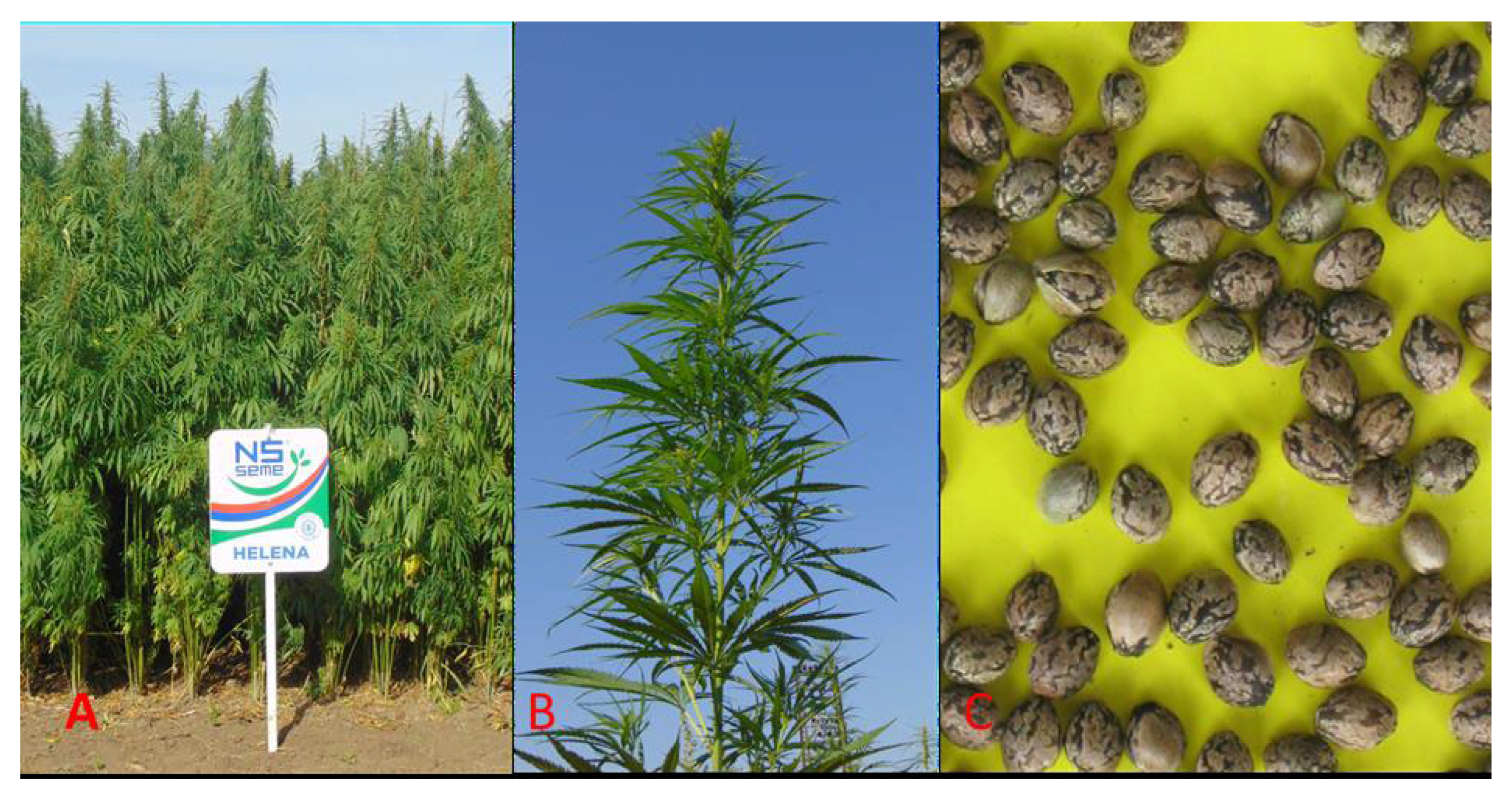
2. Materials and Methods

Cannabis sativa cv. Helena was the model plant utilized in this study. The plant was cultivated in the experimental field of the Institute for Field and Vegetable Crops in Novi Sad—Alternative Crops Department in Baški Petrovac, Serbia (45°19′28″ N 19°39′10″ E), using certified seed. The production technology recommended for the cultivation of industrial hemp in the region of Southeast Europe was applied. The main morphological characteristics of the cv. Helena include medium obovate yellow cotyledon, weak plant anthocyanin coloration, medium intensity of leaf green color, medium time of male flowering, absent or very low (less than 0.2%) inflorescence THC content, medium proportion of hermaphrodite male and female plants in population, plant natural height up to 3 m, medium stem thickness up to 30 mm, medium 1000-seed weight of 15 g, gray brown seed color testa with weak marbling (Figure 1C).

2.1. Embriologycal Analyses

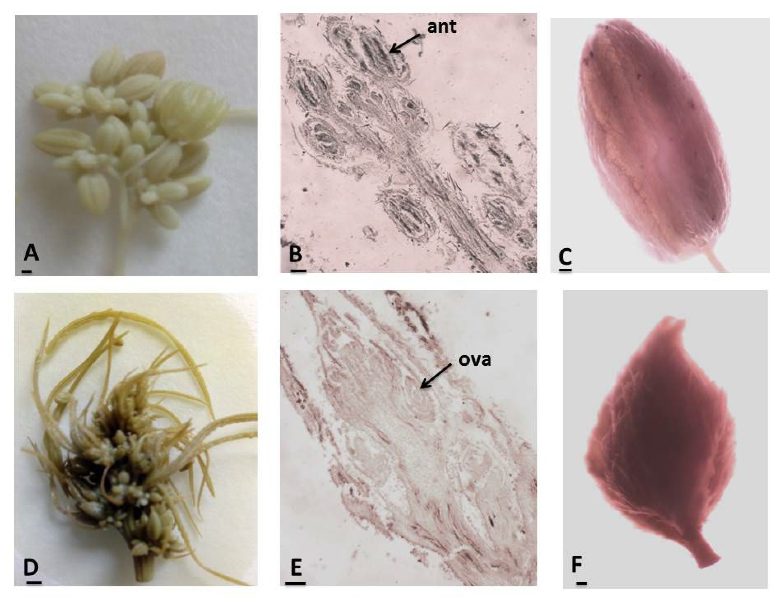
2.1.1. Male and Female Structures

For the study of the structure of reproductive organs in the male and female individuals, flower buds and flowers/50 flower buds and flowers at different stages of development per male and female individuals/were harvested from the cv. Helena and fixed in a mixture of FAA (formalin/glacial acetic acid/70% ethanol in correlation 5:5:90 parts). Subsequently, the fixed material was washed, processed by consecutive runs in a series of alcohol with an increasing percent, xylol, and embedded in paraffin. Microtome sections were cut 5–15 microns in thickness. The stains used were Heidenhain’s hematoxylin [53]. Based on the structures observed on the prepared microscope slides, the stages of development of the male and female gametophytes were determined. The description of the structure and development of the male and female gametophytes was made on the basis of the descriptions in the embryology of flowering plants by Poddubnaya-Arnoldi [54] and Batygina [55].

2.1.2. Pollen Viability

Pollen viability was estimated using the Acetocarmine test [56] on temporary slides, evaluating mature pollen grains from 30 anthers collected from flowers of different individual plants. According to the test, the cytoplasm and nuclei of viable pollen grains stain in red, while nonviable and sterile ones remain unstained. The mature pollen grains in the anthers were counted within the visible field of a light microscope at 100× magnification. A total of 7200 pollen grains were examined.

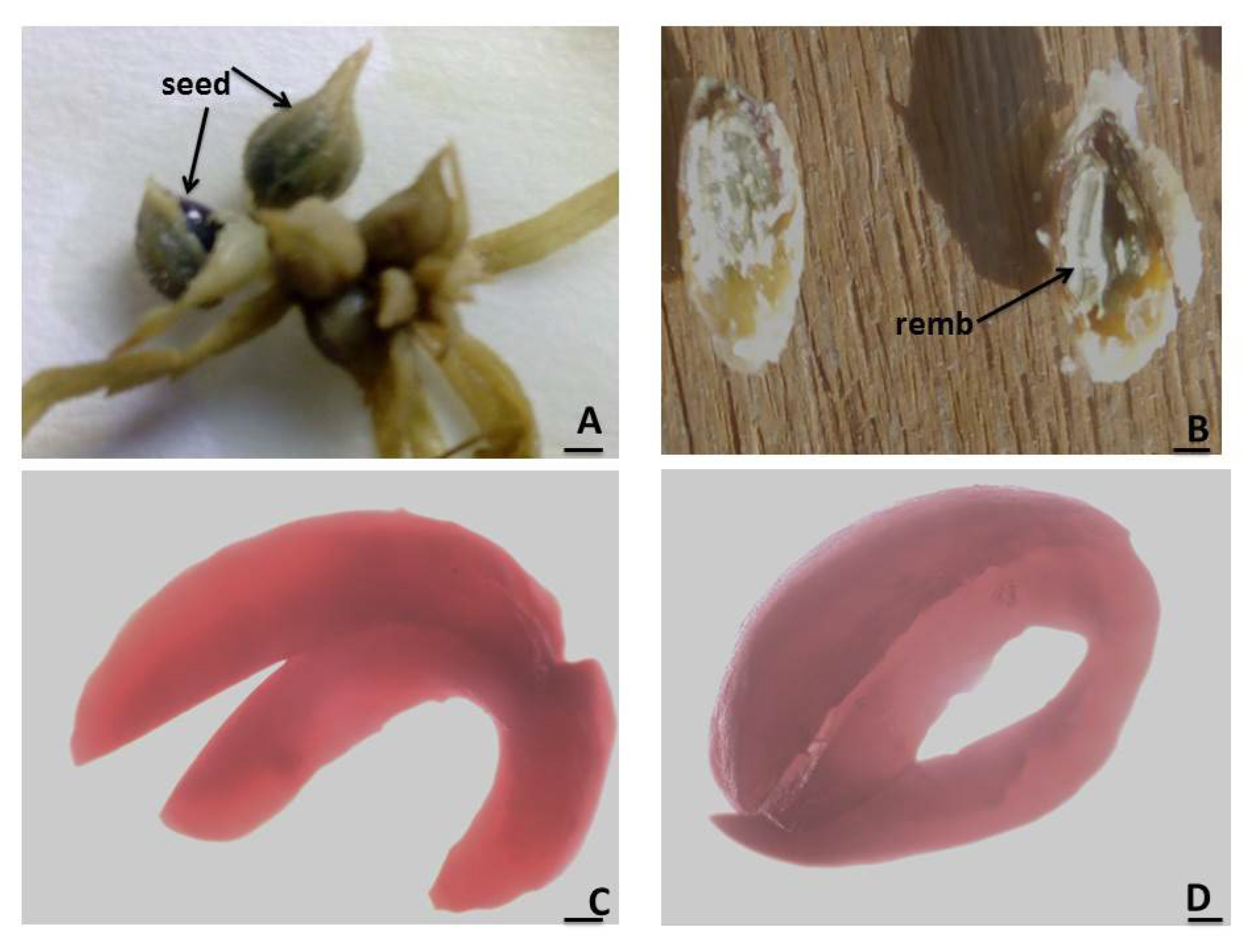
2.1.3. Seed Viability

For estimation of seed (embryo) viability, a quick tetrazolium test completed within 24 h [57] was applied. According to methods described in the literature [57], 100 mature seeds were soaked in water at 30–35 °C for 24 h. Then, the seeds’ coat was cut up at the micropylar end and seeds were incubated in a diluted 1% solution of 2,3,5-triphenyltetrazolium chloride for 24 h at 25 °C. During the test, the initially colorless tetrazolium solution turned red in contact with the hydrogen from the respiratory enzymes in the seeds. Embryos were isolated from the seeds and their viability was assessed: embryos with active respiratory activity were stained red by the tetrazolium solution and considered viable (the more intense the staining, the more active the respiratory activity of the seed), while unstained embryos were considered nonviable. The percentage of seed viability is calculated as the average of two viability tests performed.
Observations were made with a light microscope “Olympus” CX21 (Olympus Corporation, Shinjuku, Tokyo, Japan). Images were taken with a digital camera, “Infinity lite”, 1,4 Mpx mounted to the microscope (Lumenera Corporation, Ottawa, ON, Canada).

2.2. Statistical Analysis

Pollen and seed viability data were processed using descriptive statistics for Windows 10. Average values and standard deviation (±SD) were calculated.

3. Results

Since C. sativa is a naturally dioecious species, the unisexual flowers (male and female) were located on different individuals (male and female plants; Figure 2). Unisexual flowers are bored in inflorescences that are terminal at an earlier stage, and terminal or lateral in a later stage (Figure 2B,E). The staminate (male) flowers (Figure 2C) are composed of a segmented perianth and five stamens/anthers (Figure 2B). The pistillate (female) flowers (Figure 2F) consist of agamophyllous inconspicuous perianth closely embracing an ovoid ovary (Figure 2E).

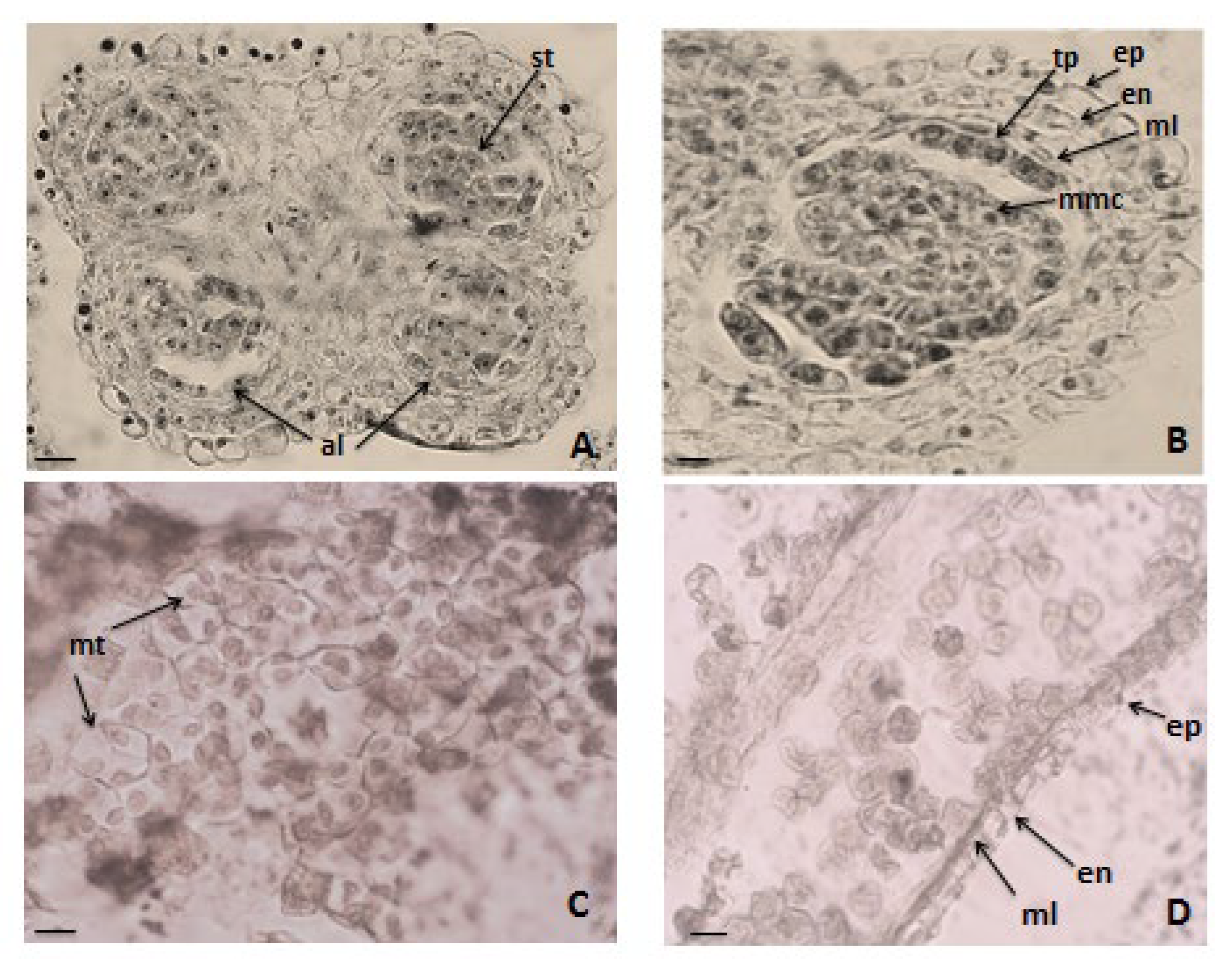
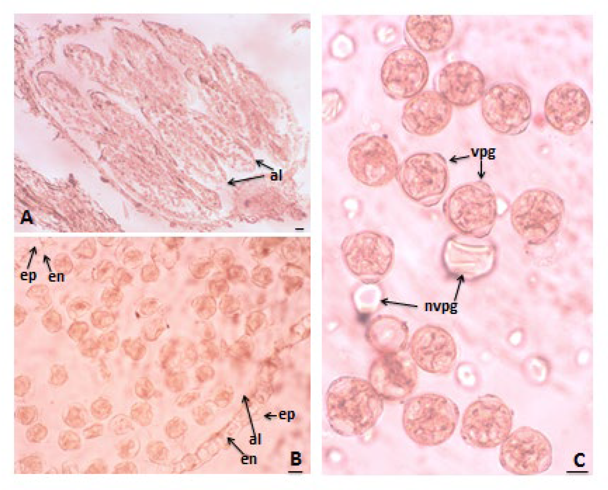
3.1. Anther and Development of Male Gametophyte

The anthers were tetrasporangiates (Figure 3A), whose wall develops by the Dicotyledonous type according to the Davis classification [29]. Its structural layers, namely, the epidermis, endothecium, two middle layers, and tapetum, were uniform at the beginning of microsporogenesis, except for the tapetum, which exhibited distinct characteristics even at this stage (Figure 3B). However, during the anther ontogenesis, these layers changed as the epidermal cells enlarged tangentially and rounded up outside. In contrast, the middle layers were ephemeral and degenerated at about the second (homeotypic) division of meiosis in microspore mother cells (MMCs). Only remnants of these middle layers remained when the pollen matured (Figure 3D). The cells of the endothecium enlarged radially and, after the stage of the microspore tetrads, developed fibrous thickenings (Figure 3D). The initially one-nucleated tapetum cells, as a result of mitotic divisions, became two-nucleated. The tapetum layer began to degenerate after the formation of the microspore tetrads. At the stage of mature pollen grains, the epidermis, endothecium, and the degenerating remains of one middle layer were observed in the anther wall (Figure 3D). The sporogenous tissue was multi-layered, composed of small cells with dense cytoplasm that initially fitted closely with each other. However, as anther ontogenesis progressed, the cell rounded up separately, and differentiated into MMCs. In the MMCs, the processes of meiotic division and pollen development proceeded regularly within all observed anthers. Simultaneous cytokinesis occurred after meiotic division, and as a result, tetrahedral microspore tetrads were formed (Figure 3C). The mature pollen grains were two-celled.

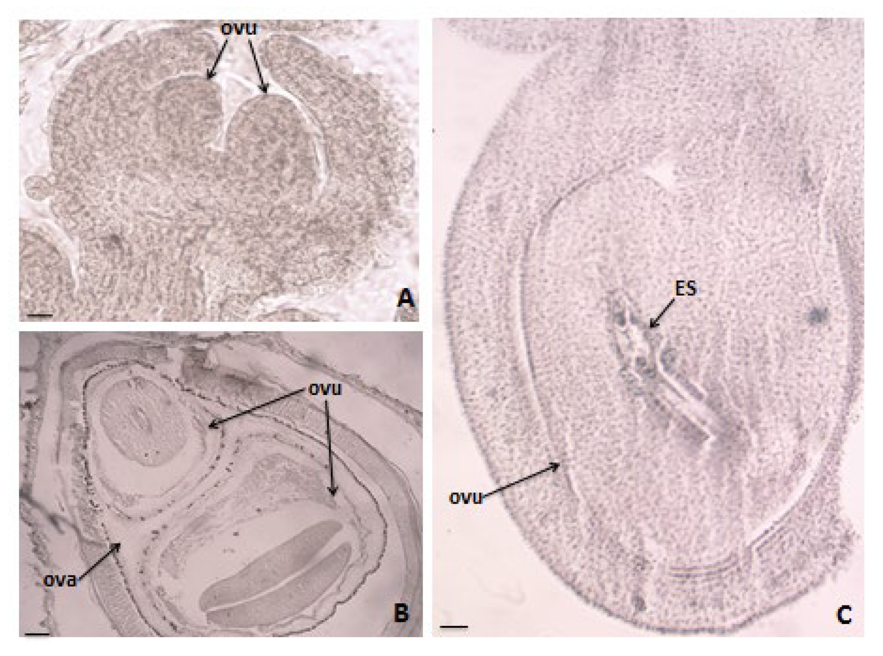
3.2. Ovule and Development of Female Gametophyte

The ovary was superior, unilocular with two ovules developed in it (Figure 4A–C). The mature ovule was anatropous, crassinucellate, bitegmic (Figure 4C). Within the still yang ovule, an unicellular archesporium formed (Figure 5A) that differentiated into a megaspore mother cell (MMC) (Figure 5B). After meiosis in the MMC, a linear tetrad formed (Figure 5C). The embryo sac (ES) developed according to the Poligonum (monosporic) type. The embryo (em) and endosperm (end) formed after porogamous double fertilization. The first division of the primary nucleus preceded that of the zygote (Figure 6C). The endosperm was ab initio nuclear and became cellular after the stage of globular embryo. During the earlier stages of embryo- and endospermogenesis, the chalazal end of the ES elongated in a radial direction, forming haustorium (Figure 6A). Mature seeds contain the embryo, which occupies the largest volume of the seeds and remnants of endosperm in a thin strip below the seed coat (Figure 6B).

3.3. Pollen Viability
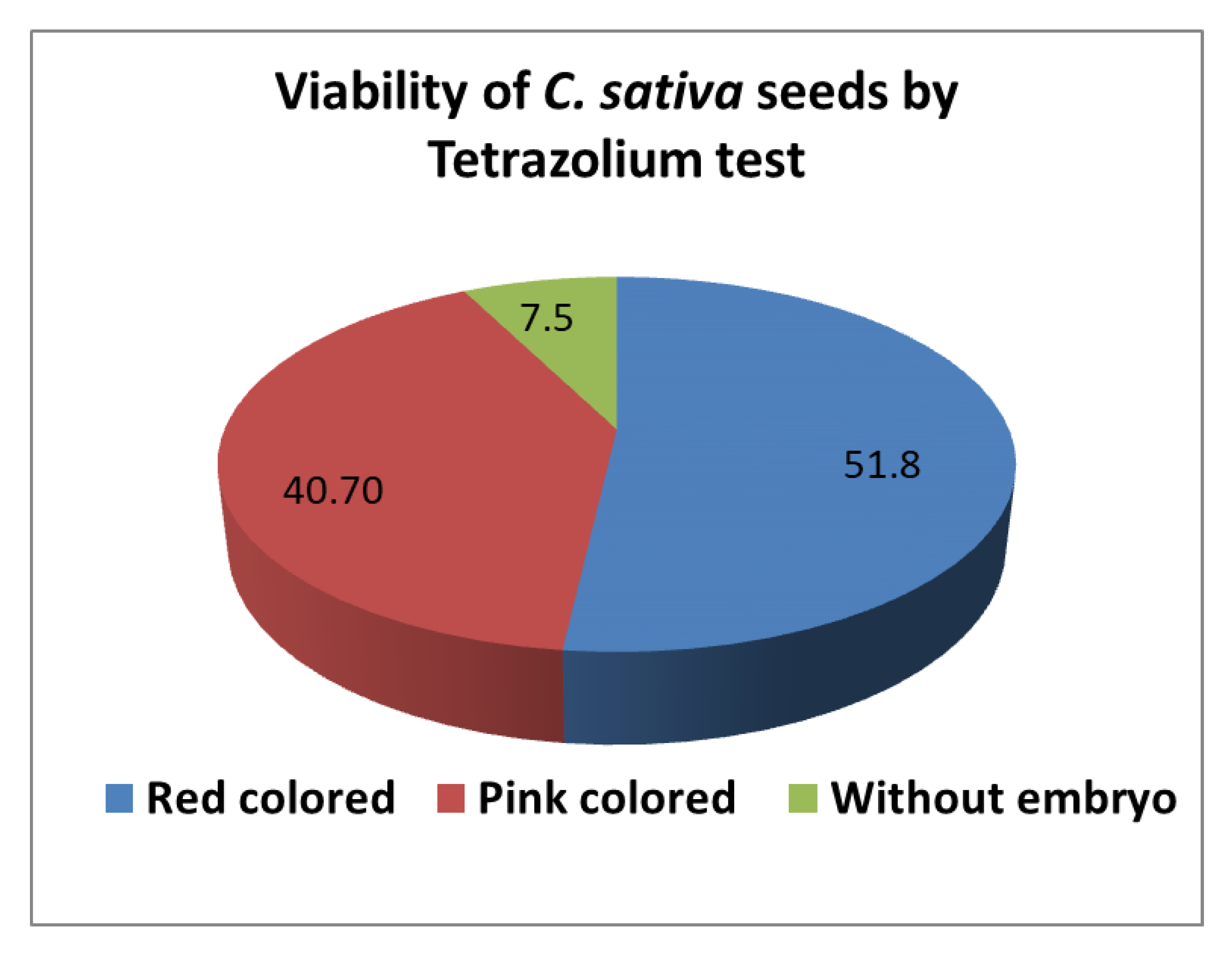
The application of the acetocarmine test technique showed a high pollen viability. A large amount of pollen grains was present in all the tested anthers (Figure 7A) and the majority of them were colored in red (Figure 7B,C). The estimated pollen viability was 95.73 ± 0.21%.

3.4. Seed Viability

Based on the results of tetrazolium testing (Figure 8 and Figure 9), the examined seeds (Figure 8A) were grouped into three classes (Figure 9): Class I: seeds with embryos stained in red (Figure 8C); Class II: seeds with pink-colored embryos (Figure 8D); and Class III: seeds with degenerated embryo (Figure 8B). According to the criteria of Moore [58] for interpreting the resulting coloring from tetrazolium testing, the seeds from Classes I and II were estimated as viable, and the resulting percent of viable seeds was 92.5 ± 0.12%.

4. Discussion

Generally, studies on the reproductive structures of C. sativa date back to the late-19th and early-20th centuries. In this context, this study provides the first comprehensive analysis of the structure and development of male and female gametophytes in C. sativa cv. Helena and, by extension, the species as a whole. Furthermore, two fundamental parameters of reproductive biology, that is, pollen and seed viability, were examined for the first time, providing crucial insights into assessing population and variety quality. The different aspects of the development of male and female gametophytes in C. sativa have also been documented in previous studies. Some of these distinctive features are discussed below. The descriptions of the structure of the female archesporium in C. sativa given in the literature are not uniform. Briosi and Tognini [23], Davis [29], and Poddubnaya-Arnoldi [30] reported a one-celled archesporium that was observed in the present study as well. Reed [27] observed a multi-celled archesporium in a number of investigated ovules. According to the latter author, the occurrence of both one-celled and multi-celled archesporium in the same species may indicate that the plant is shifting its habits.
A noteworthy characteristic revealed by this study is the established development of two ovules in the one-locular ovary. In the works dealing with the morphology of the pistil of C. sativa, the presence of more than one ovule in the ovary has not been commented on, and the presence of only one ovule in the ovary has been noted as typical for the family Cannabaceae by Poddubnaya-Arnoldi [30].
The type of ovule is a taxonomic feature that characterizes the species, genus, or entire family. In C. sativa, the ovule has been described as anatropous, orthotropous, and campylotropous [27]. In the present study, the ovule was observed to be anatropous and was reported previously as a typical one for the Cannabaceae family by Davis [29] and by Poddubnaya-Arnoldi [30].
The haustorium observed in C. sativa by different authors and in the present study is formed from nuclear cells in the chalazal end of the embryo sac (ES). It has been defined by Ram [28] as endospermal haustorium, and by Davis [29] as haustorium of the ES. Poddubnaya-Arnoldi [30] reported two types of haustorium (endospermal and ES haustorium) as a characteristic feature of the family Cannabaceae. Several haustorial structures performing nutritive functions for the ES or embryo in the ovule have been described. They may be formed by the megaspore, ES, synergid, antipodal, endosperm, suspensor, or pollen tube [59]. Maheshwari [60] cited them among the embryological features of taxonomic significance, and Poddubnaya-Arnoldi [54] considered the haustorium type as a phylogenetic and systematic feature. The structure that appears at the chalazal end of the ES of C. sativa and can be observed at the late stage of embryo formation, when the endosperm is consumed to a great degree from the embryo, is the same structure reported as endospermal haustorium and ES haustorium by the authors cited above. Davis [29] described the ES haustorium as an elongated curved base of the ES. The ES and endosperm haustoria often occur together in the same family and even in the same species [60].
Due to the importance of hemp as a high-value crop and source for numerous materials and chemicals, it is necessary to comment on the practical significance of the results of the present study.
The quantity and quality of pollen produced in each plant species are crucial factors in pollination and fertilization, directly influencing the production of viable seeds. Pollen quality is determined by its vitality, i.e., from the amount of viable (fertile) pollen grains. The high pollen viability established in the present study is a factor contributing to the effective pollination and fertilization that was evidenced by the strong sexual reproduction and successful seed formation observed in C. sativa.
In crop breeding practice, it is of great importance to determine the quality of various varieties as pollinators. The study of the structure, development, and viability of pollen allows one to determine its quality, that is, the quality of a plant variety as a pollinator. The importance of understanding the qualities of plants/pollinators has been demonstrated previously by Tursin [61]. He found that the reason for the low yield in Salvia officinalis varieties was the high number of male sterile plants and that increasing the number of plants with fertile pollen increased yields. Thus, the high amount of fertile pollen characterizes the studied cultivar of C. sativa as a good variety pollinator.
Determining the quality of mother plants is also of great importance in defining the quality of the plant cultivar. It is determined by the quality of the fruits and seeds produced as a result of the normal running of processes of development of the female gametophyte, pollination, and fertilization. The study of the structure and development of the egg cell and the embryo sac allows one to determine the quality of the cultivar mother. It is important to note that the development of the male gametophyte, the embryo sac, the pollination processes, and embryo- and endospermogenesis are influenced by external conditions. Unfavorable external conditions disrupt the normal course of these processes, which can be determined by examining them using the cytoembryological method. The established normal running of the processes of formation and development of the female gametophyte, embryo- and endospermogenesis, and the high pollen viability provide high reproductive potential for the target cultivar, and the established high seed viability is evidence of the high degree of its realization. As this realization depends on the environmental conditions (soil type, climatic conditions), the results obtained show favorable conditions for the development of the studied cultivar.

5. Conclusions

In the present study, on the reproductive capacity of C. sativa cv. Helena was evaluated to assess its quality as a cultivated plant. The established features of the structure and processes of the male and female generative sphere define it as a sexually reproducing species, which ensures a high reproductive potential for its plantations. This high reproductive potential, in turn, is the basis for creating successful agriculture. The assessed high viability of its pollen and seeds shows that, under favorable conditions, this variety can realize its reproductive ability to a high degree. All this makes C. sativa cv. Helena promising for cultivation as a crop. Revealing reproductive capacity can be used in crop breeding practice as an approach to testing the suitability of each variety.

Author Contributions

Conceptualization, V.D.Z., I.S. and E.Y.-T.; methodology, E.Y.-T.; formal analysis, E.Y.-T.; investigation, V.S. and E.Y.-T.; resources, V.S., V.D.Z. and I.S.; writing—original draft preparation, E.Y.-T., I.S. and V.D.Z.; writing—review and editing, E.Y.-T., I.S., V.S. and V.D.Z.; visualization, E.Y.-T. and V.S.; supervision, V.D.Z.; project administration, V.D.Z.; funding acquisition, V.D.Z. All authors have read and agreed to the published version of the manuscript.

Funding

This research received no external funding.

Data Availability Statement

The original contributions presented in this study are included in the article. Further inquiries can be directed to the corresponding author.

Conflicts of Interest

The authors declare no conflicts of interest.

References

  1. Haberlandt, F. Welche Einflusse bedingen das Geschlecht der HanfpflanZen? Fuhlings landw Ztg. 1877, 881. In Morphology of Cannabis sativa L.; State University of Iowa: Iowa City, IA, USA, 1914; Available online: https://www.scribd.com/document/400629834/Morphology-of-Cannabis-Sativa-L (accessed on 11 February 2025).
  2. Saccardo, P.A. Sulle cause determinanti la sessualita nslla Canapa. Bul. Soc. Veneto-Trent. Sei. Nat. Padova. 1879. In Morphology of Cannabis sativa L.; State University of Iowa: Iowa City, IA, USA, 1914; Available online: https://www.scribd.com/document/400629834/Morphology-of-Cannabis-Sativa-L (accessed on 11 February 2025).
  3. Heyer, F. Untersuchungen uber das Verhaltniss des geschlechtes bei einhausigen und Zweihausigen Pflanzen. Ber. aus d. physiolog. Laborat. d. landw. Instituts d. Universitat Halle. 1 Bd. 5. Heft. 1884. In Morphology of Cannabis sativa L.; State University of Iowa: Iowa City, IA, USA, 1914; Available online: https://www.scribd.com/document/400629834/Morphology-of-Cannabis-Sativa-L (accessed on 11 February 2025).
  4. Dusing, C.A. Die experimsntelle Prufung der Theorie von der Regulierung des Geschlechtsverbaltnisse. Jenaiehe Ztschr. f. naturwiss. 1885 19. Suppl. II. In Morphology of Cannabis sativa L.; State University of Iowa: Iowa City, IA, USA, 1914; Available online: https://www.scribd.com/document/400629834/Morphology-of-Cannabis-Sativa-L (accessed on 11 February 2025).
  5. Hoffman, H. Ueber Sexualitat. Bot. Ztg. 1885, 43. In Morphology of Cannabis sativa L.; State University of Iowa: Iowa City, IA, USA, 1914; Available online: https://www.scribd.com/document/400629834/Morphology-of-Cannabis-Sativa-L (accessed on 11 February 2025).
  6. Fisch, C. Ueber die Zahlenverhaltnisse des Geschlechtes beim Hanf. Ber. d. d. bot. Bd. V. 1887. In Morphology of Cannabis sativa L.; State University of Iowa: Iowa City, IA, USA, 1914; Available online: https://www.scribd.com/document/400629834/Morphology-of-Cannabis-Sativa-L (accessed on 11 February 2025).
  7. Noll, F. Vorlaufiger Abschluss der Versuche uber die Bestummung des Geschlechts bei dioechen Pflanzen. Sitzungsber der niederrh. Gesellsch. f. Natur-U. Heilkunde. Bonn. 1907. In Morphology of Cannabis sativa L.; State University of Iowa: Iowa City, IA, USA, 1914; Available online: https://www.scribd.com/document/400629834/Morphology-of-Cannabis-Sativa-L (accessed on 11 February 2025).
  8. Strasburger, E. Üeber geschlechbestummende Ursachen. Jahrb. Wiss. Bot. 1910, 48, 427–520. [Google Scholar]
  9. Galoch, E. The hormonal control of sex differentiation in dioecious plants of hemp (Cannabis sativa). Acta Physiol Plant 1980, 2, 31–39. [Google Scholar]
  10. Lacombe, J.P. Discrimination des sexes en fonction de caractères végétatifs précoces chez le Chanvre dioïque (Cannabis sativa L.). Physiol Vég 1980, 18, 419–430. [Google Scholar]
  11. Spitzer-Rimon, B.; Shai Duchin, S.; Bernstein, N.; Kamenetsky, R. Architecture and Florogenesis in Female Cannabis sativa Plants. Front Plant Sci. 2019, 10, 350. [Google Scholar] [CrossRef]
  12. Faeti, V.G.; Mandolino, V.G.; Ranalli, P. Genetic diversity of Cannabis sativa germplasm based on RAPD markers. Plant Breed. 1996, 115, 367–370. [Google Scholar] [CrossRef]
  13. Forapani, S.; Carboni, A.; Paoletti, C.; Moliterni, V.M.C.; Ranalli, P.; Mandolino, G. Comparison of hemp varieties using random amplified polymorphic DNA markers. Crop Sci. 2001, 41, 1682–1688. [Google Scholar] [CrossRef]
  14. Moliterni, V.M.C.; Cattivelli, L.; Ranalli, P.; Mandolino, G. The sexual differentiation of Cannabis sativa L.: A morphological and molecular study. Euphytica 2004, 140, 95–106. Available online: https://www.scribd.com/document/454759367/Euphitica2005hemp (accessed on 11 February 2025). [CrossRef]
  15. Hammond, C.T.; Mahlberg, P.G. Morphogenesis of capitate glandular hairs of Cannabis sativa (Cannabaceae). Am. J. Bot. 1977, 64, 1023–1031. [Google Scholar] [CrossRef]
  16. Andre, C.M.; Hausman, J.F.; Guerriero, G. Cannabis sativa: The plant of the thousand and one molecules. Front. Plant Sci. 2016, 7, 19. [Google Scholar] [CrossRef] [PubMed] [PubMed Central]
  17. Chandra, S.; Lata, H.; Khan, I.A.; ElSohly, M.A. Cannabis sativa L.: Botany and horticulture In Cannabis sativa L.–Botany and Biotechnology; Springer: New York, NY, USA, 2017; pp. 79–100. Available online: https://www.scribd.com/document/450709197/Cannabis-sativa-L-Botany-Biotechnology-pdf# (accessed on 11 February 2025).
  18. Raman, V.; Lata, H.; Chandra, S.; Khan, I.A.; ElSohly, M.A. Morpho-anatomy of marijuana (Cannabis sativa L.). In Cannabis sativa L.–Botany and Biotechnology; Springer: Berlin/Heidelberg, Germany, 2017. [Google Scholar]
  19. Hemphill, J.; Turner, J.; Mahlberg, P. Cannabinoid content of individual plant organs from different geographical strain of Cannabis sativa L. J. Nat. Prod. 1980, 43, 112–121. [Google Scholar] [CrossRef]
  20. Punja, Z.K.; Ni, L.; Lung, S.; Buirs, L. Total yeast and mold levels in high THC-containing cannabis (Cannabis sativa L.) inflorescences are influenced by genotype, environment, and pre-and post-harvest handling practices. Front Microbiol. 2023, 14, 1192035. [Google Scholar] [CrossRef] [PubMed] [PubMed Central]
  21. Zheljazkov, V.D.; Noller, J.S.; Dale, R.; Maggi, F. Terpenes and cannabinoids yields and profile from direct-seeded and transplanted CBD-Cannabis sativa. J. Agric. Food Chem. 2022, 70, 10417–10428. [Google Scholar] [CrossRef] [PubMed]
  22. Zinger, N. Beitrage zur Kenntniss der weiblichen Bluthen und Inflorescenzen bei Cannabineen. Flora 1898, 85, 189–253. [Google Scholar]
  23. Briosi, G.; Tognini, F. 1894–96. Intorno alla anatomia della canapa (Cannabis sativa L.); Tip. Bernardoni di C. Rebeschini e c.: Milano, Italy, 1894. [Google Scholar]
  24. Montemartini, L. Sul valore morfologico dell r ovario e del 1 ovulo della Canapa; Rendiconti Congresso Botanica di Palermo: Palermo, Italy, 1902; 8vo br.; p. VIII-224. [Google Scholar]
  25. Prain, D. On the Morphology, Teratology, and Diclinism of the Flowers of Cannabis, No, 12; Scientific Memoirs; Medical and Sanitary Departments: New Delhi, India, 1904. [Google Scholar]
  26. Modilewsky, J. Zur Samenbildung einiger Urticifloren. Flora 1908, 98, 423–470. [Google Scholar] [CrossRef]
  27. Reed, J. Morphology of Cannabis sativa L. Master’s Thesis, State University of Iowa, Ames, IA, USA, 1914. [Google Scholar] [CrossRef]
  28. Ram, M. Occurrence of endosperm haustorium in Cannabis sativa L. Ann. Bot. 1960, 24, 79–82. [Google Scholar] [CrossRef]
  29. Davis, G. Systematic Embryology of the Angiosperms; John Wiley & Sons, Inc.: New York, NY, USA, 1966. [Google Scholar]
  30. Poddubnaya-Arnoldi, V.A. Cannabaceae. In Characteristics of Angiosperms Families According to Cytoembryological Features; Nauka: Moskow, Russia, 1982; pp. 51–52. (In Russian) [Google Scholar]
  31. McPhee, H.C. Meiotic cytokinesis of Cannabis. Botan. Gazette 1924, 78, 335–341. [Google Scholar] [CrossRef]
  32. Heslop-Harrison, J. Cytoplasmic connections between angiosperm meiocytes. Annal. Bot. 1966, 30, 221–230. [Google Scholar] [CrossRef]
  33. Asanova, D.K. Specific features of microsporogenesis in the hemp of the Shu valley. Biol. Bull. Rus. Acad. Sci. 2002, 29, 545–550. [Google Scholar] [CrossRef]
  34. Heslop-Harrison, J. Origin of exine. Nature 1962, 195, 1069–1071. [Google Scholar] [CrossRef]
  35. Heslop-Harrison, J. The Pollen Wall: Structure and Development: Pollen; Butterworth-Heinemann: Oxford, UK, 1971; pp. 75–98. [Google Scholar]
  36. Bradley, D.E. The study of pollen grain surfaces in the electron microscope. New Phytol. 1958, 57, 226–229. [Google Scholar] [CrossRef]
  37. Punt, W.; Malotaux, M. Cannabaceae, moraceae and urticaceae. Rev. Palaeobot. Palynol. 1984, 42, 23–44. [Google Scholar] [CrossRef]
  38. Galán-Ávila, A.; García-Fortea, E.; Prohens, J.; Herraiz, F.J. Microgametophyte Development in Cannabis sativa L. and First Androgenesis Induction Through Microspore Embryogenesis. Front Plant Sci. 2021, 25, 669424. [Google Scholar] [CrossRef] [PubMed] [PubMed Central]
  39. Miller, N.G. The genera of the Cannabaceae in the southeastern United States. J. Arnold Arboret. 1970, 51, 185–203. [Google Scholar] [CrossRef]
  40. Wu, Z.; Zhou, Z.; Bartholomew, B. Cannabaceae. Flora China 2003, 5, 74–75. [Google Scholar]
  41. United Nations Office on Drugs and Crime (UNODC). Recommended Methods for the Identification and Analysis of Cannabis and Cannabis Products: Manual for Use by National Drug Testing Laboratories; United Nations Publications: New York, NY, USA, 2009. [Google Scholar]
  42. Hammond, C.T.; Mahlberg, P.G. Morphology of glandular hairs of Cannabis sativa from scanning electron microscopy. Am. J. Bot. 1973, 60, 524–528. [Google Scholar] [CrossRef]
  43. Livingston, S.J.; Quilichini, T.D.; Booth, J.K.; Wong, D.C.; Rensing, K.H.; Laflamme-Yonkman, J.; Castellarin, S.D.; Joerg Bohlmann, J.; Page, J.E.; Samuelset, A.L. Cannabis glandular trichomes alter morphology and metabolite content during flower maturation. Plant J. 2020, 101, 37–56. [Google Scholar] [CrossRef]
  44. Mandolino, G.; Carboni, A.; Forapani, S.; Faeti, V.; Ranalli, P. Identification of DNA markers linked to the male sex in dioecious hemp (Cannabis sativa L.). Theor. Appl. Genet. 1999, 98, 86–92. [Google Scholar] [CrossRef]
  45. Sakamoto, K.; Akiyama, Y.; Fukui, K.; Kamada, H.; Satoh, S. Characterization; Genome Sizes and Morphology of Sex Chromosomes in Hemp (Cannabis sativa L.). Cytologia 1998, 63, 459–464. [Google Scholar] [CrossRef]
  46. Kaushal, S. Impact of physical and chemical mutagens on sex expression in Cannabis sativa. Indian J. Fundam. Appl. Life Sci. 2012, 2, 97–103. [Google Scholar]
  47. Shao, H.; Song, S.-J.; Clarke, R.C. Female-Associated DNA Polymorphisms of Hemp (Cannabis sativa L.). J. Ind. Hemp. 2003, 8, 5–9. [Google Scholar] [CrossRef]
  48. Mendel, P.; Bharat Lalge, A.; Vyhnanek, T.; Trojan, V.; Kalousek, P.; Maassen, H.; Havel, L. Progress in early sex determination of cannabis plant by DNA markers. MendelNet 2016, 1, 731–735. [Google Scholar]
  49. Araméndiz-Tatis, H.; Cardona-Ayala, C.E.; Espitia-Camacho, M.; Herrera-Contreras, A.C.; Villalba-Soto, A.L. Agronomic evaluation of Cannabis sativa (L.) cultivars in northern Colombia. Rev. Colomb. Cienc. Hortícolas 2023, 17, e15695. [Google Scholar] [CrossRef]
  50. Campbell, L.G.; Peach, K.; Wizenberg, S.B. Dioecious hemp (Cannabis sativa L.) plants do not express significant sexually dimorphic morphology in the seedling stage. Sci. Rep. 2021, 11, 16825. [Google Scholar] [CrossRef] [PubMed]
  51. Farag, S.; Kayser, O. The cannabis plant: Botanical aspects. In Handbook of Cannabis and Related Pathologies: Biology, Pharmacology, Diagnosis, and Treatment; Preedy, V., Ed.; Elsevier: Amsterdam, The Netherlands, 2017; pp. 3–12. Available online: https://www.scribd.com/document/403631308/The-Cannabis-Plant-Botanical-Aspects-pdf (accessed on 11 February 2025).
  52. Mishchenko, S.; Mokher, J.; Laiko, I.; Burbulis, N.; Kyrychenko, H.; Dudukova, S. Phenological growth stages of hemp (Cannabis sativa L.): Codification and description according to the BBCH scale. Žemės ūkio mokslai 2017, 24. [Google Scholar] [CrossRef]
  53. Heidenhain, R. Eine neue Verwendung des haematoxylin. Archiv. Mikrosk. Anat. 1885, 24, 468–470. Available online: https://link.springer.com/article/10.1007/BF02960389 (accessed on 11 February 2025). [CrossRef]
  54. Poddubnaja-Arnoldi, V.A. Cytoembryology of Angiosperms; Nauka: Moscow, Russia, 1976. [Google Scholar]
  55. Batygina, T.B. Embryology of Floering Plants. Terminology and Concepts, Volume 1: Generative Organs of Flower; Science Publishers, Inc.: New York, NY, USA, 2002; p. 421. [Google Scholar]
  56. Singh, R.J. Plant Cytogenetics, 2nd ed.; CRC Press: Boca Raton, FL, USA, 2003; p. 488. Available online: https://scholar.google.com/scholar_lookup?title=Plant+Cytogenetics&author=Singh,+R.J.&publication_year=2003 (accessed on 11 February 2025).
  57. Peters, J. Tetrazolium Testing Handbook; Association of Official Seed Analysts: Ithaca, NY, USA, 2007; p. 88. Available online: https://scholar.google.com/scholar_lookup?title=Tetrazolium+Testing+Handbook&author=Peters,+J.&publication_year=2007 (accessed on 11 February 2025).
  58. Moore, R.P. Handbook on Tetrazolium Testing; International Seed Testing Association: Zurich, Switzerland, 1985. [Google Scholar]
  59. Schnarf, K. Embryologie der Angiospermen; Gebruder Bontraeger: Berlin, Germany, 1929; p. 689. [Google Scholar]
  60. Maheshwari, P. Recent Advances in the Embryology of Angiosperms; International Society of Plant Morphologists, University of Delhi: New Delhi, India, 1963; 467p. [Google Scholar]
  61. Tursin, G. Study on the Generative Organ of Salvia Officinalis. Materials All-Union Symposium “Sexual Development and Embryogenesis in Plants”; Moscow State University Publishing House: Moscow, Russia, 1973. (In Russian) [Google Scholar]
Figure 1. Peculiarities of C. sativa cv. Helena: (A) general view of cultivar; (B) the leaves and twigs; (C) the seeds. The pictures were taken by Vladimir Sikora.
Agronomy 15 00474 g001
Figure 2. Staminate and pistillate inflorescences: (AC): staminate inflorescence: (A) staminate inflorescence, (B) longitudinal section of staminate inflorescence, (C) staminate (male) flower; (D,E) pistillate inflorescence: (D) pistillate inflorescence, (E) longitudinal section of pistillate inflorescence, (F) pistillate (female) flower; ant—anther, ova—ovary. Scale bar: for (A,C,D,F) = 50 μm; for (B,E) = 100 μm.
Agronomy 15 00474 g002
Figure 3. Anther and development of male gametophyte: (A) tetrasporangiate anther; (B) MMCs and anther wall; (C) microspore tetrads; (D) mature pollen grains and anther wall; tp—tapetum, ml—middle layer, en—endothecium, ep—epidermis, mmc—microspore mother cell, mt—microspore tetrad, al—anther locule, st—sporogenous tissue. Scale bar: for (AC) = 20 μm; for (D) = 50 μm.
Agronomy 15 00474 g003
Figure 4. Ovule development: (AC): development of primordiums of two ovules in the ovary; (B) two ovules with mature embryo in the ovary; (C) anatropous ovule with developed embryo sac of Polygonum type;). ovu—ovule, ova—ovary, ES—embryo sac. Scale bar = 50 μm.
Agronomy 15 00474 g004
Figure 5. Development of female gametophyte: (A) one-celled archesporium in the ovule; (B) megaspore mother cell in the ovule; (C) megaspore tetrad in the ovule; ac—archesporial cell, mmc—megaspore mother cell, mt—megaspore tetrad. Scale bar = 20 μm.
Agronomy 15 00474 g005
Figure 6. Development of female gametophyte: (A) development of ES haustorium; (B) embryo with cotyledons and endosperm; (C) zygote and nuclear endosperm. H—haustorrium, em—embryo, end—endosperm, Z—zygote, embc-cotyledons of the embryo. Scale bar = 50 μm.
Agronomy 15 00474 g006
Figure 7. Pollen viability testing: (A,B) anther locule with pollen grains after acetocarmine staining; (C) viable (stained in red) and nonviable (unstained) pollen grains; pg—pollen grain, al—anther locule, vpg—viable pollen grain, nvpg—nonviable pollen grain; en—endothecium ep—epidermis. Scale bar: for A = 100 μm; for B = 50 μm; for C = 20 μm.
Agronomy 15 00474 g007
Figure 8. Seed viability testing: (A) untreated seed; (B) seed without embryo; (C) stained in red from tetrazolium solution viable embryo; (D) colored pink from tetrazolium solution viable embryo. remb—remains of embryo. Scale bar = 100 μm.
Agronomy 15 00474 g008
Figure 9. Estimation of seed viability by tetrazolium test: Distribution of seeds/embryos in classes based on their coloring from the tetrazolium solution: 1: Class I—embryos stained in dark red; 2: Class II—embryos stained in pink; 3: Class III—seeds without embryo.
Agronomy 15 00474 g009
Table 1. Data on morphology, phenological development, and sex differentiation in C. sativa.
AuthorsData
Zinger [22]Morphology of the pistillate flower and inflorescence of Cannabaceae;
Briosi and Tognini [23]C. sativa inflorescence, organography, organogeny, structure of megasporangium, female gametophyte, microsporangium and male gametophyte;
Montemartini [24]Morphology of the ovary and ovule of C. sativa, including their vascular supply and discussion on the origin of these parts;
Prain [25]Morphology, teratology, and diclinism of the C. sativa flowers, describing abnormalities, nature of ovary, and origin of ovule;
Modilewsky [26]Female gametophyte, fertilization, embryo development in C. sativa;
Reed [27]Morphological descriptions of C. sativa floral organs and their development;
Ram [28]Development and structure of endosperm in C. sativa;
Davis [29]Onagrad-type embryo with two-celled suspensor; suspected apomixis;
Poddubnaya-Arnoldy [30]Double fertilization of premitotic type; polyembryony;
McPhee [31]; Heslop-Harrison [32];
Asanova [33]
Meiosis in the microsporangium;
Heslop-Harrison [34,35]Development and structure of tapetum anther layer;
Bradley [36]; Punt and Malotaux [37]Exine formation and characteristics of pollen grains;
Galán-Ávila et al. [38]Microsporogenesis, and microgametogenesis and correlated microgametophyte developmental stages;
Miller [39]; Wu et al. [40]; United Nations Office on Drugs and Crime (UNODC) [41]; Raman et al. [18]Anatomy of male and female flowers;
Spitzer-Rimon et al. [11]Architecture and timing of initiation and differentiation of the inflorescence and individual flowers of different C. sativa cultivars;
Hammond and Mahlberg [42]; Spitzer-Rimon et al. [11]; Livingston et al. [43]Morphology of male flowers;
Mandolino et al. [44]; Sakamoto et al. [45]; Kausal [46]Genetic sex determination;
Shao et al. [47]; Mendel et al. [48]Development of molecular markers for sex expression in C. sativa;
Araméndiz-Tatis et al. [49]Genetic variability in vegetative and reproductive characteristics, sexual dimorphism in juvenile plants;
Farag and Kayser [51]; Mishchenko et al. [52]; Raman et al. [18]Terminology for phenological stages of C. sativa development;
Disclaimer/Publisher’s Note: The statements, opinions and data contained in all publications are solely those of the individual author(s) and contributor(s) and not of MDPI and/or the editor(s). MDPI and/or the editor(s) disclaim responsibility for any injury to people or property resulting from any ideas, methods, instructions or products referred to in the content.

