Article

Genome-Wide Analysis of NLP Genes in Peanut Reveals Significant Roles of AhNINa and AhNINb in Root Nodule Development

1
Key Laboratory of Peanut Biology, Genetic & Breeding, Ministry of Agriculture and Rural Affairs, Shandong Peanut Research Institute, Qingdao 266100, China
2
State Key Laboratory of Wheat Improvement, Shandong Agricultural University, Tai’an 271018, China
3
The Key Laboratory of Plant Development and Environmental Adaptation Biology, Ministry of Education, School of Life Sciences, Shandong University, Qingdao 266237, China
4
State Key Laboratory of Microbial Technology, Shandong University, Qingdao 266237, China
*
Authors to whom correspondence should be addressed.
Agronomy 2025, 15(10), 2377; https://doi.org/10.3390/agronomy15102377
Submission received: 5 August 2025 / Revised: 5 October 2025 / Accepted: 9 October 2025 / Published: 11 October 2025
(This article belongs to the Section Crop Breeding and Genetics)

Abstract

Nitrogen is an indispensable nutrient for plant growth and crop production, but it is not directly accessible to plants without the help of nitrogen-fixing bacteria. Legume plants can form root nodules in symbiosis with rhizobia. NODULE INCEPTION (NIN), a founding member of the NIN-like protein (NLP) family, is essential for nodulation in legume species. However, the knowledge of functional characteristics of the NLP family members in peanuts is limited. In this study, a genome-wide analysis of the NLP genes was carried out. A total of 16 NLP genes were identified in the peanut genome, including 2 AhNIN and 14 AhNLP, which were unevenly distributed on nine chromosomes of the peanut genome. Furthermore, transcriptomic profiles and expression pattern analysis showed that both AhNINa and AhNINb genes were specifically expressed in root nodules. Subcellar localization and transcriptional activity analysis revealed that both AhNINa and AhNINb encode transcriptional activators. In addition, the roots that down-regulated the expression of AhNINa and AhNINb genes failed to form nodules. These findings provide significant insights into the molecular functions of AhNINa and AhNINb genes in regulating peanut nodule development.

1. Introduction

Nitrogen (N) is an essential nutrient element and plays an important role in the growth and development of plants [1]. However, in the process of agricultural production, only 30% of soil-available nitrogen is absorbed by plant roots in the form of nitrate (NO3) and ammonium (NH4+) ions and other organic molecules [2]. The remaining 70% of nitrogen enters the soil through leaching or enters the atmosphere in gaseous form, which will also cause environmental pollution [3]. It was reported that genes and transcription factors that are involved in nitrogen signaling pathways play crucial roles in nitrate uptake and transport [4]. NIN-like proteins (NLPs) are considered to be a plant-specific transcription factor family and are involved in nitrate signaling response and symbiotic nodulation [5,6].
The NODULE INCEPTION (NIN) protein was the first identified NLP family member in Lotus japonicus, which was found to control nitrogen-mediated symbiotic nodule formation [7]. Mutants with loss of NIN orthologs function were unable to form nodules in Glycine max, Medicago truncatula, and Lotus japonicus [8,9,10]. NLP genes were named after their homology to NIN and were considered to be key regulators of nitrate signaling in land plants [5,11,12]. NLPs are DNA-binding proteins with an N-terminal RWP-RK motif, which binds to the nitrate-responsive cis-element (NRE) region of nitrate-responsive genes, and a C-terminal PB1 domain, which is responsible for protein–protein interactions [13]. Previous studies have shown that the NLP transcription factors widely exist in legume and nonlegume plants, such as Medicago sativa, Brassica napus, and Oryza sativa [14,15,16]. In Arabidopsis thaliana, nine NLP members, AtNLP1 to AtNLP9, have been identified, which can all bind the NRE and activate the expression of nitrate-responsive genes [12,17]. Loss of AtNLP7 function results in plants displaying an N-starvation phenotype due to impaired expression of nitrogen pathway-related genes, such as ARABIDOPSIS NITRATE REGULATED 1 (ANR1), NITRATE TRANSPORTER 2 (NRT2), and LATERAL ORGAN BOUNDARY DOMAIN 37 (LBD37) [18,19]. Moreover, AtNLP7 overexpression improves plant nitrogen use efficiency and photosynthetic efficiency, thereby promoting plant growth and development [20]. In addition, AtNLP7 can interact with TEOSINTE BRANCHED1/CYCLOIDEA/PROLIFERATING CELL FACTOR1-20 (AtTCP20) to regulate nitrate signaling [21]. Another NLP gene, AtNLP6, has the highest sequence similarity with AtNLP7, but disruption of AtNLP6 alone does not cause obvious phenotype defects; however, mutation of the AtNLP6 gene in the nlp7-1 mutant background exacerbates changes in gene expression and growth defects [21], suggesting that AtNLP6 and AtNLP7 play a redundant role while AtNLP8 regulates nitrate-promoted seed germination by directly binding to the promoter of an abscisic acid catabolic enzyme encoding gene CYTOCHROME P450 MONOOXYGENASE 707A2 (AtCYP707A2) and reducing abscisic acid levels in a nitrate-dependent manner [22]. Furthermore, AtNLP2 regulates rosette leaf growth and root development in response to changes in nitrate availability [23]. Therefore, NLP transcription factors play important roles in plant nitrogen metabolism, leaf and root development, and nodule formation.
Legumes are able to establish root nodule symbiosis with nitrogen-fixing soil bacteria, which are collectively called rhizobia. The root nodule symbiosis establishment involves the invasion of rhizobia in root epidermis and the formation of nodules through the root cortical cells. The most common mode of rhizobia invasion in legume, such as Vicia sp., Trifolium sp., and Pisum sp., is root hair infection [24]. Peanut (Arachis hypogea L.) is an economically important allotetraploid legume crop, serving as a source of edible vegetable oil and protein. Worldwide, peanuts are grown in 27.66 million hectares in 84 countries with an annual production of 43 million tons of pods (i.e., nuts-in-shell), with the 1590 kg ha−1 productivity. The largest producers in the world are China, India, and the United States and exports are approximately 1.25 million metric tons. Peanut can form root nodules with cognate Bradyrhizobium spp. Rhizobia enter peanut roots using developmental cracks called “crack-entry” and generate aeschynomenoid-type determinate nodules containing a uniform infection zone [25,26,27]. In peanut, rhizobia can directly penetrate into the root cortex through cracks at lateral root bases or wounding sites [28]. Previous studies have shown that the receptor-encoding gene NOD FACTOR RECEPTOR 5 (AhNFR5) and several symbiotic signaling genes, such as Ca2+/CALMODULIN DEPENDENT KINASE (AhCCaMK), HISTIDINE KINASE 1 (AhHK1), and NODULATION SIGNALING PATHWAY 2 (AhNSP2), play key roles in peanut nodulation [29,30,31,32]. However, the molecular function of the NLP gene family in peanut nodulation is largely unknown.
In this study, a genome-wide analysis of the NLP gene family in peanuts was carried out. A total of 16 NLP genes, including 2 AhNIN and 14 AhNLP, were identified in the peanut genome, and their chromosome locations, phylogenetic relationship, protein motifs, gene structure, and transcriptome profile levels in different organs were analyzed. Furthermore, we found that AhNINa and AhNINb are specifically expressed in root nodules. Simultaneous down-regulation of both AhNINa and AhNINb by RNA interference (RNAi) led to the failure of nodule formation. Therefore, this study provides detailed insights into the classification of NLPs and an in-depth understanding of the molecular function of AhNINa and AhNINb genes in nodule development in peanuts.

2. Materials and Methods

2.1. Plant Materials and Growth Conditions

The cultivated peanut variety “Shitouqi” was used as the experimental material in this study. The seeds of “Shitouqi” were collected by Shandong Peanut Research Institute (Qingdao, China). The seeds were planted into seedling pots (5 × 5 × 8 cm length, width, height) with sterilized vermiculite and grown in a growth chamber (day: 16 h, 25 °C; night: 8 h, 23 °C; relative humidity: 70%).

2.2. Root Nodule Induction

For nodule induction, the peanut seeds were planted into sterilized vermiculite. After five days, the germinated seedlings were treated with 2 mL of commercial rhizobia strain suspension (Bradyrhizobium sp. HHPB1; Colony-Forming Units (CFUs) ≥ 3 million/mL; Anhui New Simon Biotech Co., Ltd., Hefei, China) and grown in a growth chamber.

2.3. Hairy Root Transformation

For peanut hairy root transformation, the A. rhizogenes strain K599 containing the plasmids was transformed into peanut hypocotyl as described previously [33]. Briefly, healthy peanut seedlings at 7–10 days post-germination, with fully expanded cotyledon, were selected for transformation. The primary root was removed using sterile scissors, leaving a 0.5–0.8 cm segment of the hypocotyl intact. The wound site was thoroughly coated with Agrobacterium rhizogenes strain K599 harboring the corresponding vector, and the seedlings were directly inserted into moist sterilized vermiculite and grown in a growth chamber. After approximately four weeks, the transgenic hairy roots could be visually identified. Throughout this period, vermiculite moisture was consistently maintained to ensure successful root induction.

2.4. Identification of the AhNIN and AhNLP Genes in Peanut

To identify the AhNIN and AhNLP genes in the peanut genome, 1 MtNIN, 5 MtNLP, and 9 AtNLP protein sequences were used to search for their orthologs in the A. hypogaea genome database (http://peanutgr.fafu.edu.cn, accessed on 15 October 2023), with an E-value ≤ 1 × 10⁻⁵. The Arabidopsis AtNLP genes were obtained from the TAIR website (www.arabidopsis.org/, accessed on 15 October 2023) and the M. truncatula MtNIN and MtNLP genes were obtained from the MtrunA17 database (https://medicago.toulouse.inra.fr, accessed on 15 October 2023). In total, 2 AhNIN and 14 AhNLP genes were identified in A. hypogaea genome using blast with an E-valve ≤ 1 × 10⁻⁵.
The physicochemical properties of AhNLP and AhNIN proteins, including molecular weight (Mw) and isoelectric point (pI) were predicted using ExPASy ProtParam (http://web.expasy.org/protparam/, accessed on 3 June 2024). The subcellular localizations of AhNLP and AhNIN proteins were predicted using Cell-PLoc 2.0 (http://www.csbio.sjtu.edu.cn/bioinf/Cell-PLoc-2/, accessed on 27 May 2025).

2.5. Multiple Sequence Alignment and Phylogenetic Analysis of the NIN and NLP Genes

Multiple protein sequence alignment was performed using Jalview 2.10.5 software (www.jalview.org/, accessed on 25 March 2023). The phylogenetic tree of NIN and NLP proteins from A. hypogaea, A. thaliana, and M. truncatula was constructed by MEGA 7.0 using the neighbor-joining method with the following parameters: Poisson correction, pair-wise deletion, and bootstrap values in percentages with 1000 replicates.

2.6. Gene Structure, Chromosome Location, and Conserved Motif Detection

To analyze the gene structures of the AhNIN and AhNLP genes, the genomic sequences and CDS sequences were obtained from the peanut genome database. Then, the AhNIN and AhNLP genomic sequences and CDS sequences were aligned using the gene structure display server 2.0 (GSDS) website (http://gsds.cbi.pku.edu.cn/, accessed on 12 May 2025) to generate the diagrams of exon–intron structures.
For chromosome location analysis, the AhNIN and AhNLP gene information, including gene length and gene location, on the chromosome was obtained from the peanut genome database. The MapGene website (http://mg2c.iask.in/mg2c_v2.1/, accessed on 12 May 2025) was used to visualize the chromosomal distribution of these genes in the peanut genome.
Conserved motifs and domains in AhNIN and AhNLP proteins were analyzed with the online Multiple Em for Motif Elicitation (MEME) program (http://meme-suite.org/, accessed on 15 May 2025). The optimized MEME parameters were as follows: repetition number, any; maximum motif width, 200; minimum motif width, 6; and maximum motif number, 20. Multiple protein sequences alignment was carried out with Jalview software (https://jalview.software.informer.com/, accessed on 15 May 2025).

2.7. RNA Extraction and Reverse Transcription Quantitative PCR (RT-qPCR)

To detect the relative expression levels of AhNINa and AhNINb genes in different developmental stage nodules, nodules of 7, 14, and 21 days post-inoculation (dpi) with rhizobia were collected and the total RNA was extracted. Roots without inoculation with rhizobia were used as the control (0 dpi). Briefly, 20 seeds of cultivated peanut variety “Shitouqi” were planted into sterilized vermiculite. After five days of sowing, the roots of five peanuts were collected to form a pool as the control (0 dpi). Then, 2 mL of commercial rhizobia strain suspension was applied to each of the remaining peanut seedlings. Seven days post-inoculation (dpi) with rhizobia, the nodules from five peanut roots were collected to form a pool as the seven dpi experimental group. Nodules at 14 dpi and 21 dpi were also obtained by a similar method to the 7 dpi experimental group. To analyze the relative expression levels of AhNINs genes in AhNINs-RNAi transgenic hairy roots, each independent hairy root was collected after four weeks inoculation with rhizobia. The samples of roots and different developmental stage nodules were taken off and quickly stored in liquid nitrogen. Then, the total RNA of different samples was extracted using the Trizol-RT Reagent (Molecular Research Center, Inc., Cincinnati, OH, USA) according to the manufacturer’s instructions. Two micrograms of total RNA from each organ/tissue were reverse-transcribed into cDNA using the HiScript IV All-in-One Ultra RT SuperMix Kit (Vazyme, Nanjing, China). The RT-qPCR analysis was performed on Bio-Rad CFX ConnectTM using 2X M5 HiPer Realtime PCR mix (Mei5bio, Beijing, China). The relative expression levels were calculated using the 2−∆∆CT method. The AhG6PD gene was selected as an internal control for normalization [34].

2.8. Vector Construction, Subcellular Localization, and Promoter-GUS Analysis

For subcellular localization analysis, the CDS of AhNINa and AhNINb genes were PCR-amplified using gene-specific primer pairs AhNINa-CDS-F and AhNINa-CDS-R and AhNINb-CDS-F and AhNINb-CDS-R, respectively (Supplementary Table S1). Then, the PCR products were purified and cloned into the pENTR/D TOPO vector to generate the AhNINa:pENTR and AhNINb:pENTR, respectively. Finally, the pro35S:AhNINa-GFP and pro35S:AhNINb-GFP constructs were generated by gateway recombination reaction between pEarleyGate 103 and AhNINa:pENTR, and pEarleyGate 103 and AhNINb:pENTR, respectively [35]. For tobacco transformation, three-weeks-old Nicotiana benthamiana leaves were injected with Agrobacterium GV3101 (pSoup-p19) strain suspension with OD600 = 0.8 containing the pro35S:AhNINa-GFP and pro35S:AhNINb-GFP plasmids, respectively. After incubation in the dark for 24 h and then in the light for 36 h, the leaves were dissected for observation. For fluorescent imaging, a Leica LSM 880 laser scanning confocal microscope was used. The 488 nm line of an argon laser was chosen for the green fluorescent protein signal.
For gene promoter expression pattern analysis, the 1483 bp and 1474 bp promoter sequences of AhNINa and AhNINb genes were PCR-amplified using gene-specific primer pairs AhNINa-Pro-F and AhNINa-Pro-R and AhNINb-Pro-F and AhNINb-Pro-R, respectively (Supplementary Table S1). Then, the promoter sequences of AhNINa and AhNINb were subcloned into the pENTR/D TOPO vector to generate the proAhNINa:pENTR and proAhNINb:pENTR, respectively. Finally, the proAhNINa:GUS and proAhNINb:GUS constructs were generated by gateway recombination reaction between the gateway vectors pBGWFS7 and proAhNINa:pENTR, and pBGWFS7 and proAhNINb:pENTR, respectively [36].

2.9. RNAi Analysis

For AhNINa and AhNINb genes RNAi analysis, a common 308 bp CDS sequence of both AhNINa and AhNINb was cloned and subcloned into the pENTR/D TOPO vector to generate the AhNINab:pENTR. Then, the AhNINab:pENTR vector underwent a gateway recombination reaction with gateway vector pH7GWIWG2D (II) to generate the RNAi construct [36]. For root phenotype analysis, the RNAi-transformed roots were selected by eGFP fluorescence observed under Leica stereo–fluorescence microscope M205FA equipped with a Leica 34 DFC310 FX digital camera (Leica Microsystems, Wetzlar, Germany).

2.10. Transcriptional Activation Activity Analysis and Yeast Cell Transformation

For transcriptional activation activity analysis, the CDS of AhNINa and AhNINb genes were cloned using primer pairs AhNINa-CDS-F and AhNINa-CDS-R and AhNINb-CDS-F and AhNINb-CDS-R, respectively (Supplementary Table S1). Then, the PCR products were cloned into the pENTR/D TOPO vector to generate the AhNINa:pENTR and AhNINb:pENTR vectors. Finally, the pGBKT7 vector underwent a gateway recombination reaction with the AhNINa:pENTR and AhNINb:pENTR, respectively, to generate the destination vectors AhNINa:pGBKT7 and AhNINb:pGBKT7. For yeast cell transformation, vector combinations pGADT7 and AhNINa:pGBKT7, and pGADT7 and AhNINb:pGBKT7 were transformed into yeast (Saccharomyces cerevisiae) strains AH109. Then, the yeast cells containing the corresponding vectors were selected on DDO (SD/-Leu-Trp) medium and examined on TDO/X (SD/-His-Leu-Trp with X-a-Gal) according to the manufacturer’s protocol (Matchmaker, Clontech Laboratories, Mountain View, CA, USA).

2.11. Statistical Analysis

All the data are expressed as the means ± standard deviations (SDs) using Excel 2021. GraphPad Prism 5.0 was used to map the final data, and differences among gene expression was examined by Student’s t-test (*** p < 0.001).

3. Results

3.1. Genome-Wide Identification of NLP Genes in Peanut

To identify the NLP genes in peanut, a BLASTP search using the A. thaliana and M. truncatula NLP genes against the A. hypogaea database (http://peanutgr.fafu.edu.cn, accessed on 15 October 2023) was executed. A total of 16 NLP genes, including 2 AhNIN and 14 AhNLP, were obtained in the A. hypogaea genome (Table 1, Supplementary Data S1). These AhNLP genes were named based on their closest M. truncatula orthologs. The gene characteristics, as well as the gene names, accession numbers, protein lengths, molecular weight (MW), isoelectric point (pI), and predicted subcellular locations are summarized in Table 1. The protein sequence lengths of the 16 identified AhNIN and AhNLP genes ranged from 577 to 984, their MWs ranged from 64.09 to 109.25 kDa, and their pIs ranged from 4.94 to 8.84. The predicted subcellular localization results showed that most of the AhNIN and AhNLP genes were localized in the nucleus or chloroplasts, and the others were localized both in the chloroplasts and nucleus.

3.2. Phylogenetic Analysis and Chromosomal Locations of the AhNIN and AhNLP Genes

To explore the evolutionary relationships within the AhNIN and AhNLP genes, a phylogenetic tree was constructed using the 31 proteins, including 2 AhNIN, 14 AhNLP, 1 MtNIN, 5 MtNLP, and 9 AtNLP, from A. hypogaea, M. truncatula, and A. thaliana. Based on the phylogenetic analysis, the 31 proteins were divided into three different clades: clade 1 (the AhNIN and AhNLP1 clade), clade 2 (the AhNLP4 and AhNLP5 clade), and clade 3 (the AhNLP2 and AhNLP3 clade) (Figure 1). The AhNIN and AhNLP1 clade and the AhNLP4 and AhNLP5 clade contain four and three proteins, respectively. However, the AhNLP2 and AhNLP3 clade was the largest group with nine members.
The chromosomal localization information related to AhNIN and AhNLP genes was retrieved from the A. hypogaea genome database. Chromosome distribution analysis showed that the 2 AhNIN and 14 AhNLP genes were unevenly distributed on nine chromosomes of the A. hypogaea genome (Figure S1). The number of genes on chromosome 13 was the largest with four genes. Furthermore, the chromosomes 8 and 17 each have two genes, respectively. The other chromosomes, such as chromosomes 9, 10, 15, 18, and 19, contain only one member, respectively.

3.3. Conserved Motifs and Domains in AhNIN and AhNLP Proteins

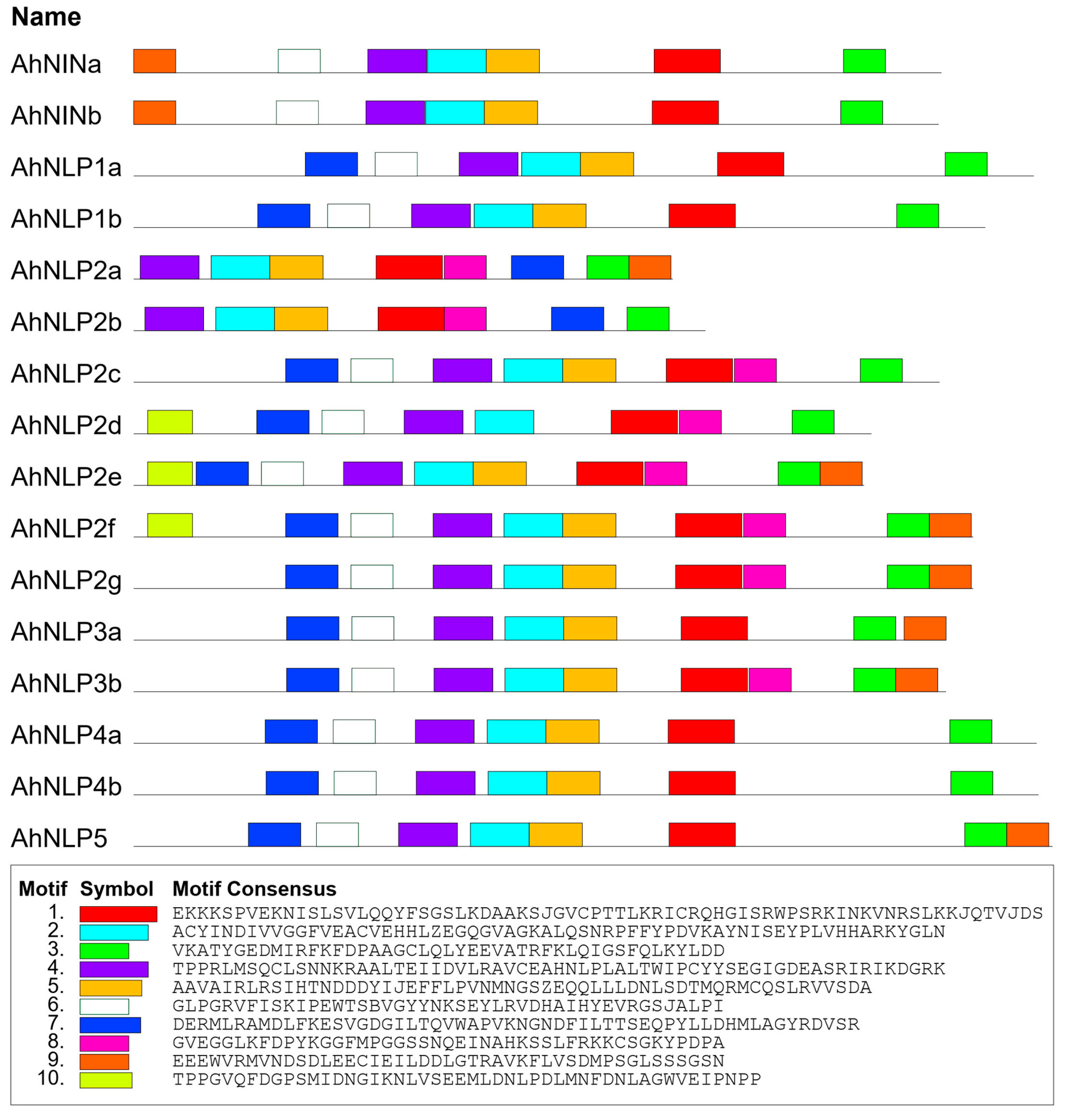
To obtain a better understanding of the protein characteristics of the AhNIN and AhNLP, a MEME search was executed to analyze the conserved motifs and domains in AhNIN and AhNLP proteins. Based on the length of the motifs and domains, the AhNIN and AhNLP proteins all contained ten different motifs/domains (Figure 2 and Figure S2). The motifs 1, 2, 3, and 4 were identified as the most conserved motif, which was present in every AhNIN and AhNLP protein. It was reported that RWP-RK domain and PB1 domain are distinctive domains of NLP genes [37,38]. Sequence alignment revealed that all the AhNIN and AhNLP proteins contain the conserved RWP-RK domain (motif 1) and the PB1 domain (motif 3) (Figure S3). However, motif 10 was only identified in the N-terminal of AhNLP2d, AhNLP2e, and AhNLP2f (Figure 2), indicating that this motif is related to the functions of these AhNLP proteins.

3.4. Gene Structure of the AhNIN and AhNLP Genes

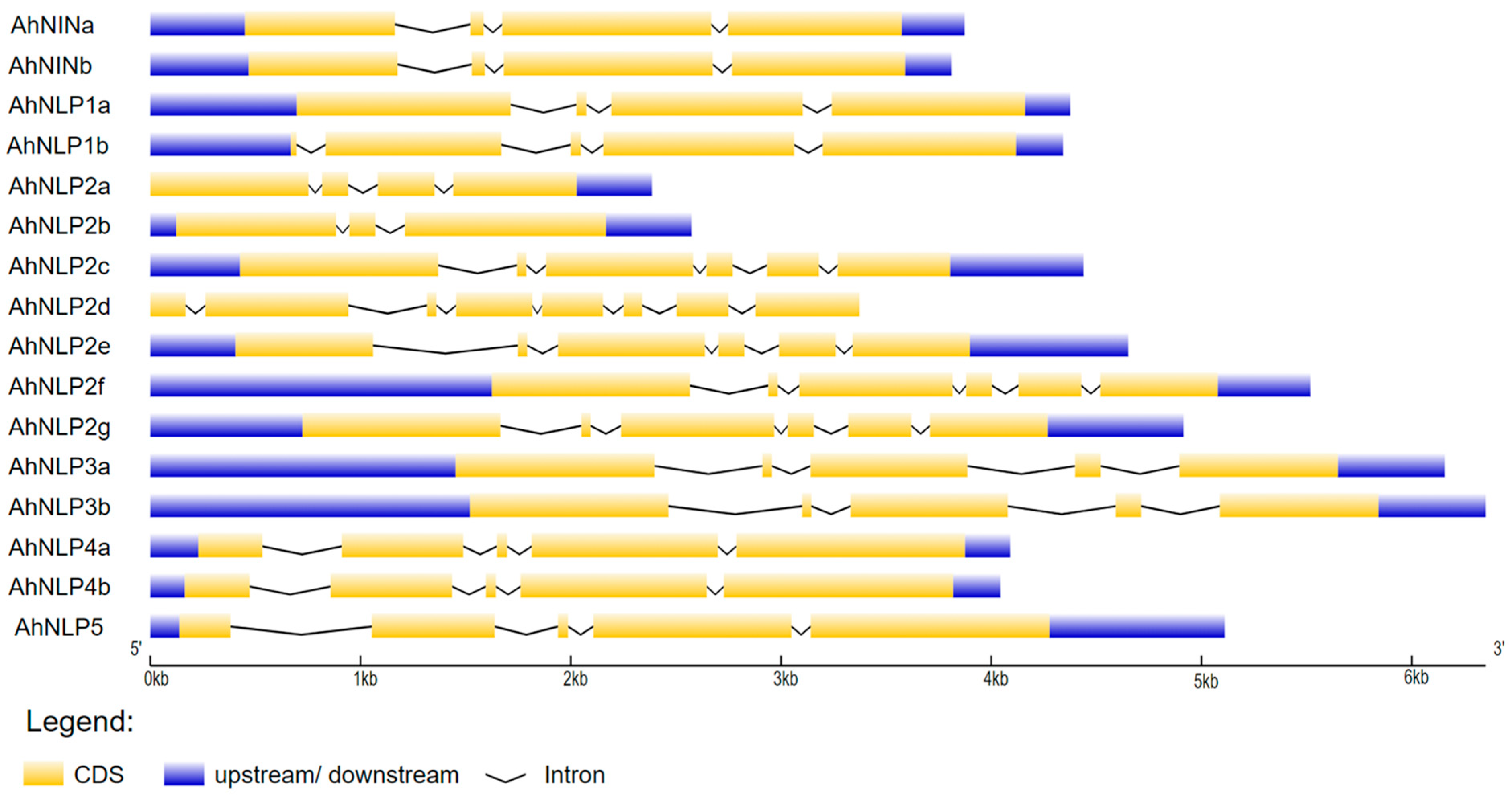
To further understand the structural diversity of the peanut AhNIN and AhNLP genes, the gene exon–intron organization analysis was carried out using the online Gene Structure Display Server tool (http://gsds.cbi.pku.edu.cn/, accessed on 12 May 2025) (Figure 3). The exon–intron structures of the 2 AhNIN and 14 AhNLP genes were generated by the alignment of their gene coding sequences with their corresponding genomic sequences. Gene structure analysis showed that the AhNLP2d gene has no 5′ upstream and 3′ downstream sequences, and AhNLP2a gene has no 5′ upstream sequence. Furthermore, the clade 2 NLP genes, AhNLP4a, AhNLP4b, and AhNLP5, have similar exon–intron organization. However, the exon numbers in clade 3 (the AhNLP2 and AhNLP3 clade) NLP genes changed from three to eight, indicating that these AhNLP2 and AhNLP3 genes may play differential functions during peanut growth and development.

3.5. Expression Profiles of the AhNIN and AhNLP Genes

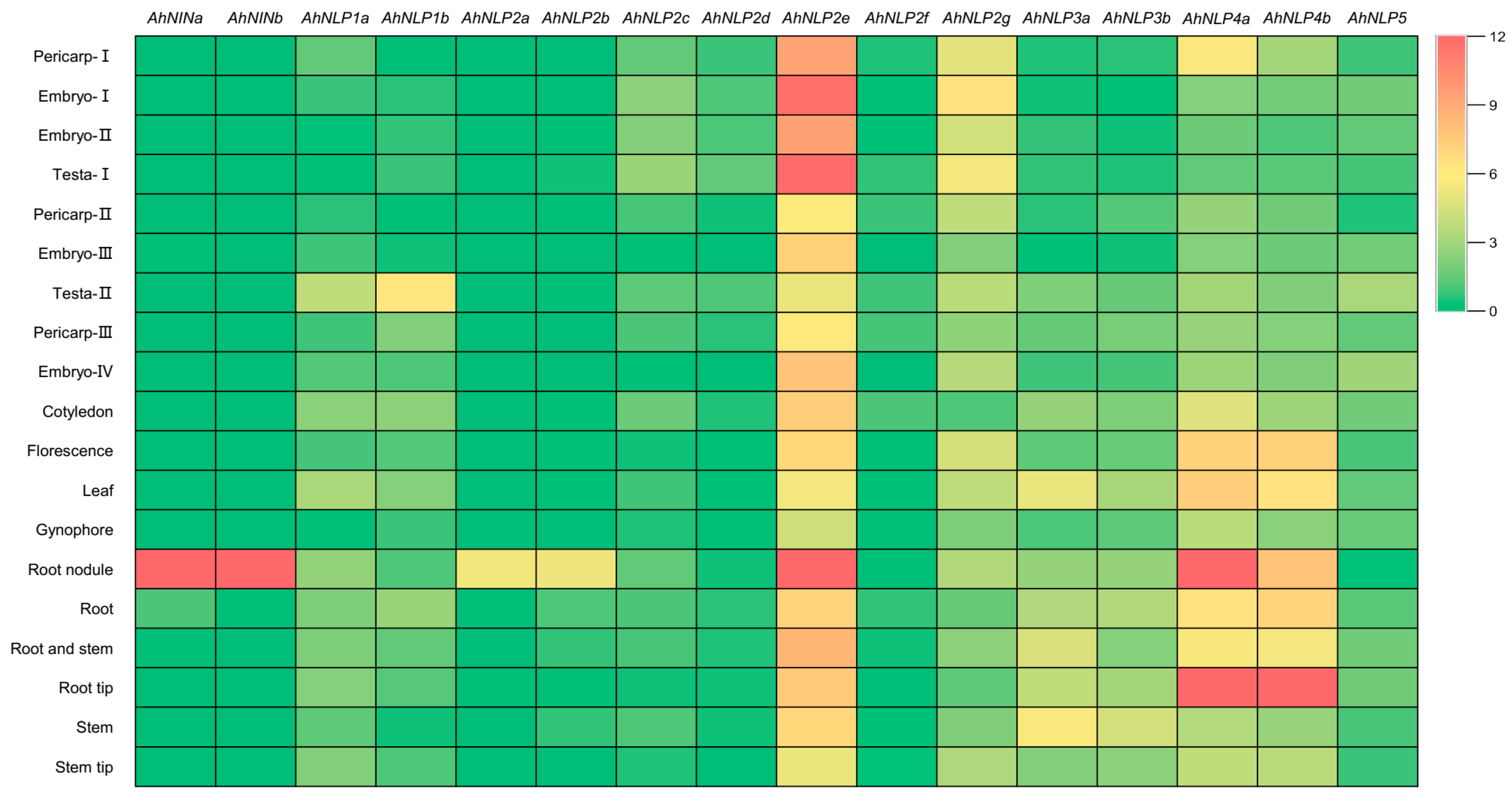
To examine the expression patterns of AhNIN and AhNLP genes, the expression profiles of the AhNIN and AhNLP genes in different organs and tissues of cultivated peanut variety “Shitouqi” were analyzed using publicly available transcriptome sequencing data (http://peanutgr.fafu.edu.cn, accessed on 1 April 2024) (Figure 4). The heat map showed that the AhNLP2e, AhNLP2g, AhNLP4a, and AhNLP4b genes were highly expressed in all tissues or organs, suggesting that they may play important roles in peanut growth and development while the other AhNLP genes were relatively lower-expressed in all tissues or organs. However, AhNINa and AhNINb were specifically highly expressed in root nodules, indicating that AhNINa and AhNINb may play crucial roles in nodule formation and development in peanut.

3.6. Nodule-Specific Expression Patterns of AhNINa and AhNINb

To gain better insights into the molecular functions of AhNINa and AhNINb genes in peanut nodule development, the expression patterns of both AhNINa and AhNINb were analyzed in detail. First, the relative expression levels of AhNINa and AhNINb in nodules at different developmental stages were measured. Nodules were harvested at 7, 14, and 21 day post-inoculation (dpi) with rhizobia strain, and roots without inoculation with rhizobia were used as the control (0 dpi). Due to the high sequence identity between AhNINa and AhNINb genes, we failed to design specific reverse transcription quantitative PCR (RT-qPCR) primers for individual AhNINa and AhNINb genes, and quantified consolidated expression of both AhNINs. RT-qPCR data showed that AhNINs were highly expressed at different developmental stages of nodules, compared with those in the root of 0 dpi (Figure 5A). Second, to analyze the expression patterns of AhNINa and AhNINb in nodules in more detail, the proAhNINa:GUS and proAhNINb:GUS hairy root transgenic plants were obtained. GUS staining showed that AhNINa and AhNINb showed a similar expression pattern and were specifically expressed in nodules (Figure 5B–E). These results demonstrate that both AhNINa and AhNINb are specifically expressed in nodules and may be involved in the development of peanut nodules.

3.7. Both AhNINa and AhNINb Act as Transcriptional Activators

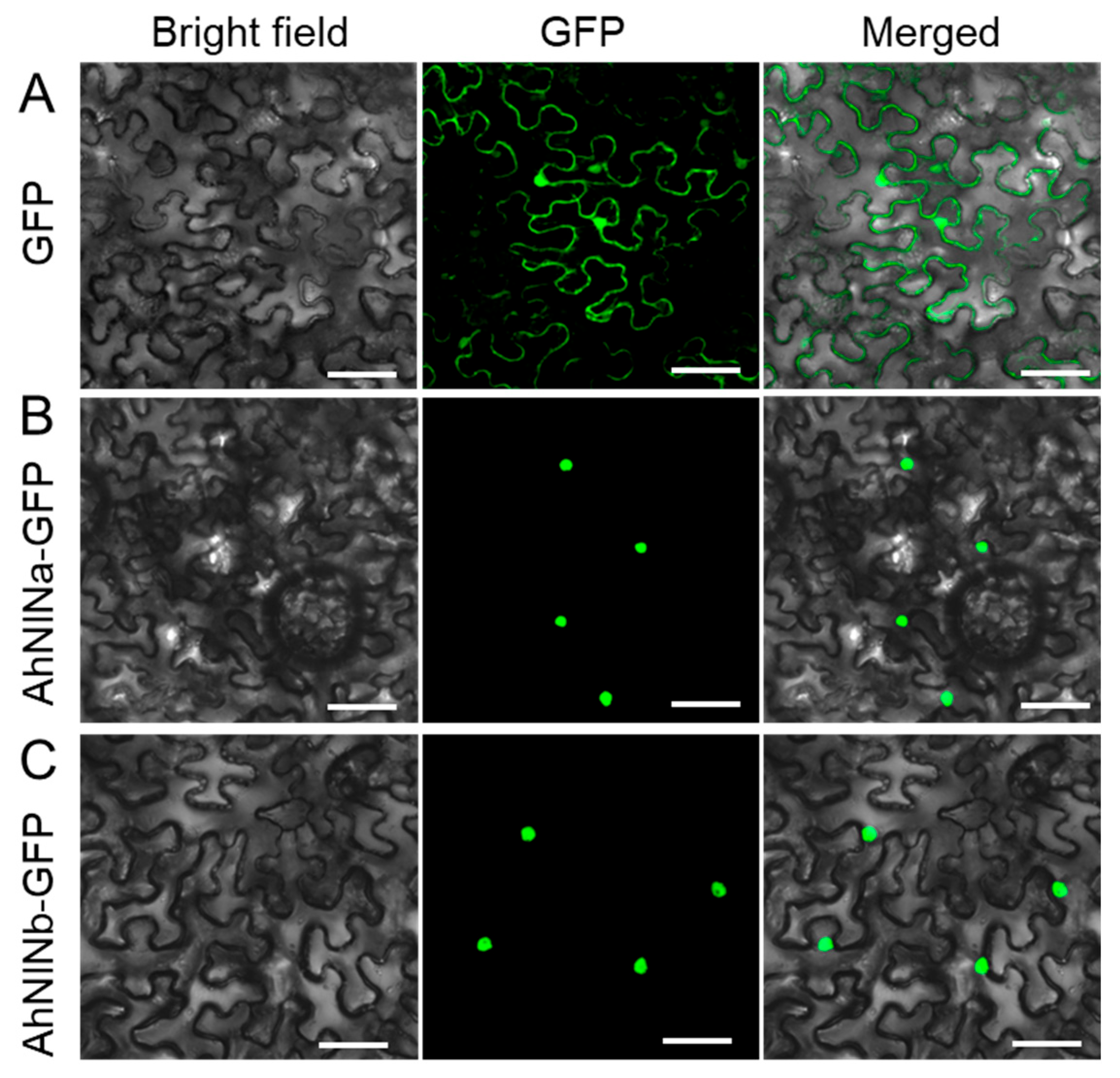
To further evaluate the possible function of AhNINa and AhNINb genes in peanut nodule development, the subcellular localization analysis of the AhNINa and AhNINb genes was performed. AhNINa and AhNINb were, respectively, fused with GFP and transformed into tobacco epidermal cells. Green fluorescence signal observation showed that compared with free GFP which is located in the cell nucleus and cytoplasm (Figure 6A), both AhNINa and AhNINb were located in the cell nucleus (Figure 6B,C), demonstrating that they are nuclear-localized transcription factors.
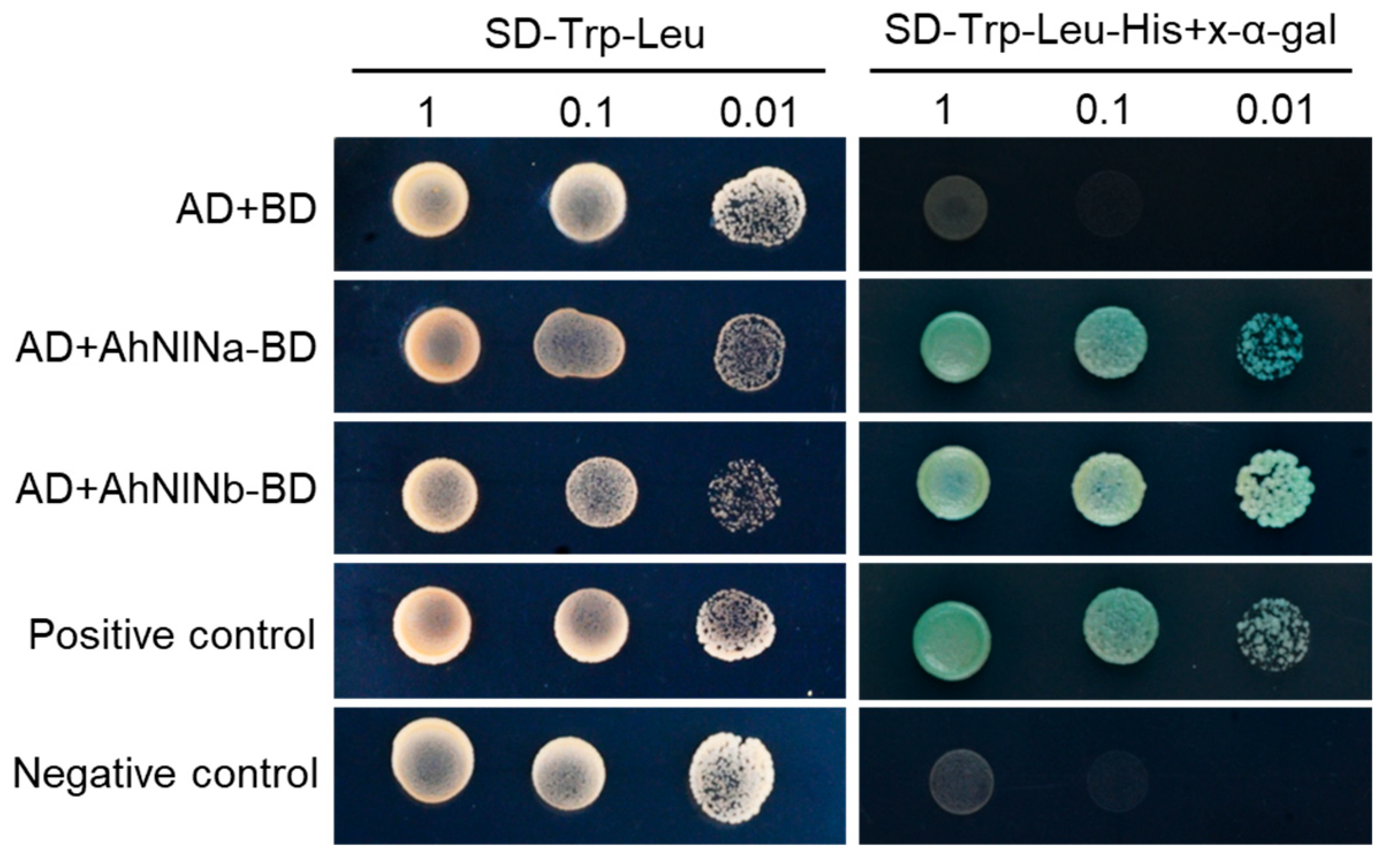
To assess the possible transcriptional activity of AhNINa and AhNINb proteins, yeast transactivation assays were performed. First, the AhNINa and AhNINb were fused with the GAL4 DNA-binding domain (BD) to generate the AhNINa:BD and AhNINb:BD constructions, respectively. Then, the GAL4 DNA-activation domain (AD) vector was cotransformed with AhNINa:BD or AhNINb:BD into yeast cells. Compared to the AD+BD control, the AhNINa or AhNINb protein alone can activate transcription (Figure 7). These results suggest that AhNINa and AhNINb may function as transcriptional activators to regulate the nodule development in peanut.

3.8. AhNINa and AhNINb Are Required for Nodule Formation in Peanut

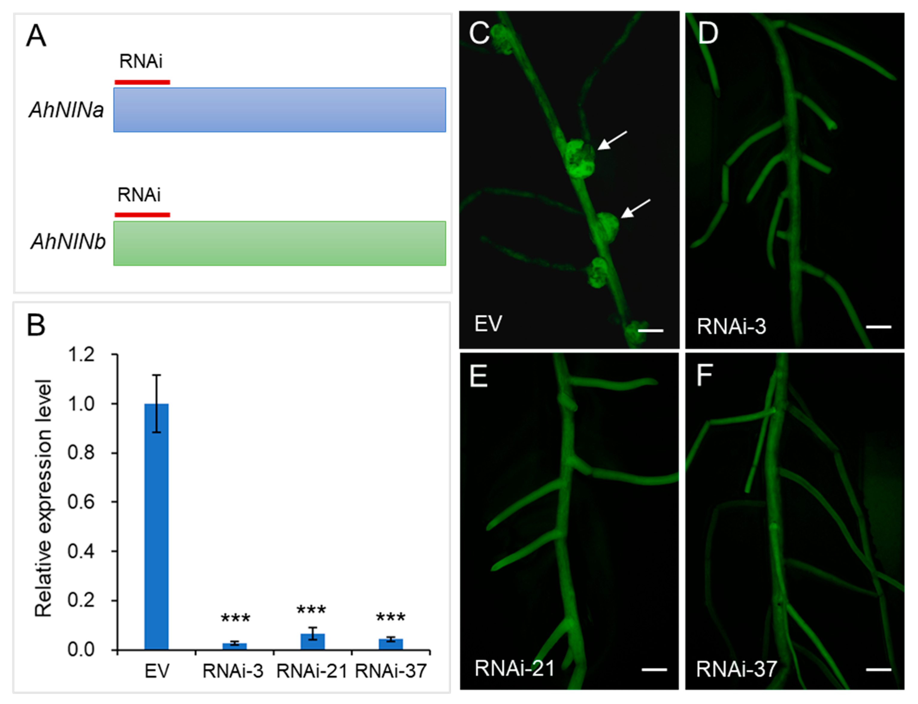
To determine whether AhNINa and AhNINb genes are involved in peanut nodule development, we knocked down the expression of AhNINa and AhNINb using an RNAi construct that targets the common sequence of both AhNINa and AhNINb (Figure 8A and Figure S4). RT-qPCR analysis showed that the expression of AhNINs was reduced by 92–96% in the RNAi roots compared to the control (EV) (Figure 8B). The root nodule phenotype observation indicated that, compared to the EV control which has nodules (Figure 8C), the AhNINs-RNAi transgenic hairy roots with significantly lower gene expression levels had no nodule formation (Figure 8B,D–F). These results demonstrate that both AhNINa and AhNINb genes are functional transcription factors and are necessary for nodule formation in peanut.

4. Discussion

Previous studies have identified 9 AtNLPs in Arabidopsis (A. thaliana) [12], 6 OsNLPs in rice (Oryza Sativa L.) [16], 6 MtNLPs in Medicago (M. truncatula) [39], 9 ZmNLPs in maize (Zea mays L.) [40] and 18 TaNLPs in wheat (Triticum aestivum L.) [41]. However, the information and function of the NLP genes in peanuts remain largely unknown. In this study, a total of 16 AhNLPs were identified in peanut genomes based on the annotation information. Compared with the number of NLP genes in Arabidopsis, rice, maize and Medicago, which are all diploid species, the number of the peanut NLP gene family members is about twice that of each species, suggesting that the NLP gene family of peanut may have undergone significant gene expansion (Table 1; Figure 1). The cultivated peanut (Arachis hypogaea L.) is of hybrid origin (two sets of ten chromosomes; mostly 2n = 2× = 20 chromosomes) and has a polyploid genome that contains essentially complete sets of chromosomes from two ancestral species [42,43]. The number of NLP genes in peanut was similar to that in hexaploidy wheat, indicating that the increase in the number of NLP genes in peanut may be due to polyploidization. In peanut, the Medicago MtNLP2 gene has seven orthologs, from AhNLP2a to AhNLP2g, implying that these genes may play a redundant role or gain some special functions in regulating the growth and development of peanut.
Phylogenetic and gene structure analysis showed that the peanut NLP family members belong to three different clades (Figure 1) and genes in the same clade display similar gene structures, except clade 3 (Figure 3). It was reported that clade 2 and 3 NLP genes originated from mosses and green algae, respectively, while group 1 NLPs appeared after the separation of monocots and dicots [6]. In Arabidopsis, the group 3 NLP gene, AtNLP7, is regulated by nitrate, and then AtNLP7 induces the expression of nitrate signaling and assimilation-related genes, such as AtNRT2.1 and NITRITE REDUCTASE (AtNIR) [12,19]. In peanut, AhNLP4a, AhNLP4a, and AhNLP5 are the closest orthologs to AtNLP6 and AtNLP7, indicating that they may play a similar function with AtNLP6 and AtNLP7, and participate in the nitrogen-regulated plant growth and development. The RWP-RK and PB1 domains enable NLP to function in multiple aspects of nitrogen metabolism, including nitrogen response, transcriptional regulation, and signal transduction [44,45]. All the AhNLP proteins contain the two conserved domains, RWP-RK and PB1 (Figure 2), which are consistent with the structural characteristics of NLP proteins from pepper (Capsicum annuum L.) [46] and Chrysanthemum lavandulifolium [47].
Transcription factors (TFs) are important regulators that control gene expression in the plant body and play important roles in cell signaling transduction and stress responses [48]. NLPs are essential TFs involved in nitrate sensing/response, symbiosis, and abiotic stress [49,50,51]. Predicted subcellular localization results showed that the AhNLPs genes were localized in the nucleus or/and chloroplasts (Table 1). In this study, the subcellular localization patterns of AhNINa and AhNINb were analyzed. AhNINa and AhNINb were located in the cell nucleus (Figure 6), demonstrating that they are nuclear-localized transcription factors.
Unlike most other plants, legumes can form root nodules in association with rhizobia, which can fix atmospheric N2 into ammonia [52,53,54,55,56]. In peanut, transcriptomic analysis showed that AhNLP2e was widely expressed in all tested tissues including the embryo, testa, and root nodules (Figure 4). In addition, AhNLP4a and AhNLP4b were highly expressed in the root tip and root nodule (Figure 4), which suggests potential functional redundancy and co-regulation. Moreover, the transactivation assay proved that AhNINa and AhNINb act as a transcriptional activator (Figure 7), indicating that they may play a positive role in peanut nodule formation. Interestingly, phylogenomic analyses revealed that the transcription factor NIN is essential for nodulation in nitrogen-fixation-clade species [57,58]. In L. japonicus and P. sativum, the expressions of LjNIN and PsNIN were detected in young and mature nodules, and the loss of the single LjNIN or PsNIN function leads to abolished nodule initiation [7,10]. Consistent with L. japonicus and P. sativum, AhNINa and AhNINb are also expressed in root nodules (Figure 4 and Figure 5A). Furthermore, the promoter-GUS staining analysis confirmed the nodule-specific expression patterns of AhNINa and AhNINb (Figure 5B–E). It was reported that AhNINa and AhNINb genes are expressed in early developmental stage nodules, such as 1, 3, and 4 dpi nodules, and play important roles during nodule initiation [59]. In this study, we found that AhNINa and AhNINb genes are also expressed in developing nodules, such as 7 and 14 dpi nodules. In soybean (Glycine max), there are four putative orthologous NIN genes, GmNIN1a, GmNIN1b, GmNIN2a, and GmNIN2b, and the Gmnin1a nin1b nin2a nin2b quadruple mutant displayed defects in root nodule formation [8]. However, there is no genetic evidence that AhNINa and AhNINb genes are involved in peanut nodule formation. In this study, the down-regulation of the expression of both AhNINa and AhNINb by RNAi also results in peanut defects in nodule formation (Figure 8), demonstrating that AhNINa and AhNINb genes play vital roles in the nodule development of peanut. Our work provides insight into the understanding of the redundant function of AhNINa and AhNINb genes in peanut nodule development.

5. Conclusions

This study provides the genome-wide characterization of the NLP family members in peanuts. A total of 16 NLP genes were identified, including 2 AhNIN and 14 AhNLP, and their chromosome locations, phylogenetic relationship, and gene function, especially the function of AhNINa and AhNINb genes, were analyzed. Both AhNINa and AhNINb genes are specifically expressed in root nodules and the silencing of AhNINa and AhNINb by RNA interference (RNAi) led to the failure of nodule formation. Future work should focus on verifying whether increasing the expression of AhNINa and AhNINb can produce more nodules and incorporate them into breeding application strategies to improve nitrogen fixation efficiency.

Supplementary Materials

The following supporting information can be downloaded at https://www.mdpi.com/article/10.3390/agronomy15102377/s1, Figure S1: Chromosome distribution of AhNIN and AhNLP genes in A. hypogaea. Figure S2: Sequences of the ten identified motifs in AhNIN and AhNLP proteins. Figure S3: Multiple sequences alignment of the AhNIN and AhNLP protein members. Figure S4: AhNINa and AhNINb gene CDS alignment. Table S1: Primers used in this study. Data S1: Protein sequences of MtNIN, MtNLP, AtNLP, AhNLP, and AhNIN used to run the BLAST analysis and used to construct the phylogenetic tree.

Author Contributions

Conceptualization, H.W. (Hongfeng Wang); methodology, Y.R., L.W. and F.Z.; software, G.C., Z.L., Y.T. and Y.W.; investigation, H.W. (Hongfeng Wang), Y.X. and H.W. (Hongwei Wang); writing—original draft preparation, H.W. (Hongfeng Wang) and H.W. (Hongwei Wang); writing—review and editing, H.W. (Hongfeng Wang) and M.Y.; supervision, H.W. (Hongfeng Wang) and M.Y. All authors have read and agreed to the published version of the manuscript.

Funding

This research was funded by the Agricultural Scientific and Technological Innovation Project of Shandong Academy of Agricultural Sciences (CXGC2025C19 and CXGC2025F19), the Taishan Scholars Program, the National Natural Science Foundation of China (32570963, 32201446, 32370336, 32401963 and 32500723), the Natural Science Foundation of Shandong Province (ZR2022QC109, ZR2024QC300 and ZR2022QC005) and the Postdoctoral Fellowship Program of CPSF (GZC20240930).

Data Availability Statement

The original contributions presented in this study are included in the article and Supplementary Materials. Further inquiries can be directed to the corresponding authors.

Conflicts of Interest

The authors declare no conflicts of interest.

Abbreviations

The following abbreviations are used in this manuscript:
NINNODULE INCEPTION
NRENitrate-responsive cis-element
ANR1ARABIDOPSIS NITRATE REGULATED 1
NRT2NITRATE TRANSPORTER 2
LBD37LATERAL ORGAN BOUNDARY DOMAIN 37
AtTCP20TEOSINTE BRANCHED1/CYCLOIDEA/PROLIFERATING CELL FACTOR1-20
CYP707A2CYTOCHROME P450 MONOOXYGENASE 707A2
NFR5NOD FACTOR RECEPTOR 5
CCaMKCa2+/CALMODULIN DEPENDENT KINASE
HK1HISTIDINE KINASE 1
NSP2NODULATION SIGNALING PATHWAY 2
NIRNITRITE REDUCTASE
RNAiRNA interference

References

  1. Tegeder, M.; Masclaux-Daubresse, C. Source and sink mechanisms of nitrogen transport and use. New Phytol. 2018, 217, 35–53. [Google Scholar] [CrossRef] [PubMed]
  2. Crawford, N.M. Nitrate: Nutrient and signal for plant growth. Plant Cell 1995, 7, 859–868. [Google Scholar]
  3. Mueller, N.D.; West, P.C.; Gerber, J.S.; MacDonald, G.K.; Polasky, S.; Foley, J.A. A tradeoff frontier for global nitrogen use and cereal production. Environ. Res. Lett. 2014, 9, 054002. [Google Scholar] [CrossRef]
  4. Choi, S.J.; Lee, Z.; Jeong, E.; Kim, S.; Seo, J.S.; Um, T.; Shim, J.S. Signaling pathways underlying nitrogen transport and metabolism in plants. BMB Rep. 2023, 56, 56–64. [Google Scholar] [CrossRef]
  5. Liu, J.; Bisseling, T. Evolution of NIN and NIN-like genes in relation to nodule symbiosis. Genes 2020, 11, 777. [Google Scholar] [CrossRef]
  6. Mu, X.; Luo, J. Evolutionary analyses of NIN-like proteins in plants and their roles in nitrate signaling. Cell. Mol. Life Sci. 2019, 76, 3753–3764. [Google Scholar] [CrossRef] [PubMed]
  7. Schauser, L.; Roussis, A.; Stiller, J.; Stougaard, J. A plant regulator controlling development of symbiotic root nodules. Nature 1999, 402, 191–195. [Google Scholar] [CrossRef]
  8. Fu, M.; Sun, J.; Li, X.; Guan, Y.; Xie, F. Asymmetric redundancy of soybean Nodule Inception (NIN) genes in root nodule symbiosis. Plant Physiol. 2022, 188, 477–489. [Google Scholar] [CrossRef] [PubMed]
  9. Marsh, J.F.; Rakocevic, A.; Mitra, R.M.; Brocard, L.; Sun, J.; Eschstruth, A.; Long, S.R.; Schultze, M.; Ratet, P.; Oldroyd, G.E. Medicago truncatula NIN is essential for rhizobial-independent nodule organogenesis induced by autoactive calcium/calmodulin-dependent protein kinase. Plant Physiol. 2007, 144, 324–335. [Google Scholar] [CrossRef]
  10. Borisov, A.Y.; Madsen, L.H.; Tsyganov, V.E.; Umehara, Y.; Voroshilova, V.A.; Batagov, A.O.; Sandal, N.; Mortensen, A.; Schauser, L.; Ellis, N.; et al. The sym35 gene required for root nodule development in pea is an ortholog of Nin from Lotus japonicus. Plant Physiol. 2003, 131, 1009–1017. [Google Scholar] [CrossRef]
  11. Alvarez, J.M.; Schinke, A.L.; Brooks, M.D.; Pasquino, A.; Leonelli, L.; Varala, K.; Safi, A.; Krouk, G.; Krapp, A.; Coruzzi, G.M. Transient genome-wide interactions of the master transcription factor NLP7 initiate a rapid nitrogen-response cascade. Nat. Commun. 2020, 11, 1157. [Google Scholar] [CrossRef]
  12. Konishi, M.; Yanagisawa, S. Arabidopsis NIN-like transcription factors have a central role in nitrate signalling. Nat. Commun. 2013, 4, 1617. [Google Scholar] [CrossRef]
  13. Chardin, C.; Girin, T.; Roudier, F.; Meyer, C.; Krapp, A. The plant RWP-RK transcription factors: Key regulators of nitrogen responses and of gametophyte development. J. Exp. Bot. 2014, 65, 5577–5587. [Google Scholar] [CrossRef]
  14. Yu, J.; Yuan, Y.; Dong, L.; Cui, G. Genome-wide investigation of NLP gene family members in alfalfa (Medicago sativa L.): Evolution and expression profiles during development and stress. BMC Genom. 2023, 24, 320. [Google Scholar] [CrossRef]
  15. Liu, M.; Chang, W.; Fan, Y.; Sun, W.; Qu, C.; Zhang, K.; Liu, L.; Xu, X.; Tang, Z.; Li, J.; et al. Genome-wide identification and characterization of NODULE-INCEPTION-Like Protein (NLP) family genes in Brassica napus. Int. J. Mol. Sci. 2018, 19, 2270. [Google Scholar] [CrossRef] [PubMed]
  16. Jagadhesan, B.; Sathee, L.; Meena, H.S.; Jha, S.K.; Chinnusamy, V.; Kumar, A.; Kumar, S. Genome wide analysis of NLP transcription factors reveals their role in nitrogen stress tolerance of rice. Sci. Rep. 2020, 10, 9368. [Google Scholar] [CrossRef] [PubMed]
  17. Konishi, M.; Yanagisawa, S. An NLP-binding site in the 3′ flanking region of the nitrate reductase gene confers nitrate-inducible expression in Arabidopsis thaliana (L.) Heynh. Soil Sci. Plant Nutr. 2013, 59, 612–620. [Google Scholar] [CrossRef]
  18. Castaings, L.; Camargo, A.; Pocholle, D.; Gaudon, V.; Texier, Y.; Boutet-Mercey, S.; Taconnat, L.; Renou, J.P.; Daniel-Vedele, F.; Fernandez, E.; et al. The nodule inception-like protein 7 modulates nitrate sensing and metabolism in arabidopsis. Plant J. 2009, 57, 426–435. [Google Scholar] [CrossRef]
  19. Marchive, C.; Roudier, F.; Castaings, L.; Bréhaut, V.; Blondet, E.; Colot, V.; Meyer, C.; Krapp, A. Nuclear retention of the transcription factor NLP7 orchestrates the early response to nitrate in plants. Nat. Commun. 2013, 4, 1713. [Google Scholar] [CrossRef]
  20. Yu, L.H.; Wu, J.; Tang, H.; Yuan, Y.; Wang, S.M.; Wang, Y.P.; Zhu, Q.S.; Li, S.G.; Xiang, C.B. Overexpression of Arabidopsis NLP7 improves plant growth under both nitrogen-limiting and -sufficient conditions by enhancing nitrogen and carbon assimilation. Sci. Rep. 2016, 6, 27795. [Google Scholar] [CrossRef] [PubMed]
  21. Guan, P.; Ripoll, J.J.; Wang, R.; Vuong, L.; Bailey-Steinitz, L.J.; Ye, D.; Crawford, N.M. Interacting TCP and NLP transcription factors control plant responses to nitrate availability. Proc. Natl. Acad. Sci. USA 2017, 114, 2419–2424. [Google Scholar] [CrossRef] [PubMed]
  22. Yan, D.; Easwaran, V.; Chau, V.; Okamoto, M.; Ierullo, M.; Kimura, M.; Endo, A.; Yano, R.; Pasha, A.; Gong, Y.; et al. NIN-like protein 8 is a master regulator of nitrate-promoted seed germination in Arabidopsis. Nat. Commun. 2016, 7, 13179. [Google Scholar] [CrossRef]
  23. Konishi, M.; Okitsu, T.; Yanagisawa, S. Nitrate-responsive NIN-like protein transcription factors perform unique and redundant roles in Arabidopsis. J. Exp. Bot. 2021, 72, 5735–5750. [Google Scholar] [CrossRef]
  24. Gage, D.J. Infection and invasion of roots by symbiotic, nitrogen-fixing rhizobia during nodulation of temperate legumes. Microbiol. Mol. Biol. Rev. 2004, 68, 280–300. [Google Scholar] [CrossRef]
  25. Delamuta, J.R.; Ribeiro, R.A.; Menna, P.; Bangel, E.V.; Hungria, M. Multilocus sequence analysis (MLSA) of bradyrhizobium strains: Revealing high diversity of tropical diazotrophic symbiotic bacteria. Braz. J. Microbiol. 2012, 43, 698–710. [Google Scholar] [CrossRef]
  26. Chen, J.Y.; Gu, J.; Wang, E.T.; Ma, X.X.; Kang, S.T.; Huang, L.Z.; Cao, X.P.; Li, L.B.; Wu, Y.L. Wild peanut Arachis duranensis are nodulated by diverse and novel bradyrhizobium species in acid soils. Syst. Appl. Microbiol. 2014, 37, 525–532. [Google Scholar] [CrossRef]
  27. Muñoz, V.; Ibañez, F.; Tonelli, M.L.; Valetti, L.; Anzuay, M.S.; Fabra, A. Phenotypic and phylogenetic characterization of native peanut bradyrhizobium isolates obtained from córdoba, argentina. Syst. Appl. Microbiol. 2011, 34, 446–452. [Google Scholar] [CrossRef]
  28. Horta Araújo, N.; Nouwen, N.; Arrighi, J.F. Nodulating another way: What can we learn from lateral root base nodulation in legumes? J. Exp. Bot. 2024, 75, 3214–3219. [Google Scholar] [CrossRef]
  29. Sharma, V.; Bhattacharyya, S.; Kumar, R.; Kumar, A.; Ibañez, F.; Wang, J.; Guo, B.; Sudini, H.K.; Gopalakrishnan, S.; DasGupta, M.; et al. Molecular basis of root nodule symbiosis between bradyrhizobium and ‘crack-entry’ legume groundnut (Arachis hypogaea L.). Plants 2020, 9, 276. [Google Scholar] [CrossRef] [PubMed]
  30. Shu, H.; Luo, Z.; Peng, Z.; Wang, J. The application of CRISPR/Cas9 in hairy roots to explore the functions of AhNFR1 and AhNFR5 genes during peanut nodulation. BMC Plant Biol. 2020, 20, 417. [Google Scholar] [CrossRef] [PubMed]
  31. Peng, Z.; Chen, H.; Tan, L.; Shu, H.; Varshney, R.K.; Zhou, Z.; Zhao, Z.; Luo, Z.; Chitikineni, A.; Wang, L.; et al. Natural polymorphisms in a pair of NSP2 homoeologs can cause loss of nodulation in peanut. J. Exp. Bot. 2021, 72, 1104–1118. [Google Scholar] [CrossRef]
  32. Kundu, A.; DasGupta, M. Silencing of putative Cytokinin Receptor Histidine Kinase1 inhibits both inception and differentiation of root nodules in Arachis hypogaea. Mol. Plant-Microbe Interact. 2018, 31, 187–199. [Google Scholar] [CrossRef] [PubMed]
  33. Kereszt, A.; Li, D.; Indrasumunar, A.; Nguyen, C.D.; Nontachaiyapoom, S.; Kinkema, M.; Gresshoff, P.M. Agrobacterium rhizogenes-mediated transformation of soybean to study root biology. Nat. Protoc. 2007, 2, 948–952. [Google Scholar] [CrossRef]
  34. Reddy, D.S.; Bhatnagar-Mathur, P.; Cindhuri, K.S.; Sharma, K.K. Evaluation and validation of reference genes for normalization of quantitative real-time pcr based gene expression studies in peanut. PLoS ONE 2013, 8, e78555. [Google Scholar] [CrossRef]
  35. Earley, K.W.; Haag, J.R.; Pontes, O.; Opper, K.; Juehne, T.; Song, K.; Pikaard, C.S. Gateway-compatible vectors for plant functional genomics and proteomics. Plant J. 2006, 45, 616–629. [Google Scholar] [CrossRef]
  36. Karimi, M.; Inzé, D.; Depicker, A. Gateway vectors for agrobacterium-mediated plant transformation. Trends Plant Sci. 2002, 7, 193–195. [Google Scholar] [CrossRef]
  37. Konishi, M.; Yanagisawa, S. The role of protein-protein interactions mediated by the PB1 domain of NLP transcription factors in nitrate-inducible gene expression. BMC Plant Biol. 2019, 19, 90. [Google Scholar] [CrossRef]
  38. Sakuraba, Y.; Zhuo, M.; Yanagisawa, S. RWP-RK domain-containing transcription factors in the Viridiplantae: Biology and phylogenetic relationships. J. Exp. Bot. 2022, 73, 4323–4337. [Google Scholar] [CrossRef]
  39. Lin, J.S.; Li, X.; Luo, Z.; Mysore, K.S.; Wen, J.; Xie, F. NIN interacts with NLPs to mediate nitrate inhibition of nodulation in Medicago truncatula. Nat. Plants 2018, 4, 942–952. [Google Scholar] [CrossRef] [PubMed]
  40. Ge, M.; Liu, Y.; Jiang, L.; Wang, Y.; Lv, Y.; Zhou, L.; Liang, S.; Bao, H.; Zhao, H. Genome-wide analysis of maize NLP transcription factor family revealed the roles in nitrogen response. Plant Growth Regul. 2018, 84, 95–105. [Google Scholar] [CrossRef]
  41. Kumar, A.; Batra, R.; Gahlaut, V.; Gautam, T.; Kumar, S.; Sharma, M.; Tyagi, S.; Singh, K.P.; Balyan, H.S.; Pandey, R.; et al. Genome-wide identification and characterization of gene family for RWP-RK transcription factors in wheat (Triticum aestivum L.). PLoS ONE 2018, 13, e0208409. [Google Scholar] [CrossRef]
  42. Pandey, M.K.; Monyo, E.; Ozias-Akins, P.; Liang, X.; Guimarães, P.; Nigam, S.N.; Upadhyaya, H.D.; Janila, P.; Zhang, X.; Guo, B.; et al. Advances in Arachis genomics for peanut improvement. Biotechnol. Adv. 2012, 30, 639–651. [Google Scholar] [CrossRef] [PubMed]
  43. Bertioli, D.J.; Jenkins, J.; Clevenger, J.; Dudchenko, O.; Gao, D.; Seijo, G.; Leal-Bertioli, S.C.M.; Ren, L.; Farmer, A.D.; Pandey, M.K.; et al. The genome sequence of segmental allotetraploid peanut Arachis hypogaea. Nat. Genet. 2019, 51, 877–884. [Google Scholar] [CrossRef] [PubMed]
  44. Sámano, M.L.; Nanjareddy, K.; Arthikala, M.K. NIN-like proteins (NLPs) as crucial nitrate sensors: An overview of their roles in nitrogen signaling, symbiosis, abiotic stress, and beyond. Physiol. Mol. Biol. Plants 2024, 30, 1209–1223. [Google Scholar] [CrossRef] [PubMed]
  45. Yan, D.; Nambara, E. Conserved and unique functions of NIN-like proteins in nitrate sensing and signaling. Plant Sci. 2023, 336, 111842. [Google Scholar] [CrossRef]
  46. Wu, Y.; Su, S.X.; Wang, T.; Peng, G.H.; He, L.; Long, C.; Li, W. Identification and expression characteristics of NLP (NIN-like protein) gene family in pepper (Capsicum annuum L.). Mol. Biol. Rep. 2023, 50, 6655–6668. [Google Scholar] [CrossRef]
  47. Zhang, Q.; Li, J.; Wen, X.; Deng, C.; Yang, X.; Dai, S. Genome-wide identification and characterization analysis of RWP-RK family genes reveal their role in flowering time of Chrysanthemum lavandulifolium. BMC Plant Biol. 2023, 23, 197. [Google Scholar] [CrossRef]
  48. Liu, Q.; Zhang, G.; Chen, S. Structure and regulatory function of plant transcription factors. Chin. Sci. Bull. 2001, 46, 271–278. [Google Scholar] [CrossRef]
  49. Scheible, W.R.; Morcuende, R.; Czechowski, T.; Fritz, C.; Osuna, D.; Palacios-Rojas, N.; Schindelasch, D.; Thimm, O.; Udvardi, M.K.; Stitt, M. Genome-wide reprogramming of primary and secondary metabolism, protein synthesis, cellular growth processes, and the regulatory infrastructure of Arabidopsis in response to nitrogen. Plant Physiol. 2004, 136, 2483–2499. [Google Scholar] [CrossRef]
  50. Wang, Z.; Zhang, L.; Sun, C.; Gu, R.; Mi, G.; Yuan, L. Phylogenetic, expression and functional characterizations of the maize NLP transcription factor family reveal a role in nitrate assimilation and signaling. Physiol. Plant. 2018. ahead of print. [Google Scholar] [CrossRef]
  51. Lin, Z.; Guo, C.; Lou, S.; Jin, S.; Zeng, W.; Guo, Y.; Fang, J.; Xu, Z.; Zuo, Z.; Ma, L. Functional analyses unveil the involvement of moso bamboo (Phyllostachys edulis) group I and II NIN-LIKE PROTEINS in nitrate signaling regulation. Plant Sci. 2021, 306, 110862. [Google Scholar] [CrossRef]
  52. Yang, J.; Lan, L.; Jin, Y.; Yu, N.; Wang, D.; Wang, E. Mechanisms underlying legume-rhizobium symbioses. J. Integr. Plant Biol. 2022, 64, 244–267. [Google Scholar] [CrossRef]
  53. Roy, S.; Liu, W.; Nandety, R.S.; Crook, A.; Mysore, K.S.; Pislariu, C.I.; Frugoli, J.; Dickstein, R.; Udvardi, M.K. Celebrating 20 years of genetic discoveries in legume nodulation and symbiotic nitrogen fixation. Plant Cell 2020, 32, 15–41. [Google Scholar]
  54. Ferguson, B.J.; Mens, C.; Hastwell, A.H.; Zhang, M.; Su, H.; Jones, C.H.; Chu, X.; Gresshoff, P.M. Legume nodulation: The host controls the party. Plant Cell Environ. 2019, 42, 41–51. [Google Scholar] [CrossRef]
  55. Wang, Q.; Liu, J.; Zhu, H. Genetic and molecular mechanisms underlying symbiotic specificity in legume-rhizobium interactions. Front. Plant Sci. 2018, 9, 313. [Google Scholar] [CrossRef]
  56. Luo, Z.; Liu, H.; Xie, F. Cellular and molecular basis of symbiotic nodule development. Curr. Opin. Plant Biol. 2023, 76, 102478. [Google Scholar] [CrossRef] [PubMed]
  57. Griesmann, M.; Chang, Y.; Liu, X.; Song, Y.; Haberer, G.; Crook, M.B.; Billault-Penneteau, B.; Lauressergues, D.; Keller, J.; Imanishi, L.; et al. Phylogenomics reveals multiple losses of nitrogen-fixing root nodule symbiosis. Science 2018, 361, eaat1743. [Google Scholar] [CrossRef] [PubMed]
  58. van Velzen, R.; Holmer, R.; Bu, F.; Rutten, L.; van Zeijl, A.; Liu, W.; Santuari, L.; Cao, Q.; Sharma, T.; Shen, D.; et al. Comparative genomics of the nonlegume parasponia reveals insights into evolution of nitrogen-fixing rhizobium symbioses. Proc. Natl. Acad. Sci. USA 2018, 115, E4700–E4709. [Google Scholar] [CrossRef]
  59. Bhattacharjee, O.; Raul, B.; Ghosh, A.; Bhardwaj, A.; Bandyopadhyay, K.; Sinharoy, S. Nodule INception-independent epidermal events lead to bacterial entry during nodule development in peanut (Arachis hypogaea). New Phytol. 2022, 236, 2265–2281. [Google Scholar] [CrossRef] [PubMed]
Figure 1. Phylogenetic analysis of NIN and NLP genes in A. hypogaea (Ah), A. thaliana (At), and M. truncatula (Mt). The neighbor-joining (NJ) phylogenetic tree was constructed using 31 protein sequences from A. hypogaea (16), A. thaliana (9), and M. truncatula (6) in MEGA 7.0 with 1000 bootstrap replicates. The NIN and NLP genes in A. hypogaea, A. thaliana, and M. truncatula are classified with blue, pink, and green dots, respectively. The black rhombuses in the phylogenetic tree indicate the different clades.
Agronomy 15 02377 g001
Figure 2. Motif composition of the AhNIN and AhNLP proteins. The conserved motifs are predicted by online MEME website (https://meme-suite.org/meme/). The different motifs are represented by boxes with different colors. Motif 1 and motif 3 represent the RWP-RK domain and the PB1 domain.
Agronomy 15 02377 g002
Figure 3. The gene structures of the AhNIN and AhNLPs genes. The 5′ UTRs and 3′ UTRs are represented by blue boxes; the exons and introns are represented by yellow boxes and black folded lines. The scale was referred to the lengths of the genes. The exon–intron structures were identified by online Gene Structure Display Server tool (http://gsds.cbi.pku.edu.cn/, accessed on 12 May 2025).
Agronomy 15 02377 g003
Figure 4. Expression profile of AhNIN and AhNLP genes in different organs and tissues of peanut. The expression heat map was constructed based on FPKM values in the A. hypogaea transcriptome dataset (http://peanutgr.fafu.edu.cn, accessed on 1 April 2024). The red represents high expression levels and the green represents low expression levels.
Agronomy 15 02377 g004
Figure 5. Expression pattern of AhNINa and AhNINb genes in peanut nodules. (A) The relative expression levels of both AhNINa and AhNINb genes in different developmental stage nodules. Values are the means and SDs of three biological replicates; *** p < 0.001. The two-sided Student’s t-test was used to estimate if the difference is significant. (BE) Promoter-GUS fusion studies of AhNINa (B,C) and AhNINb (D,E) expression in hairy root transformed nodules at 7 and 14 dpi. Dpi, days post-inoculation. Scale bars, 500 μm in (BE).
Agronomy 15 02377 g005
Figure 6. Subcellular protein localization of AhNINa and AhNINb genes. (A) The subcellular localization of free GFP protein. (B,C) The subcellular localization of AhNINa-GFP (B) and AhNINb-GFP (C) fusion proteins. Both AhNINa-GFP and AhNINb-GFP fusion proteins were localized in the nucleus of tobacco epidermal cells as observed by confocal microscopy. Free GFP was used as a control. Scale bars = 50 μm.
Agronomy 15 02377 g006
Figure 7. Analysis of the transcriptional activity of AhNINa and AhNINb in yeast cells. The transcriptional activation activities of AhNINa and AhNINb were assessed by the growth of yeast cells on DDO (SD/-Leu-Trp) medium and TDO/X (SD/-His-Leu-Trp) medium supplemented with x-α-gal.
Agronomy 15 02377 g007
Figure 8. AhNINa and AhNINb are involved in symbiotic root nodule development in peanuts. (A) Schematic illustration of AhNINa and AhNINb genes encoding sequence and the red lines indicate the same RNAi sequence in both AhNINa and AhNINb. (B) RT-qPCR analysis shows the relative expression of the AhNINs genes in control (EV) and AhNINs-RNAi roots at 28 dpi. Values are the means and SDs of three technical replicates; *** p < 0.001. (CF) Fluorescence phenotype images of EV and AhNINs-RNAi roots at 28 dpi. Arrows in (C) mark the root nodules at the base of the lateral roots. Scale bars, 2 mm in (CF).
Agronomy 15 02377 g008
Table 1. Characterization of AhNIN and AhNLP genes in peanut.
Gene NameAccession NumberLength (aa)MW (kDa)pIPredicated Localization
AhNINaAH13G60030.186595.616.33Nucleus
AhNINbAH17G00510.186295.396.37Nucleus
AhNLP1aAH08G02220.1964107.485.77Nucleus
AhNLP1bAH17G22700.1912101.895.99Nucleus
AhNLP2aAH03G04320.157764.097.63Chloroplast
AhNLP2bAH13G06340.161267.847.65Chloroplast
AhNLP2cAH03G04310.186395.925.03Chloroplast/Nucleus
AhNLP2dAH03G04850.179087.874.86Chloroplast
AhNLP2eAH10G03170.178286.775.90Chloroplast
AhNLP2fAH13G04800.189999.325.23Chloroplast/Nucleus
AhNLP2gAH13G06330.189999.505.07Chloroplast/Nucleus
AhNLP3aAH08G11530.187096.796.78Nucleus
AhNLP3bAH18G02990.187096.856.68Nucleus
AhNLP4aAH09G24840.1967107.525.70Nucleus
AhNLP4bAH19G43090.1969107.845.75Nucleus
AhNLP5AH15G36260.1984109.255.48Nucleus
Disclaimer/Publisher’s Note: The statements, opinions and data contained in all publications are solely those of the individual author(s) and contributor(s) and not of MDPI and/or the editor(s). MDPI and/or the editor(s) disclaim responsibility for any injury to people or property resulting from any ideas, methods, instructions or products referred to in the content.