Article

Occurrence of Neopestalotiopsis clavispora Causing Apple Leaf Spot in China

1
Yantai Academy of Agricultural Sciences, Yantai 265500, China
2
Shandong Agricultural Technology Extension Center, Jinan 250100, China
*
Authors to whom correspondence should be addressed.
Agronomy 2024, 14(8), 1658; https://doi.org/10.3390/agronomy14081658
Submission received: 12 June 2024 / Revised: 23 July 2024 / Accepted: 24 July 2024 / Published: 29 July 2024
(This article belongs to the Section Pest and Disease Management)

Abstract

:
Leaf spot, a major apple disease, manifests in diverse symptoms. In this study, the pathogen was isolated from diseased ‘Yanfu 3’ apple leaves in Yantai, Shandong Province, and identified as Neopestalotiopsis clavispora through morphological observation, molecular identification, and multi-gene (ITS, TEF1α, and TUB2) phylogenetic analysis. Three isolates (YTNK01, YTNK02, and YTNK03) were selected for pathogenicity tests to verify Koch’s postulates. To our knowledge, this is the first report of N. clavispora being responsible for apple leaf spots in China, and the disease has been named ‘apple Neopestalotiopsis leaf spot’. Additionally, N. clavispora was found to infect crabapple, sweet cherry, grape, peach, and pear under laboratory conditions, indicating that these fruit trees may be potential hosts for N. clavispora in the field. The in vitro toxicity of ten fungicides to the pathogen was assessed using the mycelial growth rate method. All ten fungicides were effective in inhibiting the growth of N. clavispora. Among them, those based on pylocyanonitrile, propiconazole, pyraclostrobin, tebuconazole, diphenoxazole, and osthole showed higher toxicity to N. clavispora, with EC50 values of 0.11, 0.41, 0.47, 1.32, 1.85, and 3.82 µg/mL, respectively. These fungicides could be used as alternatives to prevent this disease in production. Overall, these findings provide valuable insights into the characteristics of N. clavispora causing apple leaf spot and are crucial for developing effective management strategies.

1. Introduction

Apple (Malus pumila Mill.) is cultivated worldwide, with China being one of the leading apple producers [1]. Yantai, located in the eastern part of Shandong Province, has maintained a leading position in apple production in China, with a planting area of approximately 160,000 hectares and an output of over 5.8 million tons in 2022 [2]. However, the occurrence of diseases poses a significant challenge to the sustainable and healthy development of the local apple industry. According to Zhang’s report, there are more than 100 types of apple leaf diseases, with leaf spot diseases being the most common [3]. The similarity of their symptoms makes them difficult to distinguish, and preventing these diseases becomes more challenging. Apple leaf spot diseases have been increasing year by year in Shandong, Shaanxi, Shanxi, Gansu, and Henan provinces. These diseases can weaken tree growth and affect flower bud differentiation. Currently, the pathogens of apple leaf spot in China primarily include apple Alternaria blotch (Alternaria alternata (Fr.) Keissl.), apple Marssonina blotch (Marssonina mali (Henn.) S. Ito.), apple gray spot (Phyllosticta pyrina Sacc.), apple round spot (Phyllosticta solitaria Ellis and Everh.), and apple Glomerella leaf spot (Glomerella cingulata (G.F. Atk.) Spauld. and H. Schrenk) [4,5,6], among others.
Pestalotiopsis-like fungi (Pestalotiopsidaceae, Sordariomycetes) have been reclassified into three genera: Pestalotiopsis, Neopestalotiopsis, and Pseudopestalotiopsis [7]. Morphological identification has proven insufficient to unequivocally distinguish species within these genera, as species often have overlapping conidial measurements [8,9,10]. Molecular phylogenies based on multiple nucleotide sequences of ITS, TEF1α, and TUB2 genes have been used to delineate species within the Pestalotiopsis genera [10]. This diverse and widely distributed species-rich asexual taxon has attracted significant research interest. Numerous species of Neopestalotiopsis are known to cause diseases in various crops, and the number of related reports is increasing [11]. However, for apples, only one report has indicated that Pestalotiopsis sp. was isolated from the fruits of the ‘Red Delicious’ apple imported from Washington, USA [12].
In 2021, a new type of apple leaf spot disease was discovered on Yantai ‘Fuji’ apple trees. The apple variety in this test orchard was the 8-year-old, dwarfed, and densely planted ‘Yanfu 3’. The row spacing of the apple trees was 4 m, and the plant spacing was 3 m, with the crown size and tree growth being fairly uniform. ‘Yanfu 3’ is a Fuji bud mutant variety independently bred in the Yantai area of Shandong Province and is promoted in major apple-producing areas such as Shandong, Shaanxi, and Gansu. Reports indicate that ‘Yanfu 3’ grows vigorously, has strong resistance to apple rust, and is susceptible to apple ring rot, apple Marssonina blotch, and apple Alternaria blotch [13]. This newly discovered disease initially manifests as small brown round spots that gradually expand into near-circular, dry, concentric ring lesions. These lesions, with clear edges, were large but usually did not cause leaf shedding. Under high humidity, scattered small black viscous spots, i.e., acervuli, were visible on the lesions. The pathogen was identified as Neopestalotiopsis clavispora, marking the first report of leaf spots caused by N. clavispora in apples. The phenotype of the newly discovered leaf spot disease was significantly different from other common apple leaf spot diseases, but the differences were easily overlooked when mixed infection occurred. To effectively control apple leaf spot diseases and mitigate losses, it is crucial to accurately identify the species causing these diseases. This research aimed to identify the pathogen causing this newly discovered leaf spot disease on apples through morphological and molecular characterization, with additional objectives of determining the pathogen’s aggressiveness towards other fruit species and assessing their sensitivity to broad-spectrum fungicides. These findings may serve as a theoretical foundation for developing effective field diagnosis and prevention strategies.

2. Materials and Methods

2.1. Collection of Samples and Inoculation Materials

In September 2021, typical diseased leaves with independent lesions (symptoms as described above) and fresh healthy material for inoculations were collected from orchards in the Fushan District (37°29′ N, 121°16′ E), Yantai City, Shandong Province, China. Diseased apple leaves were collected for pathogen isolation. In the same orchard, healthy leaves and fruits from the 8-year-old ‘Yanfu 3’ apple trees were chosen for pathogenicity tests. The host range test was conducted on detached leaves of ‘Donghongguo’ crabapple, ‘Tieton’ sweet cherry, ‘Muscat Hamburg’ grape, ‘Guohong’ peach, and ‘Occidental’ pear. These samples were carefully removed from the plants, placed in clean ziplock bags, and labeled accordingly. The samples were then stored in foam boxes with ice packs and immediately transported to the laboratory for further analysis.

2.2. Isolation of Fungal Pathogens

Tissue isolation methods were employed to isolate pathogens from symptomatic apple leaves. The material was rinsed with running water, followed by two rinses with sterile water. Tissues at the margin between diseased and healthy areas were cut into 3 mm × 3 mm pieces. These pieces were surface disinfected by dipping in 70% ethanol for 30 s, 1% sodium hypochlorite for 1 min, and rinsed 3 times with sterile water. Afterward, these pieces were dried on sterile filter paper, plated on potato dextrose agar (PDA) under sterile conditions, and subsequently incubated at 25 °C in the dark. The single-spore isolation method was used to obtain pure culture for four isolates (YTNK01, YTNK02, YTNK03, and YTNK04) [14,15]. Finally, the purified isolates were transferred to a PDA slant medium and stored at 4 °C for further use.

2.3. Morphological Identification of Pathogen

The purified isolates were placed onto fresh PDA plates and incubated in the dark at 25 °C for a period of 6 to 13 days. For morphological analysis, colony and mycelial characteristics, such as color, shape, and texture, were observed after 6 days. Conidia were collected from cultures after 13 days, and their shape, length, width, and other characteristics were observed under a microscope (BA310 Digital, Motic, Xiamen, China) [16,17].

2.4. Molecular Identification and Phylogenetic Analysis

For molecular identification, fresh mycelia were collected from 7-day-old fungal cultures grown on PDA and transferred to centrifuge tubes. The genomic DNA of the pathogen was extracted using a fungal DNA extraction kit (Sangon Biotech. Co., Shanghai, China). To determine the genetic relationship between species, three gene loci sequences were used: the internal transcribed spacer (ITS), translation elongation factor 1-alpha (TEF1α), and β-tubulin (TUB2). The primer pairs ITS1/ITS4 [18], EF1-728F/EF2 [19,20], and Bt2a/Bt2b [21] were selected for PCR amplification (Table 1). The PCR reaction mixture had a total volume of 25 μL, consisting of 12.5 μL of 2× Taq Master Mix (Sangon Biotech, Shanghai, China), 1 μL of 10 μM forward primer, 1 μL of 10 μM reverse primer, 1 μL of DNA template, and 9.5 μL of ddH2O. The amplification protocol involved an initial denaturation at 94 °C for 5 min, followed by 35 cycles of denaturation at 94 °C for 30 s, annealing at the specified temperature for 30 s (Table 1), extension at 72 °C for 40 s, and final extension at 72 °C for 10 min [21]. The PCR products were examined by agarose gel electrophoresis, purified, and retrieved using the SanPrep column DNA gel extraction kit (Sangon Biotech. Co., Shanghai, China), and subsequently sent to Shanghai Sangon Biotech. Co., Ltd. for sequencing. The resulting sequences were examined using BLASTn in the NCBI databases. Several Neopestalotiopsis species available in GenBank were included in the phylogenetic analysis as reference strains based on the results of BLAST analysis. A phylogenetic tree was constructed using the partial DNA sequences of ITS, TEF1α, and TUB2 for 4 isolates (YTNK01, YTNK02, YTNK03, and YTNK04), 20 reference species, and Pestalotiopsis trachicarpicola and Pseudopestalotiopsis cocos as outgroups obtained from GenBank [11,22,23] (Table 2). Multiple sequences were aligned using MAFFT (V7.450) software to achieve maximum sequence similarity [11]. A phylogenetic tree was constructed using MEGA 11.0 based on the maximum likelihood (ML) method with 1000 bootstrap replications to assess clade stability and the reliability of the phylogenetic tree.

2.5. Pathogenicity Tests

Three representative isolates from N. clavispora (YTNK01, YTNK02, and YTNK03) were used for the pathogenicity test on ‘Yanfu 3’ apple leaves and fruits using the mycelium plug inoculation method [24]. The isolates were rejuvenated on PDA medium and cultured at 25 °C in the dark for 3 days. A 5 mm diameter sterile punch was used to collect mycelial plugs from the same position at the colony edge to ensure consistent conditions. Healthy and intact apple leaves, both young (the 3rd to 4th expanded leaves from the top) and older (the 7th to 9th expanded leaves from the top), were chosen, surface disinfected with 70% ethanol, rinsed three times with sterile distilled water and air-dried. The leaves were then inoculated with mycelial plugs using surface wound and non-wound inoculation methods [25], respectively. A sterile needle was used to puncture the leaf surface, and subsequently, the mycelial plugs were inoculated to the wounds. Leaves inoculated with sterile PDA plugs of the same size served as controls. Wet absorbent cotton was used to wrap the petioles to achieve the moisturizing effect of the leaves. Each isolate was inoculated on five leaves, with three replicates, and the inoculated samples were incubated in a growth chamber at 25 °C with 80% relative humidity and a 12 h light/dark cycle. At 24 h post-inoculation, the mycelial plugs were removed, and symptom variations were observed and recorded every 12 h. Healthy fruits were also inoculated in vitro using the same method, and the infection rate of all treatments was recorded. Lesions on the diseased tissues were re-isolated, and the characteristics of the re-isolated fungus were compared with its original culture to verify Koch’s postulates.

2.6. Host Range Tests

Due to the consistent infectivity of the various N. clavispora isolates in pathogenicity tests, we chose the representative isolate YTNK02 for the host range tests. Leaves of five main cultivated fruit species, including crabapple, sweet cherry, grape, peach, and pear, were collected from local orchards to test the host range of N. clavispora (YTNK02). All samples were rinsed under running water, sterilized with 70% ethanol, rinsed three times with sterilized water, and then air-dried. The sterilized materials were inoculated with mycelial plugs using surface wound and non-wound inoculation methods, and leaves inoculated with sterile PDA plugs of the same size were used as controls. Each treatment for each species was inoculated on four leaves, with three replicates. The inoculated samples were then incubated in a growth chamber at 25 °C with 80% relative humidity and a 12 h light/dark cycle. At 24 h post-inoculation, the mycelial plugs were removed, and symptom variations were regularly observed and recorded every 12 h. Subsequently, the incubation period (i.e., the time before lesions start to appear after inoculation), lesion diameter, and infection rate (percentage of diseased leaves) were recorded.

2.7. Toxicity Determination of Ten Fungicides

A total of ten fungicides were selected (Table 3), with six concentration gradients for each fungicide treatment based on the concentration of the active ingredient. Each concentration was replicated three times, with the experiments were conducted twice. The plates without fungicide were used as controls. The sensitivity of pathogens to fungicides was determined by the mycelial growth rate method [26]. Fungicidal suspensions were prepared by dissolving the appropriate quantities of each fungicide, which were then added to PDA plates (90 mm) to create drug-containing plates. The isolates were inoculated onto the PDA plates and incubated at 25 °C for 4 days. Mycelial plugs (5 mm in diameter) of each pathogen were obtained from the margin of the growing fungal cultures and inoculated onto the center of new PDA plates. The inoculated plates were incubated at 25 °C in the dark for 5 days. The colony diameters of each treatment were measured using the cross method, and the inhibition rate was calculated. The inhibition rate was calculated using the following formula: I = (C − T)/(C − Dm) × 100%, where I is the percentage of inhibition, C is the diameter of radial growth of the pathogen in the control plate, T is the diameter of radial growth of the pathogen in the treated plate, and Dm is the diameter of the inoculated mycelial plugs [27,28]. The median effective concentration (EC50) of different fungicides against pathogens was then calculated based on the concentration of the active ingredient [29].

2.8. Data Analysis

Data on conidia size, conidia apical appendage length, conidia basal appendage length, lesion diameter, infection rate, and the EC50 values of the N. clavispora isolate were evaluated using variance analysis. All statistical analyses were performed using Data Processing System (DPS) software (V19.05), and the least significant difference (LSD) test was used to determine differences at p ≤ 0.05 [30,31].

3. Results

3.1. The Field Disease Symptoms

The new leaf spot was first observed on ‘Yanfu 3’ apples in September 2021. The occurrence of this disease has been monitored in orchards for three years, showing an increasing trend with an incidence rate ranging from 2.55% to 6.72%. The initial lesions appeared as nearly circular spots that gradually expanded over time. The diameter of the lesions typically ranged from 1 to 2 cm and could merge into irregular necrotic spots at later stages. These lesions were primarily found at the leaf margin or in the middle part of the leaves. The front side of the lesion exhibited concentric rings with clear boundaries, while the reverse side appeared water-soaked, with a less pronounced concentric ring pattern. Under high humidity conditions, small black spots, i.e., acervuli, could be observed scattered on the lesions on the upper side of the diseased leaves. It is important to note that the phenotype of this newly discovered leaf spot disease was significantly different from other common apple leaf spot diseases, such as apple brown spot, apple Alternaria blotch, and apple Glomerella leaf spot [32,33,34] (Figure 1).

3.2. Pathogen Isolation and Morphological Characteristics

To further clarify the cause of the recently observed leaf spot, the responsible pathogens were isolated, purified, and identified based on morphology. In this study, a total of 12 purified isolates were isolated, all showing identical morphological characteristics. Four representative isolates were selected and purified for morphological analysis and further taxonomic study. The colony morphological characteristics, hyphae, and conidia characteristics of these four isolates were consistent, so they were described in a unified way. After 2 days of incubation at 25 °C, colonies exhibited a nearly round shape on the PDA medium, with aerial hyphae appearing villous and white (Figure 2A). Subsequently, the colony expanded outward from the center like petals, with irregular edges (Figure 2C,E) and pale yellow on the reverse side (Figure 2D,F). After 6 days of incubation at 25 °C, the colony’s diameter reached approximately 83.5 mm. In the later stage of growth (on the 13th day), small black spots were observed on the surface of the mycelium layer, identified as acervuli (Figure 2G). The hyphae had branches but lacked septa (Figure 2K). The conidia were fusiform and four-septate, displaying either a straight or slightly curved shape, with sizes ranging from 19.8 to 28.3 μm × 6.7 to 9.0 μm (mean ± SD = 24.2 ± 2.2 µm × 8.1 ± 0.6 µm; n = 50). Each conidium comprised five cells, with the central three cells being brown and the terminal cells nearly colorless. Among the colored cells, the first two were dark brown, while the third was light brown (Figure 2L). These conidia possessed two to three colorless, unbranched apical appendages measuring 12.8 to 32.8 μm in length (mean ± SD = 23.3 ± 5.1 μm; n = 50) and a single basal appendage ranging from 3.5 to 9.6 μm in length (mean ± SD = 6.5 ± 1.7 μm; n = 50). The acervuli were black and viscous, with scattered conidia adhering to the surface (Figure 2I,J). Based on these morphological characteristics, the pathogen has been preliminarily identified as a species within the genus Neopestalotiopsis [35].

3.3. Molecular Identification of the Pathogen

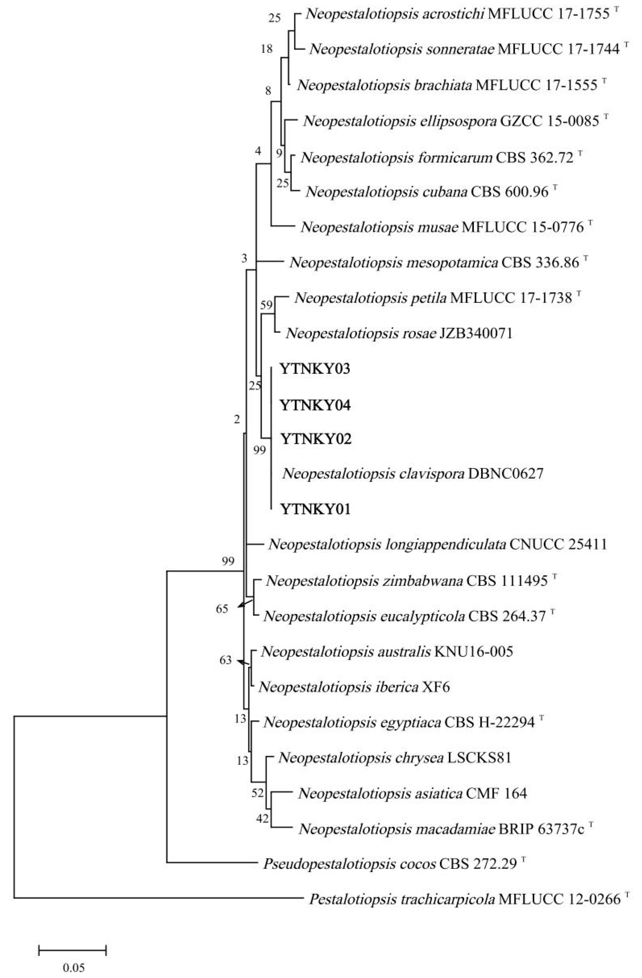
The ITS, TEF1α, and TUB2 genes of the pathogen were amplified by PCR, and the obtained sequences were subjected to BLASTn homology comparison analysis in the NCBI database. The results showed that four isolates had more than 99% sequence homology with the reference strains of Neopestalotiopsis clavispora from various sources. Based on the phylogenetic tree generated using multi-gene (ITS, TEF1α, and TUB2) sequences, the isolates YTNK01, YTNK02, YTNK03, YTNK04, and the type strain N. clavispora clustered in the same branch with a bootstrap support rate of 99%, but were distantly related to other species (Figure 3). Therefore, the pathogen was identified as N. clavispora based on morphological and molecular identification results.

3.4. Pathogenicity Tests

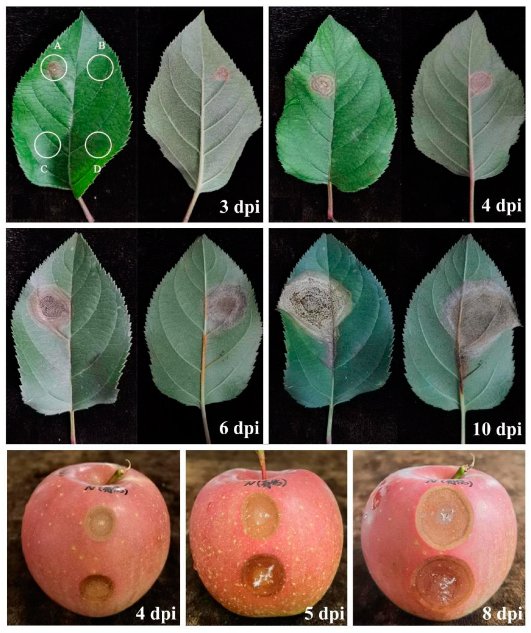
Koch’s postulates were used to further determine the pathogenicity of the pathogen to apples. The results showed that N. clavispora isolates YTNK01, YTNK02, and YTNK03 could cause typical symptoms on detached leaves and fruits upon surface wound inoculation (Figure 4), whereas the non-wound inoculation treatments and controls showed no symptoms. The symptoms of the diseased leaves mirrored those observed in the field, with all three isolates producing consistent symptoms in all inoculated tissues. Although no diseased fruit was observed in the field, the pathogen could infect detached fruits in laboratory experiments. Small, brown, and round necrotic lesions appeared at 2 days post-inoculation, with an infection rate of 100%, whereas the controls and non-wound inoculation treatments remained healthy. Continuous observation revealed brown spots with concentric rings at 3 days post-inoculation, and black acervuli were produced at 4 days post-inoculation (Figure 5). Over time, the size of leaf spots gradually expanded, reaching a diameter of about 2.0 to 3.5 cm (mean ± SD = 2.6 ± 0.4 cm; n = 15). We found no significant difference in the pathogenesis between young and old leaves. Fruit disease symptoms emerged at 2 days post-inoculation, progressing to dark halos and obvious depressions around lesions by day 4. At 5 days post-inoculation, the lesions exhibited a concentric ring pattern, and white mycelium was discovered, which was isolated and identified as N. clavispora. At 8 days post-inoculation, a small number of black spots began to appear in the center of the lesions. Non-wound inoculation and surface wound inoculation with sterile PDA both showed no pathogenicity to apple fruit (Figure 4). Finally, N. clavispora was re-isolated from these inoculated and diseased apple tissues, which confirmed Koch’s postulates.

3.5. Host Range Test of the Pathogen

The host range of N. clavispora YTNK02 was evaluated by inoculation on five fruit species. We found that the leaves of crabapple, sweet cherry, grape, peach, and pear were infected by N. clavispora approximately 2 to 3 days after surface wound inoculation, while none were infected by non-wound inoculation (Figure 6). The lesions on these leaves showed a concentric ring pattern with small black spots on the surface and developed quickly over time. Compared to the other species, the pathogenicity of N. clavispora was weaker on pear, showing a lower infection rate, smaller lesions, and slower spread speed (Table 4).

3.6. Toxicity Determination of Ten Fungicides on Neopestalotiopsis clavispora

The results of toxicity tests on the ten fungicides indicated that each of them was capable of inhibiting the growth of N. clavispora. The EC50 values indicated varying levels of toxicity of different fungicides to N. clavispora (Table 5). The toxicity of the ten fungicides based on the active ingredients, in descending order, was: pylocyanonitrile > propiconazole > pyraclostrobin > tebuconazole > diphenoxazole > osthole > procymidone > eugenol > metiram > myclobutanil. The corresponding EC50 values were 0.11 µg/mL (r = 0.964), 0.41 µg/mL (r = 0.996), 0.47 µg/mL (r = 0.995), 1.32 µg/mL (r = 0.981), 1.85 µg/mL (r = 0.960), 3.82 µg/mL (r = 0.993), 19.44 µg/mL (r = 0.998), 23.53 µg/mL (r = 0.986), 44.02 µg/mL (r = 0.975), and 83.81 µg/mL (r = 0.957), respectively. Pylocyanonitrile, propiconazole, pyraclostrobin, tebuconazole, diphenoxazole, and osthole showed strong toxicity toward N. clavispora. Therefore, these fungicides could be considered as alternative agents to control apple Neopestalotiopsis leaf spot.

4. Discussion

Over the past 20 years, the prevalence and severity of fungal plant diseases have escalated, leading to the emergence of new types of diseases [36]. The pathogens causing apple leaf spot diseases present a complex challenge due to their diversity and varied symptoms. Accurate identification of pathogens responsible for apple leaf spot diseases is thus crucial for industrial development. This study represents the first investigation of apple leaf spots caused by N. clavispora, and the disease has been preliminarily named ‘apple Neopestalotiopsis leaf spot’. These findings provide valuable insights into the identification and characterization of the pathogen responsible for the disease, laying the foundation for effective diagnosis and prevention strategies in the field.
Pestalotiopsis-like fungi, which include Neopestalotiopsis, are recognized as significant agents in plant diseases globally [37,38]. These fungi are mostly weakly parasitic or saprophytic and can cause a variety of leaf spot diseases. In some cases, they damage fruits and harvested crops [39]. In 2014, Maharachchikumbura et al. subdivided Pestalotiopsis into three genera: Pestalotiopsis, Neopestalotiopsis, and Pseudopestalotiopsis [7]. Later, some researchers proposed that the Pestalotiopsis and Neopestalotiopsis genera be classified as one genus [40]. In our study, we did not explore this taxonomic aspect in depth, as our objective was phytopathological rather than taxonomic. Morphological investigations or single-gene sequencing have proven insufficient to unequivocally distinguish species within these genera [8,9,10]. Accurate categorization of Neopestalotiopsis at the species level requires multiple nucleotide sequences of the ITS, TEF1α, and TUB2 genes [10]. In our research, we constructed a phylogenetic tree using combined sequences of ITS, TEF1α, and TUB2, which confirmed the pathogen causing apple leaf spot disease in Yantai as N. clavispora. Neopestalotiopsis spp. are widely distributed in nature and have a broad host range [41]. Specifically, N. clavispor can cause diseases in various plants, and related reports are increasing. It can harm blueberry [42], alpine azalea [43], strawberry [44], banana [45,46], Actinidia arguta Sieb. et Zucc [47], Garcinia mangostana L. [48], Taxus media Rehder [49], and other plants in China, causing symptoms such as leaf spot, branch blight, and root rot. This study revealed that apple is a new host for N. clavispora, and this pathogen-induced leaf spot disease was identified in China for the first time. From 2021 to 2023, it was observed in orchards for three consecutive years, with an increasing incidence rate. Although it has not yet caused widespread damage in the field according to the current investigation, the potential threat of N. clavispora cannot be ignored. Additionally, since there are no reports on the pathogenicity of N. clavispora in the local area, five main cultivated fruit species were selected to further explore the pathogenic range in this study. This study confirmed that N. clavispora can also quickly infect leaves of crabapple, sweet cherry, grape, peach, and pear under laboratory conditions, indicating that these fruit trees may be potential hosts for N. clavispora in the field. Under suitable or stressed conditions, the pathogen may infect the leaves of these fruit trees and become a potential threat to fruit production. Therefore, attention should be paid to field prevention and control.
Pestalotiopsis and Neopestalotiopsis species typically infect plants through insects or wounds to initiate the process of pathogenesis [50,51], but they cannot invade without a wound [25]. In our research on apple inoculation, the infection rate was 100% through the wounding method, while non-wound inoculation showed no symptoms. This finding was consistent with the aforementioned research, indicating the pathogen’s weak parasitic nature. Moreover, the experiment showed no significant difference in the pathogenicity of N. clavispora to young and old apple leaves. Fruit surface wound inoculation showed a 100% infection rate, similar to the situation caused by Pestalotiopsis in imported ‘Red Delicious’ apples in 2006 [12]. We analyzed that apple trees might have been infected with Pestalotiopsis-like fungi long ago, but these infections were confused with other leaf spot types or did not attract attention. However, natural infection of N. clavispora on fruits was not observed, possibly due to low fungal presence and unsuitable climatic conditions in the field. This finding suggests a potential risk of large-scale disease under suitable conditions. Additionally, the effects of N. clavispora on the storage period of apples and its infection mechanism warrant further investigation.
Chemical control is the most direct method [52]. Screening effective chemical agents can control the spread of these pathogens. Based on inhibitory activity under laboratory and field conditions, triazoles, carbamates, benzimidazoles, phenylpyrrole, strobilurins, and some botanical fungicides have been reported to be effective against various Pestalotiopsis species in a variety of crops [53,54,55,56]. The present study found that pylocyanonitrile, propiconazole, pyraclostrobin, tebuconazole, diphenoxazole, and osthole were effective against N. clavispora and could be used as alternative fungicides to control apple Neopestalotiopsis leaf spot. These findings are consistent with previous reports. These selected fungicides are commonly used in production to prevent apple leaf spot, ring rot, and other apple diseases, demonstrating the potential to use one fungicide to prevent multiple apple diseases. The above results were obtained through laboratory experiments, the use of fungicides in the field needs to consider factors such as local climate, product cost, and application methods, which are critical for disease prevention and control.

5. Conclusions

This study identified Neopestalotiopsis clavispora as the pathogen responsible for a new type of apple leaf spot disease through morphological observation, molecular identification, and pathogenicity testing. To our knowledge, this is the first report of N. clavispora being responsible for apple leaf spots. Additionally, N. clavispora was found to infect crabapple, sweet cherry, grape, peach, and pear under laboratory conditions. The toxicity determination of ten fungicides, based on their active ingredients, showed that pylocyanonitrile, propiconazole, pyraclostrobin, tebuconazole, diphenoxazole, and osthole had significant inhibitory effects on the mycelial growth of N. clavispora. These findings offer a theoretical foundation for developing effective strategies for disease diagnosis and prevention.
Further studies are currently underway to determine if resistance differences exist among apple varieties in China. Moving forward, the biological and ecological characteristics of N. clavispora need to be studied, as it has the potential for host transfer under favorable weather or other infection conditions. Moreover, due to its global prevalence and broad host range, the focus will be on unraveling the pathogenic nature of this cryptic genus to gain further insights into its epidemiology and disease management.

Author Contributions

Conceptualization, J.S.; Methodology, J.S.; Software, J.S. and S.W.; Validation, J.S. and B.L. (Baoyan Li); Formal analysis, J.S. and B.L. (Baoyan Li); Investigation, J.S. and W.Z.; Writing—original draft, J.S.; Writing—review & editing, Y.W. and B.L. (Baoyou Liu); Supervision, M.S.; Project administration, B.L. (Baoyou Liu); Funding acquisition, B.L. (Baoyou Liu). All authors have read and agreed to the published version of the manuscript.

Funding

This research was funded by the Shandong Province Fruit Industry Technology System (SDAIT-06-11); the Shandong Province Key R&D Program (2022CXGC020709); the Science and Technology Planning Project of Yantai (2021NYNC015, 2023YD084); and the Shandong Province Science and Technology Small and Medium Enterprises Innovation Ability Enhancement Project (2023TSGC0829, 2023TSGC0894).

Data Availability Statement

The datasets generated or analyzed in this study are included in this article. Further inquiries can be directed to the corresponding author upon reasonable request.

Conflicts of Interest

The authors declare no conflict of interest.

References

  1. Ritchie, H.; Roser, M. Agricultural Production. Our World in Data. Available online: https://ourworldindata.org/agricultural-production (accessed on 22 July 2022).
  2. Yu, H.N.; Bai, N.; Wang, G.; Guo, D.T.; Zhang, L.L. Current situation and countermeasures of apple processing industry in Yantai city. China Fruit Veg. 2023, 43, 80–84. [Google Scholar]
  3. Zhang, X.Q.; Li, D.M.; Liu, X.; Sun, T.; Lin, X.J.; Ren, Z.H. Research of segmentation recognition of small disease spots on apple leaves based on hybrid loss function and CBAM. Front. Plant Sci. 2023, 14, 1175027. [Google Scholar] [CrossRef]
  4. Deng, X.; Chen, Y.; Ma, Z.L.; Huang, X.M. Discovery of a new pathogen in early leaf falling disease of Zhaotong apple. South China Fruits 2021, 50, 124–129. [Google Scholar]
  5. Wang, K.; Liu, F.; Cai, L. A name list of common agricultural phytopathogenic fungi in China. Mycosystema 2022, 41, 361–386. [Google Scholar]
  6. Crous, P.W.; Rossman, A.Y.; Aime, C.; Allen, C.; Burgess, T.; Groenewald, J.Z.; Castlebury, L. Names of phytopathogenic fungi: A practical guide. Phytopathology 2021, 111, 1500–1508. [Google Scholar] [CrossRef] [PubMed]
  7. Maharachchikumbura, S.S.N.; Hyde, K.D.; Groenewald, J.Z. Pestalotiopsis revisited. Stud. Mycol. 2014, 79, 121–186. [Google Scholar] [CrossRef]
  8. Tejesvi, M.V.; Tamhankar, S.A.; Kini, K.R. Phylogenetic analysis of endophytic Pestalotiopsis species from ethnopharmaceutically important medicinal trees. Fungal Divers. 2009, 38, 167–183. [Google Scholar]
  9. Keith, L.M.; Velasquez, M.E.; Zee, F.T. Identification and characterization of Pestalotiopsis spp. causing scab disease of guava, Psidium guajava, in Hawaii. Plant Dis. 2006, 90, 16–23. [Google Scholar] [CrossRef]
  10. Jeewon, R.; Liew EC, Y.; Simpson, J.A. Phylogenetic significance of morphological characters in the taxonomy of Pestalotiopsis species. Mol. Phylogenetics Evol. 2003, 27, 372–383. [Google Scholar] [CrossRef]
  11. Prasannath, K.; Shivas, R.G.; Galea, V.J.; Akinsanmi, O.A. Neopestalotiopsis species associated with flower diseases of Macadamia integrifolia in Australia. J. Fungi 2021, 7, 771. [Google Scholar] [CrossRef]
  12. Yi, J.P.; Liang, W.S.; Zhou, G.L.; Ye, J.; Yin, L.P.; Qiu, S.H.; Zhou, E.X. Identification of Pestalotiopsis sp. from imported American apple. Plant Quar. 2006, 5, 269–271. [Google Scholar]
  13. Xu, X.H. Investigation on the Occurrence and Control of Apple Pests and Diseases in Yantai Area. Master’s Thesis, Shandong Agricultural University, Tai’an, China, 2019; pp. 41–50. [Google Scholar]
  14. Choi, Y.W.; Hyde, K.D.; Ho, W.H. Single spore isolation of fungi. Fungal Divers. 1999, 3, 29–38. [Google Scholar]
  15. Zhang, H.; Zhang, Z.; Xu, J.S.; Xu, J.; Zhang, L.P.; Pan, Z.C.; Tian, Q.; Feng, J. A rapid and simple method for obtaining single-spore isolates of Fusarium species-agar dilution lineation separation. Plant Prot. 2008, 34, 134–136. [Google Scholar]
  16. Lu, J.Y. Chapter XVII, Deuteromycotina (part 2), Coelomycetes. In Plant Pathogenic Mycology; Bai, J.K., Lui, G.Z., Liang, J.Y., Eds.; China Agriculture Press: Beijing, China, 2001; pp. 474–475. [Google Scholar]
  17. Ge, Q.X.; Chen, Y.X.; Xu, T. Morphological characteristics and other introduction of Pestalotiopsis clavispora. In Flora Fungorum Sinicorum (Volume 38): Pestalotiopsis; Wei, J.C., Xia, B.M., Hu, Z.Y., Zhuang, W.Y., Wu, P.C., Eds.; Science Press: Beijing, China, 2009; pp. 67–68. [Google Scholar]
  18. White, T.J.; Bruns, T.; Lee, S.; Taylor, J.W. Amplification and direct sequencing of fungal ribosomal RNA genes for phylogenetics. In PCR Protocols: A Guide to Methods and Applications; Innis, M.A., Gelfand, D.H., Sninsky, J.J., White, T.J., Eds.; Academic Press: Amsterdam, The Netherlands, 1990; pp. 315–322. [Google Scholar]
  19. O’Donnell, K.; Kistler, H.C.; Cigelnik, E.; Ploetz, R.C. Multiple evolutionary origins of the fungus causing Panama disease of banana: Concordant evidence from nuclear and mitochondrial gene genealogies. Proc. Natl. Acad. Sci. USA 1998, 95, 2044–2049. [Google Scholar] [CrossRef]
  20. Carbone, I.; Kohn, L.M. A method for designing primer sets for speciation studies in filamentous ascomycetes. Mycologia 1999, 91, 553–556. [Google Scholar] [CrossRef]
  21. Glass, N.L.; Donaldson, G.C. Development of primer sets designed for use with the PCR to amplify conserved genes from filamentous ascomycetes. Appl. Environ. Microbiol. 1995, 61, 1323–1330. [Google Scholar] [CrossRef]
  22. Daengsuwan, W.; Wonglom, P.; Arikit, S.; Sunpapao, A. Morphological and molecular identification of Neopestalotiopsis clavispora causing flower blight on Anthurium andraeanum in Thailand. Hortic. Plant J. 2021, 7, 573–578. [Google Scholar] [CrossRef]
  23. Pornsuriya, C.; Chairin, T.; Thaochan, N.; Narit, T.; Anurag, S. Identification and characterization of Neopestalotiopsis fungi associated with a novel leaf fall disease of rubber trees (Hevea brasiliensis) in Thailand. J. Phytopathol. 2020, 168, 416–427. [Google Scholar] [CrossRef]
  24. Zhang, L.; Li, X.H.; Zhou, Y.Y.; Tan, G.J.; Zhang, L.X. Identification and characterization of Colletotrichum species associated with Camellia sinensis anthracnose in Anhui province, China. Plant Dis. 2021, 105, 2649–2657. [Google Scholar] [CrossRef]
  25. Zhao, J.N.; Ma, Z.; Liu, Z.P.; Shang, Q.X.; Zhao, X.Y.; Wei, Y.M. Pestalotiopsis clavispora causing leaf spot on strawberry. J. Mycol. 2016, 35, 114–120. [Google Scholar]
  26. Zhang, Q.; Li, Y.H.; Zhao, B.; Xu, L.C.; Ma, H.Y.; Wang, M.A. Synthesis and antifungal activity of new butenolide containing methoxyacrylate scaffold. Molecules 2022, 27, 6541. [Google Scholar] [CrossRef] [PubMed]
  27. Yang, Y.L.; Fang, B.P.; Feng, S.J.; Wang, Z.Y.; Luo, Z.X.; Yao, Z.F.; Zou, H.D.; Huang, L.F. Isolation and identification of Trichoderma asperellum, the novel causal agent of green mold disease in sweetpotato. Plant Dis. 2021, 105, 1711–1718. [Google Scholar] [CrossRef] [PubMed]
  28. Wang, L.; Wang, N.; Yu, J.L.; Wu, J.; Liu, H.; Lin, K.J.; Zhang, Y.Y. Identification of pathogens causing alfalfa Fusarium Root Rot in Inner Mongolia, China. Agronomy 2023, 13, 456. [Google Scholar] [CrossRef]
  29. Addrah, E.M.; Zhang, Y.; Zhang, J.; Liu, L.; Zhou, H.Y.; Chen, W.D.; Zhao, J. Fungicide treatments to control seed-borne fungi of sunflower seeds. Pathogens 2019, 9, 29. [Google Scholar] [CrossRef] [PubMed]
  30. Li, J.L.; Liu, X.Y.; Xie, J.T.; Di, Y.L.; Zhu, F.X. A Comparison of different estimation methods for fungicide ECsub50/sub and ECsub95/sub Values. J. Phytopathol. 2015, 163, 239–244. [Google Scholar] [CrossRef]
  31. Zhou, Z.H.; Wang, Y.C.; Duan, Y.B.; He, Y.N.; Liu, S.; Chen, Y.; Deng, W.C.; Li, C.P.; Hu, W.J.; Gu, Y.J.; et al. Inhibitory Effect and Control Efficacy of Picoxystrobin against Neopestalotiopsis clavispora, Causing Vine Tea Leaf Blight. Agronomy 2023, 13, 1340. [Google Scholar] [CrossRef]
  32. Paula, A.; Cristina, A.V.; Victoria, M.; Pedro, M.; Maria, S.A.; Stadnik, M.J. Reclassification of the main causal agent of Glomerella leaf spot on apple into Colletotrichum chrysophilum in southern Brazil and Uruguay. Phytopathology 2022, 112, 1825–1832. [Google Scholar]
  33. Kumar, A.; Sharma, J. Host plant cultivar, leaf positions and nutrition affect the expression of Marssonina blotch resistance in apple. Plant Dis. Res. 2014, 29, 1–5. [Google Scholar]
  34. Sofi, A.T.; Beig, A.M.; Dar, H.G.; Ahanger, F.A.; Hamid, A.; Dar, W.A. Disease status of alternaria leaf blotch and evaluation of apple cultivars against Alternaria mali. Plant Dis. Res. 2013, 28, 239. [Google Scholar]
  35. Sajeewa, S.N.; Maharachchikumbura, S.S.N.; Guo, L.D.; Cai, L.; Chukeatirote, E.; Wu, W.P.; Sun, X.; Pedro, W.C.; Jayarama, B.D.; McKenzie, E.H.C.; et al. A multi-locus backbone tree for Pestalotiopsis, with a polyphasic characterization of 14 new species. Fungal Divers. 2012, 56, 95–129. [Google Scholar]
  36. Fisher, M.; Henk, D.A.; Briggs, C.J.; Brownstein, J.S.; Madoff, L.C.; McCraw, S.L.; Gurr, S.J. Emerging fungal threats to animal, plant and ecosystem health. Nature. 2012, 484, 186–194. [Google Scholar] [CrossRef] [PubMed]
  37. Das, R.; Chutia, M.; Das, K.; Jha, D.K. Factors affecting sporulation of Pestalotiopsis disseminata causing grey blight disease of Persea bombycina Kost., the primary food plant of muga silkworm. Crop Prot. 2010, 29, 963–968. [Google Scholar] [CrossRef]
  38. Jayawardena, R.S.; Zhang, W.H.; Liu, M.H.; Sajeewa, S.N.; Maharachchikumbura, S.S.N.; Zhou, Y.; Huang, J.B.; Nilthong, S.; Wang, Z.Y.; Li, X.H.; et al. Identifification and characterization of Pestalotiopsis-like fungi related to grapevine diseases in China. Fungal Biol. 2015, 119, 348–361. [Google Scholar] [CrossRef] [PubMed]
  39. Song, L.S. Research on taxonomy, molecular systematics and DNA barcoding of Pestalotiopsis. Ph.D. Thesis, Guangxi University, Nanning, China, 2016. [Google Scholar]
  40. Solarte, F.; Muñoz, C.G.; Maharachchikumbura, S.S.N.; Álvarez, E. Diversity of Neopestalotiopsis and Pestalotiopsis spp., causal agents of guava scab in Colombia. Plant Dis. 2018, 102, 49–59. [Google Scholar] [CrossRef] [PubMed]
  41. Reddy, M.S.; Murali, T.S.; Suryanarayanan, T.S.; Govinda, R.; Thirunavukkarasu, N. Pestalotiopsis species occur as generalist endophytes in trees of Western Ghats forests of southern India. Fungal Ecol. 2016, 24, 70–75. [Google Scholar] [CrossRef]
  42. Cao, Y.; Zhang, S.; Zhang, J.; Wang, Y.J.; Qian, S.J.; Sun, Y.; Yan, X.R. First report of Neopestalotiopsis foedans causing blueberry diseases. Acta Phytopathol. Sin. 2022, 53, 367–374. [Google Scholar]
  43. Cun, H.C.; He, P.B.; He, P.F.; Wu, Y.X.; Shahzad, M.; Tang, Z.X.; He, Y.Q. Etiology of the pathogen causing brown leaf spot on Rhododendron lapponicum. Mycosystema 2023, 42, 707–718. [Google Scholar]
  44. Chamorro, M.; Aguado, A.; Santos, B.D. First report of root and crown rot caused by Pestalotiopsis clavispora (Neopestalotiopsis clavispora) on strawberry in Spain. Plant Dis. 2016, 100, 1495. [Google Scholar] [CrossRef]
  45. Qi, Y.X.; Zhao, H.; Zhang, H. Characterization of Neopestalotiopsis clavispora, a new etiological agent of leaf spot isolated from banana. Plant Dis. Pests 2022, 13, 1–3. [Google Scholar]
  46. Qi, Y.X.; Zhang, H.; Peng, J.; Zeng, F.Y.; Xie, Y.X.; Yu, Q.F.; Zhang, X. First report of Neopestalotiopsis clavispora causing leaf spot disease on banana (Musa acuminate L.) in China. Plant Dis. 2022, 107, 220. [Google Scholar] [CrossRef]
  47. Li, K.K.; Zhang, C.L.; Wang, W.X.; Chen, C.Z.; Liu, Q.S.; Yin, H. First report of Neopestalotiopsis clavispora causing postharvest fruit rot on Actinidia arguta in Liaoning Province, China. Plant Dis. 2022, 107, 217. [Google Scholar] [CrossRef] [PubMed]
  48. Zhu, R.J.; Liu, Y.L.; Li, H.Y.; Liu, J.B.; Han, X.Y. First report of Neopestalotiopsis clavispora causing leaf spots on Garcinia mangostana in China. Plant Dis. 2022, 107, 1946. [Google Scholar] [CrossRef] [PubMed]
  49. Li, Y.; Zhang, S.Q.; Wang, T.N.; Zhou, S.G.; Wu, Y.; Huang, X.Q.; Lin, H.P.; Su, X. First report of Taxus media branch blight caused by Neopestalotiopsis clavispora in China. Plant Dis. 2022, 106, 3214. [Google Scholar] [CrossRef] [PubMed]
  50. Espinoza, J.G.; Briceño, E.X.; Keith, L.M.; Latorre, B.A. Canker and twig dieback of blueberry caused by Pestalotiopsis spp. and a Truncatella sp. in Chile. Plant Dis. 2008, 10, 1407–1414. [Google Scholar] [CrossRef] [PubMed]
  51. Chakkiyanickal, N.B.; Mohammed, F.P.; Rajan, G. Identification and characterization of Neopestalotiopsis clavispora associated with leaf blight of small cardamom (Elettaria cardamomum Maton). J. Phytopathol. 2018, 166, 532–546. [Google Scholar]
  52. Huo, H.L.; Huangfu, J.R.; Song, P.L.; Zhang, D.M.; Shi, Z.D.; Zhao, L.L.; Li, Z.Q.; Zhou, H.Y. Isolation, identification and characterization of Leptosphaerulina trifolii, the causative agent of alfalfa Leptosphaerulina Leaf spot in Inner Mongolia, China. Agronomy 2024, 14, 1156. [Google Scholar] [CrossRef]
  53. Sanjay, R.; Ponmurugan, P.; Baby, U.I. Evaluation of fungicides and biocontrol agents against grey blight disease of tea in the field. Crop Prot. 2008, 27, 689–694. [Google Scholar] [CrossRef]
  54. Saju, K.A.; Mech, S.; Deka, T.; Biswas, A.K. In vitro evaluation of biocontrol agents, botanicals and fungicides against Pestalotiopsis sp. infecting large cardamom (Amomum subulatum Roxb.). J. Spices Aromat. Crops 2011, 20, 89–92. [Google Scholar]
  55. Zhang, C.Q.; Liu, Y.H.; Wu, H.M.; Xu, B.C.; Sun, P.L.; Xu, Z.H. Baseline sensitivity of Pestalotiopsis microspora, which causes black spot disease on Chinese hickory (Carya cathayensis) to pyraclostrobin. Crop Prot. 2012, 42, 256–259. [Google Scholar] [CrossRef]
  56. Xue, D.S.; Shao, Z.H.; Li, B.H.; Wang, C.X. Indoor fungicide screening for Pestalotiopsis clavispora of blueberry. Shandong Agric. Sci. 2018, 50, 115–118. [Google Scholar]
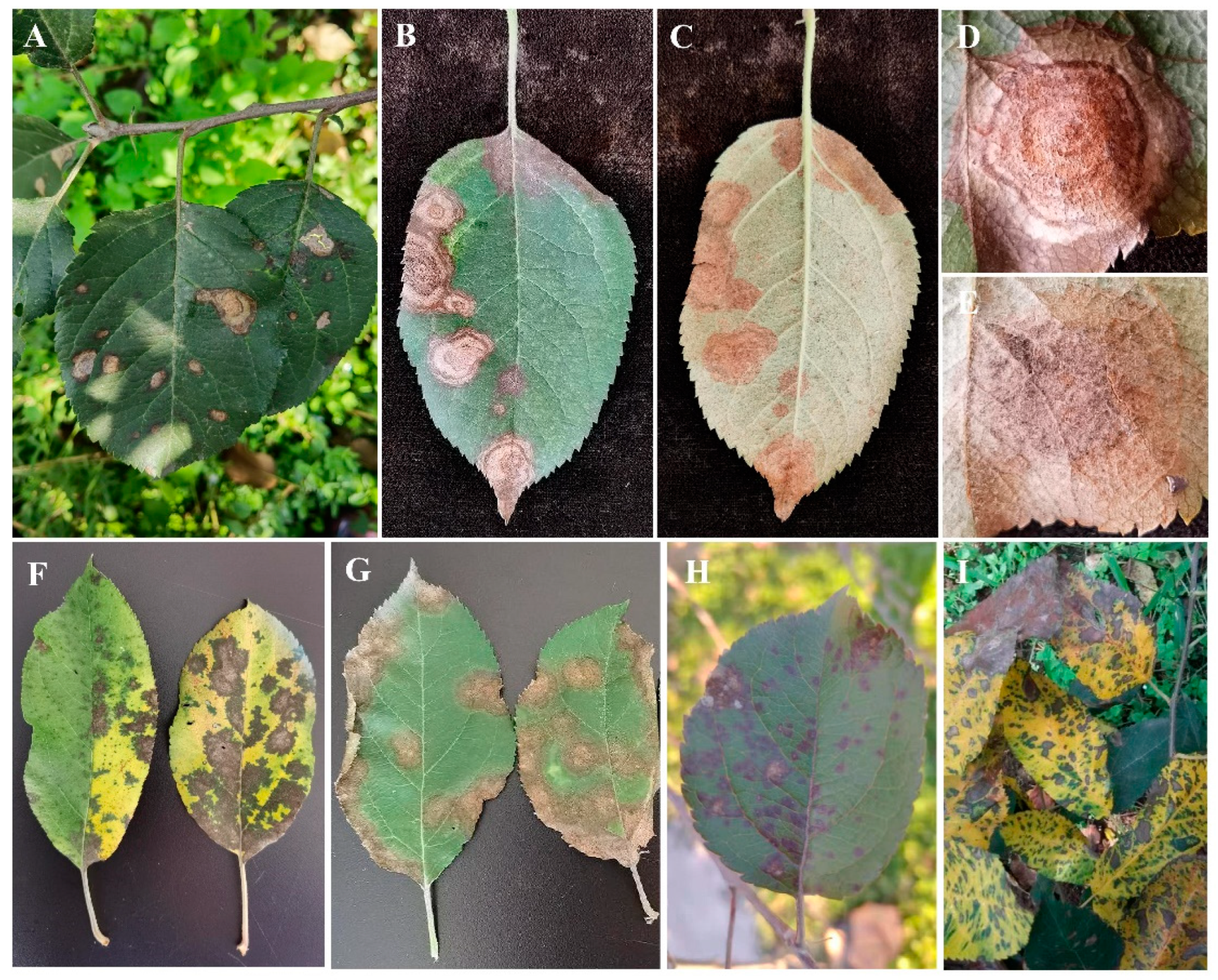
Figure 1. Symptoms of various types of apple leaf spot diseases. (AE) Apple Neopestalotiopsis leaf spot; (F,G) apple Marssonina blotch (various types); (H) apple Alternaria blotch; (I) apple Glomerella leaf spot.
Agronomy 14 01658 g001
Figure 2. Morphological characteristics of the pathogen of apple Neopestalotiopsis leaf spot (Bar = 20 μm). (AH) Colony characteristics of pathogens on PDA medium; (I,J) black viscous acervuli and their morphology observed under a microscope; (K) Hyphae; (L) Conidia.
Agronomy 14 01658 g002
Figure 3. Phylogenetic tree constructed based on concatenated sequences of ITS, TEF1α, and TUB2 genes of Neopestalotiopsis species using the Maximum Likelihood method. Pestalotiopsis trachicarpicola (MFLUCC 12-0266) and Pseudopestalotiopsis cocos (CBS 272.29) were used as outgroups. The new isolates in this study were indicated in bold. T = ex-type strains. Bar = 0.05 substitutions per nucleotide position.
Agronomy 14 01658 g003
Figure 4. Symptoms on detached apple leaves and fruits after inoculation with three Neopestalotiopsis clavispora isolates. (A-1,A-2) Symptoms induced by the YTNK01 isolate; (B-1,B-2) symptoms induced by the YTNK02 isolate; (C-1,C-2) symptoms induced by the YTNK03 isolate. Surface wound inoculation (SW); non-wound inoculation (NW); surface wound inoculation with sterile PDA (CK).
Agronomy 14 01658 g004
Figure 5. Symptoms of Neopestalotiopsis clavispora inoculation on detached apple leaves and fruits at different post-inoculation times. The presented fruit photos are of wounded fruit parts. The four white circles (A–D) in the left image of Figure 3 dpi represent four treatment sites on the same leaf. (A) Surface wound inoculation with mycelial plugs; (B) non-wound inoculation with mycelial plugs; (C) surface wound inoculation with sterile PDA; and (D) non-wound inoculation with sterile PDA. Days post-inoculation (dpi).
Agronomy 14 01658 g005
Figure 6. Lesions of infection by Neopestalotiopsis clavispora (YTNK02) on crabapple (A), sweet cherry (B), grape (C), peach (D), and pear (E) leaves at 10 days post-inoculation. The four white circles (a–d) in Figure (A) represent four treatment sites on the same leaf. (a) Surface wound inoculation with mycelial plugs; (b) non-wound inoculation with mycelial plugs; (c) surface wound inoculation with sterile PDA; (d) non-wound inoculation with sterile PDA.
Agronomy 14 01658 g006
Table 1. Primers used in this study.
GenePrimerSequence (5′-3′)Annealing Temperature/°C
ITSITS1TCCGTAGGTGAACCTGCGG53
ITS4TCCTCCGCTTATTGATATGC
TEF1αEF1-728FCATCGAGAAGTTCGAGAAGG58
EF2GGARGTACCAGTSATCATGTT
TUB2Bt2aGGTAACCAAATCGGTGCTGCTTTC59
Bt2bACCCTCAGTGTAGTGACCCTTGGC
Table 2. Sequence information for the reference strains used for phylogenetic analysis in this study.
SpeciesStrain No.GenBank Accession Numbers
ITSTEF1αTUB2
Neopestalotiopsis asiaticaCMF 164MN784703MN816249MN808590
Neopestalotiopsis australisKNU16-005KY398730KY398732KY398731
Neopestalotiopsis acrostichiMFLUCC 17-1755 TMK764273MK764317MK764339
Neopestalotiopsis brachiataMFLUCC 17-1555 TMK764274MK764318MK764340
Neopestalotiopsis chryseaLSCKS81OQ392362OQ410712OQ410711
Neopestalotiopsis clavisporaDBNC0627MT742640MT745889MT745888
Neopestalotiopsis cubanaCBS 600.96 TKM199347KM199521KM199438
Neopestalotiopsis egyptiacaCBS H-22294 TKP943747KP943748KP943746
Neopestalotiopsis ellipsosporaGZCC 15-0085 TKU500017KU500013KU500010
Neopestalotiopsis eucalypticolaCBS 264.37 TMH855907KM199551KM199431
Neopestalotiopsis formicarumCBS 362.72 TKM199358KM199517KM199455
Neopestalotiopsis ibericaXF6OM333900OM350155OM350157
Neopestalotiopsis longiappendiculataCNUCC 25411OP168149OP183977OP183981
Neopestalotiopsis macadamiaeBRIP 63737c TKX186604KX186627KX186654
Neopestalotiopsis mesopotamicaCBS 336.86 TKM199362KM199555 KM199441
Neopestalotiopsis musaeMFLUCC 15-0776 TKX789683KX789685KX789686
Neopestalotiopsis petilaMFLUCC 17-1738 TMK764276MK764320MK764342
Neopestalotiopsis rosaeJZB340071MN495978MN968335MN968343
Neopestalotiopsis sonnerataeMFLUCC 17-1744 TMK764279MK764323MK764345
Neopestalotiopsis zimbabwanaCBS 111495 TMW422813KM199545KM199456
Pestalotiopsis trachicarpicolaMFLUCC 12-0266 TJX399002JX399066JX399033
Pseudopestalotiopsis cocosCBS 272.29 TMH855069KM199553KM199467
Notes: CBS: culture collection of the Centraalbureau voor Schimmelcultures, Fungal Biodiversity Centre, Utrecht, The Netherlands; MFLUCC: Mae Fah Luang University Culture Collection, Chiang Rai, Thailand; GZCC: Guizhou Provincial Culture Collection Center, Guizhou, China; BRIP: Queensland Plant Pathology Herbarium, Australia [11]. Ex-type strains were labeled with a superscript “T”.
Table 3. Information of ten fungicides used in this study.
Active Ingredients NameCommercial NameDosage FormManufacturer Information
PylocyanonitrileHuiyou50% WPSyngenta (China) Investment Co., Ltd., Shanghai, China
DiphenoxazoleJianjingkang40% SCJiangsu Jianpai Agrochemical Co., Ltd., Yancheng, Jiangsu, China
ProcymidoneDifenkang20% SCJiangxi He Yi Chemical Co., Ltd., Jiujiang, Jiangxi, China
PyraclostrobinKairun250 g/L ECBASF(China) Co., Ltd., Shanghai, China
PropiconazoleXiute250 g/L ECSyngenta (China) Investment Co., Ltd., Shanghai, China
MyclobutanilYibaofeng40% SCJiangxi He Yi Chemical Co., Ltd., Jiujiang, Jiangxi, China
TebuconazoleHaolike430 g/L SCBayer Crop Science (China) Co., Ltd., Beijing, China
MetiramPinrun70% WGBASF (China) Co., Ltd., Shanghai, China
OstholeN/A1% EWInner Mongolia Qingyuanbao Biotechnology Co., Ltd., Bayannur, Inner Mongolia, China
EugenolShuanglang0.3% SLShandong Yijia Agricultural Chemical Co., Ltd., Shouguang, Shandong, China
Notes: WP, wettable powder; SC, suspension concentrate; EC, emulsifiable concentrate; WG, water-dispersible granules; EW, emulsion in water; SL, soluble concentrate; N/A, not available.
Table 4. The pathogenicity of Neopestalotiopsis clavispora in five kinds of fruit leaves at 10 days post-inoculation.
Fruit Name
(Scientific Name)
FamilyIncubation Period (dpi)Lesion Diameter (cm) *Infection Rate (%) *Infection Status
SWNW
Crabapple
(Malus spectabilis (Ait.) Borkh.)
Rosaceae21.53 ± 0.10 c100 ± 0 a+
Sweet cherry
(Prunus avium L.)
Rosaceae21.81 ± 0.16 b100 ± 0 a+
Peach
(Prunus persica (L.) Batsch)
Rosaceae21.23 ± 0.27 d91.67 ± 14.43 ab+
Pear
(Pyrus communis L.)
Rosaceae31.14 ± 0.18 d83.30 ± 14.43 b+
Grape
(Vitis vinifera L.)
Ampelidaceae22.41 ± 0.28 a100 ± 0 a+
Notes: * Data are expressed as mean ± SD. Different lowercase letters within the same column denote significant differences among treatments at the 0.05 significance level. dpi: days post-inoculation; SW: surface wound inoculation; NW: non-wound inoculation; +: infected; −: uninfected.
Table 5. Sensitivity of Neopestalotiopsis clavispora to fungicides under in vitro conditions.
Active Ingredients NameTreatment
Concentration (µg/mL)
Toxic Regression EquationEC50 (µg/mL) *Correlation
Coefficient (r)
Pylocyanonitrile0.039, 0.08, 0.156, 0.313, 0.625, 1.25y = 1.3009x + 6.24000.111 ± 0.082 h0.964
Diphenoxazole0.391, 0.781, 1.563, 3.125, 6.25, 12.5y = 0.7686x + 4.79441.852 ± 0.076 f0.960
Procymidone1.25, 5, 10, 20, 80, 160y = 1.3080x + 3.314519.44 ± 0.023 d0.998
Tebuconazole0.391, 0.781, 1.563, 3.125, 6.25, 12.5y = 0.8217x + 4.90121.319 ± 0.049 fg0.981
Pyraclostrobin0.195, 0.391, 0.781, 1.563, 3.125, 6.25y = 0.6665x + 5.21770.471 ± 0.020 gh0.995
Propiconazole0.156, 0.313, 0.625, 1.25, 2.5, 5y = 0.9467x + 5.36580.411 ± 0.028 gh0.996
Myclobutanil1.25, 20, 40, 80, 160, 320y = 1.4343x + 2.241583.813 ± 0.145 a0.957
Metiram5, 20, 40, 80, 160, 320y = 1.7073x + 2.193844.017 ± 0.072 b0.975
Osthole1.25, 2.5, 5, 10, 40, 80y = 0.8011x + 4.53333.824 ± 0.041 e0.993
Eugenol1.563, 3.125, 6.25, 12.5, 25, 100y = 0.8268x + 3.866023.527 ± 0.058 c0.986
Notes: * Data are expressed as mean ± SD. Different lowercase letters within the same column denote significant differences among treatments at the 0.05 significance level.
Disclaimer/Publisher’s Note: The statements, opinions and data contained in all publications are solely those of the individual author(s) and contributor(s) and not of MDPI and/or the editor(s). MDPI and/or the editor(s) disclaim responsibility for any injury to people or property resulting from any ideas, methods, instructions or products referred to in the content.