Article

In Vitro Plant Regeneration of Sulla coronaria from Floral Explants as a Biotechnological Tool for Plant Breeding

1
Department of Agricultural, Food and Forest Sciences, University of Palermo, Viale delle Scienze, Building 4, 90128 Palermo, PA, Italy
2
National Research Council of Italy (CNR), Institute of Biosciences and BioResources (IBBR), Via Ugo La Malfa 153, 90146 Palermo, PA, Italy
3
Department of Economics, Business and Statistics, University of Palermo, Viale delle Scienze, Building 13, 90128 Palermo, PA, Italy
4
National Research Council of Italy (CNR), Institute of Agricultural Biology and Biotechnology (IBBA), Via Alfonso Corti 12, 20133 Milano, MI, Italy
*
Author to whom correspondence should be addressed.
Agronomy 2024, 14(11), 2667; https://doi.org/10.3390/agronomy14112667
Submission received: 27 August 2024 / Revised: 7 November 2024 / Accepted: 11 November 2024 / Published: 13 November 2024
(This article belongs to the Section Horticultural and Floricultural Crops)

Abstract

:
Sulla coronaria L. Medik., a biennial forage legume typical of the Mediterranean basin, plays a significant role in foraging systems due to its high nutritional value, ability to increase ruminant live weight, and potential to reduce methane emissions. However, its allogamous nature complicates genetic improvement and the development of new varieties with desired traits. Given these challenges, this study aims to develop, for the first time, a successful protocol for the in vitro meristematic shoot regeneration of S. coronaria. The experiment utilizes four different flower explants (anther with filament, ovary, petals, and whole immature flower) collected from twenty distinct S. coronaria biotypes with three plant growth regulator (PGR) combinations and under both light and dark conditions. In terms of the regeneration response, the key factors appear to be the combinations of PGRs and the type of explant used. The interactions between all the factors do not seem to be significant.

1. Introduction

Sulla coronaria (L.) Medik. [1], also known as Hedysarum coronarium L., is a biennial forage legume commonly found in arable farming systems and natural environments throughout the Mediterranean region [2,3,4]. This plant plays a significant role in foraging systems, with its cultivation spanning from Portugal and Greece to North Africa (Morocco, Algeria, Tunisia), as well as Egypt, Turkey, and Lebanon [5]. It is also found in certain regions in New Zealand [6], Australia [7,8], and California [9]. Its geographical spread on a global scale was facilitated by the identification and selection of the Rhizobium sullae strain (WSM1592), as highlighted by Yates et al. [7]. This advancement was followed by the production and commercialization of the inoculum, which played a crucial role in its widespread cultivation. It is known for its excellent forage characteristics, such as an exceptional grazing resource, as well as its suitability for making hay, silage, and dehydrated products [10]. It has always been studied for its remarkable ability to increase milk production, primarily due to its high nutritional value [11] and moderate content of total phenolic compounds, especially proanthocyanidins, known as condensed tannins [12,13,14,15]. These secondary metabolites have demonstrated potential benefits for ruminant health by improving production performance. Such benefits include increased live weight, enhanced milk quality, and a reduction in methane emissions [16]. S. coronaria is primarily an allogamous species [17], a trait that influences the genetic variability of natural populations and seed production [10]. This allogamous nature complicates genetic improvement efforts aimed at developing new varieties with desired traits. Additionally, it affects the conservation of existing varieties, particularly in their natural habitats. In Italy, since 1970, when the first two varieties of sulla were added to the National Register of Varieties, a total of 15 applications for registration have been submitted. Among these, five have been rejected, one is currently in the registration phase, one has been canceled, and six are in the final stage of registration termination. As a result, only two varieties are currently registered. This scenario highlights a challenge in promoting the development and maintenance of improved varieties, given the complexity of managing the seed business. The difficulties associated with seed production are reflected by the limited interest in the production of improved varieties. Currently, much of the genetic material used for sowing S. coronaria meadows still comes from local landraces. This reliance on traditional genetic material highlights the ongoing challenges in advancing and commercializing new, improved S. coronaria varieties. However, it is also widely recognized that the effects of biotic and abiotic stresses can reduce the availability of plant resources and lead to a decrease in biomass, seeds [18], and productivity in forage crops [19]. In this context, biotechnologies offer a promising alternative to conventional breeding, aiding in the development of stress-resistant genotypes and enhancing crop yield and quality [20,21]. Plant biotechnology has proven effective in plant breeding through methods such as genomic selection, DNA-based markers, and genome editing. In vitro shoot regeneration and propagation allow for the production of new individuals from minimal starting material, thereby eliminating seasonal constraints [22]. The efficiency of in vitro plant regeneration is influenced by various factors, including genotypes, plant growth regulators (PGRs), light conditions, explant type, and the characteristics of the donor plant [23]. By manipulating these factors, the production of new varieties can be significantly enhanced [24]. Regarding the regeneration of S. coronaria, only one experiment, conducted by Arcioni et al. [25], has been reported, in which they successfully obtained regenerates from protoplasts derived from hypocotyls and cotyledons. To the best of our knowledge, no other studies on the regeneration of S. coronaria have been published. Therefore, the aim of this study was to develop a successful protocol for obtaining in vitro regeneration of meristematic shoots from floral explants. The ability to regenerate new individuals from four explant types (anther with filament, ovary, petal, and whole immature flower) was compared in 20 different biotypes of S. coronaria. Explants were cultivated in vitro under three different combinations of PGRs and under light and dark conditions.

2. Materials and Methods

2.1. Sample Collection

The flowers used in this study were collected in March 2023 in twenty different sites located in Sicily, Italy (Figure 1), and samples were kept under refrigeration until culture.
The sites were assigned to 4 different habitats (HAB1, HAB3, HAB4, HAB5) [10]. Sites were specified with 2 letters indicating the province of sampling, followed by a sequential number: Agrigento (AG), Caltanissetta (CL), Enna (EN), Messina (ME), Palermo (PA), and Trapani (TP). HAB1 denotes the mountainous area, and includes 4 sites (ME1, EN1, PA6, and PA7). HAB3 includes 4 sites (CL1, CL2, AG1, and PA5) in the inland hilly area, while HAB4 refers to the sites CL3, AG2, AG3, and AG4, which are located on the southern coast. Finally, HAB5 comprises 8 sites (AG5, PA1, PA2, PA3, PA4, TP1, TP2, and TP3), which are found in the western hilly area.

2.2. Plant Materials

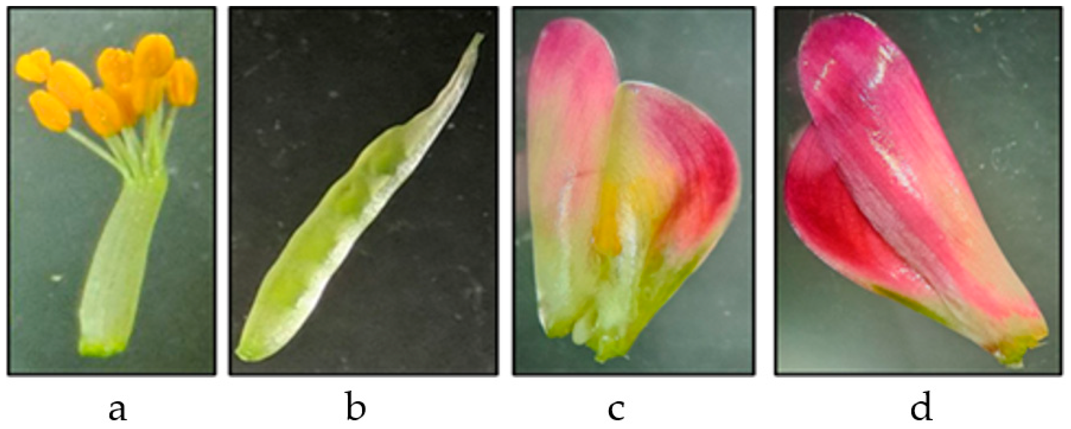
Cultures were initiated from four different explants, anther with filament (Ant), ovary (Ov), petals (Pet) and whole flower (Wf) (Figure 2) dissected from immature flowers, which were collected 15 days before anthesis from both wild plants and different biotypes.
Whole inflorescences were surface sterilized as described by Catalano et al. [26], with slight modifications. The inflorescences were first washed by immersion for 10 min in distilled water containing 0.15 mL/100 mL (approximately 3 drops) of Tween-20 under gentle shaking. Then, the flowers were disinfected under laminar flow with ethanol 70% (v/v) for 1 min and sodium hypochlorite 0.6% (w/v in water) for 7 min. The explants were then rinsed three times for 5 min in sterile distilled water. Only close flowers were used.

2.3. Culture Conditions

The explants were cultured on MS solidified medium [27] supplemented with 30 g L−1 of sucrose; the pH of the medium was adjusted to 5.7 ± 0.1 with 1N NaOH, and 7 g L−1 of agar was added to solidify the medium before autoclaving (20 min, 120 °C). The explants were plated under three different PGR combinations, as reported in Table 1.
All the PGRs were filter sterilized and added to the medium after autoclaving. Petri dishes (60 × 10 mm) were filled with 8 mL of medium and sealed with Parafilm MTM. All chemicals used in this experiment were purchased from Duchefa Biochemie, Haarlem, The Netherlands.
Four different explants were cultured (five explants per Petri dish); each treatment consisted of five replications. All the explants were incubated in a climatic chamber at 25 ± 1 °C. To evaluate the influence of light, half of the explants were incubated in dark conditions; the others were incubated under a 16 h per day length and a photosynthetic photon flux of 50 µmol m−2 s−1 provided by an Osram cool white 18 W fluorescent lamp. The explants producing callus with green spots were transferred to hormone-free MS medium and cultured for an additional month to allow for proliferation and development.

2.4. Data Collection

The explants were checked at 15-day intervals. Non-responsive explants, callus, and regenerates were evaluated as percentage of all the conditions simultaneously applied.
The effects of biotype, PGR combinations, explant type, and light conditions on callus production and the regeneration of new individuals were evaluated.

2.5. Statistical Analysis

The dataset includes multiple explants collected from 20 plants, with each explant displaying one of three possible outcomes: callus formation, meristematic shoot formation, or no response. To investigate the factors influencing these outcomes, we modeled two binary responses using logistic regression. Model 1 examined the presence vs. absence of callus formation, and model 2 analyzed the presence vs. absence of meristematic shoot formation. Since the explants represent repeated measurements taken from the same plant, it is important to account for the hierarchical structure of the data by incorporating a random effect to capture potential plant-level variability. If such variability exists and is ignored, it could result in biased parameter estimates and underestimated standard errors, as the explants from the same plant may not be independent.
To address this issue, we initially applied mixed-effects logistic regression models [28,29,30,31] for both responses (callus formation and meristematic shoot formation), incorporating plant-level random intercepts to account for potential heterogeneity among plants. In these models, biotype was included both as a fixed effect and in the random effects structure, while the other explanatory variables, such as PGR combination (3 levels), explant type (4 levels), and light condition (2 levels), were just included in the fixed part.
Contaminated explants were excluded from the analysis, as contamination was attributed to the disinfection method rather than the experimental conditions applied.
For each response, we performed model selection using the Akaike information criterion (AIC) and Bayesian information criterion (BIC) [32,33].
The general model specification, assuming no interactions between the variables, is provided by the following equation:
log Pr Y i j = 1 Pr Y i j = 0 = β 0 + β 1 Biotype i j + β 2 PGR combination i j + β 3 Explant type i j + β 4 Light condition i j + u i
where Y i j represents the binary response for the j-th explant of the i-th plant (i.e., callus formation in model 1 and meristematic shoot formation in model 2). Moreover, β 0 is the fixed intercept, while β 1 , β 2 , β 3 , β 4 represent the fixed coefficients for the explanatory variables: biotype, PGR combination, explant type, and light condition, respectively. The term u i , i.e., the random effect associated with plant i, captures the variability between individual plants. This allows the model to account for the non-independence of observations from the same plant.
The interpretation of the model’s output, particularly the estimated regression coefficients, depends on the choice of the baseline for each categorical explanatory variable. Indeed, in all factors, one level is chosen as the reference category, and the model’s coefficients reflect the comparison of other levels against this baseline.
We reported both the significance and the direction of the regression coefficients for each variable. The symbols “+” and “-” were used to indicate whether a variable had a positive or negative effect on the response (callus formation or meristematic shoots). Statistical significance was denoted as follows: * for α ≤ 0.05, ** for α ≤ 0.01, and *** for α ≤ 0.001. Statistical analysis was performed using R version 4.4.1 (14 June 2024).

3. Results

Regeneration responses were recorded after approximately 20 days from culture. A callus formation (Figure 3a) was recorded before the appearance of shoots under all the combinations tested (Figure 3b,c).

3.1. Results of the Models

The results of the percentages of callus, meristematic shoots, and non-responsive explants, under all test conditions, are reported in Table S1. Some observations have been excluded due to contamination of all the explants.

3.1.1. Callus Production

For callus production, we initially fitted the mixed-effects logistic regression model Equation (1), where Y i j represents the binary outcome, with Y i j = 1 if the j-th explant from the i-th plant exhibits callus formation, and Y i j = 0 otherwise. The models assumes that the response is Bernoulli distributed, that is:
Y i j B e r n o u l l i p i j .
Here, p i j represents the probability that the j-th explant from the i-th plant exhibits callus formation. Initially, the model included both fixed effects (biotype, PGR combination, explant type, and light conditions) and random intercepts to account for plant-level variability. However, after fitting the model, the variance associated with the random intercept was found to be near zero, indicating that plant-level heterogeneity was negligible. As a result, we excluded the random intercept in the final model, thus fitting a standard logistic regression model. Model selection on the fixed part was based on AIC and BIC, which favored the model containing only the main effects. Including interaction terms between factors led to an increase in both AIC and BIC values, indicating a poorer model fit. Additionally, the coefficients for the interaction terms were not statistically significant, further justifying their exclusion. Thus, the final model is defined as:
log Pr Y j = 1 Pr Y j = 0 = β 0 + β 1 Biotype j + β 2 PGR combination j + β 3 Explant type j + β 4 Light condition j
The baseline for the final model was set as: Biotype = “AG1”, PGR combination = “S.c. 1”, Explant type = “Ant”, and Light condition = “Dark”. The results are summarized in Table 2.
Table 2 shows that the Biotype AG1, which was used as the reference category, is the most favorable for callus formation. Due to the corner-point parameterization, all other biotypes showed negative coefficients compared to AG1, indicating that they produced fewer calli. These negative coefficients were statistically significant for all other biotypes, suggesting a clear difference in callus production across biotypes. PGR combinations also play a role, with S.c. 3 reducing callus formation significantly compared to S.c. 1, while S.c. 2 does not show significant differences compared to the baseline. The explant type is a key factor in affecting the probability of callus formation. Specifically, Ant (baseline) shows the highest probability of callus formation, while Ov, Pet, and Wf are associated with significantly lower odds of callus production. Finally, light conditions have a small but significant negative effect, indicating that light exposure decreases the probability of callus formation. All estimates reflect the difference in log-odds of callus production between a given level of a factor and its reference level. Hence, AG1 biotype, Ant explants, S.c. 1 PGR combination, and no light exposure represent the most favorable conditions for callus production according to this model.

3.1.2. Meristematic Shoots

For meristematic shoots, we initially fitted the mixed-effect logistic regression model defined in Equation (1), where Y i j is 1 if the j-th explant from the i-th plant exhibits meristematic shoot formation, and 0 otherwise. The models assumes that the response is Bernoulli distributed, that is:
Y i j B e r n o u l l i p i j .
where p i j represents the probability that the explant exhibits meristematic shoots. This probability was modeled as a function of the fixed effects (such as biotype, PGR combination, explant type, and light conditions) and random effects (biotype) to account for plant-level variability. During model selection for the fixed effects, the variables biotype and light condition were found to be non-significant. Excluding these variables led to improvements in AIC and BIC, indicating a better model fit. However, the variance associated with the random intercept for biotype was remarkable (equals to 3.455), indicating non-negligible heterogeneity across plants, thus justifying the inclusion of the random effect in the final model. Thus, the final model includes only the significant main effects of PGR combination and explant type, along with the random intercept for plant-level variability. It is important to note that while the variable biotype did not show significance as a fixed effect, it proves valuable as a random effect due to the variability observed in the response across different biotypes. This phenomenon can be attributed to the substantial variance of the random component associated with biotype, suggesting that while individual biotypes may not consistently influence shoot formation, the differences among biotypes still contribute to the overall model by accounting for unobserved heterogeneity. Moreover, although meristematic shoots were observed in only seven biotypes, the model includes all biotypes for analysis. Indeed, including all biotypes in the model ensures a robust analysis without prematurely excluding any groups based on limited outcomes. As regards model selection, when interaction terms between covariates were included, both the AIC and BIC values increased, indicating a decline in model performance. Moreover, the interaction terms were not statistically significant, further justifying their exclusion. Thus, the final model is specified as follows:
log Pr Y i j = 1 Pr Y i j = 0 = β 0 + β 1 PGR combination i j + β 3 Explant type i j + u i
The model’s baseline was set at: PGR combination = “S.c. 1”, Explant type = “Ant”. The results of the model are summarized in Table 3.
Notably, the PGR combinations demonstrated a significant impact on the response, with both S.c. 2 and S.c. 3 showing significant positive effects on shoot formation. Specifically, the results indicate that using the PGR combination S.c. 1 results in fewer meristematic shoots compared to S.c. 2 and S.c. 3, which produced the highest number of shoots. Additionally, the type of explant significantly influenced shoot development, with petals showing a notable negative effect, suggesting that they are less effective for shoot regeneration compared to the reference condition. In contrast, other explant types, such as ovary and whole flower, did not exhibit significant differences compared to the baseline.

4. Discussion

We report, for the first time to our knowledge, an experimental procedure for meristematic shoot regeneration of S. coronaria starting from flower explants. Although using flowers imposes a temporal limitation on collection, flower tissues are easy to sterilize and have demonstrated high regenerative potential in several species [34,35,36,37,38,39]. Additionally, immature tissues have been identified as the most suitable explant for somatic embryogenesis and in vitro regeneration in many species, including Leguminosae [40].
In our experimental procedure, callus and shoots were obtained under all the combinations tested. The best performance in meristematic shoot production was recorded with medium S.c. 3 (10 μM NOA + 4.4 μM BA) and medium S.c. 2 (8.8 μM BA + 1.07 μM NAA). In both cases, a combination of auxin and cytokinin was used to induce regeneration. Auxins and cytokinins together determine the regeneration response due to their crucial roles in cell cycle regulation and cell division [41]. The presence of auxin NAA is generally essential for initiating competent callus for plant regeneration [42]. Both NOA and NAA have been noted for their beneficial impact on promoting significant regeneration rates in various species, including grapevine [34,38] and sesame [43]. BA has also been widely used for in vitro regeneration processes, yielding successful results [44]. The addition of different concentrations of BA in combination with auxins such as NAA to different media significantly affects callus induction and tissue quality [45]. Our results confirm previous reports, but concerning S. coronaria, they are novel.
The effectiveness of most tissue culture studies is largely dependent on the type of explant [46], even though among all the explants tested, only the petals showed a negative effect on the production of meristematic shoots. Several authors have reported the effectiveness of anthers, which are quite simple to handle, especially in the early stages of development when the ability to respond positively is greater [47]. In vitro regeneration from anthers has been documented in other plant species, suggesting that these explants might be more competent for regeneration compared to other tissue types [48,49,50]. Genotype is a crucial factor influencing how plants respond to tissue culture, with variations observed across species, cultivars, and even among individuals within the same species. Certain genotypes exhibit a higher propensity for in vitro regeneration, as demonstrated in various species such as Crocus sp., Rosa sp., Glycine max L. Merr., Feijoa sellowiana O. Berg, and Carthamus tinctorius L. [51]. In our study, shoots were obtained in only seven S. coronaria biotypes tested, but based on statistical analysis, this does not appear to be a significant factor. Environmental conditions influence the regeneration outcome, and the light regime (e.g., dark vs. light exposure) usually affects both the type of callus formed and subsequent regeneration [24]. Some species require dark treatments to improve regeneration percentages, as reported for grapevine [52], while for other species, continuous light presence is important in every regeneration phase [53]. In our study, no significant differences were found between explants incubated in the dark and those in light regarding meristematic shoot production, but this seems to be important for callus production. Interactions between factors can strongly impact the process [54]. Unfortunately, not all interactions in the regeneration process are well documented in the literature. Fiuk & Rybczyński [51] emphasized the importance of considering the interaction between genotype and PGR combinations, but based on our data, this does not appear to be valid for shoot formations in S. coronaria. Additionally, genotype and PGR combinations seem to be primary sources of variation in the in vitro culture responses of cereals and forages [55], as we observed for PGR combinations. Pinto et al. [56] reported no statistically significant interaction between light conditions and culture media, which is in line with our study. However, the triple interaction between genotype, explant, and culture medium was investigated by Bedir et al. [46], who found that each explant from each genotype responded differently to each culture medium, contrary to our results in which there were no significant interactions.

5. Conclusions

This study is the first to explore the potential for in vitro regeneration from floral explants of S. coronaria, providing a fundamental contribution to improve the knowledge of the factors involved in the process. Four different factors—biotype, light conditions, PGR combinations, and explant type—were tested for their effects on the in vitro regeneration of sulla. Regarding the development of meristematic shoots, only the PGR combinations and the explant types appear to be important for the regeneration process. In particular, the use of PGR combinations S.c. 2 and S.c. 3 showed a positive effect on the process, while using petals as explants had a negative effect. These results could be useful for the selection of new varieties and to better understand, with breeding techniques such as DNA-based markers, which genes are responsible for the desired trait. Research is currently ongoing to verify these data, while additional analyses are necessary to evaluate the ploidy and the genetic assessments of regenerates.

Supplementary Materials

The following supporting information can be downloaded at: https://www.mdpi.com/article/10.3390/agronomy14112667/s1, Table S1: Percentages of callus, meristematic shoots and non-responsive explants for all combinations tested.

Author Contributions

Conceptualization, G.D.M., A.C. and C.C.; methodology, A.C., C.C., M.A. and A.M.; software, N.I. and A.A.; validation, A.C. and C.C.; formal analysis, A.C., C.C., N.I. and A.A.; investigation, A.C., C.C., M.A. and A.M.; resources, G.D.M. and A.C.; data curation, A.C., C.C., M.A. and A.A.; writing—original draft preparation, M.A., A.C., C.C., N.I. and N.L.; writing—review and editing, G.D.M., A.C., M.A., A.A. and N.L.; visualization, G.D.M., A.C., N.I. and A.A.; supervision, G.D.M. and A.C.; project administration, G.D.M. and A.C.; funding acquisition, G.D.M. All authors have read and agreed to the published version of the manuscript.

Funding

This study was funded by REGIONE SICILIANA, grant number: G31J20000120009 and G31J20000110009—“Operazione 10.2.a and 4.4.a—PSR Sicilia 2014 2020”.

Data Availability Statement

The original contributions presented in the study are included in the article; further inquiries can be directed to the corresponding author.

Acknowledgments

The authors would like to thank Antonino Orlando for his contribution to the collection of samples of S. coronaria plants in different Sicilian sites.

Conflicts of Interest

The authors declare no conflicts of interest.

References

  1. Choi, B.H.; Ohashi, H. Generic criteria and an infrageneric system for Hedysarum and related genera (Papilionoideae–Leguminosae). Taxon 2003, 52, 567–576. [Google Scholar] [CrossRef]
  2. Amato, G.; Giambalvo, D.; Frenda, A.S.; Mazza, F.; Ruisi, P.; Saia, S.; Di Miceli, G. Sulla (Hedysarum coronarium L.) as Potential Feedstock for Biofuel and Protein. BioEnergy Res. 2016, 9, 711–719. [Google Scholar] [CrossRef]
  3. Annicchiarico, P.; Ruisi, P.; Di Miceli, G.; Pecetti, L. Morpho-Physiological and Adaptive Variation of Italian Germplasm of Sulla (Hedysarum coronarium L.). Crop Pasture Sci. 2014, 65, 206–213. [Google Scholar] [CrossRef]
  4. Zullo, B.A.; Ciafardini, G. Potential of Rhizobium sullaeSulla coronaria Symbiotic Biological Nitrogen Fixation to Supplement Synthetic Mineral Nitrogen in Olive Tree Fertilization. Agronomy 2020, 10, 270. [Google Scholar] [CrossRef]
  5. Rossi, R.; Amato, M.; Claps, S. Sulla (Hedysarum coronarium L.) Response to Drought Stress during Early Vegetative Stage. Plants 2023, 12, 3396. [Google Scholar] [CrossRef]
  6. Douglas, G.B.; Keogh, R.G.; Foote, A.G. Development of Sulla (Hedysarum coronarium) for Better Adaptation to Grazing. Proc. N. Z. Grassl. Assoc. 1998, 60, 173–179. [Google Scholar] [CrossRef]
  7. Yates, R.; Howieson, J.; De Meyer, S.E.; Tian, R.; Seshadri, R.; Pati, A.; Woyke, T.; Markowitz, V.; Ivanova, N.; Kyrpides, N.; et al. High-Quality Permanent Draft Genome Sequence of Rhizobium sullae Strain WSM1592; a Hedysarum coronarium Microsymbiont from Sassari, Italy. Stand. Genom. Sci. 2015, 10, 44. [Google Scholar] [CrossRef] [PubMed]
  8. Howieson, J.G.; O’hara, G.W.; Carr, S.J. Changing Roles for Legumes in Mediterranean Agriculture: Developments from an Australian Perspective. Field Crops Res. 2000, 65, 107–122. [Google Scholar] [CrossRef]
  9. Mitchell, J.P.; Thomsen, C.D.; Graves, W.L.; Shennan, C. Cover Crops for Saline Soils. J. Agron. Crop Sci. 1999, 183, 167–178. [Google Scholar] [CrossRef]
  10. Ruisi, P.; Siragusa, M.; Di Giorgio, G.; Graziano, D.; Amato, G.; Carimi, F.; Giambalvo, D. Pheno-Morphological, Agronomic and Genetic Diversity among Natural Populations of Sulla (Hedysarum coronarium L.) Collected in Sicily, Italy. Genet. Resour. Crop Evol. 2011, 58, 245–257. [Google Scholar] [CrossRef]
  11. Bonanno, A.; Di Grigoli, A.; Stringi, L.; Di Miceli, G.; Giambalvo, D.; Tornambè, G.; Vargetto, D.; Alicata, M.L. Intake and Milk Production of Goats Grazing Sulla Forage under Different Stocking Rates. Ital. J. Anim. Sci. 2007, 6, 605–607. [Google Scholar] [CrossRef]
  12. Tibe, O.; Meagher, L.P.; Fraser, K.; Harding, D.R.K. Condensed Tannins and Flavonoids from the Forage Legume Sulla (Hedysarum coronarium). J. Agric. Food Chem. 2011, 59, 9402–9409. [Google Scholar] [CrossRef] [PubMed]
  13. Terrill, T.H.; Douglas, G.B.; Foote, A.G.; Purchas, R.W.; Wilson, G.F.; Barry, T.N. Effect of Condensed Tannins upon Body Growth, Wool Growth and Rumen Metabolism in Sheep Grazing Sulla (Hedysarum coronarium) and Perennial Pasture. J. Agric. Sci. 1992, 119, 265–273. [Google Scholar] [CrossRef]
  14. Douglas, G.B.; Stienezen, M.; Waghorn, G.C.; Foote, A.G.; Purchas, R.W. Effect of Condensed Tannins in Birdsfoot Trefoil (Lotus corniculatus) and Sulla (Hedysarum coronarium) on Body Weight, Carcass Fat Depth, and Wool Growth of Lambs in New Zealand. N. Z. J. Agric. Res. 1999, 42, 55–64. [Google Scholar] [CrossRef]
  15. Piluzza, G.; Bullitta, S.; Deroma, M.; Odoardi, M. The Accumulation of Condensed Tannins in Local Populations of Sulla. Cah. Options Méditerranéennes 2000, 45, 199–202. [Google Scholar]
  16. Bonanno, A.; Di Miceli, G.; Di Grigoli, A.; Frenda, A.S.; Tornambè, G.; Giambalvo, D.; Amato, G. Effects of Feeding Green Forage of Sulla (Hedysarum coronarium L.) on Lamb Growth and Carcass and Meat Quality. Animal 2011, 5, 148–154. [Google Scholar] [CrossRef]
  17. Yagoubi, N.; Chriki, A. Estimation of Mating System Parameters in Hedysarum coronarium L. (Leguminoseae, Fabaceae). Agronomie 2000, 20, 933–942. [Google Scholar] [CrossRef]
  18. Dresselhaus, T.; Hückelhoven, R. Biotic and Abiotic Stress Responses in Crop Plants. Agronomy 2018, 8, 267. [Google Scholar] [CrossRef]
  19. Araújo, S.S.; Beebe, S.; Crespi, M.; Delbreil, B.; González, E.M.; Gruber, V.; Lejeune-Henaut, I.; Link, W.; Monteros, M.J.; Prats, E.; et al. Abiotic Stress Responses in Legumes: Strategies Used to Cope with Environmental Challenges. Crit. Rev. Plant Sci. 2015, 34, 237–280. [Google Scholar] [CrossRef]
  20. Ochatt, S.J. Agroecological Impact of an In Vitro Biotechnology Approach of Embryo Development and Seed Filling in Legumes. Agron. Sustain. Dev. 2015, 35, 535–552. [Google Scholar] [CrossRef]
  21. Altman, A. From Plant Tissue Culture to Biotechnology: Scientific Revolutions, Abiotic Stress Tolerance, and Forestry. In Vitro Cell. Dev. Biol. Plant 2003, 39, 75–84. [Google Scholar] [CrossRef]
  22. Radomir, A.-M.; Stan, R.; Florea, A.; Ciobotea, C.-M.; Bănuță, F.M.; Negru, M.; Neblea, M.A.; Sumedrea, D.I. Overview of the Success of In Vitro Culture for Ex Situ Conservation and Sustainable Utilization of Endemic and Subendemic Native Plants of Romania. Sustainability 2023, 15, 2581. [Google Scholar] [CrossRef]
  23. Bidabadi, S.S.; Jain, S.M. Cellular, Molecular, and Physiological Aspects of In Vitro Plant Regeneration. Plants 2020, 9, 702. [Google Scholar] [CrossRef] [PubMed]
  24. Rodríguez-Sahagún, A.; Acevedo-Hernández, G.; Rodríguez-Domínguez, J.M.; Rodríguez-Garay, B.; Cervantes-Martínez, J.; Castellanos-Hernández, O.A. Effect of Light Quality and Culture Medium on Somatic Embryogenesis of Agave tequilana Weber var. Azul. Plant Cell Tissue Organ Cult. 2011, 104, 271–275. [Google Scholar] [CrossRef]
  25. Arcioni, S.; Mariotii, D.; Pezzotti, M. Hedysarum coronarium L. In Vitro Conditions for Plant Regeneration from Protoplasts and Callus of Various Explants. J. Plant Physiol. 1985, 121, 141–148. [Google Scholar] [CrossRef]
  26. Catalano, C.; Carra, A.; Carimi, F.; Motisi, A.; Sajeva, M.; Butler, A.; Lucretti, S.; Giorgi, D.; Farina, A.; Abbate, L. Somatic Embryogenesis and Flow Cytometric Assessment of Nuclear Genetic Stability for Sansevieria spp.: An Approach for In Vitro Regeneration of Ornamental Plants. Horticulturae 2023, 9, 138. [Google Scholar] [CrossRef]
  27. Murashige, T.; Skoog, F. A Revised Medium for Rapid Growth and Bio Assays with Tobacco Tissue Cultures. Physiol. Plant. 1962, 15, 473–497. [Google Scholar] [CrossRef]
  28. Snijders, T.A.; Bosker, R. Multilevel Analysis: An Introduction to Basic and Advanced Multilevel Modeling, 2nd ed.; Sage Publishing: London, UK, 2012; xii+368p, ISBN 9781849202015 (pbk). [Google Scholar]
  29. Gelman, A. Data Analysis Using Regression and Multilevel/Hierarchical Models; Cambridge University Press: New York, NY, USA, 2007. [Google Scholar]
  30. Raudenbush, S.W. Hierarchical Linear Models: Applications and Data Analysis Methods; Advanced Quantitative Techniques in the Social Sciences Series; SAGE: Thousand Oaks, CA, USA, 2002. [Google Scholar]
  31. McCulloch, C.E.; Searle, S.R. Generalized, Linear, and Mixed Models, 1st ed.; Wiley Series in Probability and Statistics; Wiley: Hoboken, NJ, USA, 2000; ISBN 978-0-471-19364-7. [Google Scholar]
  32. Burnham, K.P.; Anderson, D.R. (Eds.) Model Selection and Multimodel Inference; Springer: New York, NY, USA, 2004; ISBN 978-0-387-95364-9. [Google Scholar]
  33. Hastie, T.; Tibshirani, R.; Friedman, J. The Elements of Statistical Learning; Springer Series in Statistics; Springer: New York, NY, USA, 2009; ISBN 978-0-387-84857-0. [Google Scholar]
  34. Carimi, F.; Barizza, E.; Gardiman, M.; Schiavo, F.L. Somatic Embryogenesis from Stigmas and Styles of Grapevine. In Vitro Cell. Dev. Biol. Plant 2005, 41, 249–252. [Google Scholar] [CrossRef]
  35. Carra, A.; De Pasquale, F.; Ricci, A.; Carimi, F. Diphenylurea Derivatives Induce Somatic Embryogenesis in Citrus. Plant Cell Tiss Organ Cult. 2006, 87, 41–48. [Google Scholar] [CrossRef]
  36. Sajeva, M.; Carra, A.; De Pasquale, F.; Carimi, F. Somatic Embryogenesis and Plant Regeneration from Pistil Transverse Thin Cell Layers of Lemon (Citrus limon). Plant Biosyst. Int. J. Deal. All Asp. Plant Biol. 2008, 142, 199–203. [Google Scholar] [CrossRef]
  37. Wojciechowicz, M.K. Organogenesis and Somatic Embryogenesis Induced in Petal Cultures of Sedum Species. Acta Biol. Cracoviensia Ser. Bot. 2009, 51, 83–90. [Google Scholar]
  38. Vidal, J.R.; Rama, J.; Taboada, L.; Martin, C.; Ibañez, M.; Segura, A.; González-Benito, M.E. Improved Somatic Embryogenesis of Grapevine (Vitis vinifera) with Focus on Induction Parameters and Efficient Plant Regeneration. Plant Cell Tiss Organ Cult. 2009, 96, 85–94. [Google Scholar] [CrossRef]
  39. Prado, M.J.; Grueiro, M.P.; González, M.V.; Testillano, P.S.; Domínguez, C.; López, M.; Rey, M. Efficient Plant Regeneration Through Somatic Embryogenesis from Anthers and Ovaries of Six Autochthonous Grapevine Cultivars from Galicia (Spain). Sci. Hortic. 2010, 125, 342–352. [Google Scholar] [CrossRef]
  40. Lakshmanan, P.; Taji, A. Somatic Embryogenesis in Leguminous Plants. Plant Biol. 2000, 2, 136–148. [Google Scholar] [CrossRef]
  41. Jiménez, V.M.; Thomas, C. Participation of Plant Hormones in Determination and Progression of Somatic Embryogenesis. In Somatic Embryogenesis; Mujib, A., Šamaj, J., Eds.; Plant Cell Monographs; Springer: Berlin/Heidelberg, Germany, 2006; Volume 2, pp. 103–118. ISBN 978-3-540-28717-9. [Google Scholar] [CrossRef]
  42. Nguyen, Q.V.; Sun, H.J.; Boo, K.H.; Lee, D.; Lee, J.-H.; Lim, P.O.; Lee, H.Y.; Riu, K.-Z.; Lee, D.-S. Effect of Plant Growth Regulator Combination and Culture Period on In Vitro Regeneration of Spinach (Spinacia oleracea L.). Plant Biotechnol. Rep. 2013, 7, 99–108. [Google Scholar] [CrossRef]
  43. Jeya Mary, R.; Jayabalan, N. Influence of Growth Regulators on Somatic Embryogenesis in Sesame. Plant Cell Tissue Organ Cult. 1997, 49, 67–70. [Google Scholar] [CrossRef]
  44. Carra, A.; Sajeva, M.; Abbate, L.; Siragusa, M.; Sottile, F.; Carimi, F. In Vitro Plant Regeneration of Caper (Capparis spinosa L.) from Floral Explants and Genetic Stability of Regenerants. Plant Cell Tissue Organ Cult. 2012, 109, 373–381. [Google Scholar] [CrossRef]
  45. Guo, G.; Jeong, B.R. Explant, Medium, and Plant Growth Regulator (PGR) Affect Induction and Proliferation of Callus in Abies koreana. Forests 2021, 12, 1388. [Google Scholar] [CrossRef]
  46. Bedir, H.; Ari, E.; Vural, G.E.; Seguí-Simarro, J.M. Effect of the Genotype, Explant Source and Culture Medium in Somatic Embryogenesis and Organogenesis in Vaccaria hispanica (Mill.) Rauschert. Plant Cell Tissue Organ Cult. 2022, 150, 329–343. [Google Scholar] [CrossRef]
  47. Pathirana, R.; Carimi, F. Studies on Improving the Efficiency of Somatic Embryogenesis in Grapevine (Vitis vinifera L.) and Optimising Ethyl Methanesulfonate Treatment for Mutation Induction. Plants 2023, 12, 4126. [Google Scholar] [CrossRef]
  48. Jiang, W.; Hua, S.; Zhou, X.; Han, P.; Lu, Q.; Qiu, Y. Assessment of Genetic Stability and Analysis of Alkaloids Potential in Micropropagated Plants of Croomia japonica Miquel, an Endangered, Medicinal Plant in China and Japan. Plant Cell Tissue Organ Cult. 2018, 135, 1–12. [Google Scholar] [CrossRef]
  49. Deo, P.C.; Tyagi, A.P.; Taylor, M.; Harding, R.; Becker, D. Factors Affecting Somatic Embryogenesis and Transformation in Modern Plant Breeding. S. Pac. J. Nat. Appl. Sci. 2010, 28, 27–40. [Google Scholar] [CrossRef]
  50. Croser, J.S.; Lülsdorf, M.M.; Davies, P.A.; Clarke, H.J.; Bayliss, K.L.; Mallikarjuna, N.; Siddique, K.H.M. Toward Doubled Haploid Production in the Fabaceae: Progress, Constraints, and Opportunities. Crit. Rev. Plant Sci. 2006, 25, 139–157. [Google Scholar] [CrossRef]
  51. Fiuk, A.; Rybczyński, J.J. Genotype and Plant Growth Regulator-Dependent Response of Somatic Embryogenesis from Gentiana spp. Leaf Explants. In Vitro Cell. Dev. Biol. Plant 2008, 44, 90–99. [Google Scholar] [CrossRef]
  52. Zhang, M.; Wang, A.; Qin, M.; Qin, X.; Yang, S.; Su, S.; Sun, Y.; Zhang, L. Direct and Indirect Somatic Embryogenesis Induction in Camellia oleifera Abel. Front. Plant Sci. 2021, 12, 644389. [Google Scholar] [CrossRef]
  53. Gutiérrez-Mora, A.; González-Gutiérrez, A.G.; Rodríguez-Garay, B.; Ascencio-Cabral, A.; Li-Wei, L. Plant Somatic Embryogenesis: Some Useful Considerations. In Embryogenesis; InTech: Rijeka, Croatia, 2012; pp. 229–248. [Google Scholar]
  54. Aalifar, M.; Arab, M.; Aliniaeifard, S.; Dianati, S.; Zare Mehrjerdi, M.; Limpens, E.; Serek, M. Embryogenesis Efficiency and Genetic Stability of Dianthus caryophyllus Embryos in Response to Different Light Spectra and Plant Growth Regulators. Plant Cell Tiss Organ Cult. 2019, 139, 479–492. [Google Scholar] [CrossRef]
  55. Colomba, E.L.; Grunberg, K.; Griffa, S.; Ribotta, A.; Mroginski, L.; Biderbost, E. The Effect of Genotype and Culture Medium on Somatic Embryogenesis and Plant Regeneration from Mature Embryos of Fourteen Apomictic Cultivars of Buffel Grass (Cenchrus ciliaris L.). Grass Forag. Sci. 2006, 61, 2–8. [Google Scholar] [CrossRef]
  56. Pinto, G.; Park, Y.-S.; Silva, S.; Neves, L.; Araújo, C.; Santos, C. Factors Affecting Maintenance, Proliferation, and Germination of Secondary Somatic Embryos of Eucalyptus globulus Labill. Plant Cell Tissue Organ Cult. 2008, 95, 69–78. [Google Scholar] [CrossRef]
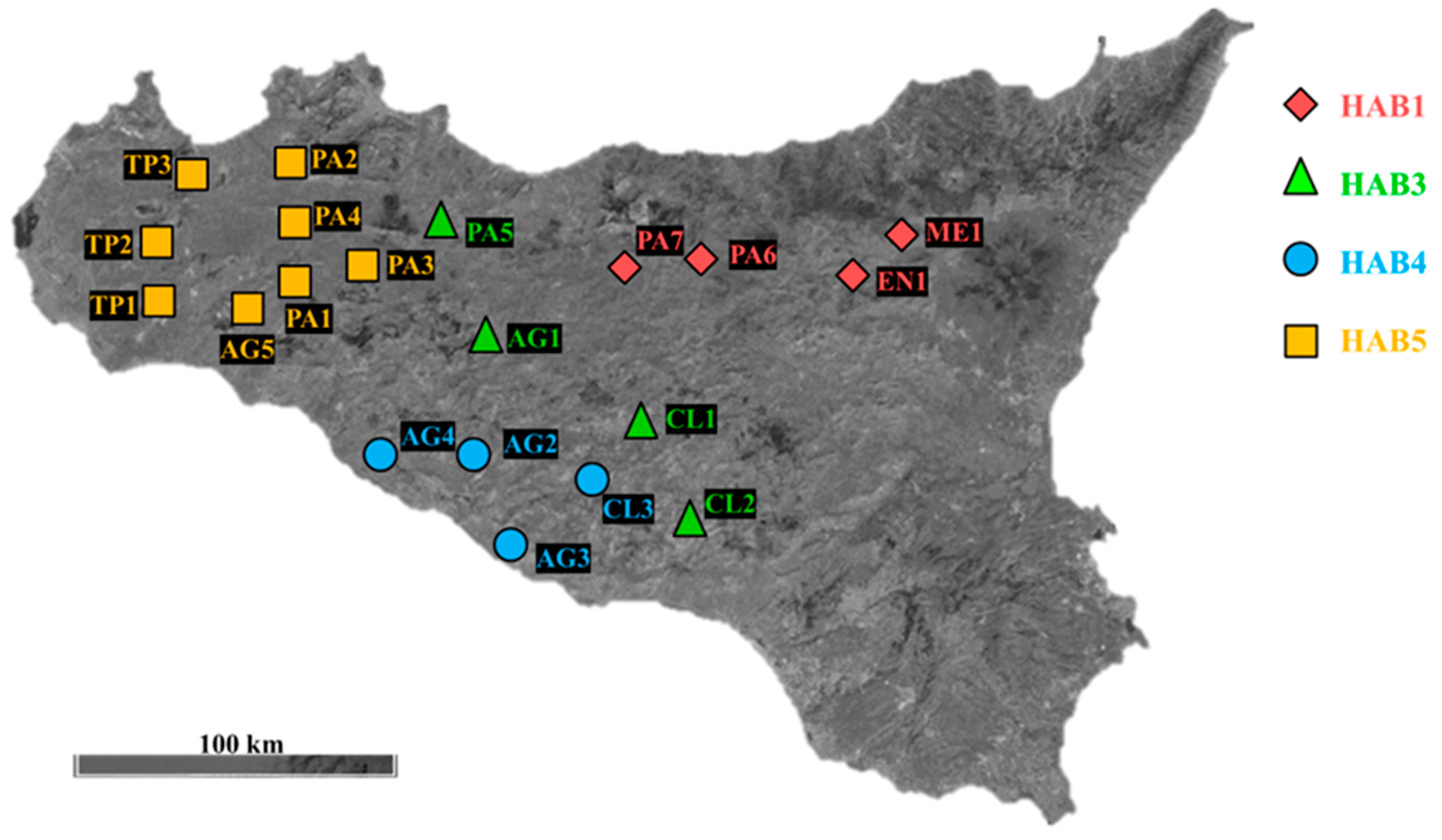
Figure 1. Geographical locations of the 20 sampling sites of the Sulla coronaria landraces. The 2 letters indicate the province of sampling: AG: Agrigento, PA: Palermo, TP: Trapani, CL: Caltanissetta, EN: Enna, ME: Messina, followed by a sequential number. Different symbols indicate the habitats assigned according to Ruisi et al. [10].
Agronomy 14 02667 g001
Figure 2. Floral explants used in this study. Anthers with filament (a), ovary (b), petals (c), whole flower (d).
Agronomy 14 02667 g002
Figure 3. Callus (a) and shoots (b,c) obtained from whole flower and ovaries. Respectively, (a) was obtained from ME1 cultured on S.c. 1 medium, whole flower, light conditions; (b,c) from TP1 and TP3 both from ovary, cultured on S.c. 3 medium, incubated in light regime. Pictures were taken after 1 month of culture.
Agronomy 14 02667 g003
Table 1. PGR combinations used in this study.
PGR CombinationsRef.
(S.c. 1) 5 µM N-(2-chloro-4-pyridyl)-N′-phenylurea (4-CPPU) + 5 µM 2,4-dichlorophenoxyacetic acid (2,4-D)[26]
(S.c. 2) 8.8 µM 6-banzylaminopurine (BA) + 1.07 µM 1-Naphthalene acetic acid (NAA)[27]
(S.c. 3) 10 µM naphthoxyacetic acid (NOA) + 4.4 µM 6-banzylaminopurine (BA)[26]
Table 2. Effects of biotypes, PGR combinations, explant type, and light conditions on callus formation.
VariablesCategorySignificance for Callus ProductionType of Effect
BiotypesAG1   Reference category
AG2***-
AG3**-
AG4***-
AG5**-
CL1***-
CL2***-
CL3**-
EN1***-
ME1***-
PA1***-
PA2***-
PA3*-
PA4***-
PA5***-
PA6*-
PA7***-
TP1***-
TP2**-
TP3***-
PGR CombinationsS.c. 1   Reference category
S.c. 2n.s.n.s.
S.c. 3**-
Explant TypeAnt   Reference category
Ov***-
Pet***-
Wf***-
Light ConditionsDark   Reference category
Light*-
The baseline model is Biotype = “AG1”, PGR combinations = “S.c. 1”, Explant = “Ant”, and Light condition = “Dark”. Significance levels: n.s. = not significant; * = significant at α ≤ 0.05, ** = significant at α ≤ 0.01, *** = significant at α ≤ 0.001. +: positive interaction; -: negative interaction. Ant: Anther, Ov: Ovary, Pet: Petals, Wf: Whole flower.
Table 3. Effects of PGR combinations and explant type on meristematic shoots.
VariablesCategorySignificance for
Meristematic Shoots
Type of Effect
PGR CombinationsS.c. 1   Reference category
S.c. 2*+
S.c. 3**+
Explant TypeAnt   Reference category
Ovn.s.n.s.
Pet*-
Wfn.s.n.s.
The baseline model is PGR = “S.c. 1”, Explant type = “Ant”. Significance levels: n.s. = not significant; * = α ≤ 0.05; ** = α ≤ 0.01.
Disclaimer/Publisher’s Note: The statements, opinions and data contained in all publications are solely those of the individual author(s) and contributor(s) and not of MDPI and/or the editor(s). MDPI and/or the editor(s) disclaim responsibility for any injury to people or property resulting from any ideas, methods, instructions or products referred to in the content.