Article

Comparison of Nutritional and Medicinal Ingredients Between Ganoderma leucocontextum and G. lucidum

1
Department of Vegetables, College of Horticulture, China Agricultural University, Haidian District, Beijing 100193, China
2
Biotechnology and Genetic Germplasm Resources Research Institute, Yunnan Academy of Agricultural Sciences, Kunming 650205, China
*
Author to whom correspondence should be addressed.
These authors contributed equally to this work.
Agronomy 2024, 14(11), 2523; https://doi.org/10.3390/agronomy14112523
Submission received: 13 September 2024 / Revised: 24 October 2024 / Accepted: 25 October 2024 / Published: 27 October 2024

Abstract

:
The genus Ganoderma is a widely used medicinal fungus in East Asia. The main medicinal components are triterpenoids, polyphenols, and polysaccharides. Bitterness is an important commercial trait for Ganoderma. White Lingzhi (G. leucocontextum) is less bitter. But the characteristics of its nutritional and medicinal ingredients are still unclear, which undoubtedly limits its commercialization. In this study, the medicinal ingredients of Lingzhi and white Lingzhi were extracted and quantified. The structure and antioxidant activities of purified polysaccharides were determined. At the same time, their nutritional differences were compared. White Lingzhi contains more medicinal ingredients and its polysaccharide is more active. The higher protein content may be one of the reasons for weaker bitterness in white Lingzhi. The nutritional and medicinal traits of white Lingzhi were described for the first time in this study, which provides fundamental knowledge to support the development of white Lingzhi.

1. Introduction

Ganoderma lucidum, commonly known as Lingzhi, is a traditional Chinese fungus that is widely used in China, Japan, Korea, and other Asian countries. It possesses a wide range of bioactivities, including anti-tumor properties [1,2], immunomodulatory effects [3], antimicrobial and antiviral activities [4], liver-protective effects [5], anti-aging properties [6], and potential in diabetes control [7]. Lingzhi has been extensively studied due to its numerous beneficial properties.
In 2015, G. leucocontextum, also known as white Lingzhi, was identified as a new species [8]. White Lingzhi exhibits significant morphological differences compared to Lingzhi. While Lingzhi has a semi-circular or kidney-shaped cap and off-white to light brown hard flesh, white Lingzhi has a semi-circular or fan-shaped cap and white soft flesh. Additionally, white Lingzhi has been found to promote neurite outgrowth [9] and possess immunomodulatory effects [10].
The medicinal value of Ganoderma fungi primarily stems from the presence of bioactive substances, including polysaccharides, triterpenoids, polyphenols, and proteins [11]. However, Lingzhi’s practical applications are hindered by its poor palatability due to its strong bitter taste. Furthermore, there have been reports of adverse reactions associated with Lingzhi consumption, such as dry mouth, nausea [12], diarrhea [13], sore throat [14], and insomnia [1].
In contrast, white Lingzhi exhibits relatively less bitterness and causes milder hypersensitivity reactions, making it a more promising candidate for broader applications. These characteristics of white Lingzhi make it more palatable and tolerable for individuals, potentially increasing its acceptability and usage.
Overall, the discovery of white Lingzhi as a new species and its distinct morphological features, along with its unique bioactivities, suggest that white Lingzhi has the potential to contribute to the development of new therapeutic agents. Further research is warranted to explore the full range of its medicinal properties and potential applications.
In this study, our focus was on examining differences in the main nutritional and medicinal ingredients between white Lingzhi and Lingzhi. We extracted the main medicinal ingredients and quantified these medicinal and nutritional components. Additionally, we characterized the structure of the polysaccharide. Furthermore, to gain a clearer understanding of the bioactivities, we investigated the antioxidant capacity in vitro of the polysaccharides, phenolics, and triterpenoids.
These investigations into the differences between white Lingzhi and Lingzhi may explain the milder and less bitter taste of white Lingzhi. The details obtained from this study will serve as a foundation for the more accurate application of white Lingzhi, providing valuable insights into its potential benefits and uses.

2. Materials and Methods

2.1. Materials and Chemicals

Fresh fruiting bodies of Lingzhi and white Lingzhi were collected from the Biotechnology and Genetic Germplasm Resources Research Institute, Kunming, China. 1,1-diphenyl-2-picrylhydrazyl (DPPH), pyrogallic acid, and ferrozine were obtained from Sigma-Aldrich. All the reagents were analytical grade.

2.2. Preparation of Sample

The fruiting bodies were dried in an oven at 40 °C until a constant weight was achieved. Subsequently, they were ground into a fine powder (40 mesh) using a ball mill (model PM 100; Retsch GmbH; Haan, Germany) operating at a speed of 350 rpm for 1.5 h. The ball mill operated for 5 min followed by a 10 min interval during which the rotational direction was changed. These powder samples were stored in a desiccator for the following experiments.

2.3. Determination of Protein Content

The content of crude protein was quantified by the Kjeldahl method [15]. A total of 1 g sample of powder was mixed with 12 mL H2SO4 and 6.4 g catalytic mix (K2SO4:CuSO4 = 15:1, w/w). The sample was heated to 400 °C to completely fix the protein nitrogen into ammonium. In total, 60 mL H2O and 50 mL 40% NaOH solution were added to the digestion solution and heated for distillation. The distillate was absorbed by 2.5% H3BO3, which was then titrated with 0.05 mol/L HCl standardized for the quantification of total nitrogen (N). For the protein content, the value of N was converted to N × 4.38 fungal protein [16].

2.4. Determination of Amino Acids Composition

The amino acid composition was determined by the ninhydrin method with minor modification [17]. The sample (50 mg) was hydrolyzed by 6 M HCl for 24 h at 110 °C in vacuo. The supernatant was transferred and concentrated after cooling in the dark. The concentrated solution was diluted with sodium citrate buffer (0.2 M, pH 2.2) and filtered through a 0.2 μm filter for analysis via an amino acid analyzer (Biochrom; Cambridge, UK). A mixture of 17 kinds of amino acids was used as a working standard. The concentration of each amino acid was 100 nmol/mL. The content of valine was determined by an HPLC system equipped with a C18 column and UV detector. Tryptophan content was determined using the p-dimethylaminobenzaldehyde (p-DAB) colorimetric method [18].

2.5. Determination of Triterpenoid Content

Triterpenoids were extracted using methanol as the solvent. The sample was evenly dispersed in methanol at a ratio of 1:10 (w/v). The mixture was then shaken for 12 h at room temperature to ensure complete extraction.
The content of total triterpenoids (Tr) was represented by ursolic acid equivalents [19]. In brief, 100 μL of the extracting solution was mixed with 150 μL of 5% vanillin solution (dissolved in glacial acetic acid) and 500 μL of a 70% perchloric acid solution. The resulting mixture was heated for 45 min at 60 °C and then cooled on ice. As it cooled, 2.25 mL glacial acetic acid was added. The absorbance of the solution was measured at 548 nm against a blank (methanol). To prepare a calibration curve, different concentrations of ursolic acid (ranging from 0.1 to 0.5 mg/mL) dissolved in methanol were used as standards.

2.6. Determination of Phenolic Content

Phenolics were extracted using methanol as the solvent. The sample was evenly dispersed in methanol at a ratio of 1:10 (w/v), and the mixture was shaken for 12 h to ensure full extraction.
The content of total phenolics (Ph) was represented by gallic acid equivalents [20]. A total of 500 μL of the extract solution was mixed with 2.5 mL of Folin–Ciocalteu reagent (previously diluted ten times in water) and 2.0 mL of Na2CO3 solution (75 g/L). The mixture was vortexed and incubated for 30 min at 40 °C to develop color. The absorbance of the solution was measured at 765 nm against a methanol blank. To prepare a calibration curve, different concentrations of gallic acid (ranging from 0.1 to 0.5 mg/mL) dissolved in methanol were used as standards.

2.7. Analysis of Polysaccharide

2.7.1. Extraction of Crude Polysaccharide

The sample powder was dispersed in water at a ratio of 1:15 (w/v) and heated at 95 °C for 4 h. After centrifugation at 8000 rpm for 15 min, the supernatant was mixed with ethanol at a ratio of 1:3 (v/v) and left to stand for 10 h. The sediment was collected by centrifugation and dissolved in water. To remove proteins, Sevage reagent was used [21].
The polysaccharide content of the supernatant was determined using the 3,5-dinitrosalicylic (DNS) colorimetric method, as described by [22]. Prior to analysis, the supernatant was dialyzed against water by using a membrane with a molecular weight cutoff of 3500 Dalton. The dialyzed solution was then subjected to the DNS colorimetric method to quantify the polysaccharide content.

2.7.2. Purification of Polysaccharide

The polysaccharides were purified following the protocols of Feng et al. [23]. After dialysis against phosphate buffer (0.05 M, pH 7.4), the crude polysaccharide was purified on a DEAE-Sepharose column. Elution was performed stepwise with phosphate buffer (0.05 M, pH 7.4), 0.15 M NaCl in phosphate buffer (0.05 M, pH 7.4), 0.5 M NaCl in phosphate buffer (0.05 M, pH 7.4), and 1 M NaCl in phosphate buffer (0.05 M, pH 7.4). The fractions with high polysaccharide content, eluted by phosphate buffer (0.05 M, pH 7.4) and 0.5 M NaCl in phosphate buffer (0.05 M, pH 7.4), were collected for further analysis.
Subsequently, the collected fractions were subjected to a CM-Sepharose column and eluted with acetate buffer (0.05 M, pH 4.6). The unbound fraction was collected and subsequently freeze-dried to obtain pure polysaccharides. This purification process ensured the isolation of polysaccharides with high purity from the crude extract.

2.7.3. Characterization of Polysaccharide

The molecular weight of the polysaccharide was determined using high-performance gel permeation chromatography (HPGPC) with an Agilent 1200 HPLC system equipped with a PL aquagel-OH 50 column (7.7 × 300 mm) and a differential refractive index detector following the method described by [24]. Phosphate buffer (0.2 M, pH 7.5) was used as the flow phase at a flow rate of 0.5 mL/min. The weight-average molecular weights (Mw) and number-average molecular weights (Mn) were estimated from a calibration equation based on PL pullulan polysaccharide standards.
A thermogravimetric analysis (TGA) of polysaccharide was conducted with a thermal analyzer (model STA 2500; Netzsch; Selb, Germany) under a nitrogen atmosphere, and heated from 30 to 800 °C with a heating rate of 10 °C/min [25].
The polysaccharide was hydrolyzed by trifluoroacetic acid (TFA, 2.0 M) [26] and then analyzed using a high-performance anion exchange chromatography (HPAEC) system (Dionex ISC 3000, Sunnyvale, CA, USA) with pulsed amperometric detector (PAD), CarboPac PA20 column (4 × 250 mm, Dionex), and PA-20 guard column (3 × 30 mm). Calibration was performed using the standard solutions of L-arabinose, D-xylose, D-glucose, D-mannose, D-galactose, glucuronic acid, and galacturonic acid.
Fourier-transform infrared spectroscopy (FTIR) was performed on an FTIR spectrometer (Alpha; Bruker; Optics, Germany) with KBr disks containing 1% finely ground samples [27]. The spectrum was recorded in the range of 4000 to 400 cm−1.
For nuclear magnetic resonance (NMR) spectroscopy, the polysaccharide was dissolved in D2O, and solution-state 1H, 13C, and heteronuclear single quantum coherence (HSQC) NMR spectra were acquired at 298 K from an Avance III HD 500 spectrometer (Bruker; Optics, Germany) [28].

2.8. Antioxidant Capacity In Vitro

In this experiment, polysaccharides and phenolics were dissolved in phosphate buffer (0.1 M, pH 7.4), while triterpenoids were dissolved in 50% methanol. Antioxidant capacity was determined as follows:
The ability to scavenge H2O2 was evaluated following [29] with minor modifications. Sample solution (1.0 mL) with different concentrations was added to a mixture containing 0.4 mL H2O2 (0.3%) and 2.0 mL phosphate buffer (0.1 M, pH 7.4). The absorbance of H2O2 at 230 nm was determined after 10 min. H2O2 scavenging ability = (A − As + Ac)/A × 100, where A was the absorbance of the reaction solution without the sample, As was the absorbance of the reaction solution with the sample and Ac was that of the solution replaced H2O2 with solvent.
DPPH radical scavenging activity was assayed following [30]. Sample solution (1 mL) with different concentrations was added to 3 mL 75 μM DPPH in 50% methanol solution. The mixture was kept in the dark at room temperature for 30 min. Absorbance was measured at 517 nm. DPPH scavenging ability (%) = (A − As + Ac)/A × 100, where A was the absorbance of the solution including DPPH and methanol, As was the absorbance of the sample and DPPH solution, and Ac was the absorbance of the solution including the sample and methanol.
The binding of Fe2+ was assessed following [31] with minor modification. Sample solution (800 μL) with different concentrations was added to a reaction mixture containing 2.0 mL deionized water, 40 μL FeCl2 (2 mM), and 160 μL ferrozine solution (5 mM). Absorbance at 562 nm was determined after 10 min. The binding rate of Fe2+ = (A − As + Ac)/A × 100, where A was the absorbance of the reaction solution without the sample, As was the absorbance of the reaction solution with the sample, and Ac was that of the solution including the sample and water only.

2.9. Statistical Analysis

The independent experiments were performed in triplicate, and all the data were expressed as Means ± Standard Deviation (M ± S.D.). Statistical analyses were performed by one-way ANOVA using the software program GraphPad Prizm 8.0. The FTIR and TGA spectra were plotted with Origin 2019. The NMR spectra were analyzed with the software program MestreNova 14.0.

3. Results

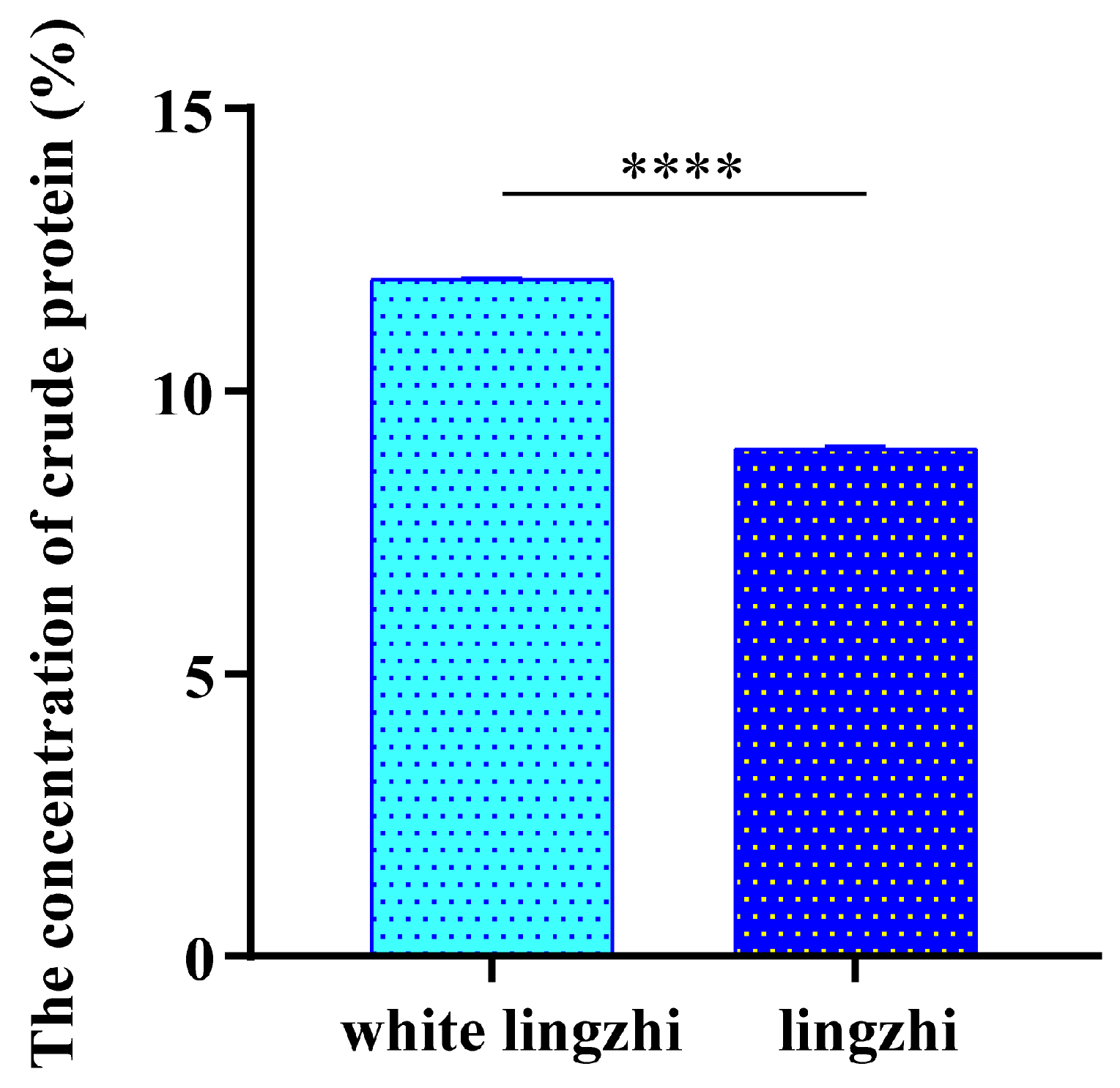
3.1. Difference in Crude Protein Concentration

The crude protein concentration of white Lingzhi was 11.98%, while Lingzhi was only 8.98% (Figure 1). In a wider study of Chinese Lingzhi, the mean protein concentration was 9.9% [32], which falls between our values for Lingzhi and white Lingzhi.

3.2. Differences in Amino Acid Composition

For a more detailed comparison of protein, the amino acid compositions were determined. We classified 18 amino acids into four groups (Table 1). The total amino acid concentration of white Lingzhi exceeded that of Lingzhi, which was consistent with the protein results. There were differences in the relative proportions of amino acids. The concentrations of monosodium glutamate-like (MSG-like) amino acids were higher in white Lingzhi, while the overall percentage of sweet amino acids was the highest in Lingzhi. However, there was no significant difference in the relative proportions of the four amino acid groups.

3.3. Differences in Concentrations and Antioxidant Activities of Triterpenoids

Triterpenoid concentrations and DPPH scavenging ability are shown in Figure 2. The triterpenoid concentration of white Lingzhi was significantly higher than that of Lingzhi. Ganderma triterpenoids were strong scavengers on DPPH. However, these two triterpenoid extracts showed little activity in H2O2 removal or Fe2+ chelation. This may be due to the location of the hydroxyl group in their structure. And, it indicates that the mode of action of triterpenoids does not involve Fe2+ chelation to any significant degree.

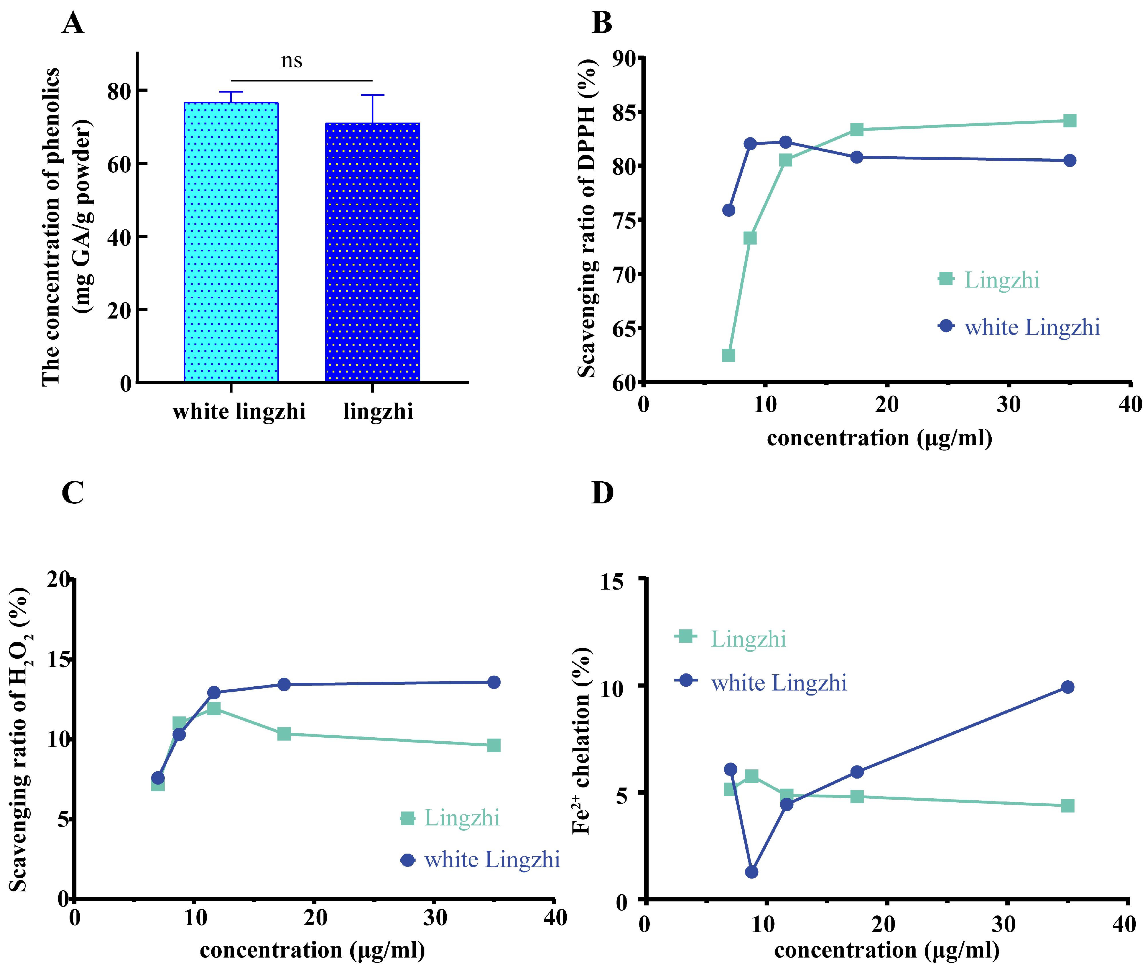
3.4. Differences in Concentrations and Antioxidant Activities of Total Phenolics

No significant difference in the concentration of phenolics between white Lingzhi and Lingzhi was observed. These phenolics are able to scavenge DPPH and H2O2 and can chelate Fe2+. But, the relationships between antioxidant capability and concentration were inconsistent. Compared with the phenolics from Lingzhi, those from white Lingzhi had a stronger initial capacity to scavenge H2O2 and chelate Fe2+. In addition, the scavenging ability of white Lingzhi phenolics on DPPH was relatively stable, and less affected by their concentration (Figure 3).

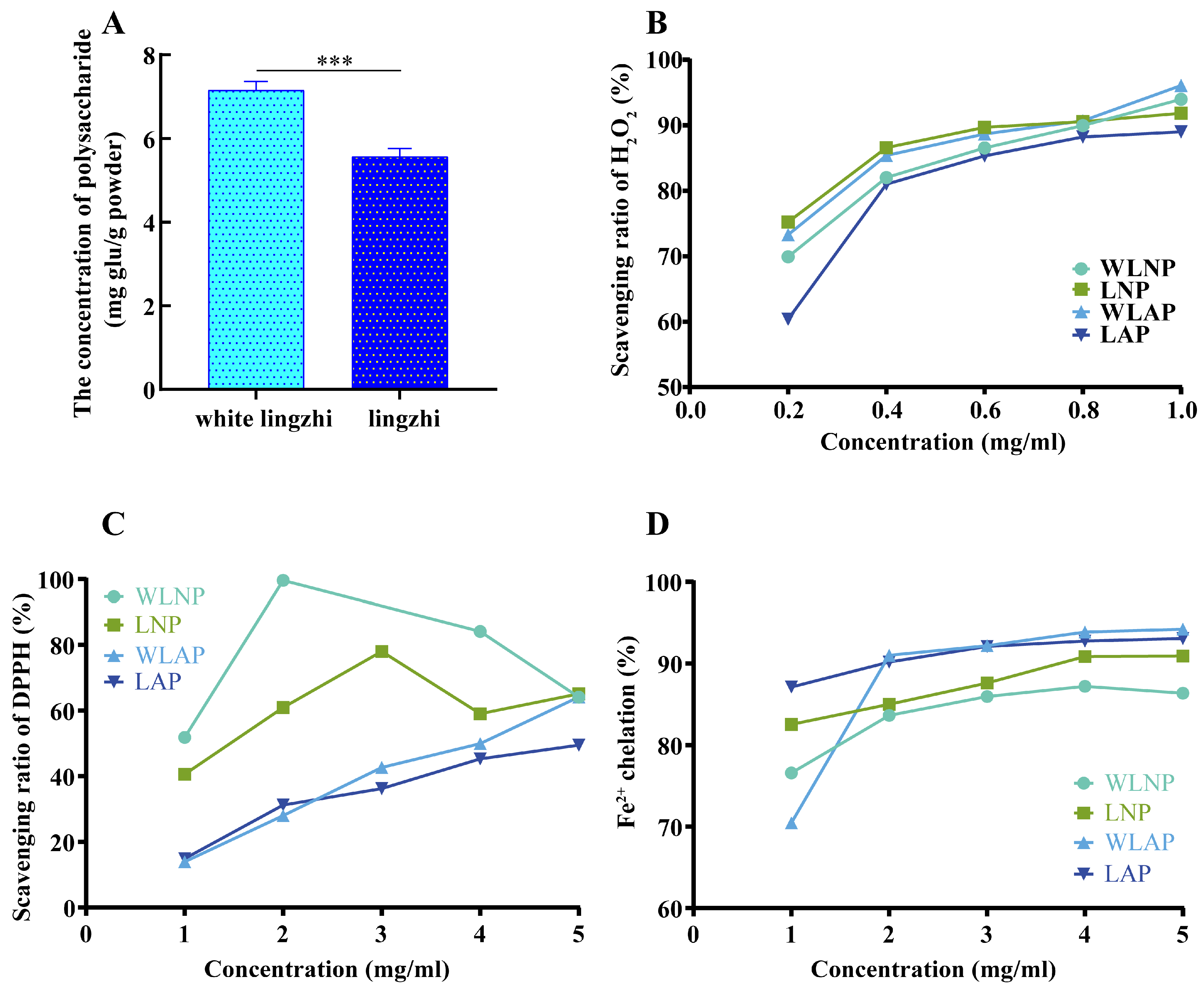
3.5. Differences in Characters and Antioxidant Activities of Polysaccharides

A DEAE-sepharose column was used to separate the polysaccharides into acidic and neutral fractions. The fraction bound to the column was identified as an acidic polysaccharide, while the unbound fraction was identified as a neutral polysaccharide. The purified fractions will be referred to as white Lingzhi neutral polysaccharide (WLNP), white Lingzhi acidic polysaccharide (WLAP), Lingzhi neutral polysaccharide (LNP), and Lingzhi acidic polysaccharide (LAP).

3.5.1. The Molecular Weights of Polysaccharides

Table 2 presents the molecular weights (Mw and Mn) of the four polysaccharides. The polydispersity (PD) values ranged between 1.55 and 1.75, indicating that the molecular weights of the polysaccharides were relatively uniform. Furthermore, it can be observed from Table 2 that the molecular weight of the neutral polysaccharide was higher than that of the acidic polysaccharide.

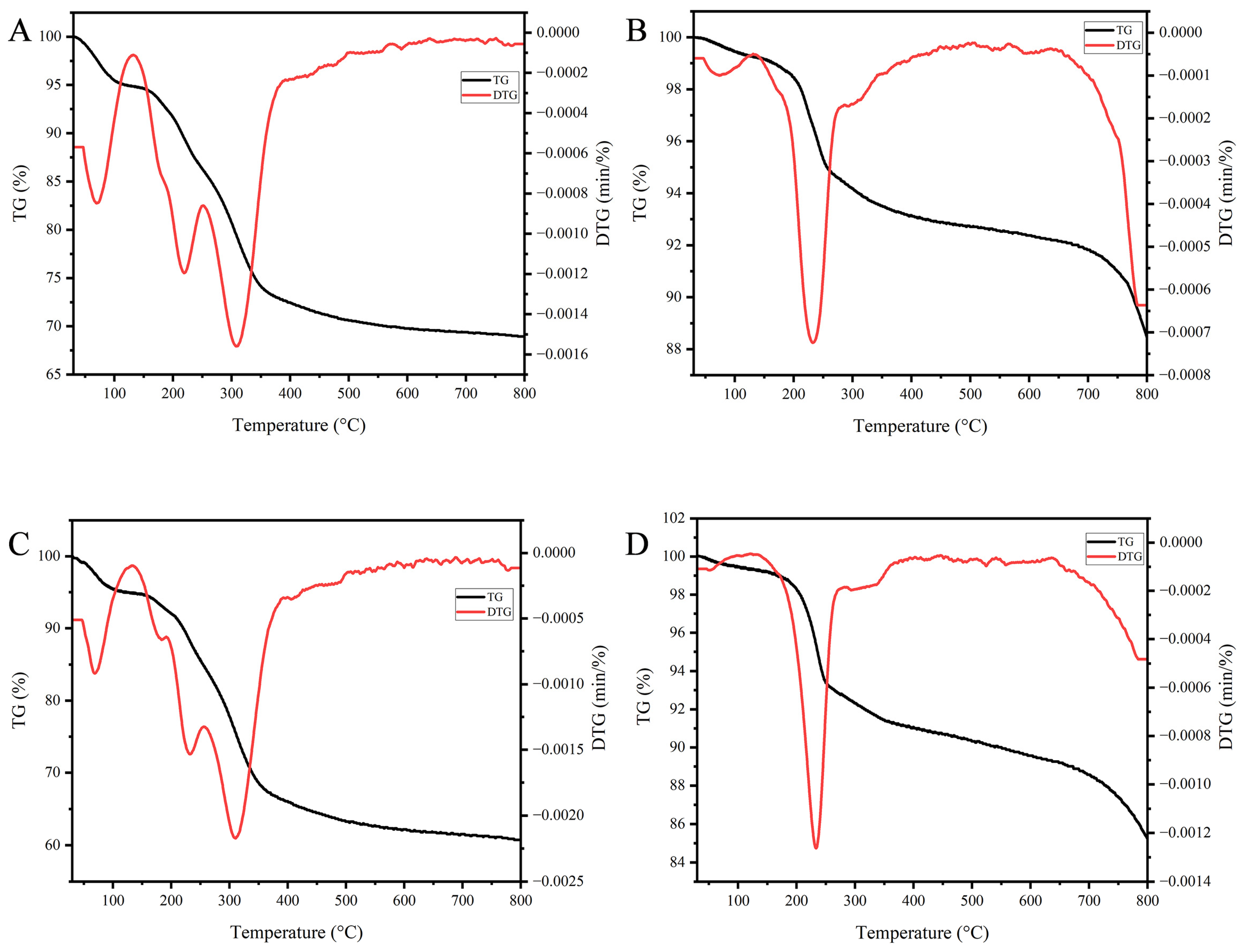
3.5.2. Thermogravimetric Analysis

The thermal stability of biomolecules plays a vital role in their suitability for various biological applications. In this study, the thermogravimetry (TG) and derivative thermogravimetry (DTG) curves of four polysaccharides were examined, as depicted in Figure 4. As the temperature increased, the residues gradually decreased. Interestingly, the acidic polysaccharides exhibited higher residue levels than did neutral polysaccharides at 800 °C. Moreover, the DTG curves of all four polysaccharides displayed a peak below 100 °C, which can be attributed to the evaporation of residual moisture [33]. However, it is worth noting that the weight loss peak for the acidic polysaccharides was smaller, indicating weaker water absorption and retention capabilities compared to the neutral polysaccharides. The remaining peaks observed in the curves were a result of thermal decomposition [34]. Notably, the thermal decomposition pattern differed between the acidic and neutral polysaccharides. The neutral polysaccharides exhibited significant weight loss between 150 and 400 °C, whereas the acidic polysaccharides displayed a minor, but more abrupt weight loss between 200 and 300 °C, followed by further weight loss above 700 °C. Our TG findings suggest that the acidic polysaccharides possess greater stability at high temperatures. Additionally, when comparing the amount of residues at 800 °C, it was observed that the white Lingzhi polysaccharide exhibited slightly better stability, although the difference was not statistically significant.

3.5.3. Monosaccharide Components of Polysaccharides

Based on our HPAEC-PAD analyses, the composition of the acidic polysaccharide was very simple, while the neutral polysaccharides were more complex (Table 3). The four polysaccharides were mostly composed of glucose (Glc), strongly suggesting that it is the major unit of the main backbone for Ganoderma polysaccharides. In addition, the neutral polysaccharides were composed of a large variety of monosaccharides, which suggests they may have a wider range of biological activities. The proportion of Glc in white Lingzhi was lower, with greater balance among the various component monosaccharides, which may be one of the reasons for the mild pharmacological properties of white Lingzhi.

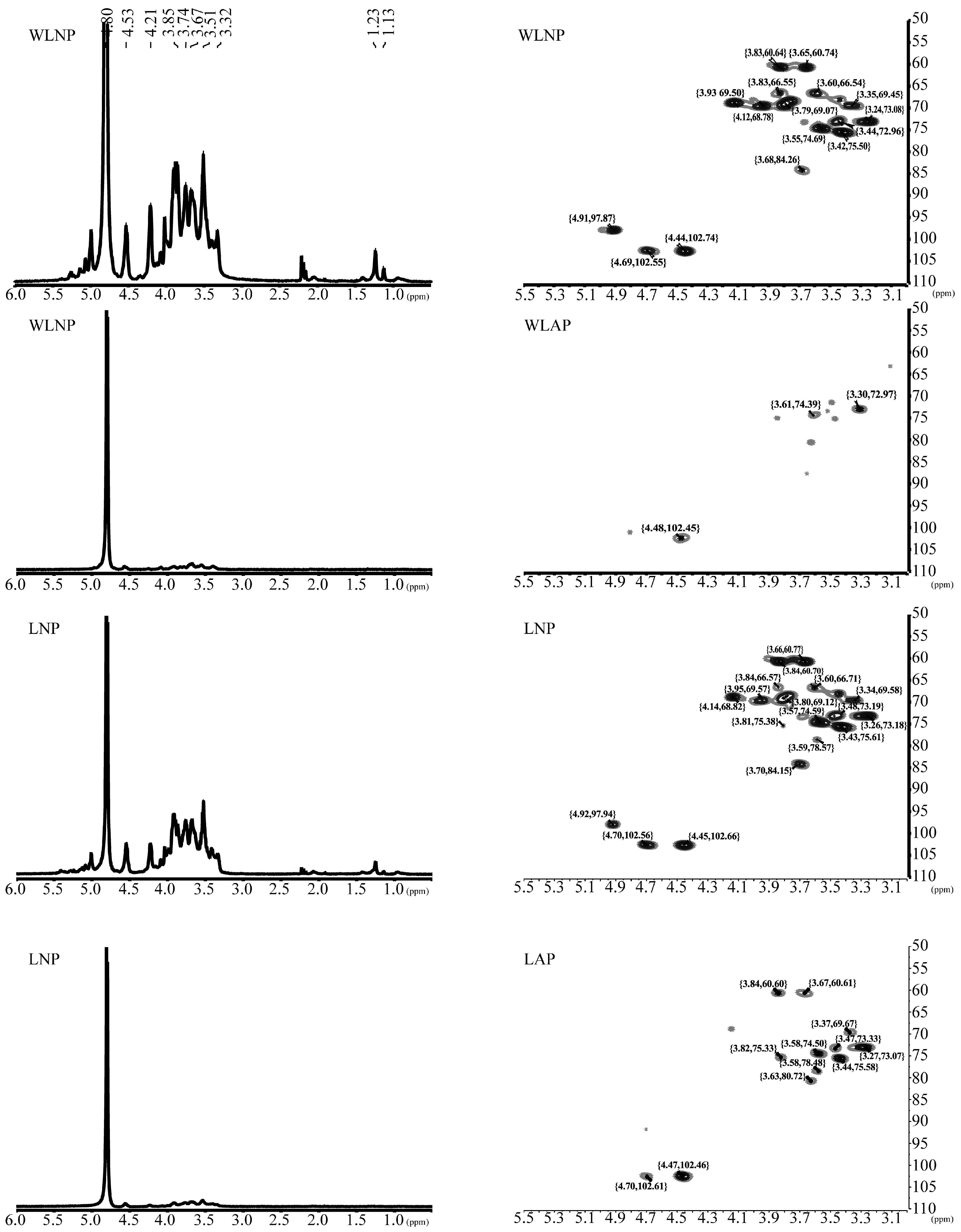
3.5.4. Polysaccharide Structural Analysis

The structure of the polysaccharides was analyzed using the Fourier-transform infrared spectroscopy (FTIR) and nuclear magnetic resonance (NMR) techniques. The FTIR spectra, presented in Figure 5, facilitate the determination of the chemical bonds and molecular structure of the polysaccharides, serving as complementary methods for structural analysis [35].
Several absorption peaks were observed in the FTIR spectra. The peaks at 3444.24 cm−1 and 2891.74 cm−1 can be attributed to the stretching vibration of hydroxyl groups and C-H groups, respectively [36,37]. The absorption peaks within the 1700–1300 cm−1 range indicate the presence of carboxyl groups in the polysaccharides. Specifically, the peak at 1635 cm−1 reflects C=O linkages [38], while the peak at 1362 cm−1 corresponds to carboxylate groups [39]. The peak observed at 1159 cm−1 reflects the stretching of α-(1,4) glycosidic linkages [40]. Additionally, the peak at 1074 cm−1 confirms the presence of a pyranose ring structure. Furthermore, the peaks observed at 946 cm−1, 861 cm−1, and 547 cm−1 represent β-glycosidic bonds, α-glycosidic bonds, and in-plane C=O bending, respectively [41,42,43]. Notably, the α-glucosidic bonds in the neutral polysaccharide were not clearly evident in the FTIR spectra.
In summary, the FTIR analysis provided valuable insights into the chemical bonds and molecular structure of the polysaccharides, highlighting the presence of hydroxyl groups, C-H groups, carboxyl groups, α-(1,4) glycosidic linkages, and various other functional groups.
The NMR spectra provide detailed information about the structural characteristics of the polysaccharide polymers. Both the 1H NMR and HSQC NMR spectra are presented in Figure 6. The 1H NMR spectra reveal several signals that indicate the presence of specific sugar residues. These include β-D-glucose (3.57 and 3.65 ppm), β-D-mannose (3.76 ppm), α-L-aranitol (4.12 ppm), and β-D-xylan (4.44 and 3.23 ppm). Further details can be observed in the HSQC spectra.
In the HSQC spectra, a δCH signal at 3.83/60.64 ppm corresponds to the C6-H of (1 → 4)-linked α-D-glucan [44]. The presence of different Glc configurations in the polysaccharide is evident from the 1H/13C chemical shifts. The signals at δCH 3.44/72.96 and 3.65/60.74 ppm are assigned to H-5/C-5 and H-6/C-6 of →3)-β-D-Glcp-(1→ residues [23]. The cross-peak at δCH 3.37/69.45 ppm is assigned to →3,6)-β-D-Glcp-(1→ [45,46,47]. The signal at δCH 4.91/97.87 ppm is associated with →6)-β-D-Glcp-(1→ [47]. Similarly, the cross-peaks at δCH 4.12/68.78 and 3.77/68.59 ppm are assigned to H-5/C-5 and H-6/C-6 of →6)-β-D-Manp-(1→ residues [23]. The δCH 4.69/102.55 ppm signal is associated with →3)-β-D-Galp-(1→ [45]. Additionally, the 1H/13C chemical shifts at δCH 4.44/102.74 and 3.25/73.08 ppm are assigned to C1-H and C2-H of (1 → 4)-linked β-D-xylan [44].
In summary, the neutral polysaccharides from white Lingzhi and Lingzhi were found to be composed of (1 → 4) α-glucan, →3)-β-D-Glcp-(1→, →3,6)-β-D-Glcp-(1→, →6)-β-D-Glcp-(1→, →6)-β-D-Manp-(1→, →3)-β-D-Galp-(1→, and (1 → 4)-linked β-D-xylan. This is consistent with the information obtained from the FTIR spectrum. The spectra of WLNP and LNP are very similar, with the exception that LNP has an additional peak (δCH 3.70/84.15 ppm) representing →3,6)-β-D-Galp-(1→ [45]. This indicates that LNP has an additional branch type, which may have an impact on its functionality. In comparison to the neutral polysaccharides, the structure of the acidic polysaccharides, particularly WLAP, appears to be simpler.

3.5.5. Antioxidant Activities

The polysaccharide concentration of white Lingzhi significantly exceeded that of Lingzhi. Ganoderma polysaccharides had different antioxidant patterns for different free radicals, and the properties of the four polysaccharides varied slightly (Figure 7). There was no significant difference in the scavenging effect of the four polysaccharides on H2O2 or Fe2+, and it was obviously dose-dependent. However, the dose required to scavenge H2O2 was much smaller. WLNP was the most effective in scavenging DPPH radicals at concentrations between 1 and 4 mg/mL.

4. Discussion

We compared the antioxidant activities of several components of Ganoderma, among which polysaccharides were the strongest, suggesting that the antioxidant activities in white Lingzhi and Lingzhi may be derived mostly from their polysaccharides.
Neutral and acidic polysaccharides of high Mw (>105 Da) were obtained from our extractions. Ganoderma polysaccharides with larger or smaller Mw have been obtained [48,49], which could be related to different varieties, extractions, and purification procedures [50]. The relationships between Mw and antioxidant activity were inconclusive. One viewpoint is that Mw in polysaccharides is positively correlated with its bioactivity [51]. However, some studies have shown that degraded polysaccharides have stronger antioxidant activity [49]. The two outcomes may not be contradictory. A possible explanation is that the antioxidant activity of polysaccharides shows two peaks in the molecular weight distribution. Another explanation is that the antioxidant activity of the purified polysaccharides can be positively correlated with Mw, and degradation further enhances their activity.
In light of the intended applications of these polysaccharide extracts, their stability must be considered. Polysaccharides must be subjected to intense heat in certain manufacturing processes. High thermal stability suggests that polysaccharides can be used for functionalization and many other chemical modifications [52] to enhance their biological activity. Additionally, the high stability of polysaccharides can reduce the costs associated with storage and transportation. The application scenario of a polysaccharide depends on its water retention and stability. For example, heat-stable acidic polysaccharides can be used in food processing, while neutral polysaccharides with good water retention can be added to cosmetics.
Reactive oxygen species (ROS) are reactive molecules produced by normal metabolism in the human body. Excessive levels of ROS can cause many metabolic disorders and diseases, such as cancer, liver injury, coronary heart disease, and hypertension [53]. Therefore, the balance between the production and removal of ROS is crucial to human health. Many studies have reported that polysaccharides can alleviate oxidative damage by scavenging free radicals, such as Sanqi polysaccharide, Agaricus bisporus polysaccharide, and G. lucidum polysaccharide [54,55,56]. The four kinds of Ganoderma polysaccharides have clear antioxidant activities. Although the antioxidant effect of WLNP is only slightly higher than that of LNP, the concentrations of triterpenoids and total phenolics in white Lingzhi are significantly higher. Similarly to polysaccharides, these triterpenoids and phenolics can also scavenge ROS [19]. And, our results showed that the triterpenoids and phenolics of white Lingzhi were superior to Lingzhi in antioxidant activity. Overall, the effective doses of white Lingzhi may be smaller than those of Lingzhi, which should reduce undesirable side effects.
The structure of polysaccharides is critically associated with their activity. Heteropolysaccharides exhibit superior anti-tumor and immunomodulatory properties because they contain a variety of monosaccharides [57]. Mannose, one of the most critical components, can activate the immune response effectively by mediating the mannose receptor [58]. Neutral residues like →3)-β-D-Galp-(1→ and →3,6)-β-D-Galp-(1→ may suggest a compositional basis for anti-aging activity [59].
We must comprehensively consider the efficacy and taste of G. lucidum because it is well established in Chinese traditional medicine. It is clear that the taste of Lingzhi reduces its consumption. Researchers are working on methods to reduce the bitterness and make Lingzhi better suited to consumer needs [42]. Our analysis of amino acid composition showed that the relative proportions of bitter amino acids did not differ between white Lingzhi and Lingzhi, but in absolute terms were higher in white Lingzhi, as were its triterpenoid. But, people who consume white Lingzhi agree that it is less bitter, contradicting previous studies reporting that bitterness was attributed to the content of triterpenoids [60]. Here, the ratio of triterpenoids to proteins (T/P) was introduced to represent the bitterness. Although the triterpenoid and protein levels in white Lingzhi were higher than those in Lingzhi, the T/P was significantly lower (the value of T/P was 0.58 for white Lingzhi and 0.64 for Lingzhi). Protein is often tasteless but may interfere with taste buds’ perception of white Lingzhi as less bitter [61].

5. Conclusions

In conclusion, the differences in the nutritional and medicinal ingredients of white Lingzhi and Lingzhi were compared. There is little difference in the nutritional ingredients of the two Ganoderma, but the concentrations of the medicinal ingredients in white Lingzhi are higher than those in Lingzhi. The triterpenoid levels in white Lingzhi are 20% higher than those in Lingzhi, and the polysaccharide fraction is 28% higher. White Lingzhi neutral polysaccharide has better antioxidant activity than do the other three polysaccharides, with one less branch than the Lingzhi neutral polysaccharide. The factors that affect the taste of G. lucidum are complex, and we use the ratio of triterpenoid to protein (T/P) to track the degree of bitterness. The T/P value of Lingzhi is 10% higher than that of white Lingzhi, which may help explain why white Lingzhi has a less bitter taste. Our findings provide a basis for the commercialization and further development of white Lingzhi.

Author Contributions

P.W.: data curation, formal analysis, investigation, methodology, resources, and writing—original draft. F.F.: data curation, formal analysis, investigation, methodology, resources, writing—original draft, and writing—review and editing. C.Y.: data curation, formal analysis, investigation, methodology, resources, and validation. Q.T.: data curation, formal analysis, investigation, methodology, and resources. G.T.: data curation, formal analysis, and writing—review and editing. L.H.: data curation, formal analysis, and writing—original draft. Y.B.: formal analysis and writing—review and editing. Q.L.: conceptualization, data curation, formal analysis, funding acquisition, investigation, methodology, project administration, resources, supervision, validation, writing—original draft, and writing—review and editing. All authors have read and agreed to the published version of the manuscript.

Funding

This research was funded by Yunnan Liu Qinghong Specialist Station (202305AF150164).

Data Availability Statement

The original contributions presented in the study are included in the article, further inquiries can be directed to the corresponding author.

Conflicts of Interest

The authors declare no conflicts of interest.

References

  1. Jin, X.; Ruiz Beguerie, J.; Sze, D.M.; Chan, G.C. Ganoderma lucidum (Reishi mushroom) for cancer treatment. Cochrane Database Syst. Rev. 2016, 4, Cd007731. [Google Scholar] [CrossRef] [PubMed]
  2. Liu, X.; Xu, Y.; Li, Y.; Pan, Y.; Sun, Z.; Zhao, S.; Hou, Y. Ganoderma lucidum fruiting body extracts inhibit colorectal cancer by inducing apoptosis, autophagy, and G0/G1 phase cell cycle arrest in vitro and in vivo. Am. J. Transl. Res. 2020, 12, 2675–2684. [Google Scholar] [PubMed]
  3. Lin, Z.B. Cellular and molecular mechanisms of immuno-modulation by Ganoderma lucidum. J. Pharmacol. Sci. 2005, 99, 144–153. [Google Scholar] [CrossRef]
  4. Wu, S.J.; Zhang, S.Y.; Peng, B.; Tan, D.C.; Wu, M.Y.; Wei, J.C.; Wang, Y.T.; Luo, H. Ganoderma lucidum: A comprehensive review of phytochemistry, efficacy, safety and clinical study. Food Sci. Hum. Wellness 2024, 13, 568–596. [Google Scholar] [CrossRef]
  5. Ahmad, M.F.; Ahmad, F.A.; Zeyaullah, M.; Alsayegh, A.A.; Mahmood, S.E.; AlShahrani, A.M.; Khan, M.S.; Shama, E.; Hamouda, A.; Elbendary, E.Y.; et al. Ganoderma lucidum: Novel insight into hepatoprotective potential with mechanisms of action. Nutrients 2023, 15, 1874. [Google Scholar] [CrossRef]
  6. Wang, J.; Cao, B.; Zhao, H.; Feng, J. Emerging roles of Ganoderma lucidum in anti-aging. Aging Dis. 2017, 8, 691–707. [Google Scholar] [CrossRef]
  7. Ma, H.T.; Hsieh, J.F.; Chen, S.T. Anti-diabetic effects of Ganoderma lucidum. Phytochemistry 2015, 114, 109–113. [Google Scholar] [CrossRef]
  8. Li, T.H.; Hu, H.P.; Deng, W.Q.; Wu, S.H.; Wang, D.M.; Tsering, T. Ganoderma leucocontextum, a new member of the G. lucidum complex from southwestern China. Mycoscience 2015, 56, 81–85. [Google Scholar] [CrossRef]
  9. Chen, H.; Zhang, J.; Ren, J.; Wang, W.; Xiong, W.; Zhang, Y.; Bao, L.; Liu, H. Triterpenes and meroterpenes with neuroprotective effects from Ganoderma leucocontextum. Chem. Biodivers. 2018, 15, e1700567. [Google Scholar] [CrossRef]
  10. Gao, X.; Qi, J.; Ho, C.T.; Li, B.; Mu, J.; Zhang, Y.; Hu, H.; Mo, W.; Chen, Z.; Xie, Y. Structural characterization and immunomodulatory activity of a water-soluble polysaccharide from Ganoderma leucocontextum fruiting bodies. Carbohydr. Polym. 2020, 249, 116874. [Google Scholar] [CrossRef]
  11. Batra, P.; Sharma, A.K.; Khajuria, R. Probing Lingzhi or Reishi medicinal mushroom Ganoderma lucidum (higher Basidiomycetes): A bitter mushroom with amazing health benefits. Int. J. Med. Mushrooms 2013, 15, 127–143. [Google Scholar] [CrossRef] [PubMed]
  12. Klupp, N.L.; Kiat, H.; Bensoussan, A.; Steiner, G.Z.; Chang, D.H. A double-blind, randomised, placebo-controlled trial of Ganoderma lucidum for the treatment of cardiovascular risk factors of metabolic syndrome. Sci. Rep. 2016, 6, 29540. [Google Scholar] [CrossRef]
  13. Chen, X.; Hu, Z.P.; Yang, X.X.; Huang, M.; Gao, Y.; Tang, W.; Chan, S.Y.; Dai, X.; Ye, J.; Ho, P.C.; et al. Monitoring of immune responses to a herbal immuno-modulator in patients with advanced colorectal cancer. Int. Immunopharmacol. 2006, 6, 499–508. [Google Scholar] [CrossRef]
  14. Chu, T.T.; Benzie, I.F.; Lam, C.W.; Fok, B.S.; Lee, K.K.; Tomlinson, B. Study of potential cardioprotective effects of Ganoderma lucidum (Lingzhi): Results of a controlled human intervention trial. Br. J. Nutr. 2012, 107, 1017–1027. [Google Scholar] [CrossRef]
  15. Nascimento, F.d.S.; Santos, C.A.F.; Figueiredo Neto, A.; Bezerra, L.G.V.; da Silva, S.A.M.F. Crude proteion content in pigeon pea grain lines. Rev. Contemp. 2024, 4, e3615. [Google Scholar] [CrossRef]
  16. Fraile-Fabero, R.; Ozcariz-Fermoselle, M.V.; Oria-de-Rueda-Salgueiro, J.A.; Garcia-Recio, V.; Cordoba-Diaz, D.; Del P Jiménez-López, M.; Girbés-Juan, T. Differences in antioxidants, polyphenols, protein digestibility and nutritional profile between Ganoderma lingzhi from industrial crops in asia and Ganoderma lucidum from cultivation and Iberian origin. Foods 2021, 10, 1750. [Google Scholar] [CrossRef] [PubMed]
  17. Fountoulakis, M.; Lahm, H.W. Hydrolysis and amino acid composition of proteins. J. Chromatogr. A 1998, 826, 109–134. [Google Scholar] [CrossRef] [PubMed]
  18. Gao, J.Z.; Fang, H. Spectrophotometric determination of L-tryptophan by condensation with p-dimethylaminobenzaldehyde. J. Cap. Norm. Univ. 1999, 1999, 48–52. [Google Scholar]
  19. Oludemi, T.; Barros, L.; Prieto, M.A.; Heleno, S.A.; Barreiro, M.F.; Ferreira, I. Extraction of triterpenoids and phenolic compounds from Ganoderma lucidum: Optimization study using the response surface methodology. Food Funct. 2018, 9, 209–226. [Google Scholar] [CrossRef]
  20. Heleno, S.A.; Barros, L.; Martins, A.; Queiroz, M.; Santos-Buelga, C.; Ferreira, I. Fruiting body, spores and in vitro produced mycelium of Ganoderma lucidum from Northeast Portugal: A comparative study of the antioxidant potential of phenolic and polysaccharidic extracts. Food Res. Int. 2012, 46, 135–140. [Google Scholar] [CrossRef]
  21. Ding, J.; Du, H.; Tan, H.; Li, J.; Wang, L.; Li, L.; Zhang, Y.; Liu, Y. Optimization of protein removal process of Lonicera japonica polysaccharide and its immunomodulatory mechanism in cyclophosphamide-induced mice by metabolomics and network pharmacology. Food Sci. Nutr. 2022, 11, 364–378. [Google Scholar] [CrossRef] [PubMed]
  22. Yuan, Z.; Cong, G.; Zhang, J. Effects of exogenous salicylic acid on polysaccharides production of Dendrobium officinale. S. Afr. J. Bot. 2014, 95, 78–84. [Google Scholar] [CrossRef]
  23. Feng, X.; Wang, P.; Lu, Y.; Zhang, Z.; Yao, C.; Tian, G.; Liu, Q. A novel polysaccharide from Heimioporus retisporus displays hypoglycemic activity in a diabetic mouse model. Front. Nutr. 2022, 9, 964948. [Google Scholar] [CrossRef] [PubMed]
  24. Sun, L.; Wang, C.; Shi, Q.; Ma, C. Preparation of different molecular weight polysaccharides from Porphyridium cruentum and their antioxidant activities. Int. J. Biol. Macromol. 2009, 45, 42–47. [Google Scholar] [CrossRef]
  25. Chen, Y.; Xue, Y. Purification, chemical characterization and antioxidant activities of a novel polysaccharide from Auricularia polytricha. Int. J. Biol. Macromol. 2018, 120, 1087–1092. [Google Scholar] [CrossRef]
  26. Yu, Z.; Liu, L.; Xu, Y.; Wang, L.; Teng, X.; Li, X.; Dai, J. Characterization and biological activities of a novel polysaccharide isolated from raspberry (Rubus idaeus L.) fruits. Carbohydr. Polym. 2015, 132, 180–186. [Google Scholar] [CrossRef] [PubMed]
  27. Wang, P.; Feng, X.; Lv, Z.; Liu, J.; Teng, Q.; Chen, T.; Liu, Q. Temporal dynamics of lignin degradation in Quercus acutissima sawdust during Ganoderma lucidum cultivation. Int. J. Biol. Macromol. 2024, 268, 131686. [Google Scholar] [CrossRef] [PubMed]
  28. Abeygunawardana, C.; Williams, T.C.; Sumner, J.S.; Hennessey, J.P., Jr. Development and validation of an NMR-based identity assay for bacterial polysaccharides. Anal. Biochem. 2000, 279, 226–240. [Google Scholar] [CrossRef]
  29. Ganie, S.A.; Haq, E.; Masood, A.; Hamid, A.; Zargar, M.A. Antioxidant and protective effect of ethyl acetate extract of podophyllum hexandrum rhizome on carbon tetrachloride induced rat liver injury. Evid.-Based Complement. Altern. Med. 2011, 2011, 238020. [Google Scholar] [CrossRef]
  30. Li, C.; Huang, Q.; Fu, X.; Yue, X.J.; Liu, R.H.; You, L.J. Characterization, antioxidant and immunomodulatory activities of polysaccharides from Prunella vulgaris Linn. Int. J. Biol. Macromol. 2015, 75, 298–305. [Google Scholar] [CrossRef]
  31. Dinis, T.C.P.; Madeira, V.M.C.; Almeida, L.M. Action of phenolic derivatives (acetaminophen, salicylate, and 5-aminosalicylate) as inhibitors of membrane lipid peroxidation and as peroxyl radical scavengers. Arch. Biochem. Biophys. 1994, 315, 161–169. [Google Scholar] [CrossRef] [PubMed]
  32. Stojković, D.S.; Barros, L.; Calhelha, R.C.; Glamočlija, J.; Ćirić, A.; van Griensven, L.J.; Soković, M.; Ferreira, I.C. A detailed comparative study between chemical and bioactive properties of Ganoderma lucidum from different origins. Int. J. Food Sci. Nutr. 2014, 65, 42–47. [Google Scholar] [CrossRef] [PubMed]
  33. Liang, X.; Hu, Q.; Wang, X.; Li, L.; Dong, Y.; Sun, C.; Hu, C.; Gu, X. Thermal kinetics of a lignin-based flame retardant. Polymers 2020, 12, 2123. [Google Scholar] [CrossRef]
  34. Rozi, P.; Abuduwaili, A.; Mutailifu, P.; Gao, Y.; Rakhmanberdieva, R.; Aisa, H.A.; Yili, A. Sequential extraction, characterization and antioxidant activity of polysaccharides from Fritillaria pallidiflora Schrenk. Int. J. Med. Mushrooms 2019, 131, 97–106. [Google Scholar] [CrossRef]
  35. Zhao, H.; Li, H.; Lai, Q.; Yang, Q.; Dong, Y.; Liu, X.; Wang, W.; Zhang, J.; Jia, L. Antioxidant and hepatoprotective activities of modified polysaccharides from Coprinus comatus in mice with alcohol-induced liver injury. Int. J. Biol. Macromol. 2019, 127, 476–485. [Google Scholar] [CrossRef]
  36. Liu, D.; Sun, Q.; Xu, J.; Li, N.; Lin, J.; Chen, S.; Li, F. Purification, characterization, and bioactivities of a polysaccharide from mycelial fermentation of Bjerkandera fumosa. Carbohydr. Polym. 2017, 167, 115–122. [Google Scholar] [CrossRef]
  37. Seedevi, P.; Moovendhan, M.; Viramani, S.; Shanmugam, A. Bioactive potential and structural chracterization of sulfated polysaccharide from seaweed (Gracilaria corticata). Carbohydr. Polym. 2017, 155, 516–524. [Google Scholar] [CrossRef] [PubMed]
  38. Wang, H.; Gao, T.; Du, Y.; Yang, H.; Wei, L.; Bi, H.; Ni, W. Anticancer and immunostimulating activities of a novel homogalacturonan from Hippophae rhamnoides L. berry. Carbohydr. Polym. 2015, 131, 288–296. [Google Scholar] [CrossRef]
  39. Bociek, S.M.; Welti, D. The quantitative analysis of uronic acid polymers by infrared spectroscopy. Carbohydr. Res. 1975, 42, 217–226. [Google Scholar] [CrossRef]
  40. Yang, J.P.; Hsu, T.; Lin, F.; Hsu, W.; Chen, Y. Potential antidiabetic activity of extracellular polysaccharides in submerged fermentation culture of Coriolus versicolor LH1. Carbohydr. Polym. 2012, 90, 174–180. [Google Scholar] [CrossRef]
  41. Jiang, J.; Kong, F.; Li, N.; Zhang, D.; Yan, C.; Lv, H. Purification, structural characterization and in vitro antioxidant activity of a novel polysaccharide from Boshuzhi. Carbohydr. Polym. 2016, 147, 365–371. [Google Scholar] [CrossRef] [PubMed]
  42. Ru, Y.; Chen, X.; Wang, J.; Guo, L.; Lin, Z.; Peng, X.; Qiu, B.; Wong, W.-L. Structural characterization, hypoglycemic effects and mechanism of a novel polysaccharide from Tetrastigma hemsleyanum Diels et Gilg. Int. J. Med. Mushrooms 2019, 123, 775–783. [Google Scholar] [CrossRef] [PubMed]
  43. Wang, W.F.; Stevenson, A.; Reuter, D.C.; Sirota, J.M. Absolute band intensities in the ν1923 (530 cm−1) and ν7 (777 cm−1) bands of acetone ((CH3)2CO) from 232 to 295 K. Spectrochim. Acta Part A Mol. Biomol. Spectrosc. 2000, 56, 1111–1116. [Google Scholar] [CrossRef]
  44. Li, M.F.; Fan, Y.M.; Xu, F.; Sun, R.C. Structure and thermal stability of polysaccharide fractions extracted from the ultrasonic irradiated and cold alkali pretreated bamboo. J. Appl. Polym. Sci. 2011, 121, 176–185. [Google Scholar] [CrossRef]
  45. Choi, J.W.; Synytsya, A.; Capek, P.; Bleha, R.; Pohl, R.; Park, Y.I. Structural analysis and anti-obesity effect of a pectic polysaccharide isolated from Korean mulberry fruit Oddi (Morus alba L.). Carbohydr. Polym. 2016, 146, 187–196. [Google Scholar] [CrossRef]
  46. Kono, H.; Kondo, N.; Hirabayashi, K.; Ogata, M.; Totani, K.; Ikematsu, S.; Osada, M. NMR spectroscopic structural characterization of a water-soluble β-(1→3, 1→6)-glucan from Aureobasidium pullulans. Carbohydr. Polym. 2017, 174, 876–886. [Google Scholar] [CrossRef] [PubMed]
  47. Liu, X.; Liu, D.; Chen, Y.; Zhong, R.; Gao, L.; Yang, C.; Ai, C.; El-Seedi, H.R.; Zhao, C. Physicochemical characterization of a polysaccharide from Agrocybe aegirita and its anti-ageing activity. Carbohydr. Polym. 2020, 236, 116056. [Google Scholar] [CrossRef]
  48. Ooi, L.S.M.; Ooi, V.E.C.; Fung, M.C. Induction of gene expression of immunomodulatory cytokines in the mouse by a polysaccharide from Ganoderma lucidum (Curt.: Fr.) P. Karst. (Aphyllophoromycetideae). Int. J. Med. Mushrooms 2002, 4, 27–35. [Google Scholar] [CrossRef]
  49. Xu, Y.; Zhang, X.; Yan, X.H.; Zhang, J.L.; Wang, L.Y.; Xue, H.; Jiang, G.C.; Ma, X.T.; Liu, X.J. Characterization, hypolipidemic and antioxidant activities of degraded polysaccharides from Ganoderma lucidum. Int. J. Biol. Macromol. 2019, 135, 706–716. [Google Scholar] [CrossRef]
  50. Yang, Y.; Qiu, Z.; Li, L.; Vidyarthi, S.K.; Zheng, Z.; Zhang, R. Structural characterization and antioxidant activities of one neutral polysaccharide and three acid polysaccharides from Ziziphus jujuba cv. Hamidazao: A comparison. Carbohydr. Polym. 2021, 261, 117879. [Google Scholar] [CrossRef]
  51. Kang, Q.; Chen, S.; Li, S.; Wang, B.; Liu, X.; Hao, L.; Lu, J. Comparison on characterization and antioxidant activity of polysaccharides from Ganoderma lucidum by ultrasound and conventional extraction. Int. J. Biol. Macromol. 2019, 124, 1137–1144. [Google Scholar] [CrossRef] [PubMed]
  52. Xu, C.; Yang, C.; Mao, D. Fraction and chemical analysis of antioxidant active polysaccharide isolated from flue-cured tobacco leaves. Pharmacogn. Mag. 2014, 10, 66–69. [Google Scholar] [CrossRef] [PubMed]
  53. Ma, J.S.; Liu, H.; Han, C.-R.; Zeng, S.J.; Xu, X.J.; Lu, D.J.; He, H.J. Extraction, characterization and antioxidant activity of polysaccharide from Pouteria campechiana seed. Carbohydr. Polym. 2020, 229, 115409. [Google Scholar] [CrossRef] [PubMed]
  54. Feng, S.L.; Cheng, H.R.; Xu, Z.; Feng, S.C.; Yuan, M.; Huang, Y.; Liao, J.Q.; Ding, C.B. Antioxidant and anti-aging activities and structural elucidation of polysaccharides from Panax notoginseng root. Process Biochem. 2019, 78, 189–199. [Google Scholar] [CrossRef]
  55. Li, S.; Liu, H.; Wang, W.; Wang, X.; Zhang, C.; Zhang, J.; Jing, H.; Ren, Z.; Gao, Z.; Song, X.; et al. Antioxidant and anti-aging effects of acidic-extractable polysaccharides by Agaricus bisporus. Int. J. Biol. Macromol. 2018, 106, 1297–1306. [Google Scholar] [CrossRef]
  56. Ma, C.; Feng, M.; Zhai, X.; Hu, M.; You, L.; Luo, W.; Zhao, M. Optimization for the extraction of polysaccharides from Ganoderma lucidum and their antioxidant and antiproliferative activities. J. Taiwan Inst. Chem. Eng. 2013, 44, 886–894. [Google Scholar] [CrossRef]
  57. Ruthes, A.C.; Smiderle, F.R.; Iacomini, M. Mushroom heteropolysaccharides: A review on their sources, structure and biological effects. Carbohydr. Polym. 2016, 136, 358–375. [Google Scholar] [CrossRef]
  58. Zhu, X.-l.; Lin, Z.-b. Effects of Ganoderma lucidum polysaccharides on proliferation and cytotoxicity of cytokine-induced killer cells. Acta Pharmacol. Sin. 2005, 26, 1130–1137. [Google Scholar] [CrossRef]
  59. Liang, X.; Liu, M.; Wei, Y.; Tong, L.; Guo, S.; Kang, H.; Zhang, W.; Yu, Z.; Zhang, F.; Duan, J.-a. Structural characteristics and structure-activity relationship of four polysaccharides from Lycii fructus. Int. J. Biol. Macromol. 2023, 253, 127256. [Google Scholar] [CrossRef]
  60. Hennicke, F.; Cheikh-Ali, Z.; Liebisch, T.; Maciá-Vicente, J.G.; Bode, H.B.; Piepenbring, M. Distinguishing commercially grown Ganoderma lucidum from Ganoderma lingzhi from Europe and East Asia on the basis of morphology, molecular phylogeny, and triterpenic acid profiles. Phytochemistry 2016, 127, 29–37. [Google Scholar] [CrossRef]
  61. Bohin, M.C.; Roland, W.S.; Gruppen, H.; Gouka, R.J.; van der Hijden, H.T.; Dekker, P.; Smit, G.; Vincken, J.P. Evaluation of the bitter-masking potential of food proteins for EGCG by a cell-based human bitter taste receptor assay and binding studies. J. Agric. Food Chem. 2013, 61, 10010–10017. [Google Scholar] [CrossRef] [PubMed]
Figure 1. Crude protein concentration in white Lingzhi and Lingzhi. **** indicates that p < 0.0001.
Agronomy 14 02523 g001
Figure 2. Concentrations and antioxidant activities of triterpenoids. The concentration (A) of triterpenoids and capacity to scavenging DPPH (B), H2O2 (C) and chelate Fe2+ (D) in white Lingzhi and Lingzhi. *** indicates that p < 0.001.
Agronomy 14 02523 g002
Figure 3. Concentrations and antioxidant activities of phenolics. The concentration (A) of phenolics and capacity to scavenging DPPH (B), H2O2 (C) and chelate Fe2+ (D) in white Lingzhi and Lingzhi. ns indicates that no significance.
Agronomy 14 02523 g003
Figure 4. TG and DTG curves for four different samples: (A) WLNP, white Lingzhi neutral polysaccharide; (B) WLAP, white Lingzhi acidic polysaccharide; (C) LNP, Lingzhi neutral polysaccharide; and (D) LAP, Lingzhi acidic polysaccharide.
Agronomy 14 02523 g004
Figure 5. FTIR spectra of WLNP (white Lingzhi neutral polysaccharide), WLAP (white Lingzhi acidic polysaccharide), LNP (Lingzhi neutral polysaccharide), and LAP (Lingzhi acidic polysaccharide).
Agronomy 14 02523 g005
Figure 6. 1H (left) and HSQC (right) NMR spectra of WLNP (white Lingzhi neutral polysaccharide), WLAP (white Lingzhi acidic polysaccharide), LNP (Lingzhi neutral polysaccharide), and LAP (Lingzhi acidic polysaccharide).
Agronomy 14 02523 g006
Figure 7. Concentrations and antioxidant activities of polysaccharides. The concentration (A) of polysaccharides and capacity to scavenging DPPH (B), H2O2 (C) and chelate Fe2+ (D) in white Lingzhi and Lingzhi. *** indicates that p < 0.001.
Agronomy 14 02523 g007
Table 1. Amino acid composition of white Lingzhi and Lingzhi.
Amino AcidAmino Acid Concentration (mg g−1)
White LingzhiLingzhi
MSG-likeAspartic acid0.96 ± 0.020.77 ± 0.02
Glutamic acid1.44 ± 0.020.80 ± 0.02
Total/percentage2.40/25.2%1.57/22.6%
SweetAlanine0.64 ± 0.000.50 ± 0.01
Glycine0.50 ± 0.010.40 ± 0.02
Serine0.47 ± 0.010.40 ± 0.02
Threonine0.51 ± 0.010.47 ± 0.02
Total/percentage2.12/22.3%1.77/25.4%
BitterArginine0.65 ± 0.020.32 ± 0.01
Histidine0.17 ± 0.000.13 ± 0.01
Leucine0.88 ± 0.010.69 ± 0.02
Phenylalanine0.45 ± 0.000.34 ± 0.01
Methionine0.15 ± 0.010.10 ± 0.00
Cysteine0.15 ± 0.000.18 ± 0.00
Valine0.60 ± 0.010.48 ± 0.01
Total/percentage3.05/32.0%2.24/32.2%
TastelessIsoleucine0.44 ± 0.010.35 ± 0.01
Lysine0.55 ± 0.010.36 ± 0.01
Proline0.47 ± 0.010.35 ± 0.01
Tryptophan0.15 ± 0.00l0.09 ± 0.00
Tyrosine0.34 ± 0.000.23 ± 0.01
Total/percentage1.95/20.5%1.38/19.8%
Total amino acids9.526.96
Table 2. Molecular weight of polysaccharides.
PolysaccharideMwMnPD
WLNP131,12776,3721.72
WLAP123,86974,1051.67
LNP139,14188,4651.57
LAP104,00661,4171.69
Note: WLNP, white Lingzhi neutral polysaccharide; WLAP, white Lingzhi acidic polysaccharide; LNP, Lingzhi neutral polysaccharide; and LAP, Lingzhi acidic polysaccharide. PD is an abbreviation for polydispersity and is calculated as the ratio of Mw to Mn.
Table 3. The monosaccharide components of polysaccharides.
SampleFucose (Fuc)Galactose (Gal)Glucose (Glc)Xylose (Xyl)Glucuronic Acid (GlcA)
LAP0.000.0078.07%0.0021.93%
WLAP0.000.0066.91%0.0033.09%
LNP3.65%12.79%70.84%7.51%5.21%
WLNP5.05%19.21%66.32%6.00%3.42%
Disclaimer/Publisher’s Note: The statements, opinions and data contained in all publications are solely those of the individual author(s) and contributor(s) and not of MDPI and/or the editor(s). MDPI and/or the editor(s) disclaim responsibility for any injury to people or property resulting from any ideas, methods, instructions or products referred to in the content.

