Article

Identification and Functional Analysis of PR Genes in Leaves from Variegated Tea Plant (Camellia sinensis)

State Key Laboratory of Tea Plant Biology and Utilization, Anhui Agricultural University, 130 Changjiang West Road, Hefei 230036, China
*
Author to whom correspondence should be addressed.
These authors contributed equally to this work.
Agronomy 2024, 14(1), 156; https://doi.org/10.3390/agronomy14010156
Submission received: 5 December 2023 / Revised: 3 January 2024 / Accepted: 6 January 2024 / Published: 10 January 2024
(This article belongs to the Special Issue Secondary Metabolism in Tea Plants)

Abstract

:
Tea is a healthy beverage made from the leaves of the tea plant [Camellia sinensis (L.) O. Kuntze]. The tea plant is a perennial evergreen plant that is widely distributed in tropical and subtropical regions. PR proteins (pathogenesis-related proteins, PRs) are defense-related proteins induced under pathogenesis-related conditions. Currently, there are a few studies on PRs in plants. Tea leaf color mutants have been of wide interest to scientists as special materials. A tea plant displaying variegated spontaneous albinism found in a local tea plantation in Huangshan City, Anhui Province, China, was used as the material sample in this study. An analysis of preexisting transcriptomic and proteomic data revealed the significant upregulation of three classes of PRs. In order to investigate the correlation between PRs and variegated tea plants, a series of studies were conducted on PR genes. The results revealed the identification of 17 CsPR1, 3 CsPR4 and 31 CsPR5 genes in tea plants using bioinformatics methods, and their structures and promoter sequences. The expression of three PR1, two PR4 and one PR5 genes was determined to be induced in stress treatment experiments involving mechanical damage, insect bites, low temperature treatment, and fungal infections. Additionally, the ribonuclease activity of CsPR4 was successfully verified. This is the first study to report the ribonuclease activity of CsPR4 in tea plants. The results can serve as a reference for future studies on PRs in tea plants, offering new insights into information on albinism in tea plants.

1. Introduction

Biotic stress causes two types of immune responses in plants: pattern-triggered immunity (PTI) and effector-triggered immunity (ETI). PTI occurs in response to pathogen-associated molecular patterns (PAMPs) in microbial or pathogenic structures. The secretion of microbial pathogens that are recognized by resistance (R) proteins in plant cells leads to ETI. Finally, salicylic acid (SA) or jasmonic acid (JA) signaling pathways are activated, resulting in the production of pathogenesis-related (PR) proteins [1], which are produced by plants exposed to some abiotic stresses, such as low temperature and mechanical damage [2,3]. In addition, PRs are secreted when the plant matures or undergoes senescence [4]. However, it has also been reported that PRs accumulate during the growth and development of plants such as tobacco, mung beans and tomatoes [5]. Initially, PRs were defined as a class of defensive proteins on the base of their production under pathogenesis-related conditions. However, some researchers determined that the expression of PRs increased in plants during infection with pathogenic bacteria; therefore, PRs were believed to play a crucial role in the defense response. PRs were first discovered in tobacco plants when infected with the tobacco mosaic virus [6]. Later, PRs with high expression were reported in plants when infected with bacteria, fungi, viruses or when attacked by insects or nematodes [7].
To date, PRs have been divided into 17 families [8]. PR1 has been mostly studied for its anti-fungal properties. For example, 16 PR1 genes have been identified in garlic, and involved in the defense against Fusarium infection [9]. A PR1 gene was identified in oats, being activated in response to many abiotic (drought and heat treatment) and hormonal (JA, SA, abscisic acid (ABA), and indole-3-acetic acid) treatments [10]. The PR4 family is composed of low-molecular-weight acidic proteins that can be divided into two categories on the basis of their structure. These proteins are found in both dicotyledons and monocotyledons [11,12]. Studies have identified the functions of PR4 proteins: nuclease activity, chitinase activity and antifungal activity. A PR4 gene was identified in cocoa, and the PR4 protein was determined to exhibit nuclease activity and antifungal effects [13]. Two types of PR4 genes were identified in jelly fig (Ficus awkeotsang Makino) achenes. The purified proteins exhibited ribonuclease and chitinase activities, as well as antifungal activity [14]. The amino acid sequence and structure of the PR5 protein are similar to those of an intensely sweet-tasting protein, namely thaumatin, also called a sweet-like protein [15]. The PR5 protein possesses antifungal properties. The transgenic tomato with a PR5 gene knockout showed lower resistance to pathogenic fungi compared to the wild-type tomato [16]. A PR5 gene was recently identified in Camellia sinensis and transgenically transferred into Arabidopsis thaliana. Compared to the wild-type plant, the transgenic A. thaliana plant exhibited higher seed yield and viability, as well as higher drought tolerance [17].
The tea plant [C. sinensis (L.) O. Kuntze] is one of the most important nonalcoholic beverage crops. Under stimulation by the external environment, tea leaves produce a series of mutations to survive and reproduce. Among them, those causing variations in leaf color are of major concern. Albinism is a common variation in plants, and some albino mutants have been found in rice [18], Arabidopsis [19], and maize [20]. In most crops, albinism can cause adverse effects, such as decreased yield and quality. These effects are also caused by the damage of photosynthetic organs and the imbalance of nitrogen and carbon in plants [21]. However, the albino varieties of tea plants are popular because of their freshness and high theanine content. In tea plants, theanine and catechins are the main secondary metabolites which contribute to the flavor of tea. They also have healthy and medicinal values. Currently, chlorosis tea plants can be divided into albino (white leaves), etiolated (yellow leaves), and variegated types in accordance with light-sensitive, temperature-sensitive, and ecologically insensitive factors. The material used in this study is a variegated tea plant with an albino phenotype that does not change, regardless of ecological factors.
Studies related to PR1 and PR5 in tea plants have been conducted, and the stress resistance of PR genes in the tea plant has been reported. These studies have determined that CsPR1 responds to blister-blight disease stress [22] and that CsPR5 enhances seed production and viability in A. thaliana under drought stress [17]. Transfer of a PR5 gene from a tea plant into a potato plant improved its resistance [23]. However, no study on the expression of PRs in albino tea plants has been conducted. In this study, we collected a tea plant sample exhibiting variegated albinism from Furong Valley, Huangshan City, Anhui Province, China. The sample, which was subjected to transcriptomic and proteomic analyses, was described in our previous publications [24]. A significant upregulation of three classes of PRs was revealed after the analysis of pre-existing transcriptomic and proteomic data. Then, PR genes were identified and cloned in tea plants and their physicochemical properties, gene structures, and cis-acting elements were determined. Moreover, functional studies of the PRs were also performed. The obtained results can give some basic information on PR genes in a tea plant with a phenotype of variegated albinism.

2. Materials and Methods
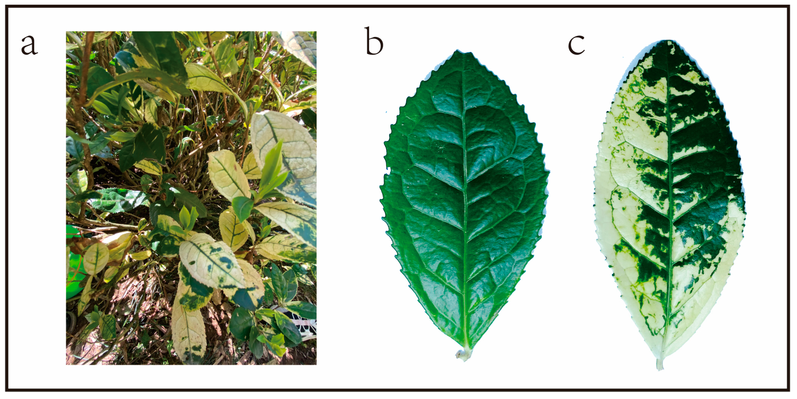
2.1. Plant Material

A variegated tea tree, approximately 10 years old (Figure 1a), growing in a local tea plantation in Furong Valley, Huangshan City, Anhui Province, China, was reported in our previous publications [24]. Some of the tissues in the variegated leaves appear white and the veins appear green. This tea plant has normal green-colored leaves (Figure 1b), as well as variegated leaves (Figure 1c). Three biological replicates of the leaves were collected from this tea plant at 3 stages of maturity, namely young leaves/control young leaves (YL/CYL), mature leaves/control mature leaves (DL/CDL), and old leaves/control old leaves (OL/COL). For the determination of PR genes expression, CK leaves from a normal tea plant with no albino phenotype, which was grown in the same tea plantation, were also used together with DL and CDL samples. Six-month-old cuttings of LongJing 43 (Camellia sinensis) were cultivated in a light incubator under a 12 h alternating light/dark cycle at 70% relative humidity, light intensity of 10,000 lx at 25 °C. After a week of growth in the greenhouse, the tea plants were subjected to the following treatments: low temperature, mechanical damage, insect feeding, and pathogenic fungal infection. For the low temperature treatment, tea plants were placed in a light incubator at 4 °C and subjected to the aforementioned treatments. The second leaf samples were collected at 0, 12, 24, and 48 h after the 4 °C treatment. For the mechanical damage treatment, the second leaf of the tea plants was damaged using scissors, and the plants were placed in the greenhouse as described above. The second leaf samples were collected at 0, 12, and 24 h after the mechanical damage treatment. For the insect (Ectropis obliqua) feeding treatment, the insect was placed on the second leaf of the tea plants. We waited for the insect to bite and a wound knot to appear, and then removed the worm. This tea plant was placed in the greenhouse as described above. The second leaf samples were collected at 0, 12 and 24 h after the insect feeding treatment [25]. The tea plants were inoculated with a pathogenic fungus (Colletotrichum camalliae) using the pinprick method [26]. The complete culture of PDA pathogenic fungus plate was cut into small pieces, the back of the leaf was scratched with a needle, and the mycelial piece of the small fungus block was attached to the wound. The block was removed by moistening for 24 h. The second leaf samples were collected at 0, 12, 24, 48, and 72 h after inoculation. Three pots of tea seedlings were set up for each treatment at each time point. All the samples were processed in liquid nitrogen and stored in a −80 °C refrigerator.

2.2. Identification of CsPR1, CsPR4, and CsPR5 Genes

The hidden Markov model profiles of PR1, PR4, and PR5 domains were obtained from the Pfam database (http://pfam.sanger.ac.uk/, accessed on 5 August 2022). The nonredundant sequences of PRs were identified by searching the conserved domains database [27] to validate the presence of PR domains. To identify all sequences of the PR family, the PRs of A. thaliana were used as queries in the BLAST program in TBtools [28] against the genomes of C. sinensis. The structures and the conserved motifs were analyzed using Simple MEME Wrapper in TBtools software (V 1.046). The exon–intron structure was identified from the Tea Plant Genome Database [29]. The structure was visualized using TBtools software (V 1.046).

2.3. Physicochemical Characterization of CsPRs

The ExPASy tool [30] was used to predict the properties of CsPRs. The subcellular localization of CsPRs was predicted using Plant-mPLoc ProtParam tools [31]. In addition, signal peptides were predicted using the SignalP 5.0 Server (https://services.healthtech.dtu.dk/services/SignalP-5.0/, accessed on 5 August 2022).

2.4. Promoter Analysis

The 2000 bp DNA upstream sequences of CsPR were extracted to predict the cis-acting regulatory elements by using Plant CARE (http://bioinformatics.psb.ugent.Be/webtools/plantcare/html/, accessed on 5 August 2022). These elements were visualized using TBtools software (V 1.046) [28].

2.5. Phylogenetic Analysis of CsPR1, CsPR4, and CsPR5

The amino acid sequences of PRs from C. sinensis and A. thaliana (as a model dicot plant) were used to construct a phylogenetic tree. All the PR sequences of the two species were aligned using MEGA 7.0 software with the software parameters (Poisson distribution, pairwise deletion, and a bootstrap value of 1000). [32]. A phylogenetic tree was constructed using the neighbor-joining method with Poisson distribution, pairwise deletion, and a bootstrap value of 1000 [32]. ITOL was used to modify the tree (https://itol.embl.de/, accessed on 5 August 2022).

2.6. RNA Isolation and Real-Time Quantitative Polymerase Chain Reaction

Total RNA was extracted using the RNA Prep Pure Plant Plus Kit (Tiangen, Beijing, China) in accordance with the manufacturer’s instructions. RNA was used for the synthesis of first-strand cDNA using the RTIII All-in-One Mix with dsDNase (Monad, Hefei, China). Real-time quantitative polymerase chain reaction (qPCR) was performed using the ChemoHS qPCR Mix (Monad, Hefei, China) on the CFX96 real-time PCR detection system (Bio-Rad Laboratories) with the following protocol: 95 °C for 10 min; 40 cycles at 95 °C for 10 s, and 60 °C for 30 s. The relative expression level was calculated using the 2−ΔΔCt method. CsActin (Genbank ID: XM_028240324.1) was used as the internal reference gene.

2.7. Expression and Purification of Recombinant CsPRs

CsPRs were first amplified using the amplification primers listed in Supplementary Table S1 (from NCBI and Tea Plant Information Archive, TPIA, http://tpia.teaplants.cn/, accessed on 1 July 2022), with the sample tea tree cDNA as the template; the CsPRs were ligated into the pEasy-Blunt cloning vector and sequencing was performed through colony PCR to screen for positive clones. Plasmids with correct sequences were extracted from single colonies and stored for backup. Primers were designed by adding the BamHI enzyme (New England Biolabs (Beijing) LTD, Beijing, China) cleavage site upstream of the target CDS (coding sequence) and the SalI enzyme (New England Biolabs (Beijing) LTD, Beijing, China) cleavage site downstream of the gene. The plasmid from the single colonies with correct sequences obtained after the sequencing of the aforementioned clone was used as a template, and the target gene, along with the enzyme cleavage site, was cloned again. The resulting PCR product was purified and used to ligate the vector. Subsequently, the pMAL-C2E prokaryotic expression vector (Laboratory preservation) was double digested with BamHI and SalI and then purified. The target gene fragment with the enzyme cleavage site was ligated to the expression vector fragment by using homologous recombinase and then transferred into Escherichia coli Trans1-T1 (Shanghai Weidi Biotechnology Co., Ltd., Shanghai, China). Positive clones were screened through colony PCR and sequenced to ensure in-frame insertion. The successfully ligated recombinant expression vector was transferred into E. coli BL21 (DE3) (Shanghai Weidi Biotechnology Co., Ltd., Shanghai, China). Protein expression in E. coli was examined as described previously [33]. Target proteins were eluted using the indicated protein purification columns. The column was eluted with a buffer comprising 200 mM NaCl (sodium chloride) (Sinopharm Chemical Reagent Co., Ltd., Shanghai, China), 1 mM EDTA (ethylene diamine tetraacetic acid) (Shanghai yuanye Bio-Technology Co., Ltd., Shanghai, China), 20 mM Tris (hydroxymethyl)-HCL (aminomethane hydrochloride) (Beijing Solarbio Science & Technology Co., Ltd., Beijing, China), and 1 mM DTT (dithiothreitol) (Beijing Solarbio Science & Technology Co., Ltd., Beijing, China) buffer (pH 7.5), followed by elution with the same buffer containing an additional 0.36 g of maltose (Aladdin Industrial Corporation, Shanghai, China). The target proteins were analyzed through 12% sodium dodecyl sulfate polyacrylamide gel electrophoresis.

2.8. Statistical Data Analysis

The heat map of proteins with significant differential expression, identified through proteomic data analysis, was generated using TBtools (V 1.046). The data were normalized using log2 transformation. All statistical analyses were performed with the SPSS 25.0 statistical package. The Kolmogorov–Smirnov test was used to inspect the normality and homogeneity of variance of all the data. A p > 0.1 was considered as an indication of normality in the data. For multi-group comparisons, p values were derived from one-way ANOVA and Duncan’s multiple range test. For all comparisons, p < 0.05 was considered statistically significant, and statistical significance was indicated using different letters.

3. Results

3.1. Proteomic and Transcriptomic Analyses

The samples (normal green color leaves and variegated leaves) were subjected to transcriptomic and proteomic analyses as described in our previous study [24]. We screened differentially expressed proteins (DEPs) on the basis of changes in expression by more than 1.2 times and by focusing on proteins with high expression in the proteome; a p value of <0.05 was considered statistically significant. DEPs were acquired from the comparison group DL-VS-CDL, which included 20 upregulated proteins. Figure 2a presents the quantitative proteomic statistics as a heatmap. We determined that the protein annotated as PR4 had significant differences in protein expression. The expression of PR4 protein was higher in DL than in CDL. Transcriptomic data for PR genes were also obtained. We identified more differentially expressed PR genes in the transcriptome data, including PR4, which was screened from the proteome data using the same method. The results showed that the expression of either PR genes or proteins was higher in DL than in CDL. As presented in Figure 2b, the expression of 21 PR genes exhibited significant differences in the transcriptome data. The genes marked with asterisks in the figure were screened for further research on the basis of their high expression levels and high significance.

3.2. Identification of the PR1, PR4, and PR5 Gene Family Members in C. sinensis

A total of 17 CsPR1, 3 CsPR4, and 31 CsPR5 genes were retrieved from the tea genome database (TPIA) [29]. All the PR genes were named as CsPR1-1 to CsPR1-17, CsPR4-1 to CsPR4-3, and CsPR5-1 to CsPR5-31 (Table 1). The CsPR1 protein had 159–376 amino acids and a molecular weight of 17.52–41.14 k Da. The pI (isoelectric point) values ranged from 4.88 to 10.07, indicating that the CsPR1 protein has both acidic and basic amino acids. Hydrophilicity (GRAVY) analysis revealed that all the CsPR1 had negative GRAVY values; thus, they were highly hydrophilic. The CsPR4 protein had 156–310 amino acids and a molecular weight of 16.82–34.43 k Da. The pI values ranged from 4.53 to 6.23, indicating that the CsPR4 protein had both acidic and basic amino acids. Hydrophobicity (GRAVY) analysis revealed that CsPR4 had negative and positive GRAVY values and was both hydrophilic and hydrophobic. The CsPR5 protein had 144–637 amino acids and a molecular weight of 15.79–70.56 k Da. The pI value ranged from 3.83 to 9.08, indicating that the CsPR5 protein had both acidic and basic amino acids. Hydrophobicity (GRAVY) analysis revealed that CsPR5 had both negative and positive GRAVY values and was both hydrophilic and hydrophobic. More detailed information is listed in Table 1.

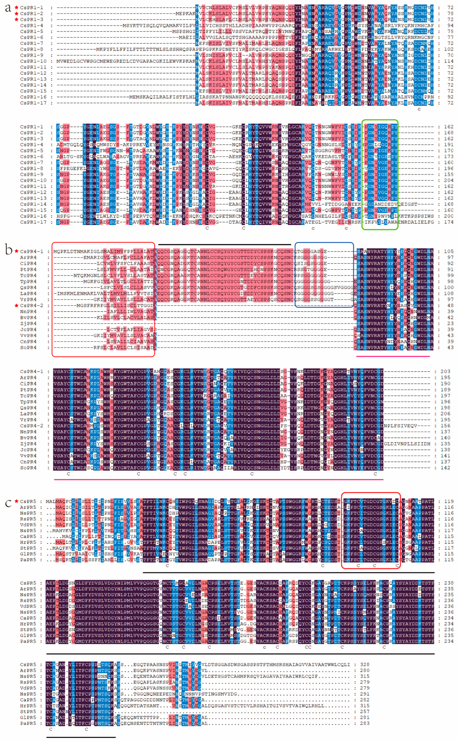
3.3. Evolutionary Relationships and Sequence Alignment of PR1, PR4, and PR5 Gene Family Members in C. sinensis

On the basis of amino acid sequences, we compared 17 CsPR1, 3 CsPR4, and 31 CsPR5 genes with 58 PR1, 3 PR4, and 5 PR5 genes from A. thaliana to analyze the genetic evolutionary relationships. The results clearly revealed that the three types of PRs can be categorized into classes I, II, and III, denoting PR1, PR4, and PR5, respectively (Figure 2c). The same type of PRs in Arabidopsis and C. sinensis clustered together within the same large branch. In the second group, two structures of PR4 were clearly delineated. Figure 3a presents the multiple sequence alignment of 17 PR1 proteins in C. sinensis, with five conserved semi-fatty acid sites, and the green boxes denote the regions contributing to their functions. Figure 3b presents the multiple sequence comparison of PR4 in C. sinensis and that in other species, with two PR4 types found. They both had a signal peptide at the front end and a Barwin structure in the middle. The main difference between the two PR4s was that the first type of PR4 had a chitin-binding domain structure between the signal peptide and the Barwin structure. The red box denotes the signal peptide, the black line indicates the chitin-binding domain structure, and the blue box represents a hinge. The pink line indicates the Barwin structural domain. Figure 3c illustrates the multiple sequence comparison of PR5 from C. sinensis and 10 other species. The red box represents the area contributing to its function, and the black line represents the entire structural domain.

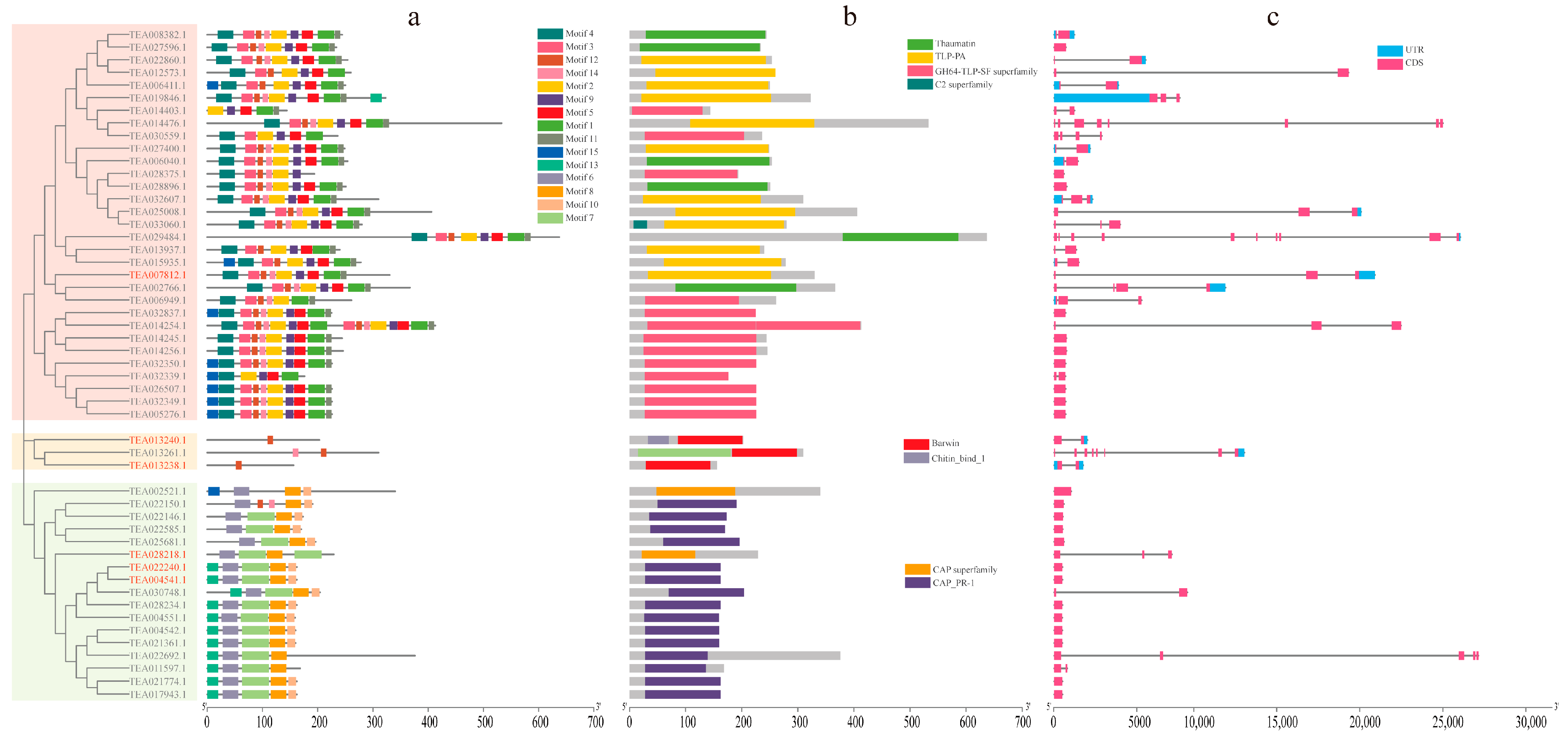
3.4. Gene Structures and Conserved Protein Motifs of CsPRs

The 51 members of PRs in C. sinensis were divided into three groups (Figure 4). On the basis of the sequences of CsPRs, 15 conserved themes were predicted using MEME in TBtools software. As presented in Figure 4a, 31 CsPR5 contained 6–10 motifs, 3 CsPR4 contained only 1 or 2 motifs, and 17 CsPR1 contained 4 or 5 motifs. The PR5 gene had the most motifs, which might be related to the length of its conserved structural domain. We visualized the conserved structural domains of genes to determine the length of the gene expression region occupied by structural domains (Figure 4b). PR5 had the TLP (thaumatin-like protein) conserved domain, PR4 had the Barwin domain, and PR1 had the CAP conserved domain. Expressed and non-expressed regions were also visualized (Figure 4c).

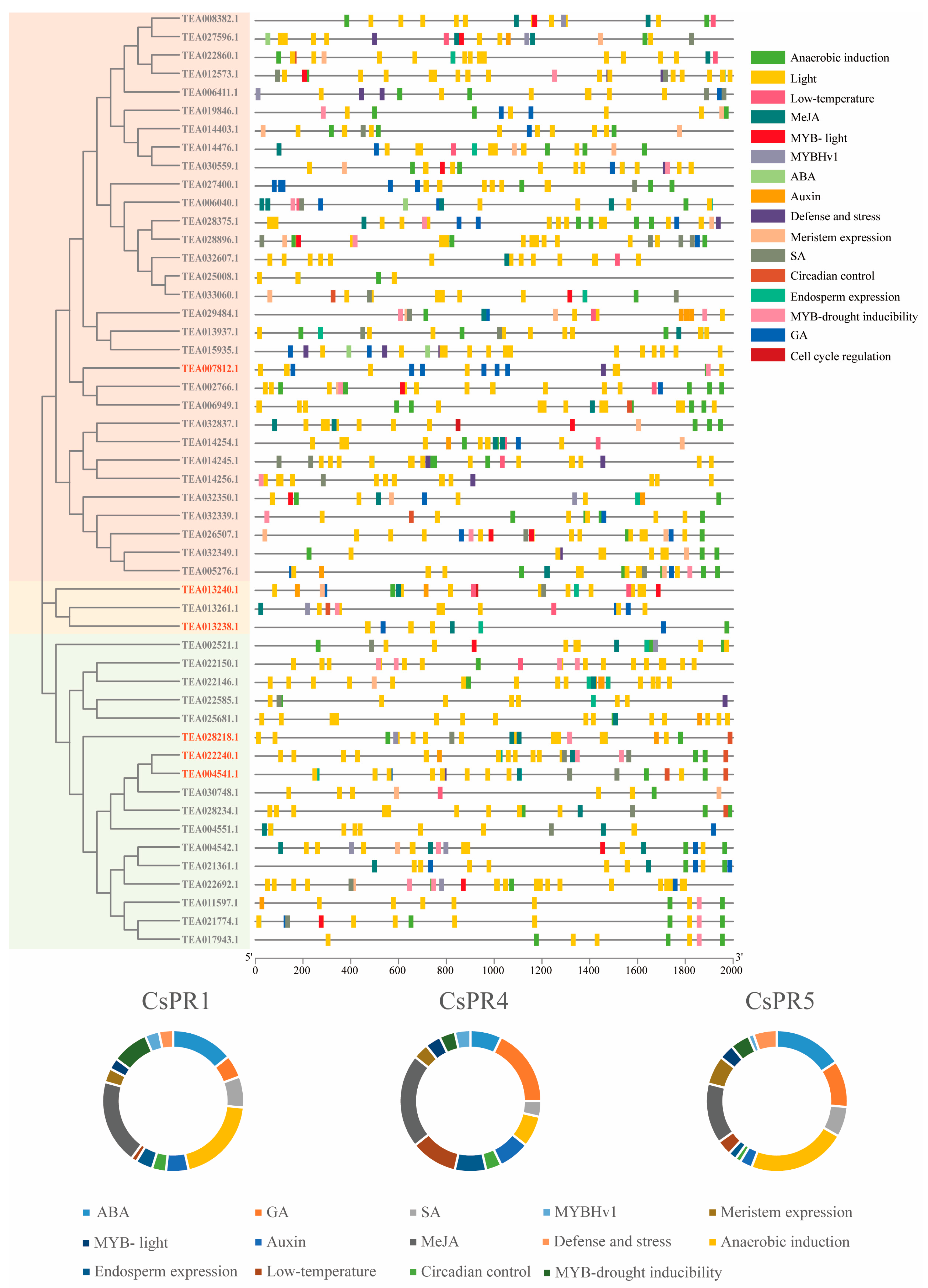
3.5. Analysis of Cis Elements of the CsPR Genes

To explore the regulatory network of CsPRs involved in plant growth and development, the 2 kb 5′ upstream regions of 51 CsPRs were analyzed (Figure 5). These regions could be divided into four categories: hormone response, plant growth and development, stress response, and transcription factors. The hormone response category included methyl jasmonate (31.50%), ABA (29.13%), gibberellin (18.50%), SA (13.39%), and auxin (7.48%). The plant growth and development category contained the endosperm expression element (27.45%), the meristem expression element (52.94%), and the circadian control element (19.61%). The stress response category included the anaerobic induction element (73.43%), low-temperature responsiveness (11.19%), and defense and stress (15.38%). However, defense and stress were not found in CsPR4. The transcription factor category contained MYB-drought inducibility (51.85%), MYB-light (29.63%), and MYBHv1 (18.52%). A large number of light-responsive elements were observed in the promoters of all CsPRs.

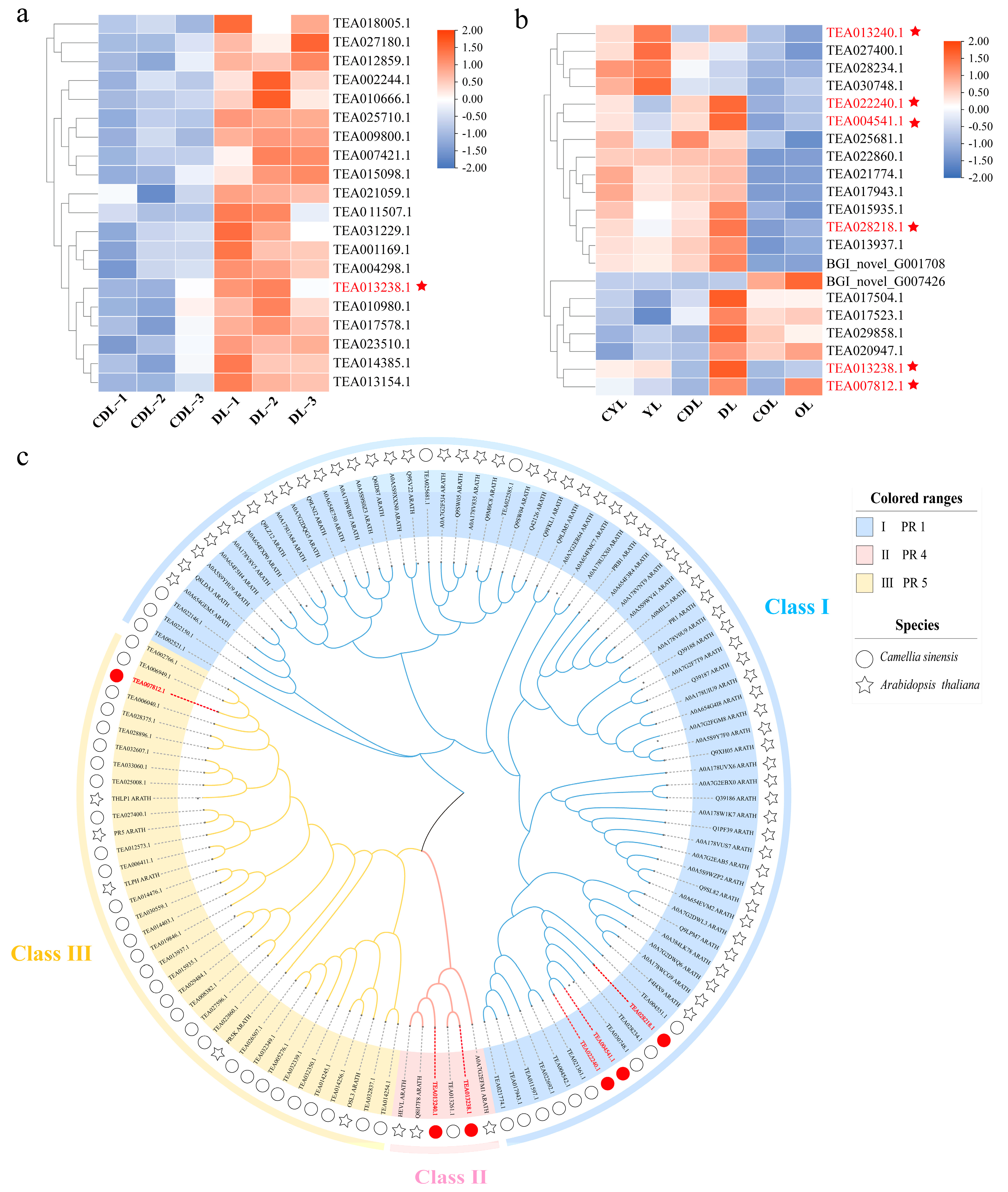
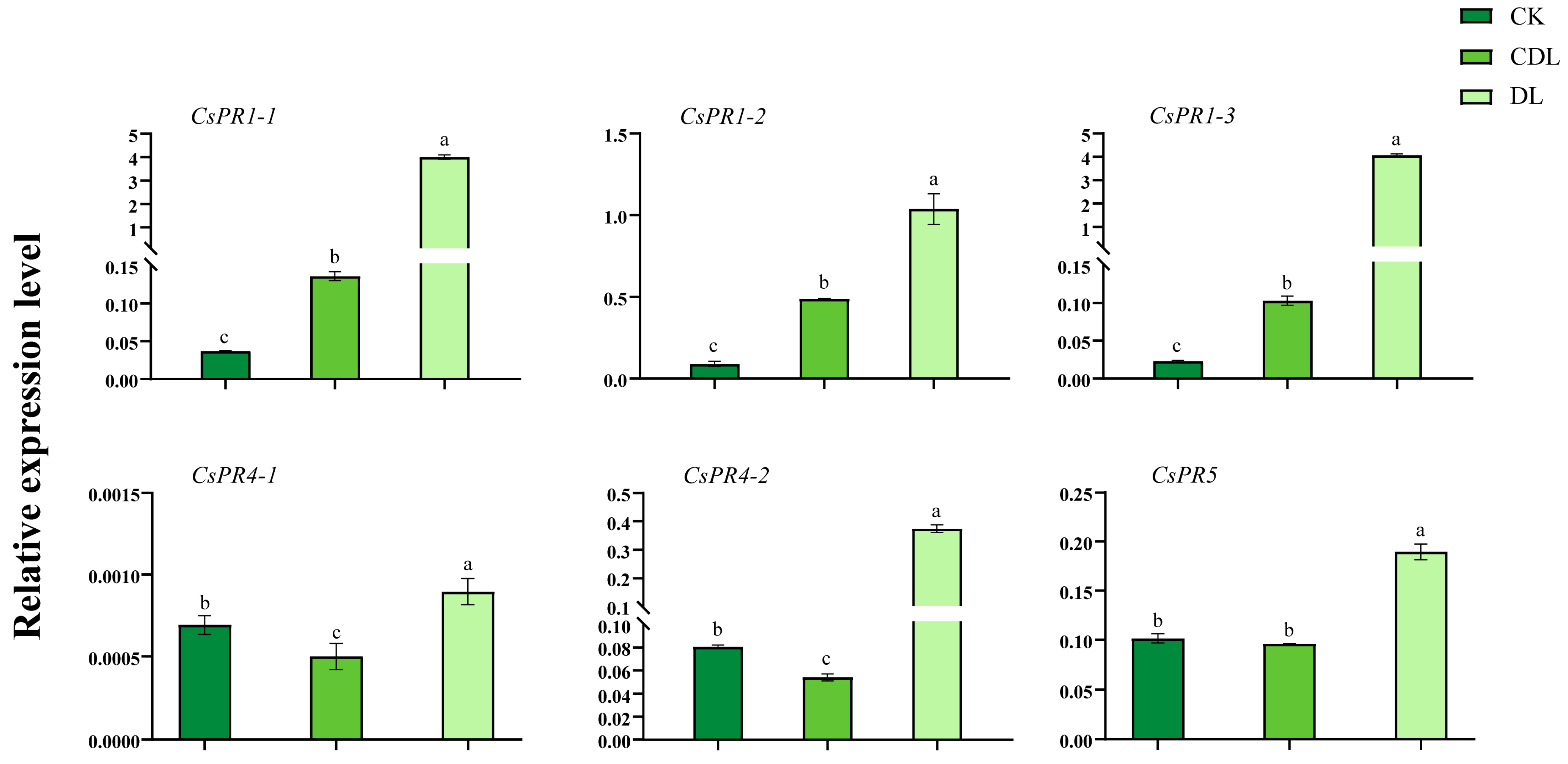
3.6. CsPR Gene Expression in Variegated Tea Leaves and Expression Profiles under Biotic and Abiotic Stresses

As presented in Figure 6, these six genes were found to have the most pronounced changes in expression in both proteomic and transcriptomic data, and were considered the most important in this study. We measured the expression of six PR genes in the available samples. The results revealed that the gene expressions in DL leaves were significantly higher than those in the control leaves of CK and CDL. However, the expression of CsPR1 was the highest, and that of CsPR4-1 was the lowest. The tea plant Longjing 43 was subjected to insect bites, mechanical damage, low temperature (4 °C), and pathogenic fungal infection treatments (Figure 7). The expression of all the six CsPR genes was induced by insect bites and mechanical damage. The expression of these genes peaked at 12 h after insect bites, and mechanical damage caused the CsPR gene expression to remain elevated. At 4 °C, the expression of CsPR1-1, CsPR1-2, CsPR1-3, and CsPR4-2 genes all showed an increase followed by a decrease. The expression of CsPR4-1 and CsPR5 demonstrated an increase and then a decrease, followed by an increase. The infection of the leaves with pathogenic fungus resulted in an increase in the expression of CsPR1-1 and CsPR1-2; an increase and then a decrease in the expression of CsPR4-1, CsPR4-2, and CsPR5; and an increase followed by a decrease and then again by an increase in the expression of CsPR1-3. These results indicate that low temperature, mechanical damage, insect bites, and pathogenic fungal infection could all induce PR gene expression.

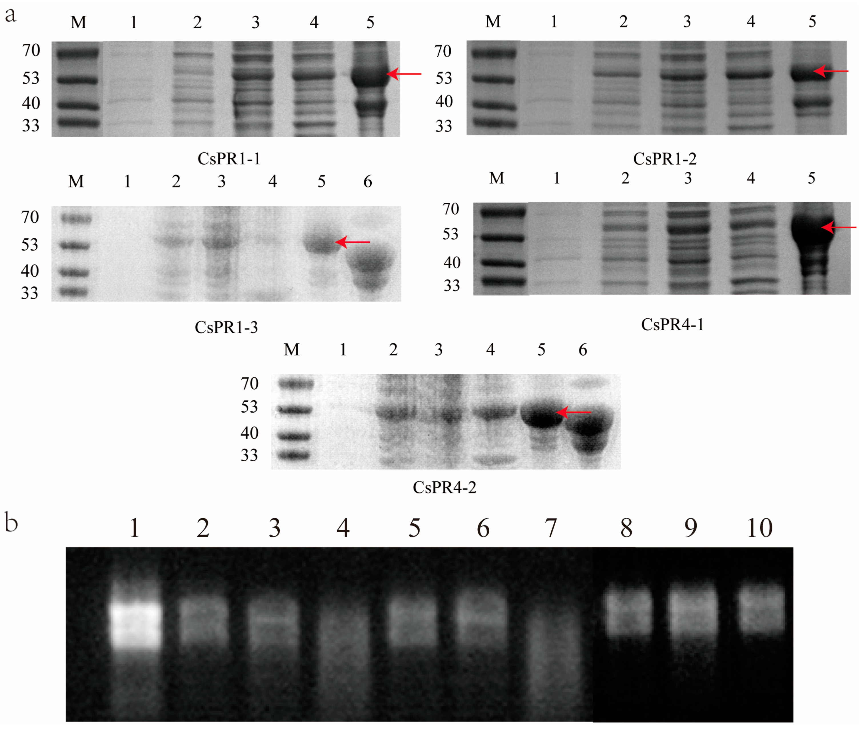
3.7. Expression and Ribonuclease Activity Determination of the Recombinant CsPR1, CsPR4, and CsPR5 Proteins

The CsPR genes (PR1-1, PR1-2, PR1-3, PR4-1, PR4-2, PR5-1) were cloned in the pMAL-C2E plasmid and successfully expressed in E. coli BL21 (DE3) (Figure 8a, lanes 2 to 5). No bands were observed in the control (Figure 8a, lane 1). The protein was highly expressed at 16 °C. We determined that 0.5 M isopropyl β-d-1-thiogalactopyranoside and 16 °C (overnight) were the optimal conditions for CsPR expression. The molecular mass of the recombinant protein was 56–61 kDa, which was close to the predicted molecular mass (40 kDa with MBP maltose-binding protein). CsPRs were successfully purified using a dedicated resin (Figure 8a, lane 5).
We determined the ribonuclease activities of the purified recombinant CsPRs. For the controls, untreated RNA (2 μL) was introduced into the No. 1 reaction. Diluted RNA (2 μL RNA + 38 μL RNase-free ddH2O) was added to the No. 2 and No. 5 reactions, respectively. After that, 5 μg RNA was separately added to the other reactions. And 10 μg of protein from two CsPR4s, after being treated at 100 °C for 10 min, was separately loaded to the No. 3 and 6 reactions; untreated 10 μg protein of two CsPR4s was separately added to the No. 4 and 7 reactions; 10 μg protein from three purified CsPR1s was loaded to the No. 8, 9, and 10 reactions. The total volume of all reaction mixtures was 40 μL, and the reactions were incubated for 50 min at 37 °C. After the reaction, all the mixtures were loaded into lanes 1–10 and analyzed on the gel electrophoresis. Significant degradation of RNA that was catalyzed by CsPR4 was detected. However, no degradation function was found for CsPR1 (Figure 8b).

4. Discussion

In this study, we found that the expression of the CsPR1, CsPR4, and CsPR5 genes was significantly higher in the variegated leaves than in normal control leaves. In previous studies, the expressions of the PRs genes were induced to increase resistance by biotic or abiotic stresses in plants. Therefore, we suspected that variegated leaves were probably in a stressed environment in comparison to normal control leaves. A total of 17 PR1, 3 PR4, and 31 PR5 genes were identified, and their gene structures, conserved motifs, and cis-elements were analyzed, providing a scientific basis for future functional studies of PRs. PR1 belongs to the CAP family (cysteine-rich secretory proteins, antigen 5, and PR1 proteins). PR4 belongs to the Barwin family, and class I contains a ChtBD1 structure, which is not present in class II. PR5 belongs to the thaumatin family. All three structures were determined to possess antifungal properties [27]. The PR1, PR4, and PR5 families, highly conserved, were present in most of the studied species.
The findings from the promoter analysis revealed that many cis-acting elements were activated for the regulation of CsPR genes, including phytohormone, stress, growth and development, as well as MYB families. Thus, we performed a quantitative analysis of six PR genes. We determined gene expression patterns in response to different treatments, including low temperature, mechanical damage, insect bites, and pathogenic bacteria infection. This result supports previous findings indicating that biotic and abiotic stresses induce the expression of PRs. Under low temperature induction at 4 °C, the expression of the PR gene showed a trend of initial increase followed by a decrease. It was assumed that the tea plant activated the defense mode pathway to induce the expression of PRs when resisting external temperature changes. Additionally, it was expected that PRs would not be overexpressed when the tea plant had adapted to the temperature environment.
In this study, six PR genes were screened and successfully cloned, and five PRs were successfully expressed, including two types of CsPR4 proteins. The PR4 genes can be divided into class I and II depending on their structures. It is widely distributed among other species and plays a role in plant defense [13,14,34]. We found that the PR4 protein showed a ribonuclease activity. This is the first report indicating that the PR4 protein has a ribonuclease activity in the tea plant. Some studies examined PR4 in other species, and most of them observed antifungal properties along with ribonuclease activity. It was demonstrated that PR4 exhibits ribonuclease activity and anti-fungal properties against Moniliophthora perniciosa in cocoa beans [13]. Another PR4 was identified to display ribonuclease activity and antifungal properties against Magnaporthe grisea in Lycoris radiata [34]. In addition, a PR4 with ribonuclease activity and antifungal properties was found in figs [14]. However, the antifungal properties of CsPR4 were not verified in our present investigation. The PR1 and PR5 proteins were reported to have antifungal properties in other plants. For example, PR1 was identified in wheat and exhibited antibacterial activity in vitro experiments against eight bacteria and fungi [35]. A PR5 gene was identified in Prunus domestica, and the purified protein also displayed antifungal activity [36]. We found that PR1, PR4, and PR5 showed high expression in response to pathogenic bacteria infection. However, the antifungal properties of PR1 and PR5 proteins were not verified in this study.
Whether the upregulation of CsPR gene expression could lead to leaves with a variegated phenotype was not verified in our study. We speculated that some external or internal stimulus would contribute to the formation of variegated leaves, and that this external or internal stimulus would also lead to the high expression of CsPRs. Based on previous studies, there were no PR genes reported in albino tea plants. To some extent, we doubted that the high expression of PRs might have a certain relation with the variegated phenotype in albino tea leaves. The obtained results could serve as a reference for future studies on PRs in tea plants, offering basic information to understand variegated albinism in tea plants.

5. Conclusions

A significant upregulation of three classes of PRs was revealed by the analysis of pre-existing transcriptomic and proteomic data in the variegated phenotype tea leaves. The PR1, PR4, and PR5 genes were identified in tea plants, including the gene structure, motifs, intron–exon distribution, cis-element features, and phylogenetic relationships. PRs were expressed in E. coli, and the proteins were successfully purified. This was the first time we found that CsPR4 exhibits ribonuclease activity. The expression patterns of six PR genes were screened and then determined under the treatments of low temperature, mechanical damage, insect bites, and pathogenic fungal infection. The obtained results could serve as a reference for future studies on PRs in tea plants, especially in the context of variegated albinism in tea plants.

Supplementary Materials

The following supporting information can be downloaded at: https://www.mdpi.com/article/10.3390/agronomy14010156/s1, Table S1. Primers were used for quantitative reverse transcription-polymerase chain reaction and amplification of PR genes.

Author Contributions

Z.X. prepared the experiments and analyzed the data. Z.W., D.K., Y.L., J.M., H.J. and M.L. participated in data analysis as well as in image revision and completion. Z.X., H.J. and W.-W.D. were responsible for drafting and revising the manuscript. W.-W.D. guided this research. All authors have read and agreed to the published version of the manuscript.

Funding

This study was supported by the Natural Science Foundation of Anhui Province, Outstanding Youth Project (2008085J18) and the National Natural Science Foundation of China (NSFC, Grant No. 31870679).

Data Availability Statement

All data are available on request to corresponding author.

Conflicts of Interest

The authors declare that they have no competing interests.

References

  1. Ali, S.; Ganai, B.A.; Kamili, A.N.; Bhat, A.A.; Mir, Z.A.; Bhat, J.A.; Tyagi, A.; Islam, S.T.; Mushtaq, M.; Yadav, P. Pathogenesis-related proteins and peptides as promising tools for engineering plants with multiple stress tolerance. Microbiol. Res. 2018, 212, 29–37. [Google Scholar] [CrossRef] [PubMed]
  2. Kim, Y.J.; Lee, H.J.; Jang, M.G.; Kwon, W.S.; Kim, S.Y.; Yang, D. Cloning and characterization of pathogenesis-related protein 4 gene from Panax ginseng. Russ. J. Plant Physiol. 2014, 61, 664–671. [Google Scholar] [CrossRef]
  3. Gaudet, D.A.; Laroche, A.; Frick, M.; Huel, R.; Puchalski, B. Cold induced expression of plant defensin and lipid transfer protein transcripts in winter wheat. Physiol. Plant. 2010, 117, 195–205. [Google Scholar] [CrossRef]
  4. Buchanan-Wollaston, V.; Earl, S.; Harrison, E.; Mathas, E.; Navabpour, S.; Page, T.; Pink, D. The molecular analysis of leaf senescence–a genomics approach. Plant Biotechnol. J. 2003, 1, 3–22. [Google Scholar] [CrossRef]
  5. van Loon, L.C.; Rep, M.; Pieterse, C.M. Significance of inducible defense-related proteins in infected plants. Annu. Rev. Phytopathol. 2006, 44, 135–162. [Google Scholar] [CrossRef]
  6. Van Loon, L. Polyacrylamide disc electrophoresis of the soluble leaf proteins from Nicotiana tabacum var.‘Samsun’and ‘Samsun NN’: IV. Similarity of qualitative changes of specific proteins after infection with different viruses and their relationship to acquired resistance. Virology 1975, 67, 566–575. [Google Scholar] [PubMed]
  7. Van Loon, L.C. Occurrence and properties of plant pathogenesis-related proteins. In Pathogenesis-Related Proteins in Plants; CRC Press: Boca Raton, FL, USA, 1999; pp. 1–19. [Google Scholar]
  8. Sels, J.; Mathys, J.; De Coninck, B.M.; Cammue, B.P.; De Bolle, M.F. Plant pathogenesis-related (PR) proteins: A focus on PR peptides. Plant Physiol. Biochem. 2008, 46, 941–950. [Google Scholar] [CrossRef]
  9. Anisimova, O.K.; Shchennikova, A.V.; Kochieva, E.Z.; Filyushin, M.A. Pathogenesis-related genes of PR1, PR2, PR4, and PR5 families are involved in the response to Fusarium infection in garlic (Allium sativum L.). Int. J. Mol. Sci. 2021, 22, 6688. [Google Scholar] [CrossRef]
  10. AlHudaib, K.A.; Alanazi, N.A.; Ghorbel, M.; El-Ganainy, S.M.; Brini, F. Isolation and Characterization of a Novel Pathogenesis-Related Protein-1 Gene (AvPR-1) with Induced Expression in Oat (Avena sativa L.) during Abiotic and Hormonal Stresses. Plants 2022, 11, 2284. [Google Scholar] [CrossRef]
  11. Hejgaard, J.; Jacobsen, S.; Bjørn, S.E.; Kragh, K.M. Antifungal activity of chitin-binding PR-4 type proteins from barley grain and stressed leaf. FEBS Lett. 1992, 307, 389–392. [Google Scholar] [CrossRef]
  12. Van Damme, E.J.; Charels, D.; Roy, S.; Tierens, K.; Barre, A.; Martins, J.C.; Rougé, P.; Van Leuven, F.; Does, M.; Peumans, W.J. A gene encoding a hevein-like protein from elderberry fruits is homologous to PR-4 and class V chitinase genes. Plant Physiol. 1999, 119, 1547–1556. [Google Scholar] [CrossRef] [PubMed]
  13. Pereira Menezes, S.; de Andrade Silva, E.M.; Matos Lima, E.; Oliveira de Sousa, A.; Silva Andrade, B.; Santos Lima Lemos, L.; Peres Gramacho, K.; da Silva Gesteira, A.; Pirovani, C.P.; Micheli, F. The pathogenesis-related protein PR-4b from Theobroma cacao presents RNase activity, Ca2+ and Mg2+ dependent-DNase activity and antifungal action on Moniliophthora perniciosa. BMC Plant Biol. 2014, 14, 161. [Google Scholar] [CrossRef] [PubMed]
  14. Lu, H.-C.; Lin, J.-H.; Chua, A.C.; Chung, T.-Y.; Tsai, I.-C.; Tzen, J.T.; Chou, W.-M. Cloning and expression of pathogenesis-related protein 4 from jelly fig (Ficus awkeotsang Makino) achenes associated with ribonuclease, chitinase and anti-fungal activities. Plant Physiol. Biochem. 2012, 56, 1–13. [Google Scholar] [CrossRef]
  15. van der Wel, H.; Loeve, K. Isolation and characterization of thaumatin I and II, the sweet-tasting proteins from Thaumatococcus daniellii Benth. Eur. J. Biochem. 1972, 31, 221–225. [Google Scholar] [CrossRef] [PubMed]
  16. Zhai, X.; Kong, Q.; An, P.; Ren, X. The function and mechanism of pathogenesis-related 5 protein resistance in cherry tomato in response to Alternaria alternata. Food Biotechnol. 2018, 32, 178–190. [Google Scholar] [CrossRef]
  17. Muoki, R.C.; Paul, A.; Kaachra, A.; Kumar, S. Membrane localized thaumatin-like protein from tea (CsTLP) enhanced seed yield and the plant survival under drought stress in Arabidopsis thaliana. Plant Physiol. Biochem. 2021, 163, 36–44. [Google Scholar] [CrossRef] [PubMed]
  18. Jiang, H.; Li, M.; Liang, N.; Yan, H.; Wei, Y.; Xu, X.; Liu, J.; Xu, Z.; Chen, F.; Wu, G. Molecular cloning and function analysis of the stay green gene in rice. Plant J. 2007, 52, 197–209. [Google Scholar] [CrossRef]
  19. Næsted, H.; Holm, A.; Jenkins, T.; Nielsen, H.B.; Harris, C.A.; Beale, M.H.; Andersen, M.; Mant, A.; Scheller, H.; Camara, B. Arabidopsis VARIEGATED 3 encodes a chloroplast-targeted, zinc-finger protein required for chloroplast and palisade cell development. J. Cell Sci. 2004, 117, 4807–4818. [Google Scholar] [CrossRef] [PubMed]
  20. Asakura, Y.; Hirohashi, T.; Kikuchi, S.; Belcher, S.; Osborne, E.; Yano, S.; Terashima, I.; Barkan, A.; Nakai, M. Maize mutants lacking chloroplast FtsY exhibit pleiotropic defects in the biogenesis of thylakoid membranes. Plant Cell 2004, 16, 201–214. [Google Scholar] [CrossRef] [PubMed]
  21. Abbo, S.; Berger, J.; Turner, N.C. Evolution of cultivated chickpea: Four bottlenecks limit diversity and constrain adaptation. Funct. Plant Biol. 2003, 30, 1081–1087. [Google Scholar] [CrossRef]
  22. Zhang, Q.; Guo, N.; Zhang, Y.; Yu, Y.; Liu, S. Genome-wide characterization and expression analysis of pathogenesis-related 1 (PR-1) gene family in tea plant (Camellia sinensis (L.) O. Kuntze) in response to blister-blight disease stress. Int. J. Mol. Sci. 2022, 23, 1292. [Google Scholar] [CrossRef] [PubMed]
  23. Acharya, K.; Pal, A.K.; Gulati, A.; Kumar, S.; Singh, A.K.; Ahuja, P.S. Overexpression of Camellia sinensis thaumatin-like protein, CsTLP in potato confers enhanced resistance to Macrophomina phaseolina and Phytophthora infestans infection. Mol. Biotechnol. 2013, 54, 609–622. [Google Scholar] [CrossRef]
  24. Lu, M.; Li, Y.; Jia, H.; Xi, Z.; Gao, Q.; Zhang, Z.-Z.; Deng, W.-W. Integrated proteomics and transcriptome analysis reveal a decreased catechins metabolism in variegated tea leaves. Sci. Hortic. 2022, 295, 110824. [Google Scholar] [CrossRef]
  25. Wang, Y.; Tang, L.; Hou, Y.; Wang, P.; Yang, H.; Wei, C. Differential transcriptome analysis of leaves of tea plant(Camellia sinensis) provides comprehensive insights into the defense responses to Ectropis oblique attack using RNA-Seq. Funct. Integr. Genom. 2016, 16, 383–398. [Google Scholar] [CrossRef] [PubMed]
  26. Chen, W.; Ye, T.; Sun, Q.; Niu, T.; Zhang, J. Arbuscular mycorrhizal fungus alleviates anthracnose disease in tea seedlings. Front. Plant Sci. 2023, 13, 1058092. [Google Scholar] [CrossRef]
  27. Marchler-Bauer, A.; Derbyshire, M.K.; Gonzales, N.R.; Lu, S.; Chitsaz, F.; Geer, L.Y.; Geer, R.C.; He, J.; Gwadz, M.; Hurwitz, D.I. CDD: NCBI’s conserved domain database. Nucleic Acids Res. 2015, 43, D222–D226. [Google Scholar] [CrossRef] [PubMed]
  28. Chen, C.; Chen, H.; Zhang, Y.; Thomas, H.R.; Frank, M.H.; He, Y.; Xia, R. TBtools: An integrative toolkit developed for interactive analyses of big biological data. Mol. Plant 2020, 13, 1194–1202. [Google Scholar] [CrossRef]
  29. Xia, E.; Tong, W.; Hou, Y.; An, Y.; Chen, L.; Wu, Q.; Liu, Y.; Yu, J.; Li, F.; Li, R. The reference genome of tea plant and resequencing of 81 diverse accessions provide insights into its genome evolution and adaptation. Mol. Plant 2020, 13, 1013–1026. [Google Scholar] [CrossRef] [PubMed]
  30. Gasteiger, E.; Gattiker, A.; Hoogland, C.; Ivanyi, I.; Appel, R.D.; Bairoch, A. ExPASy: The proteomics server for in-depth protein knowledge and analysis. Nucleic Acids Res. 2003, 31, 3784–3788. [Google Scholar] [CrossRef] [PubMed]
  31. Chou, K.-C.; Shen, H.-B. Plant-mPLoc: A top-down strategy to augment the power for predicting plant protein subcellular localization. PLoS ONE 2010, 5, e11335. [Google Scholar] [CrossRef]
  32. Kumar, S.; Stecher, G.; Tamura, K. MEGA7: Molecular evolutionary genetics analysis version 7.0 for bigger datasets. Mol. Biol. Evol. 2016, 33, 1870–1874. [Google Scholar] [CrossRef]
  33. Zhang, J.; Jia, H.; Zhu, B.; Li, J.; Yang, T.; Zhang, Z.-Z.; Deng, W.-W. Molecular and biochemical characterization of jasmonic acid carboxyl methyltransferase involved in aroma compound production of methyl jasmonate during black tea processing. J. Agric. Food Chem. 2021, 69, 3154–3164. [Google Scholar] [CrossRef]
  34. Li, X.; Xia, B.; Jiang, Y.; Wu, Q.; Wang, C.; He, L.; Peng, F.; Wang, R. A new pathogenesis-related protein, LrPR4, from Lycoris radiata, and its antifungal activity against Magnaporthe grisea. Mol. Biol. Rep. 2010, 37, 995–1001. [Google Scholar] [CrossRef]
  35. Lu, S.; Friesen, T.L.; Faris, J.D. Molecular characterization and genomic mapping of the pathogenesis-related protein 1 (PR-1) gene family in hexaploid wheat (Triticum aestivum L.). Mol. Genet. Genom. 2011, 285, 485–503. [Google Scholar] [CrossRef]
  36. El-Kereamy, A.; El-Sharkawy, I.; Ramamoorthy, R.; Taheri, A.; Errampalli, D.; Kumar, P.; Jayasankar, S. Prunus domestica pathogenesis-related protein-5 activates the defense response pathway and enhances the resistance to fungal infection. PLoS ONE 2011, 6, e17973. [Google Scholar] [CrossRef]
Figure 1. The variegated albino tea plant with normal and variable leaves. (a) Variegated tea plant, (b) normal mature leaf (CDL), and (c) variegated mature leaf (DL).
Agronomy 14 00156 g001
Figure 2. (a) Heat map of proteins with significant differential expression identified in proteomic data analysis. Proteins marked with red stars show the differentially highly expressed PRs that were selected. (b) Heat map of differentially expressed genes identified in the transcriptome data analysis. Genes with red stars show the differentially highly expressed PR genes that were selected. (c) Phylogenetic analysis of 115 PR genes in Camellia sinensis and Arabidopsis. PR genes in C. sinensis are indicated by circles, those in Arabidopsis are indicated by pentagrams, and the six differentially expressed PR genes are marked in red. Three different types of PR genes in C. sinensis are labeled with different shades of blue, pink, and yellow.
Agronomy 14 00156 g002
Figure 3. Multiple sequence alignments of PR1, PR4, and PR5 proteins using GENEDOC software (Version 2.7). (a) The amino acid sequence alignments of 17 PR1s in the tea plant. (b) The amino acid sequence alignments of PR4s in the tea plant and other species. (c) The amino acid sequence alignments of PR5s in the tea plant and other species. The main target genes in this study are marked with red stars. The red box denotes the signal peptide; the blue box represents a hinge; the green box denotes the region contributing to the function; the black line indicates the chitin-binding do-main structure; the pink line indicates the Barwin structural domain.
Agronomy 14 00156 g003
Figure 4. Conserved motifs (a), conserved structural domains (b), expressed and non-expressed regions (c) of CsPRs. In the leftmost evolutionary tree, red represents CsPR5, yellow represents CsPR4, and green represents CsPR1.
Agronomy 14 00156 g004
Figure 5. Analysis of cis-regulatory elements distribution in CsPR promoters. In the leftmost evolutionary tree, red represents CsPR5, yellow represents CsPR4, and green represents CsPR1. The types of cis-elements are presented on the right, and the pie chart located below indicates the proportions of cis-elements distributed.
Agronomy 14 00156 g005
Figure 6. Real-time quantitative polymerase chain reaction (qPCR) of six CsPRs in DL (variegated mature leaf), CDL (normal mature leaf), and CK (leaves from a normal tea plant with no albino phenotype) samples. Data represent the means ± SD (n = 3) of three biological samples. Different letters above bars represent significant differences at p < 0.05.
Agronomy 14 00156 g006
Figure 7. Real-time quantitative polymerase chain reaction (qPCR) of 6 CsPRs in the leaves of Longjing 43 under insect bites (a), mechanical damage (b), low temperature at 4 °C (c), and anthracnose disease stress (d). Expression was analyzed at different time periods. Data represent the means ± SD (n = 3) of three biological samples. Different letters above bars represent significant differences at p < 0.05.
Agronomy 14 00156 g007
Figure 8. (a) CsPRs expression in E. coli, and purification. The red arrow positions indicate successfully purified bands. M: marker; 1: pre-inducible protein; 2: post-induction protein; 3: induced supernatant; 4: induced precipitation; 5: purified protein; 6: purified MBP protein. (b) Ribonuclease activity verification of the successfully purified protein CsPR4s (lanes 4 and 7). Lane 1, untreated RNA for control; lanes 2 and 5, diluted RNA for control; lanes 4 and 7, the reaction mixture of two purified CsPR4 proteins; lanes 3 and 6, the reaction mixture of two purified CsPR4 proteins (treated at 100 °C for 10 min first); lanes 8, 9, and 10, the reaction mixture of three purified CsPR1 proteins.
Agronomy 14 00156 g008
Table 1. Gene and protein features of PR1, PR4, and PR5 gene family members in Camellia sinensis.
Protein SymbolLength (aa)MW (kD)pISPSLGRAVYSequence ID
CsPR1−116217.886.86YVacuole−0.297TEA022240.1
CsPR1−216818.538.10YVacuole−0.354TEA028218.1
CsPR1−316217.878.62YVacuole−0.345TEA004541.1
CsPR1−419121.539.16YVacuole−0.340TEA022150.1
CsPR1−517019.259.22YVacuole−0.375TEA022585.1
CsPR1−617318.606.81YVacuole−0.227TEA022146.1
CsPR1−716017.584.96YVacuole−0.235TEA004542.1
CsPR1−837641.144.88YVacuole−0.574TEA022692.1
CsPR1−919622.606.09YVacuole−0.479TEA025681.1
CsPR1−1015917.528.47YVacuole−0.270TEA004551.1
CsPR1−1120422.595.66NVacuole−0.257TEA030748.1
CsPR1−1216218.269.16YVacuole−0.399TEA028234.1
CsPR1−1316217.938.74YVacuole−0.213TEA021774.1
CsPR1−1416217.938.74YVacuole−0.213TEA017943.1
CsPR1−1516818.084.90YVacuole−0.239TEA011597.1
CsPR1−1616017.615.38YVacuole−0.269TEA021361.1
CsPR1−1734038.2810.07YVacuole−1.089TEA002521.1
CsPR4−120321.456.23YVacuole−0.300TEA013240.1
CsPR4−215616.824.53YCell wall0.128TEA013238.1
CsPR4−331034.435.35NCytoplasm−0.034TEA013261.1
CsPR5−132834.224.74NCytoplasm0.089TEA007812.1
CsPR5−231032.614.70YCytoplasm−0.073TEA032607.1
CsPR5−314415.795.22NCytoplasm−0.062TEA014403.1
CsPR5−453358.556.85NCytoplasm−0.020TEA014476.1
CsPR5−524425.746.05YCytoplasm0.046TEA008382.1
CsPR5−632334.524.60YCytoplasm−0.011TEA019846.1
CsPR5−722624.174.75YVacuole−0.146TEA026507.1
CsPR5−824925.614.53YCytoplasm0.019TEA027400.1
CsPR5−919420.468.62YCytoplasm0.046TEA028375.1
CsPR5−1025428.008.60NCytoplasm−0.374TEA022860.1
CsPR5−1124025.975.29YCytoplasm0.053TEA013937.1
CsPR5−1225027.039.00YCytoplasm0.004TEA006411.1
CsPR5−1327830.178.62NCytoplasm−0.049TEA015935.1
CsPR5−1422524.619.08YVacuole−0.312TEA032837.1
CsPR5−1536738.734.63NCytoplasm−0.187TEA002766.1
CsPR5−1625126.575.75YCytoplasm0.112TEA028896.1
CsPR5−1723425.018.73NCytoplasm−0.047TEA027596.1
CsPR5−1817618.904.73YCytoplasm0.069TEA032339.1
CsPR5−1922624.194.65YVacuolar−0.152TEA032349.1
CsPR5−2022624.615.09YVacuolar−0.304TEA032350.1
CsPR5−2123625.745.14YVacuolar−0.005TEA030559.1
CsPR5−2240641.934.32YCytoplasm−0.086TEA025008.1
CsPR5−2326027.678.92NNuclear−0.323TEA012573.1
CsPR5−2424425.943.83YVacuolar−0.061TEA014245.1
CsPR5−2541344.908.34Ncytoplasmic−0.577TEA014254.1
CsPR5−2624626.224.54YVacuolar−0.105TEA014256.1
CsPR5−2763770.568.49YVacuolar0.133TEA029484.1
CsPR5−2825426.194.67YVacuolar0.139TEA006040.1
CsPR5−2926127.114.98YVacuolar0.094TEA006949.1
CsPR5−3028029.715.03NCytoplasmic −0.371TEA033060.1
CsPR5−3122624.144.97YVacuolar−0.140TEA005276.1
MW: molecular weight; SP: signal peptide; SL: subcellular localization.
Disclaimer/Publisher’s Note: The statements, opinions and data contained in all publications are solely those of the individual author(s) and contributor(s) and not of MDPI and/or the editor(s). MDPI and/or the editor(s) disclaim responsibility for any injury to people or property resulting from any ideas, methods, instructions or products referred to in the content.

