Article

Isolation and Molecular Characterization of Two Arabinosyltransferases in Response to Abiotic Stresses in Sijichun Tea Plants (Camellia sinensis L.)

1
Department of Agronomy, National Chung Hsing University, Taichung 40227, Taiwan
2
Graduate Institute of Biotechnology, National Chung Hsing University, Taichung 40227, Taiwan
3
Department of Accounting, Chinese Culture University, Taipei 11114, Taiwan
4
Smart Sustainable New Agriculture Research Center (SMARTer), National Chung Hsing University, Taichung 40227, Taiwan
*
Author to whom correspondence should be addressed.
These authors contributed equally to this work.
Agronomy 2023, 13(6), 1476; https://doi.org/10.3390/agronomy13061476
Submission received: 29 April 2023 / Revised: 20 May 2023 / Accepted: 25 May 2023 / Published: 26 May 2023
(This article belongs to the Special Issue Crop Tolerance under Biotic and Abiotic Stresses)

Abstract

:
The modification of secondary metabolites is crucial to the function of metabolites in tea (Camellia sinensis L.). The arabinan deficient (ARAD) encodes an arabinosyltransferase and is involved in the arabinan biosynthesis pathway. Two full-length sequences of CsARADs were cloned and obtained from tea plants through the rapid amplification of cDNA ends and named CsARAD1 and CsARAD2. CsARAD1 and CsARAD2 are predicted to be 2 membrane proteins containing N-glycosylation, phosphorylation, and N-myristoylation sites and are 2 homologs of the glycosyltransferases (GT) 47 family, according to various bioinformatic analyses. CsARADs showed higher transcription levels in nonlignified tissues (e.g., buds and young leaves) than in old leaves and stems. CsARADs also exhibited the highest expression level in autumn, indicating that CsARAD regulation is affected by environmental factors. The transcript levels of CsARADs were changed after various abiotic stress treatments, and CsARAD1 and CsARAD2 displayed different regulation patterns in temperature stress, saline, and drought-like conditions. CsARAD1 and CsARAD2 were both significantly downregulated after tea seedlings were treated with an ethylene precursor and abscisic acid. In addition, CsARAD2 was downregulated after being treated with methyl jasmonate and gibberellin. Collectively, our findings on the function of arabinosyltransferase serve as a basis for further research and breeding applications.

1. Introduction

Tea tree (Camellia sinensis L.) is a perennial, woody plant of the Camellia family. It is a key and special crop with considerable economic value in Taiwan. Tea is the most popular beverage in the world. Green tea and oolong tea are mostly consumed in Asia, and black tea is more commonly consumed in Europe and North America [1]. Tea is rich in secondary metabolites such as polyphenols, alkaloids, and flavonoids. Flavonoids and flavonol glycosides create the beneficial health properties of tea, including its antioxidation, antiobesity, anticancer, and hypoglycemic properties. Flavonoids (e.g., kaempferol, quercetin, myricetin, and apigenin) can form flavonol glycosides with one or more glycosidic bonds with glucose, galactose, rhamnose, and arabinose, resulting in various molecular functions [2]. A total of 19 types of flavonol glycosides and 28 types of acylated glycosylated flavonols have been identified in tea and the postprocessed fermented products of various tea species [3].
Glycosyltransferases (GTs) can transfer active sugars from donor compounds to acceptor compounds to form glycosidic bonds. The glycosylation of GTs increases the variety and diversity of the secondary metabolites in plants; such glycosylation changes the molecular properties of glycosyl compounds and substantially influences the biofunctions of plant metabolism (e.g., protecting plants from pests and pathogens) [4]. Glycosylation also regulates the activity of hormones such as auxin, abscisic acid, cytokinin, salicylic acid (SA), and brassinosteroids [5]. Studies have revealed that the glycosylation of arabinose can prevent seed dormancy by regulating the downstream genes of the abscisic acid signaling pathway during seed germination [6]. Higher plants have 91 GT families [7], and the protein in the GT47 subfamily contains a highly-conserved region of β-glucuronyltransferase and exhibits xyloglucan β-galactosyltransferase and arabinan α-L-arabinosyltransferase activities that are involved in the composition of cellulose, hemicellulose, pectin, and cell walls [8]. In tea, the catalytic activity of arabinosyltransfrease is essential to the formation of numerous tea catechins and flavonols [9], which decides the flavor and beneficial health effect of tea.
Arabinan deficient (ARAD) is an arabinosyltransferase involved in the biosynthesis of pectic arabinans, which are key ingredients of the cell wall [10]. Studies have highlighted that arabinan deficient 1 (ARAD1) participates in the biosynthesis of rhamnogalacturonan I, which is a major component of the pectin in cell walls. The expression of AtARAD1 increases polysaccharide content (e.g., the pectin in cell) and helps strengthen primary cell walls and improve cell adhesion, stomatal function, and the defense response of cell walls [11,12]. AtARAD2 is a homologous protein of AtARAD1 that helps promote the participation of various glycosyltransferases and AtARAD1 in the synthesis of the arabinose backbone, and AtARAD2 participates in the synthesis of the arabinose side chains. AtARAD1 and AtARAD2 are synergistic proteins with different functions [13]. An experiment on AtARAD mutants revealed that the arabinose content of plants is related to their tolerance to mechanical stress [14], suggesting that ARAD plays a key role in stress resistance.
Considering the importance of glycose transferases on tea metabolites and the crucial role of ARAD in plant stress resistance, it is indispensable to investigate the function of ARAD in tea. However, studies on arabinosyltransferases have mostly focused on the composition of cell wall structures [15,16]; few studies have explored the biological function and regulation of ARADs. This study aims to explore the molecular characterization of ARAD, providing further details of the function and regulation of ARAD in tea plants. The ARAD sequence was cloned from the Sijichun cultivar (a tea plant variety from Taiwan) on the basis of the sequence information of the tea cultivar Shuchazao, which was obtained from the National Center for Biotechnology Information (NCBI) database. Two independent, full-length sequences of the CsARADs of Sijichun tea plants were obtained and named CsARAD1 and CsARAD2. The amino acid sequences of CsARADs were analyzed, and the gene expression patterns of CsARADs at various tissue sites and abiotic stress were analyzed through a quantitative, real-time polymerase chain reaction (qRT-PCR). A higher expression level of CsARADs was detected in nonlignified tissue (e.g., young leaves and stems) than in old leaves and stems. The number of CsARADs transcribed in various seasons and abiotic stress conditions indicated that CsARADs are regulated by several environmental factors such as temperature and humidity. Abscisic acid (ABA) and ethylene signaling are also involved in the regulation of CsARADs. In addition, CsARAD1 and CsARAD2 exhibited disparate regulation patterns. This study produced key findings on the bioinformation of CsARAD1 and CsARAD2 and the molecular regulatory mechanism of CsARADs, providing a foundation for subsequent studies.

2. Materials and Methods

2.1. Plant Materials

Eight-year-old tea (C. sinensis (L.) Kuntze) cultivars of Sijichun, which were plucked from the tea cultivated in Nantou, Taiwan, were used as plant material. The sampling time started from September 2019 to June 2020 once every three months for the whole year, representing samples from different seasons. Tea samples were plucked from different individuals of the same garden. The shoots (buds), young leaves (yellow-green tender leaves (YL)), old leaves (dark-green mature leaves (OL)), young stems (green nonlignified tender stem (YS)), old stems (brown lignified hard stem (OS)) tissues were cut from randomly selected tea trees, immediately frozen with liquid nitrogen, and then stored in a refrigerator at −80 °C for further analysis. One-year-old Sijichun seedlings were purchased from a tea breeder in Nantou, Taiwan.

2.2. Cloning and Rapid Amplification of cDNA Ends

The amino acid sequence of the ARAD in the tea cultivar Shuchazao was obtained from the NCBI database and used as the template sequence for the cloning of CsARADs in Sijichun. Primer pairs comprising full-ARAD1s and full-ARAD2s were designed (Table S1), and PCRs were conducted using the Platinum II Hot-Start PCR Master Mix Kit (Invitrogen, Waltham, MA, USA) to obtain the partial sequence of CsARADs. The rapid amplification of cDNA ends (RACE) was then applied with the SMARTer RACE 5′/3′ Kit (Takara, San Jose, CA, USA) to obtain the full-length sequences of CsARADs. To acquire the complimentary cDNA sequences of CsARADs, 1 µL of the total RNA of tea was mixed with 9 µL of sterile H2O and 1 µL of the 5′CDS of primer A or the 3′CDS of primer A in a 0.2 mL test tube to synthesize the 5′ and 3′ ends of CsARADs separately. Reactions were conducted at 72 °C for 3 min and at 42 °C for 2 min, and centrifugation was then performed at 14,000× g for 10 s after cooling. Subsequently, 4 µL of first-strand buffer, 0.5 µL of dithiothreitol (100 mM), 1 µL of deoxynucleotide triphosphate (20 mM), 0.5 µL of ribonuclease inhibitor (40 U/µL), and 2 µL of SMARTScribe reverse transcriptase (100 U) were added to 5′ RACE and 3′ RACE tubes and mixed well. Thereafter, reactions were conducted at 42 °C for 90 min and then at 70 °C for 10 min. Specific primers (Table S1) were designed on the 5′ and 3′ ends of the CsARADs to amplify the obtained sequences (Figure S1). Next, 2.5 µL of cDNA, 5 µL of 10X Universal Primer Mix, 1 µL of primers, 15.5 µL of PCR-grade H2O, 25 µL of SeqAmpTM buffer, and 1 µL of SeqAmp DNA polymerase were mixed in a 0.2 mL test tube. The PCR comprised 5 cycles at 94 °C for 30 s and at 72 °C for 3 min; 5 cycles at 94 °C for 30 s and at 70 °C for 30 s; and 25 cycles at 94 °C for 30 s, at 68 °C for 30 s, and at 72 °C for 3 min. After sequence amplification, gel electrophoresis was performed on 1% TBE gel to separate the target sequences. A NucleoSpin Gel and PCR Clean-up Kit (Takara, USA) was then used to purify and collect the target sequences. The target sequences were constructed on vector pCR 2.1-TOPO (Invitrogen, USA) and transformed into hose cells. The Escherichia coli strain (DH5α) was used as the host for cloning. The plasmid DNA of the transformed colonies was purified using the Plasmid Miniprep Purification Kit (GeneMark, Taipei, Taiwan) and sent for sequencing. After sequencing was completed, the full-length sequences of CsARAD1 and CsARAD2 were acquired.

2.3. Bioinformatics Analysis of CsARAD Gene

To perform the bioinformatics analysis of the CsARAD sequences, several online resources were used. ExPASy Translate (https://web.expasy.org/translate/ (accessed on 1 June 2020)) was used to estimate the amino acid sequence translated by a nucleotide. The molecular weight and isoelectric point of the CsARADs were acquired using ExPASy-Compute pI/Mw (https://www.expasy.org/resources/compute-pi-mw (accessed on 1 June 2020)). The amino acid compositions of proteins were analyzed using the website ExPASy-ProtParam (https://www.expasy.org/resources/protparam (accessed on 1 July 2020)). ExPASy-ProtScale (https://www.expasy.org/resources/protscale (accessed on 1 July 2020)) was used to predict the hydrophilicity and hydrophobicity of the proteins. SignalP 5.0 Server (http://www.cbs.dtu.dk/services/SignalP/ (accessed on 1 July 2020)) was used to predict whether a protein had a signaling peptide. The subcellular localization of proteins was performed using DeepLoc 1.0 (http://www.cbs.dtu.dk/services/DeepLoc/ (accessed on 1 October 2020)). The transmembrane regions of proteins were predicted using TMHMM Server v. 2.0 (http://www.cbs.dtu.dk/services/TMHMM/ (accessed on 1 October 2020)). The ARAD amino acid sequences of higher plants (e.g., almond, Spanish cork oak, bayberry, and tobacco) were obtained from the NCBI (https://www.ncbi.nlm.nih.gov/ (accessed on 1 June 2020)) database; subsequently, they were analyzed by using the EMBOSS Stretcher (www.ebi.ac.uk/Tools/psa/emboss_stretcher/ (accessed on 1 June 2020)) website to conduct a multiple-sequence alignment analysis, and the results were presented using the GeneDoc software (version 2.7.0.0) (Pittsburgh, PA, USA). Motif Scan (https://myhits.sib.swiss/cgi-bin/motif_scan (accessed on 1 July 2020)) was used to determine the structural and functional regions of the proteins. Dendrogram analysis was performed using the MEGA-X software (version 10.0.5) (Philadelphia, PA, USA), and neighbor joining and bootstrapping were used to perform analyses and calculations, respectively.

2.4. Abiotic Stress and Hormone Treatments on Tea Seedlings

The 1-year-old Sijichun seedlings were subjected to abiotic stress treatments (heat, cold, drought, and high salinity stress). The control group was placed in a growth chamber at 22 ± 1 °C under a 16–8 h light–dark photoperiod with daily irrigation (50 mL/day). Plants were placed in a growth chamber at 35 °C and 4 °C for 24 h for heat and cold stress treatments with normal irrigation, respectively. The irrigation of the plants in the drought group was withheld for 3 days, and 50 mL of 300 mM NaCl was provided instead of water to the high-salinity group every day for 3 days. Every treatment group was compared to the corresponding control group. After the stress treatments, the bud tissues of each group were frozen by using liquid nitrogen and stored in a refrigerator at −80 °C for further analysis. For hormone treatments, 10 mL of 100 μM ABA, SA, methyl jasmonate (MeJA), gibberellic acid (GA), and 1-aminocyclopropane-1-carboxylic acid (ACC) solutions were prepared and sprayed on the young leaves of the tea seedlings. The treatment time was 8 h; spraying was performed hourly during the first 3 h of treatment, and the tissues of young leaves were collected after an additional 5 h of standing and stored at −80 °C for further analysis.

2.5. Total RNA Extraction and qRT-PCR

Tea leaf samples weighing 0.1 g were ground into powder in liquid nitrogen, and their total RNA was then extracted using a Plant Total RNA Purification Kit (GeneMark, Taiwan). A Nanodrop Lite Spectrophotometer was used to assess the purity and quantity of the extracted RNA. After total RNA extraction, DNase digestion was conducted with 1 μL of TURBO DNase (2 U/L; Invitrogen, USA) and 30 μL of 1 DNase buffer (diluted from 10 buffer with diethylpyrocarbonate-treated water) per sample at 37 °C for 30 min. The M-MLV first-strand synthesis kit (Invitrogen, Waltham, MA, USA) was used to reverse transcribe the extracted total RNA to obtain cDNA.
For the gene expression analysis, the CFX ConnectTM Real-Time System (Bio-Rad, Hercules, CA, USA) was used to amplify the cDNA for qRT-PCR. Next, 1 µL of 10X PCR buffer (GeneDireX, Las Vegas, NV, USA), 1 µL of 0.5 mM dNTP (GeneDireX, Las Vegas, NV, USA), 1.2 µL of primer sets (10 µM), 0.05 µL of Taq DNA polymerase (Wizpure, Seongnam, Republic of Korea), 1 µL of 1000X SYBR dye (Invitrogen, USA), and 2.75 µL of ddH2O were mixed to obtain a 10 µL solution for reactions. The 18S rRNA of tea was used as the internal control. The thermal cycle applied for all qRT-PCR reactions was 5 min at 94 °C followed by 40 cycles at 94 °C for 30 s, 55 °C for 30 s, and 72 °C for 30 s. Transcript levels were calculated using the Bio-Rad software (Taiwan Branch, U.S.A.) and the 2−ΔΔCt method, and the average values of at least three separate replicates were acquired.

2.6. Statistics

Statistical analyses were conducted using the Statistical Analysis System (SAS) statistical analysis software (version 9.4) (North Carolina State University, Raleigh, NC, USA). The data were subjected to an analysis of variance, and multiple comparisons were performed by applying Duncan’s test. The different letters indicate significant differences between treatments (p < 0.05). The differences between abiotic stress and hormonal treatments were tested by performing Student’s t-test at a significance level of 5%, and * indicates significant differences between treatments. All experimental data are expressed as means ± standard deviations (SDs).

3. Results

3.1. Bioinformatics Analysis of CsARAD Gene and Amino Acid Sequences of C. sinenesis L.

The CsARADs sequences were cloned from the Sijichun tea plant. These nucleic acid sequences were then submitted to the GenBank repository with accession numbers ON234110 and ON234111. For CsARAD1 and CsARAD2, the full lengths of their cDNA sequences were 1705 and 1637 bp, respectively, and their untranslated regions were 78 and 116 bp, respectively. The open reading frames of CsARAD1 and CsARAD2 included 1503 and 1443 nucleotide sequences, which encode 500 and 480 amino acid sequences, respectively (Figure S2).
The BLAST function in the NCBI database was used to compare the two CsARAD amino acid sequences with those of other higher plants such as almond (PdARAD), Spanish cork oak (QsARAD), bayberry (MrARAD), and tobacco (NtARAD). The similarity between CsARAD1 and PdARAD1, QsARAD1, MrARAD1, and NtARAD1 was 71.6%, 69.4%, 69.29%, and 68.35%, respectively, and that between CsARAD2 and the ARAD2s of almond, Spanish cork oak, bayberry, and tobacco was 76.81%, 74.95%, 75.31%, and 77.5%, respectively. The CsARADs and the ARADs of other higher plants exhibited the sequences XCX, G, XXX, CXPV, and XX, which are the conserved sequences of GT47 [12] (Figure 1). To further analyze the functional sites on the CsARAD1 and CsARAD2 sequences, motif scanning was used to predict the structural and functional regions of the proteins. The functional sites of N-glycosylation, cAMP- and cGMP-dependent protein kinase phosphorylation, casein kinase II phosphorylation, N-myristoylation, and protein kinase C phosphorylation were detected on the amino acid sequences of both CsARAD1 and CsARAD2, which could have the catalytic function (Figure 1).
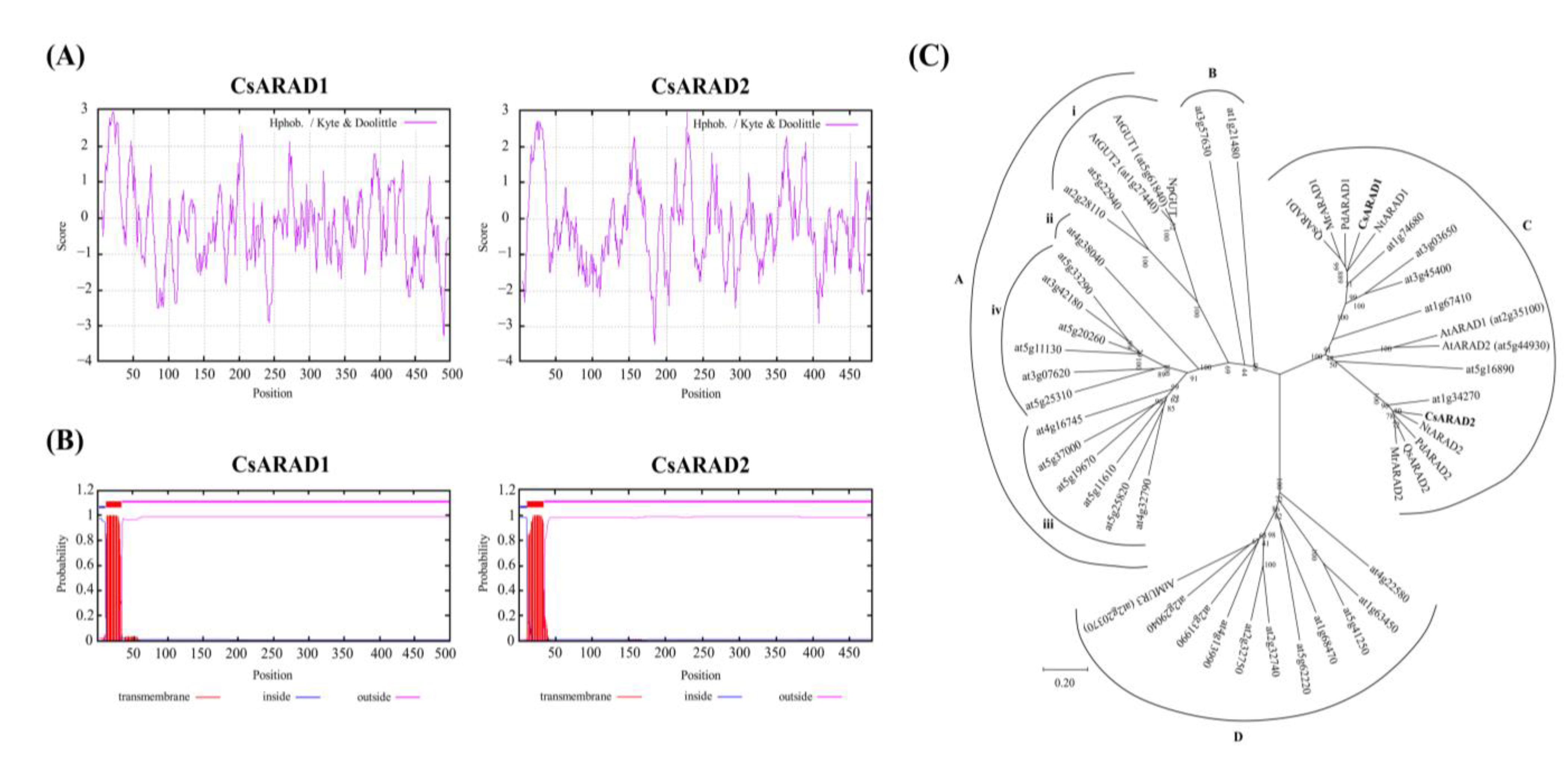
The molecular weights and isoelectric points of CsARAD1 and CsARAD2 were analyzed by compute pI/Mw with the values of 9.17 (molecular weight of CsARAD1), 8.52 (molecular weight of CsARAD2), 57.07 (isoelectric point of CsARAD1), and 54.23 kDa (isoelectric point of CsARAD2), respectively. Amino acid compositions were analyzed using ProtParam. CsARAD1 was 10.4% leucine, 9.6% serine, 6.4% isoleucine, 6.2% alanine, 6.2% arginine, and 6.2% valine; CsARAD2 was 10.6% leucine, 9.4% serine, 7.3% valine, and 6.5% proline (Table 1). ExPASy-ProtScale was used to calculate the hydropathic index of the proteins in the Hphob/Kyte and Doolittle mode. The proteins of both CsARAD1 and CsARAD2 had hydrophobic ends at the N-terminus. Transmembrane protein regions were predicted using TMHMM Server v. 2.0, and the results indicated that the transmembrane regions of CsARAD1 and CsARAD2 proteins were located between the 12th and 34th amino acid sites (Figure 2A,B). Based on the prediction results for the hydropathic index and transmembrane protein region, CsARAD1 and CsARAD2 proteins were determined to have a hydrophobic end and a transmembrane region at the N-terminus. Therefore, the two proteins were inferred to be type II membrane proteins. Furthermore, CsARAD1 and CsARAD2 were also predicted to be with no signaling peptides and were speculated to be located in the Golgi bodies by the utilization of SignalP 5.0 and DeepLoc 1.0 (Table S2).
One study conducted a phylogenetic tree analysis of GT47 sequences in Arabidopsis and demonstrated that they can be divided into four groups, namely subclades A, B, C, and D [11]. Subclade A contains xyloglucan galactosyltransferases, subclade B contains putative arabinan arabinosyltransferases, subclade C contains at least one xylogalacturonan xylosyltransferase (and probably other activities), and subclade D contains members with unknown activity that are putatively involved in xylan biosynthesis [13]. Notably, the two ARAD proteins AtARAD1 (At2g35100) and AtARAD2 (At5g44930) in Arabidopsis belong to Group B in the GT47 family [12]. In order to test the genetic relationships of CsARAD1 and CsARAD2 with the other members of the GT47 family of Arabidopsis and other higher plants, the MEGA-X software was used. The results indicated that CsARAD1 and CsARAD2 also belong to the B group of the GT47 family (Figure 2C).

3.2. High Level of CsARAD Expression in Nonlignified Tissues of Tea Plants

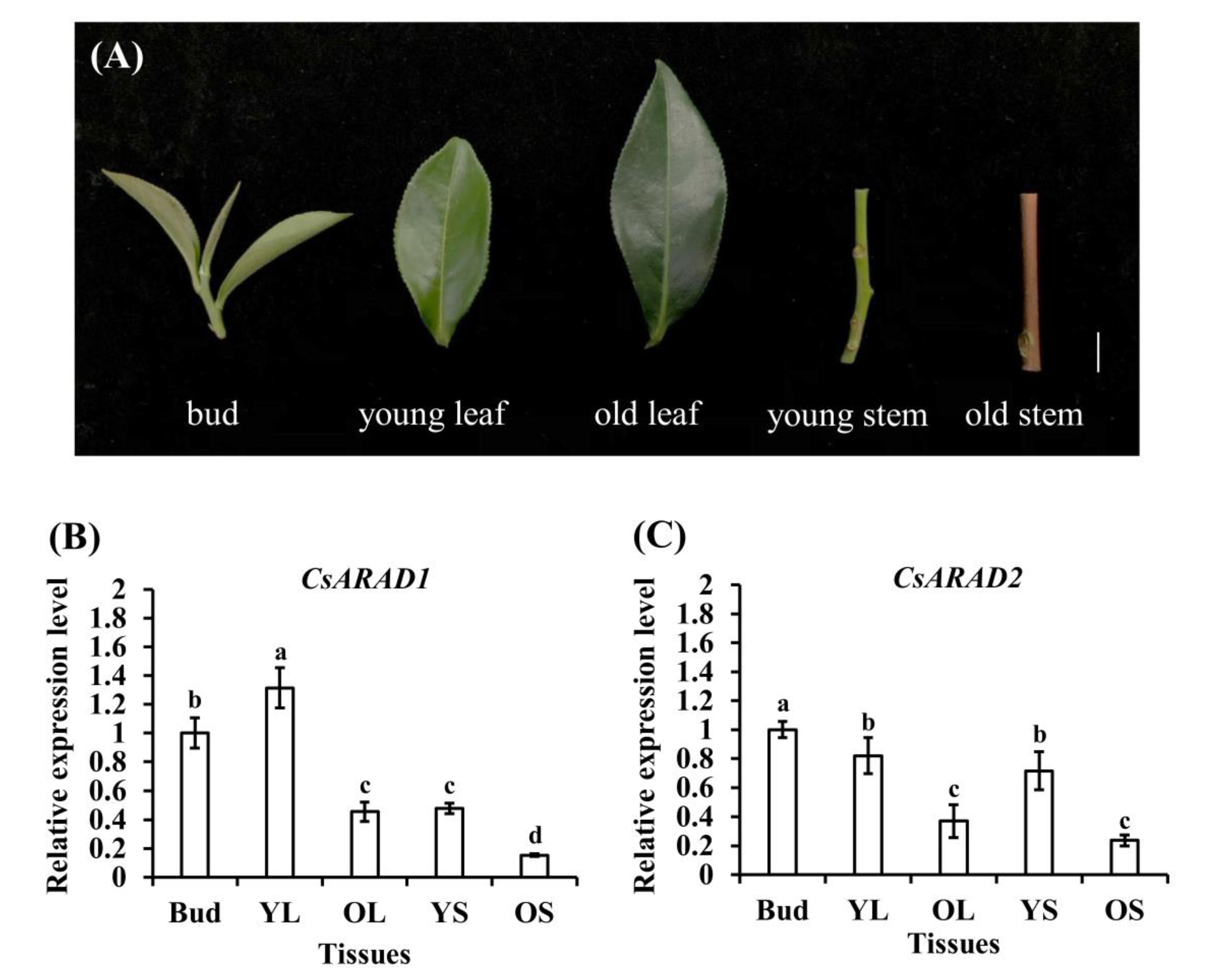
A study reported that the transcript level of AtARAD1 is highly expressed in the mature roots and stems of Arabidopsis thaliana [17]. To compare the expression patterns of CsARAD1 and CsARAD2 in various tea plant tissues, samples of buds, young leaves, old leaves, young stems, and old stems were collected from 8-year-old Sijichun tea plant and used as test materials (Figure 3A). The total RNA of various tissues was extracted and then reverse transcribed to cDNA. The transcript levels of CsARAD1 and CsARAD2 genes in the tissues were analyzed through qRT-PCR. The results indicated that the transcript level of CsARAD1 was higher in young buds and young leaves than in old leaves, young stems, and old stems (Figure 3B), whereas the relative expression level of CsARAD2 was higher in young buds, young leaves, and young stems than in old leaves and old stems (Figure 3C). On the basis of these results, both CsARAD1 and CsARAD2 were inferred to exhibit high relative expression levels in the nonlignified young tissues of tea plants.

3.3. Transcript Level of CsARADs Was Affected by Environmental Factors and Hormonal Signals

Climate change affects the yield and quality of crops, and the quality of crops can be regulated by managing plant nutrition, mineral content, and primary and secondary metabolites [18]. We investigated whether seasonal changes affect the expression levels of CsARAD1 and CsARAD2 genes in tea plants. Bud tissues were collected in March, June, September, and December 2020 and treated as spring, summer, autumn, and winter samples, respectively. The total RNA of the samples was extracted and reverse transcribed to cDNA for further analysis. The qRT-PCR results indicated that CsARAD1 had the highest and lowest transcript levels in September and June, respectively (Figure 4A). CsARAD2 had the highest transcript level in September and lower transcript levels in June and December (Figure 4B). Based on these results and meteorological data from the Taiwan Central Meteorological Administration (Figure S3), the expression of CsARADs was affected by environmental factors such as temperature, rainfall, and relative humidity. Our results suggest that the bud tissues in tea plants are affected by multiple factors (e.g., temperature, relative humidity, solar radiation, and precipitation) that change in each month, resulting in differences in the expression of CsARAD1 and CsARAD2 across seasons.

3.4. Abiotic Stresses and Hormonal Signals Regulate CsARAD Expression

To investigate the regulation of CsARADs under abiotic stresses, 1-year-old tea seedlings were treated with high-temperature (35 °C), low-temperature (4 °C), high-salinity (300-mM NaCl solution), and drought conditions (absence of irrigation for 3 days) in a growth chamber. Next, total RNA was extracted from the one-tip-two-leaf tissues of Sijichun seedlings, and a qRT-PCR analysis was performed. The results indicated that, compared with the expression of CsARAD1 in the control group, the expression levels of CsARAD1 were lower in the experimental groups that were exposed to high temperature, low temperature, high salinity, and drought stress (Figure 5A). However, compared with the control group, the expression levels of CsARAD2 were higher in the experimental groups that were exposed to high temperature and low temperature stresses and lower in those that were exposed to salinity and drought stress (Figure 5B).
When plants experience biotic or abiotic stress, they release stress-related hormonal signals to initiate their defense mechanism and improve their resistance to harmful situations. We treated 1-year-old tea seedlings with 100 μM of plant hormones (i.e., ACC, SA, MeJA, ABA, and GA) for 8 h and, subsequently, collected one-tip-two-leaf tissues to analyze CsARAD expression through qRT-PCR. The results indicated that, compared with the control group, CsARAD1 was downregulated in the experimental groups that received ACC and ABA signals (Figure 5C), and CsARAD2 was downregulated in the experimental groups that received ACC, ABA, and GA signals (Figure 5D). These results suggest that CsARADs participate in the ABA and ethylene signaling pathways and that CsARAD1 and CsARAD2 exhibit different hormonal regulation patterns.

4. Discussion

The GT superfamily contains enzymes that synthesize polysaccharides such as starch and cell wall polysaccharides. The function of GT47 proteins is reported as a pectin β-glucuronyltransferase and a xyloglucan β-galactosyltransferase, which are both essential to the development of plant cell walls [12]. The mutation of pectin β-glucuronyltransferase causes a defect in pectin synthesis, resulting in the development of calluses with an abnormal phenotype in Nicotiana plumbaginifolia mutants [19]. GT47 family members have highly conserved domains of XCX, G, XXX, CXPV, and XX in their amino acid sequences. In this study, the CsARADs obtained from Sijichun also exhibited these domains in their amino sequences (Figure 1). Thus, CsARAD1 and CsARAD2 were inferred to be members of the GT47 superfamily in tea trees. Cell wall polysaccharides are synthesized by the enzymatic complexes in two major subcellular locations. Pectin and hemicellulose are formed in the Golgi body and, subsequently, transferred to cell walls, whereas cellulose microfibrils and callose are generated at the plasma membrane surface [20]. The GT47 proteins of A. thaliana and Populus trichocarpa are type II transmembrane proteins located in the Golgi apparatus of cells [13,21]. The subcellular localization analysis of Sijichun CsARAD1 and CsARAD2 also indicated that they were located in the Golgi body, suggesting that their protein functions are identical to those of the members of these plants and involved in cell wall synthesis (Table S2).
The GT47 proteins of p. trichocarpa were mainly expressed in leaves, petioles, and stems, especially in primary xylem and phloem fibers. The GT47s were speculated to be related to the growth of xylem and fibers in stems and roots [21]. Another study used a GUS assay to investigate the AtARAD1 expression patterns of various tissues, and it reported that AtARAD1 exhibited the highest expression levels in the root system and primary leaves of 3-day-old seedlings and were mildly expressed in the vascular cambium and secondary phloem of 9-week-old plants. AtARAD1 was inferred to be mainly expressed in the nonlignified immature tissues of A. thaliana [17]. In the Sijichun tea cultivar, both CsARAD1 and CsARAD2 exhibited higher transcript levels in young tissues than in old tissues (Figure 3). These similar expression patterns (i.e., tender tissues exhibit higher levels of CsARAD expression than do lignified tissues) are consistent with the findings of studies on other species, suggesting that ARADs contribute to the metabolism and growth of young tissues. Moreover, arabinan was reported to present more in the young tissue in plants [22]. The coordinate pattern of the arabinan content and transcript level of ARAD might imply the arabinan-related function of ARADs in plant cells. The expression patterns of CsARAD1 and CsARAD2 differed slightly. Reasons for this may be that the metabolite composition and gene expression regulation in different growth stages and the functions of the two proteins differ. No study has explored whether other CsARAD-related homologous proteins exist in tea trees; therefore, our understanding of the function of CsARADs is still limited, and further investigation is warranted.
The plant cell wall determines plant morphogenesis and architecture, provides mechanical support for the plant body, conducts water and nutrients, and defends against biotic and abiotic stresses [23]. The composition of a cell wall can change to enable plants to survive under various stressful conditions [24,25]. ARADs were reported to produce the essential material arabinan of cell walls [26]. The chemical composition of cell walls would be affected by a stressful environment [27], indicating that the expression of ARAD might also differ under stress conditions. A study has shown that overexpression of arabinan synthase could lead to increased stomatal openings in Arabidopsis, which is an important moisture regulation approach in plants [28]. In our results, the expression levels of CsARAD1 and CsARAD2 were both down-regulated in drought condition. This might imply that CsARADs were turned off to prevent the stomata from opening under drought conditions. Our results indicate that temperature, high salinity, and drought stress can affect the gene expression of CsARADs in tea shoots (Figure 4C,D). Therefore, both CsARAD1 and CsARAD2 participate in numerous defensive responses to abiotic stress in plants. However, the two genes exhibit different expression patterns, indicating that they have different regulatory mechanisms for stress. CsARAD1 was downregulated in both heat and cold stress condition; in contrast, CsARAD2 showed the opposite regulation pattern under these stress conditions. Furthermore, among these two genes, CsARAD2 was downregulated by the GA signal (Figure 5C,D). A similar pattern was not observed in CsARAD1, which could be considered direct evidence that, though CsARAD1 and CsARAD2 are two homolog genes in tea, their functions and signaling may vary.
In addition to increasing the solubility and stability of glycosyl derivatives, glycosylation plays a role in regulating the hormones in plants by promoting hormonal catabolism [5,29]. Phytohormones such as ethylene, GA, and ABA are involved in the anthocyanin biosynthesis pathway in plants. The ethylene signal inhibits the accumulation of anthocyanins in plants subjected to stressful conditions [30,31]. In annual Sijichun seedlings, the transcription levels of CsARAD1 and CsARAD2 were both downregulated by ACC and ABA treatments. CsARADs were assumed to be regulated by the ethylene and abscisic acid signals associated with the physiological responses of tea plants.
GT plays a key role in plant responses to stressful conditions and the hormonal signals of plants. Studies have pointed out that the cis-acting elements of the GT47 family promoters of different cotton (Gossypium spp.) varieties were highly related to hormone signals such as salicylic acid, methyl jasmonate, auxin, estrogen, and auxin [8]. The mRNA level of ABA-inducible GTase gene obtained from the UDP-GTase homologs in the adzuki bean (Vigna angularis) was increased with ABA treatment and by water and wounding stress [32]. Ten GTs in quinoa (Chenopodium quinoa Willd.) were discovered to respond to ethylene and salt stress [33]. Previous study have indicated that the cell wall structure would be loosened with ABA during fruit ripening, and the cell wall degrading enzyme, such as rhamnogalacturonan I lyase and β-Galactosidase, were regulated with ABA signaling [34]. Based on this point, the regulation of ARAD, the putative arabinan producer in the cell wall, might also be affected by the ABA signal. In our results, CsARAD1 and CsARAD2 were significantly downregulated under ABA treatment and regulated by related abiotic stresses such as highly saline and drought-like conditions (Figure 5C,D); therefore, both CsARAD1 and CsARAD2 may participate in the ABA-dependent signaling pathway. In this study, the relationships of CsARADs with abiotic stress and hormonal signaling were investigated, and the results provide a basis for further research.

5. Conclusions

The sequences of CsARAD1 and CsARAD2 from the Sijichun tea cultivar were cloned and analyzed. CsARAD1 and CsARAD2 were mainly expressed in young tissues and regulated by environmental changes and ethylene and ABA signals. The CsARADs share similar sequences; however, the minor differences in their regulation patterns suggest that their functions differ slightly. The regulation of CsARAD by environmental factors and hormones holds great potential for informing cultivation management practices, which could greatly impact tea metabolites and prove beneficial to farmers. This study provides basic information regarding key enzymes involved in the secondary metabolism of tea plants for further research and breeders. By understanding these aspects, researchers and breeders could gain insights into how to modulate the secondary metabolite profiles to elevate tea infusion taste and quality through targeted breeding or biotechnological approaches.

Supplementary Materials

The following supporting information can be downloaded at: https://www.mdpi.com/article/10.3390/agronomy13061476/s1, Table S1: list of primers used in this study; Table S2: subcellular localization analysis of CsARAD1 and CsARAD2; Figure S1: cloning strategy and primers positions on CsARAD1 and CsARAD2; Figure S2: the correspondence table of the nucleotide sequence and deduced amino acid sequence of tea tree CsARAD1 and CsARAD2; Figure S3: the meteorological data of Nantou during 2019 and 2020.

Author Contributions

Data curation, C.-T.C. and T.-C.L.; formal analysis, M.-C.W. and S.-L.O. investigation, C.-T.C. and T.-C.L.; methodology, M.-C.W., S.-L.O., J.T.C.T. and C.-Y.Y.; project administration, C.-Y.Y.; supervision, J.T.C.T. and C.-Y.Y.; validation, C.-T.C. and T.-C.L.; writing—original draft C.-T.C.; writing—review and editing, C.-Y.Y. All authors have read and agreed to the published version of the manuscript.

Funding

This research was funded by the Ministry of Science and Technology, Taiwan, under grant numbers 108-2313-B-005-037 and partially supported by the National Science and Technology Council under Grant Number NSTC 111-2634-F-005-001-project Smart Sustainable New Agriculture Research Center (SMARTer).

Data Availability Statement

Not applicable.

Conflicts of Interest

The authors declare no conflict of interest.

References

  1. Zhang, L.; Ho, C.T.; Zhou, J.; Santos, J.S.; Armstrong, L.; Granato, D. Chemistry and biological activities of processed Camellia sinensis teas: A comprehensive review. Compr. Rev. Food Sci. Food Saf. 2019, 18, 1474–1495. [Google Scholar] [CrossRef] [PubMed]
  2. Bai, W.-X.; Wang, C.; Wang, Y.-J.; Zheng, W.-J.; Wang, W.; Wan, X.-C.; Bao, G.-H. Novel acylated flavonol tetraglycoside with inhibitory effect on lipid accumulation in 3T3-L1 cells from Lu’an GuaPian tea and quantification of flavonoid glycosides in six major processing types of tea. J. Agric. Food Chem. 2017, 65, 2999–3005. [Google Scholar] [CrossRef] [PubMed]
  3. Jiang, L.; Shen, X.; Shoji, T.; Kanda, T.; Zhou, J.; Zhao, L. Characterization and activity of anthocyanins in Zijuan tea (Camellia sinensis var. kitamura). J. Agric. Food Chem. 2013, 61, 3306–3310. [Google Scholar] [CrossRef] [PubMed]
  4. Louveau, T.; Orme, A.; Pfalzgraf, H.; Stephenson, M.J.; Melton, R.; Saalbach, G.; Hemmings, A.M.; Leveau, A.; Rejzek, M.; Vickerstaff, R.J. Analysis of two new arabinosyltransferases belonging to the carbohydrate-active enzyme (CAZY) glycosyl transferase family1 provides insights into disease resistance and sugar donor specificity. Plant Cell 2018, 30, 3038–3057. [Google Scholar] [CrossRef] [PubMed]
  5. Gachon, C.M.; Langlois-Meurinne, M.; Saindrenan, P. Plant secondary metabolism glycosyltransferases: The emerging functional analysis. Trends Plant Sci. 2005, 10, 542–549. [Google Scholar] [CrossRef]
  6. Yan, J.; Fang, L.; Yang, L.; He, H.; Huang, Y.; Liu, Y.; Zhang, A. Abscisic acid positively regulates l-arabinose metabolism to inhibit seed germination through ABSCISIC ACID INSENSITIVE4-mediated transcriptional promotions of MUR4 in Arabidopsis thaliana. New Phytol. 2020, 225, 823–834. [Google Scholar] [CrossRef]
  7. Hansen, S.F.; Harholt, J.; Oikawa, A.; Scheller, H.V. Plant glycosyltransferases beyond CAZy: A perspective on DUF families. Front. Plant Sci. 2012, 3, 59. [Google Scholar] [CrossRef]
  8. Wu, A.; Hao, P.; Wei, H.; Sun, H.; Cheng, S.; Chen, P.; Ma, Q.; Gu, L.; Zhang, M.; Wang, H. Genome-wide identification and characterization of glycosyltransferase family 47 in cotton. Front. Genet. 2019, 10, 824. [Google Scholar] [CrossRef]
  9. Ma, Y.; Ding, S.; Fei, Y.; Liu, G.; Jang, H.; Fang, J. Antimicrobial activity of anthocyanins and catechins against foodborne pathogens Escherichia coli and Salmonella. Food Control 2019, 106, 106712. [Google Scholar] [CrossRef]
  10. Porchia, A.C.; Sørensen, S.O.; Scheller, H.V. Arabinoxylan biosynthesis in wheat. Characterization of arabinosyltransferase activity in Golgi membranes. Plant Physiol. 2002, 130, 432–441. [Google Scholar] [CrossRef]
  11. Geshi, N.; Harholt, J.; Sakuragi, Y.; Jensen, J.K.; Scheller, H.V. Glycosyltransferases of the GT47 family. Annu. Plant Rev. Plant Polysacch. Biosynth. Bioeng. 2010, 41, 265–283. [Google Scholar]
  12. Zhong, R.; Ye, Z.-H. Unraveling the functions of glycosyltransferase family 47 in plants. Trends Plant Sci. 2003, 8, 565–568. [Google Scholar] [CrossRef] [PubMed]
  13. Harholt, J.; Jensen, J.K.; Verhertbruggen, Y.; Søgaard, C.; Bernard, S.; Nafisi, M.; Poulsen, C.P.; Geshi, N.; Sakuragi, Y.; Driouich, A. ARAD proteins associated with pectic Arabinan biosynthesis form complexes when transiently overexpressed in planta. Planta 2012, 236, 115–128. [Google Scholar] [CrossRef] [PubMed]
  14. Verhertbruggen, Y.; Marcus, S.E.; Chen, J.; Knox, J.P. Cell wall pectic arabinans influence the mechanical properties of Arabidopsis thaliana inflorescence stems and their response to mechanical stress. Plant Cell Physiol. 2013, 54, 1278–1288. [Google Scholar] [CrossRef] [PubMed]
  15. Petersen, B.L.; MacAlister, C.A.; Ulvskov, P. Plant protein O-arabinosylation. Front. Plant Sci. 2021, 12, 645219. [Google Scholar] [CrossRef]
  16. Zhang, L.; Zhao, Y.; Gao, Y.; Wu, L.; Gao, R.; Zhang, Q.; Wang, Y.; Wu, C.; Wu, F.; Gurcha, S.S. Structures of cell wall arabinosyltransferases with the anti-tuberculosis drug ethambutol. Science 2020, 368, 1211–1219. [Google Scholar] [CrossRef]
  17. Harholt, J.; Jensen, J.K.; Sørensen, S.O.; Orfila, C.; Pauly, M.; Scheller, H.V. Arabinan deficient 1 is a putative arabinosyltransferase involved in biosynthesis of pectic arabinan in Arabidopsis. Plant Physiol. 2006, 140, 49–58. [Google Scholar] [CrossRef]
  18. Ahmed, S.; Griffin, T.S.; Kraner, D.; Schaffner, M.K.; Sharma, D.; Hazel, M.; Leitch, A.R.; Orians, C.M.; Han, W.; Stepp, J.R. Environmental factors variably impact tea secondary metabolites in the context of climate change. Front. Plant Sci. 2019, 10, 939. [Google Scholar] [CrossRef]
  19. Iwai, H.; Masaoka, N.; Ishii, T.; Satoh, S. A pectin glucuronyltransferase gene is essential for intercellular attachment in the plant meristem. Proc. Natl. Acad. Sci. USA 2002, 99, 16319–16324. [Google Scholar] [CrossRef]
  20. Saulnier, L.; Guillon, F.; Chateigner-Boutin, A.-L. Cell wall deposition and metabolism in wheat grain. J. Cereal Sci. 2012, 56, 91–108. [Google Scholar] [CrossRef]
  21. Zhou, G.-K.; Zhong, R.; Richardson, E.A.; Morrison, W.H., III; Nairn, C.J.; Wood-Jones, A.; Ye, Z.-H. The poplar glycosyltransferase GT47C is functionally conserved with Arabidopsis Fragile fiber8. Plant Cell Physiol. 2006, 47, 1229–1240. [Google Scholar] [CrossRef]
  22. Wilmowicz, E.; Kućko, A.; Alché, J.D.D.; Czeszewska-Rosiak, G.; Florkiewicz, A.B.; Kapusta, M.; Karwaszewski, J. Remodeling of Cell Wall Components in Root Nodules and Flower Abscission Zone under Drought in Yellow Lupine. Int. J. Mol. Sci. 2022, 23, 1680. [Google Scholar] [CrossRef] [PubMed]
  23. Zhang, B.; Gao, Y.; Zhang, L.; Zhou, Y. The plant cell wall: Biosynthesis, construction, and functions. J. Integr. Plant Biol. 2021, 63, 251–272. [Google Scholar] [CrossRef] [PubMed]
  24. Barajas-Lopez, J.d.D.; Tiwari, A.; Zarza, X.; Shaw, M.W.; Pascual, J.; Punkkinen, M.; Bakowska, J.C.; Munnik, T.; Fujii, H. Early response to dehydration 7 Remodels Cell Membrane Lipid Composition during Cold Stress in Arabidopsis. Plant Cell Physiol. 2020, 62, 80–91. [Google Scholar] [CrossRef] [PubMed]
  25. Ganie, S.A.; Ahammed, G.J. Dynamics of cell wall structure and related genomic resources for drought tolerance in rice. Plant Cell Rep. 2021, 40, 437–459. [Google Scholar] [CrossRef]
  26. Mariette, A.; Kang, H.S.; Heazlewood, J.L.; Persson, S.; Ebert, B.; Lampugnani, E.R. Not Just a Simple Sugar: Arabinose Metabolism and Function in Plants. Plant Cell Physiol. 2021, 62, 1791–1812. [Google Scholar] [CrossRef]
  27. Hura, T.; Hura, K.; Ostrowska, A. Drought-Stress Induced Physiological and Molecular Changes in Plants. Int. J. Mol. Sci. 2022, 23, 4698. [Google Scholar] [CrossRef]
  28. Carroll, S.; Amsbury, S.; Durney, C.H.; Smith, R.S.; Morris, R.J.; Gray, J.E.; Fleming, A.J. Altering arabinans increases Arabidopsis guard cell flexibility and stomatal opening. Curr. Biol. 2022, 32, 3170–3179.e4. [Google Scholar] [CrossRef]
  29. Aksamit-Stachurska, A.; Korobczak-Sosna, A.; Kulma, A.; Szopa, J. Glycosyltransferase efficiently controls phenylpropanoid pathway. BMC Biotechnol. 2008, 8, 25. [Google Scholar] [CrossRef]
  30. Blanco-Ulate, B.; Vincenti, E.; Powell, A.L.; Cantu, D. Tomato transcriptome and mutant analyses suggest a role for plant stress hormones in the interaction between fruit and Botrytis cinerea. Front. Plant Sci. 2013, 4, 142. [Google Scholar] [CrossRef]
  31. Jeong, S.-W.; Das, P.K.; Jeoung, S.C.; Song, J.-Y.; Lee, H.K.; Kim, Y.-K.; Kim, W.J.; Park, Y.I.; Yoo, S.-D.; Choi, S.-B. Ethylene suppression of sugar-induced anthocyanin pigmentation in Arabidopsis. Plant Physiol. 2010, 154, 1514–1531. [Google Scholar] [CrossRef] [PubMed]
  32. Xu, Z.-J.; Nakajima, M.; Suzuki, Y.; Yamaguchi, I. Cloning and Characterization of the Abscisic Acid-Specific Glucosyltransferase Gene from Adzuki Bean Seedlings. Plant Physiol. 2002, 129, 1285–1295. [Google Scholar] [CrossRef] [PubMed]
  33. Ma, Q.; Su, C.; Dong, C.-H. Genome-Wide Transcriptomic and Proteomic Exploration of Molecular Regulations in Quinoa Responses to Ethylene and Salt Stress. Plants 2021, 10, 2281. [Google Scholar] [CrossRef] [PubMed]
  34. Moya-León, M.A.; Mattus-Araya, E.; Herrera, R. Molecular events occurring during softening of strawberry fruit. Front. Plant Sci. 2019, 10, 615. [Google Scholar] [CrossRef]
Figure 1. Alignment analysis of amino acid sequences of ARAD from tea and other higher plants. (A) Amino acid sequence analysis of CsARAD1, almond, Spanish cork, bayberry, and tobacco. (B) Amino acid sequence analysis of CsARAD2, almond, Spanish cork, bayberry, and tobacco. Almond PdARAD1 (Prunus dulcis; probable arabinosyltransferase ARAD1; XP_034217676.1) and PdARAD2 (probable arabinosyltransferase ARAD2; XP_034196771.1), Spanish cork oak QsARAD1 (Quercus suber; probable arabinosyltransferase ARAD1; XP_023886550.1; XP_023890514.1), Yangmei MrARAD1 (Morella rubra; putative arabinosyltransferase ARAD1; KAB1209453.1) and MrARAD2 (putative arabinosyltransferase ARAD2; KAB1220702.1), and Tobacco NtARAD1 (Nicotiana tabacum; probable arabinosyltransferase ARAD1; probable arabinosyltransferase ARAD2; XP_016467921.1). Sequence alignment analysis was performed using the Clustal Omega computing mode and graphically presented using the GeneDoc software. The black background indicates 100% identification of conserved sequences, the dark-gray background indicates 80% identification of conserved sequences, and the light-black background indicates 60% identification of conserved sequences. Conserved sequences XCX, G, XXX, CXPV, and XX of GT47 family of sequences are marked with a black line.
Agronomy 13 01476 g001
Figure 2. Hydrophobicity and transmembrane region analysis of tea CsARAD proteins and dendrogram of GT47 family members. (A) Hydrophilic and hydrophobic analysis of CsARADs. (B) Analysis of protein transmembrane region of CsARADs. (C) Dendrogram of protein sequences include Camellia sinensis (Cs), Arabidopis thaliana (At), Prunus dulcis (Pd), Quercus suber (Qs), Morella rubra (Mr), Nicotiana tabacum (Nt), and Nicotiana plumbaginifolia (Np). Hydrophobicity analysis was performed using the ExPASy-ProtScale website and hydrophobicity scores of Hphob/Kyte and Doolittle. Transmembrane region analysis was performed using the TMHMM Server v. 2.0 website. The horizontal axis indicates the position of the amino acid; positive values represent hydrophobicity levels, and negative values represent hydrophilicity levels.
Agronomy 13 01476 g002
Figure 3. Transcript levels of CsARADs in various tissues of tea plants. (A) YL, OL, YS, and OS; bar = 1 cm. (B) Relative expression levels of CsARAD1 in various tissues of tea plants. (C) Relative expression level of CsARAD2 in various tissues of tea plants. Expression levels of CsARAD1 and CsARAD2 in tissues of tea plants were analyzed through qRT-PCR. Total RNA was extracted from various tissues. Relative expression levels were calculated using 18S rRNA as an internal control. The values represent the average ± SE of at least four independent experiments. SAS software version 9.4 was used to perform Duncan’s operation analysis; the letters represent significant differences in performance measures of upper tissues in different regions (p < 0.05).
Agronomy 13 01476 g003
Figure 4. Transcript levels of CsARADs each month. (A,B) Expression levels of CsARAD1 and CsARAD2 in tea buds each month. Transcript levels of CsARADs in buds were analyzed through qRT-PCR. Relative expression levels were calculated using 18S rRNA as an internal control. The values represent the average ± SE of at least four independent experiments. SAS software version 9.4 was used to perform Duncan’s operation analysis; letters represent significant differences between samples (p < 0.05).
Agronomy 13 01476 g004
Figure 5. Transcript levels of CsARADs under abiotic stresses and hormones treatment. (A,B) Expression level of CsARADs in tea seedlings under various abiotic stresses. (C,D) Regulation of CsARAD1 and CsARAD2 in tea seedlings treated with various hormones. Transcript levels of CsARADs in buds were analyzed through qRT-PCR. Relative expression levels were calculated using 18S rRNA as an internal control. The values represent the average ± SE of at least three independent experiments. SAS software version 9.4 was used to perform Duncan’s operation analysis; letters represent significant differences between samples (p < 0.05). Abbreviations: Control check, CK; 1-aminocyclopropane-1-carboxylic acid, ACC; salicylic acid, SA; methyl jasmonate, MeJA; abscisic acid, ABA; gibberellin acid, GA.
Agronomy 13 01476 g005
Table 1. The amino acid composition of CsARADs.
ProteinCsARAD1
(57.07 kDa)
CsARAD2
(54.23 kDa)
Amino Acid
Alanine (A)6.2%5.0%
Araginine (R)6.2%5.8%
Asparagine (N)4.8%4.2%
Asparate (D)5.4%5.4%
Cysteine (C)1.2%1.5%
Glutamin (Q)4.2%3.3%
Glutamate (E)4.6%5.2%
Glycine (G)5.2%5.8%
Histidine (H)3.0%2.7%
Isoleucine (I)6.4%5.4%
Leucine (L)10.4%10.6%
Lysine (K)6.0%5.6%
Methionine (M)2.4%1.9%
Phenylalainie (F)5.6%4.8%
Proline (P)5.0%6.5%
Serine (S)9.6%9.4%
Threonine (T)2.4%4.6%
Tryptophan (W)1.4%1.9%
Tyrosine (Y)3.8%3.1%
Valine (V)6.2%7.3%
Pyrrolysine (O)0.0%0.0%
Selenocysteine (U)0.0%0.0%
Disclaimer/Publisher’s Note: The statements, opinions and data contained in all publications are solely those of the individual author(s) and contributor(s) and not of MDPI and/or the editor(s). MDPI and/or the editor(s) disclaim responsibility for any injury to people or property resulting from any ideas, methods, instructions or products referred to in the content.