Article

Nutritional Characterization Based on Vegetation Indices to Detect Anthocyanins, Carotenoids, and Chlorophylls in Mini-Lettuce

by
Andressa Alves Clemente
1,
Gabriel Mascarenhas Maciel
2,
Ana Carolina Silva Siquieroli
3,*,
Rodrigo Bezerra de Araujo Gallis
4,
José Magno Queiroz Luz
5,
Fernando César Sala
6,
Lucas Medeiros Pereira
1 and
Rickey Yoshio Yada
7
1
Postgraduate Program in Agriculture and Geospatial Information, Institute of Agrarian Sciences, Federal University of Uberlândia, Monte Carmelo 38500-000, Brazil
2
Institute of Agrarian Sciences, Federal University of Uberlândia, Monte Carmelo 38500-000, Brazil
3
Institute of Biotechnology, Federal University of Uberlândia, Monte Carmelo 38500-000, Brazil
4
Institute of Geography, Federal University of Uberlândia, Monte Carmelo 38500-000, Brazil
5
Institute of Agrarian Sciences, Federal University of Uberlândia, Uberlândia 38410-337, Brazil
6
Center of Agrarian Sciences, Federal University of São Carlos, Araras 13604-900, Brazil
7
Faculty of Land and Food Systems, University of British Columbia, Vancouver, BC V6T 1Z4, Canada
*
Author to whom correspondence should be addressed.
Agronomy 2023, 13(5), 1403; https://doi.org/10.3390/agronomy13051403
Submission received: 20 March 2023 / Revised: 20 April 2023 / Accepted: 25 April 2023 / Published: 19 May 2023

Abstract

:
When obtaining new cultivars or monitoring the nutritional composition of lettuce, new techniques are necessary given the high cost and time required to conduct laboratory analyses of plant composition by conventional methods. The objective of this study was to evaluate different vegetation indices for the estimation of anthocyanin, chlorophyll, and carotenoids in mini-lettuce genotypes with different leaf colors and different typologies from red, green, and blue (RGB) images. The contents of pigments were evaluated in 15 lettuce genotypes, in addition to the soil plant analysis development (SPAD) index and vegetation indices in the visible range. The variability among genotypes was confirmed by the Scott-Knott test (p < 0.05) and multivariate analysis. Linear regressions were obtained between the green leaf index (GLI) and leaf pigments. GLI was a good predictor for estimating the contents of anthocyanin (r = −0.83; r2 = 0.75), carotenoid (r = −0.59; r2 = 0.43), chlorophyll a (r = −0.69; r2 = 0.48), chlorophyll b (r = −0.62; r2 = 0.39), and total chlorophyll (r = −0.77; r2 = 0.65) in red and green mini-lettuce. The high-performance phenotyping technique can be used to evaluate leaf pigments in breeding programs, as well as in crops for monitoring biofortification levels in lettuce.

1. Introduction

Nutritional deficiency is a serious problem worldwide [1,2], and its main cause is unbalanced diets. Currently, this is the main factor in the global risk of death [3]. In the post-COVID-19 pandemic world, malnutrition will remain one of the main challenges for humanity [4]. These problems have stimulated the population to seek nutritionally rich foods with beneficial effects on human health and longevity [5].
Biofortification is one of the means to nutritionally enrich edible parts of plants and obtain functional foods [6]. The justification for using biofortification in food, since the emergence of the technique, lies in its potential to combat micronutrient deficiencies, which affect a large part of the world population [7]. Thus, the intake of biofortified foods can meet the nutritional requirements of humans [8].
Lettuce (Lactuca sativa L.) is a promising crop for biofortification due to its worldwide popularity [9], being the most produced and consumed leafy vegetable in Brazil. There is a very large variation among commercial cultivars in terms of shape, size, and color [10]. In addition, it is one of the most efficient species in micronutrient absorption [11]. It is considered one of the most important vegetables in human diet and health due to its pleasant taste, nutritional quality, and ease of acquisition, having significant economic importance in Brazil [10]. As lettuce is a food that is increasingly present in the dishes of the population, besides being easy to prepare and low cost, any strategy aiming at enhancing biofortification can result in several health benefits.
Most lettuce varieties used are green in color, but a small group is red due to the presence of anthocyanins [12], whose contents may vary according to environmental and genetic conditions [13]. Anthocyanins and other plant pigments, such as chlorophylls and carotenoids, have a strong antioxidant action and are essential in plant physiology [14].
Research has revealed that anthocyanins, carotenoids, and chlorophylls can prevent the onset of chronic diseases such as cancer or cardiovascular disease, in addition to increasing the immune response [15,16]. In addition to the relationship with physical well-being, these plant pigments promote mental well-being when they are part of a regular healthy diet [17]. Since then, leafy vegetables and colorful fruits have gained greater attention.
Lettuce varieties have significantly different genetic, phenotypic, and commercial characteristics, and nutritional quality is one of the traits with great variability [18,19]. Compositional analysis of each variety is limited. Among the factors that limit the nutritional characterization of lettuce varieties and genetic improvement studies aimed at the selection of nutrient-rich plants are the high cost and the time required to conduct laboratory tests [20].
Studies have shown the potential of vegetation indices calculated from visible spectrum images (RGB) to evaluate photosynthetic parameters in several species [21,22,23,24,25,26]. The use of image phenotyping presented a high correlation with carotenoids and chlorophyll in experiments with green lettuce germplasm from the smooth and curly segments [27] and anthocyanins, chlorophylls, and carotenoids in red lettuce from the smooth and curly segments [20,28]. However, due to the greater complexity, studies evaluating anthocyanins, carotenoids, and chlorophylls by remote sensing in lettuce genotypes with different leaf colors and different typologies are rare, even more so when it comes to mini-type lettuces as proposed here.
Therefore, the objective of this study was to evaluate different vegetation indices for the estimation of anthocyanin, chlorophyll, and carotenoids in mini-lettuce genotypes with different leaf colors and different typologies through red, green, and blue (RGB) images.

2. Materials and Methods

The study was conducted at the Vegetable Experimental Station of the Federal University of Uberlândia (Figure 1), in the municipality of Monte Carmelo (873 m altitude), Minas Gerais, Brazil, from June to September 2019. The climate of the region is tropical, characterized by hot and humid summers and cold and dry winters, according to the Köppen classification.
Thirteen mini-lettuce genotypes of the Biofortified and Tropicalized Lettuce Breeding Program of UFU were evaluated and registered in the software program BG α BIOFORT INPI BR512019002403-6 [29]. The genotypes evaluated were: Purpurita; 2: UFU-215#2#2; 3: UFU-215#3#2;4: UFU-104#1#1; 5: UFU-215#6#5; 6: UFU-215#1#2; 7: UFU-215#7#3; 8: UFU MC MINIBIOFORT2; 9: UFU-215#10#2; 10: UFU-215#13#1; 11: UFU-66#4#2; 12: UFU-66#3#1; 13: UFU-66#8#1; 14: UFU-66#7; 15: Vitória, obtained after hybridization between the cultivars Pira 72 and Uberlândia 10,000, biofortified [30]. Purpurita and Vitória cultivars were used as commercial controls, totaling 15 treatments in the experiment (Figure 1).
Sowing was performed in June 2019 in expanded polystyrene trays with 200 cells filled with a commercial substrate based on coconut fiber. After sowing, the trays remained in an arched greenhouse, with dimensions of 6 m × 5 m × 3.5 m, covered with 150-micron transparent polyethylene film, protected against ultraviolet rays, and with anti-aphid white screen side curtains. Thirty-seven days after sowing (DAS), when the lettuce plant had 4 to 5 leaves, transplantation to the field was performed. The spacing adopted was 0.25 × 0.25 m. The experimental design was randomized blocks, with three replicates of 20 plants, in a total of 45 plots (Figure 1).
The methodological steps for the quantification of leaf pigments of the lettuce genotypes, image processing, and data analysis are presented in the flowchart of Figure 2.
Thirty-two days after transplanting to the field, the flight to obtain the images was carried out using a remotely piloted aircraft (RPA) Phantom 4 Advanced model (SZ DJI Technology Co., Ltd., Shenzhen, China). The parameters used in the flight were: a height of 20 m, longitudinal overlap of 80%, and side overlap of 75%. The flight was carried out automatically with the proprietary software DroneDeploy (DroneDeploy Inc, San Francisco, CA, USA). The Phantom 4 camera (model FC6310) configuration parameters were defined before the mission was carried out, with no possibility of changing them mid-flight. The camera was adjusted in its white balance to the sunny option. The R, G, and B values were not calibrated. The values of spectral sensitivity of the Phantom 4 Pro camera are red (594 ± 32.5 nm), green (532 ± 58 nm), and blue (468 ± 47 nm) [31]. The collected images were processed and orthorectified in Pix4Dmapper software version 1.1.38 (Pix4D SA, Prilly, Switzerland) to generate the orthomosaic.
Two days after collecting the images, the leaf contents of anthocyanins (AT), total carotenoids (CN), chlorophyll a (CA), chlorophyll b (CB), and total chlorophyll (CT) were evaluated. Three leaves from the middle third of the four central plants of each plot were collected and sent to the laboratory. The still-fresh leaves were washed and crushed after removal of the midrib. For AT, a solution composed of 95% ethanol and hydrochloric acid (85:15) was added to the plant tissue. For CN, CA, CB, and CT, an ether solution of oil and acetone (1:1) was added. After 24 h of reaction in the absence of light, the absorbance of the supernatant extract was read in a UV-5100 digital spectrophotometer (Kalstein Co., Paris, France). The wavelengths employed were: 535 nm for AT, 645, 652, and 663 nm for CA, CB, CT, and 470 nm for CN. From the absorbances, the contents of leaf pigments (ug·g−1 of fresh tissue) were calculated according to refs. [32,33,34].
The Soil Plant Analysis Development (SPAD) index was evaluated the morning after the extraction of leaf pigments in the laboratory. Three readings per plant were performed in the four central plants of the plot, thus obtaining the average of the plot. The readings were performed with the chlorophyll meter Minolta SPAD-502 CFL1030 (Konica Minolta Inc., Tokyo, Japan), which measures the transmittance of the leaves in red and infrared wavelengths [35].
In image processing, vegetation indices (Table 1) were tested in the 45 plots for the creation of a mask layer and vegetation segmentation.
The main result of the segmentation process was a binary image (mask layer), where the vegetation was separated from the other objects [43]. This procedure aimed to remove the pixels referring to the background of the image (soil) to better study the behavior of vegetation (Figure 3).
After segmentation, the vegetation indices described in Table 1 were calculated, and the average index of each plot was extracted for analysis of leaf pigment contents by image.
Image analyses were performed in R software version 3.6.3 [44] using the R FieldImageR package [45]. The data were statistically analyzed in R software version 3.6.3 [44]. The statistical significance of the differences in the obtained data was evaluated by the analysis of variance and the F test (p ≤ 0.05), and the means were compared by the Scott-Knott test (p ≤ 0.05) using the R ExpDesp.pt package [46].
Multivariate analyses of genetic dissimilarity between genotypes were based on the Euclidean distance. Genetic dissimilarity was represented by a dendrogram obtained by the hierarchical method of unweighted pair group method with arithmetic mean (UPGMA). The cophenetic correlation coefficient (CCC) was calculated to test the efficiency of UPGMA clustering and the relative importance of the variables was analyzed by the method of Singh [47]. These analyses were performed using the packages R Vegan [48], NbClust [49], and Biotools [50].
Pearson’s correlation matrix was calculated at a 5% significance level between the leaf pigments evaluated in the laboratory, the SPAD index measured in the field, and vegetation indices obtained via remote sensing using the R Hmisc package version 5.0-1 [51].
The vegetation index that showed the highest correlation with leaf pigment contents was used to calculate the models for estimating anthocyanin, carotenoid, chlorophyll a, chlorophyll b, and total chlorophyll.

3. Results and Discussion

3.1. Nutritional Characterization of Mini-Lettuce Genotypes and Confirmation of Genetic Dissimilarity for Leaf Pigments

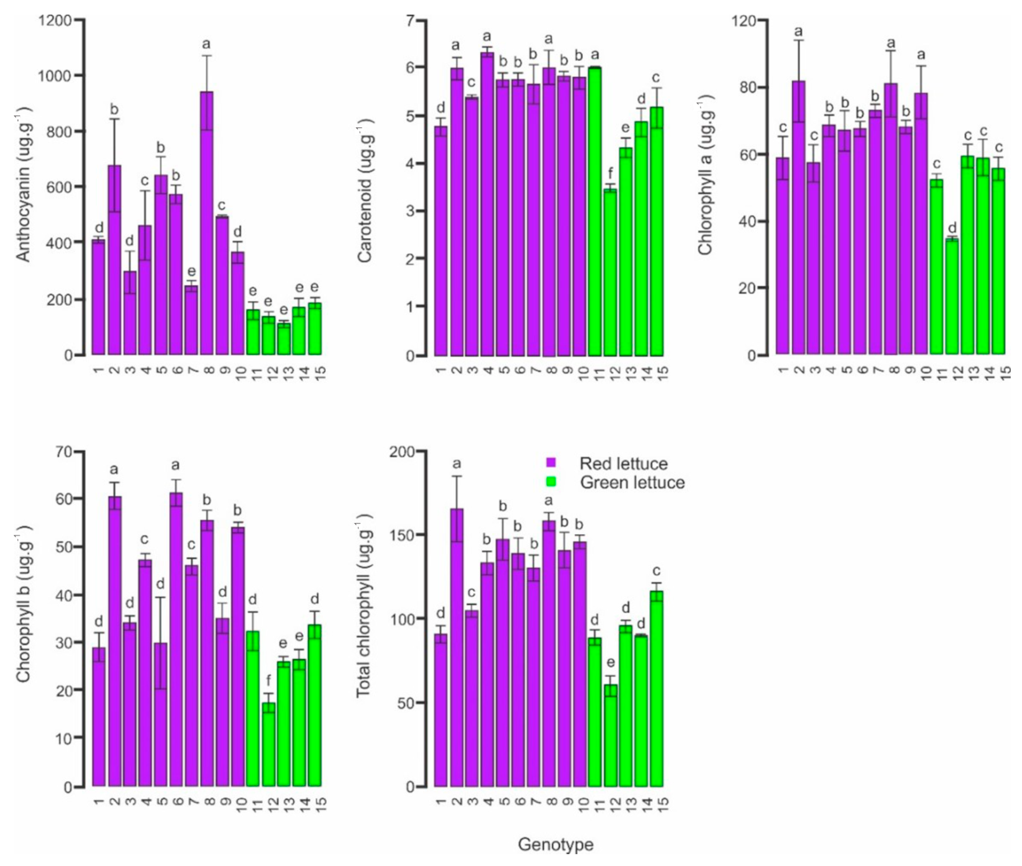
Based on the analysis of variance (ANOVA), there were significant differences determined by the F test (p ≤ 0.05) among lettuce genotypes for the contents of anthocyanin, carotenoid, chlorophyll a, chlorophyll b, and total chlorophyll. Therefore, the means were compared using the Scott-knott test (p ≤ 0.05) (Figure 4).
The anthocyanin content ranged from 108.7 for UFU-66#8#1 to 937.9 to ug·g−1 of fresh tissue for UFU MC MINIBIOFORT2, biofortified cultivar registered in BG α BIOFORT software [29]. Anthocyanin is strongly correlated with antioxidant activity [52,53,54].
Red lettuce genotypes were generally rich in anthocyanin, except for UFU-215#7#3. This genotype is classified as red but showed anthocyanin content statistically equivalent to that of green genotypes. The behavior of this genotype can be explained by the expression of red and green colors simultaneously in the leaves of the plant (variegate leaves) [55]. This result is in agreement with the fact that anthocyanin is the pigment that gives a red color to leaves [12,15,32,56].
For carotenoids, the variation among genotypes was 45%. The genotypes UFU-215#2#2, UFU-104#1#1, UFU MC MINIBIOFORT2, and UFU-66#4#2 were noticeable, reaching 6.3 ug·g−1 of fresh tissue. In addition to having antioxidant action, carotenoids can exist as various forms of provitamin A that, when ingested, can be converted into vitamin A [57]. Vitamin A deficiency is a public health problem worldwide, being the cause of disorders such as xerophthalmia and increased risk of infectious diseases [57,58]. Thus, lettuce varieties biofortified with carotenoids are desirable. The selection of genotypes with higher contents of vitamin A can be an excellent strategy for their use as commercial varieties, as well as being used in other breeding programs in Brazil and worldwide.
The genotypes UFU-215#2#2 and UFU MC MINIBIOFORT2 showed the highest concentrations of chlorophyll a, chlorophyll b, and total chlorophyll. The chlorophyll concentration in genotype UFU-215#2#2 reached 165.6 ug·g−1 of fresh tissue, while the genotype UFU-66#3#1 had only 40% of this value (59.8 ug·g−1 of fresh tissue). High chlorophyll content can favor their use in the food and pharmaceutical industry, as consumption of chlorophyll-rich vegetables can help prevent obesity and improve glucose tolerance [59]. Chlorophyll has potential health benefits, such as antioxidant, anti-inflammatory, antimutagenic, and anticancer activity [60].
High chlorophyll varieties can also benefit farmers due to the higher photosynthetic capacity of vegetables [61]. Plants with more chlorophyll tend to have a higher photosynthetic rate, but it does not necessarily result in higher biomass accumulation [62]. Lettuce with a color that best pleases and attracts the attention of the final consumer is important to meet the needs of an increasingly dynamic and demanding market [63,64]. In the breeding of lettuce, some research studies aim at varieties with a more intense green color (higher chlorophyll content) and with brightness in their leaves [65,66].
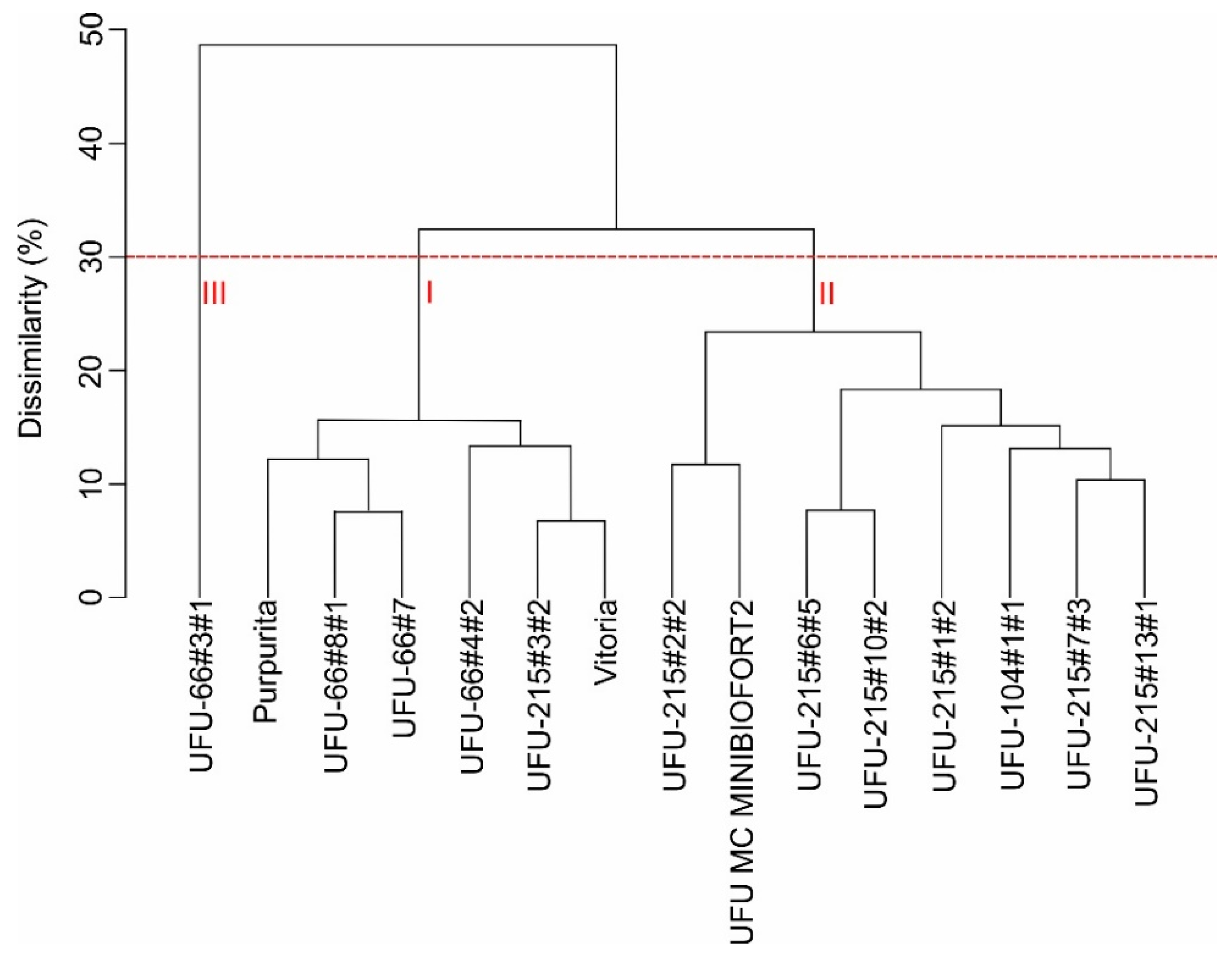
The Euclidean distance between the 15 treatments ranged from 0.67 (UFU-215#3#2 and Vitória) to 7.34 (UFU MC MINIBIOFORT2 and UFU-66#3#1), indicating genetic diversity. The clusters formed in the UPGMA dendrogram (Unweighted Pair Group Method with Arithmetic Mean) (Figure 5) showed a cophenetic correlation coefficient of 0.79 (t-test, p < 0.01). Thus, the dendrogram satisfactorily reflected the matrix data and subsequent clusters.
The number of clusters was established as indicated by most of the indices tested by ref. [67]. Cluster I consisted of six genotypes; among them were the two commercial controls (Purpurita and Vitória). Cluster II comprised eight genotypes (red leaves), which stood out for their high levels of leaf pigments. Cluster III contained the genotype UFU-66#3#1, characterized by low contents of all leaf pigments evaluated (Figure 5).
UFU MC MINIBIOFORT2, a biofortified variety, was grouped in cluster II. As it is a biofortified cultivar, it was suggested that the other genotypes grouped in cluster II are also nutritionally promising.
The relative importance of traits based on the criterion proposed by ref. [47] indicated that anthocyanin was the compound with the greatest contribution to genetic dissimilarity in green and red lettuce germplasm (27.7%), followed by carotenoids (24%) and chlorophyll a (20.1%). The lowest contributions came from chlorophyll b (16.3%) and total chlorophyll (14%).

3.2. Segmentation of Plants for RGB Images

The application of images obtained with remotely piloted aircraft (RPA) in agriculture depends on image processing to extract the appropriate information to achieve the desired objectives [43]. For lettuce analysis, the process of vegetation segmentation or background removal should be the initial step to achieve the objective. This process is easily accomplished using multispectral cameras. However, the use of multispectral cameras for RPA has the disadvantage of the high cost, while RGB sensors are low cost and favor the adoption of the technology. Thus, it was necessary to develop a method to perform the segmentation of red and green lettuce in an automated way using RGB images.
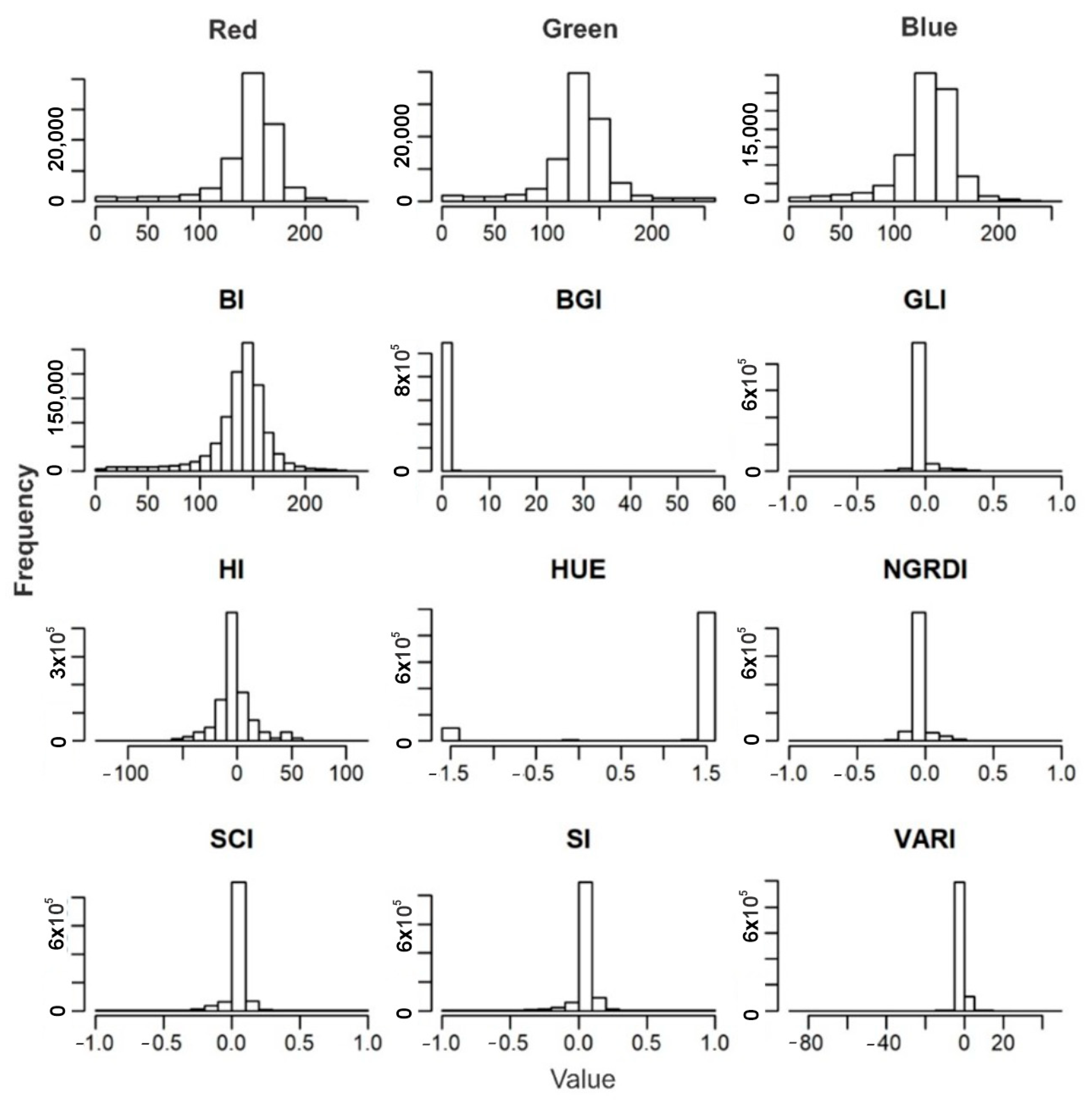
After calculating the vegetation indices described in Table 1, the distribution of the values of each index in pixels was analyzed by means of histograms (Figure 6).
HUE (overall hue index) was the index that best discriminated against the soil of green and red lettuces (Figure 6). In the HUE histogram, there were two extremes in the distribution of the digital number values of the pixels. The first extreme was related to the plants, and the last, with a greater number of pixels, to the soil, confirming the need for segmentation. In an experiment with different types of vegetation cover, it was observed that HUE contrasted the vegetation against the non-plant elements in the image [68].
To create the mask layer, a cutoff value of 1.52 in the HUE index was determined. Pixels with a HUE index above the cutoff value were counted for the mask layer referring to the soil. This cutoff value in the HUE index promoted excellent vegetation segmentation.
The procedure of removal of the image background (soil) was important to reduce noise in the analysis of leaf pigments and improve the reproducibility of image analysis.

3.3. Validation of High-Performance Phenotyping for Red and Green Lettuce Genotypes

Of the vegetation indices (Table 1) analyzed for segmentation (Figure 6), the five with the highest standard deviation were selected after removing the pixels from the image referring to the soil. The indices selected were: brightness index (BI), green leaf index (GLI), normalized green/red difference index (NGRDI), soil color index (SCI), and spectral slope saturation index (SI).
The quantification of anthocyanin, carotenoid, chlorophyll a, chlorophyll b, and total chlorophyll in the 15 treatments of red and green lettuce required 18 h and a team of four people (72 h of work), with consumption of 1.8 L of high-cost and dangerous reagents for the RPA. On the other hand, the SPAD Index measurement with the SPAD-502 chlorophyll meter was performed in 2.5 h by two people, reducing the evaluation time by 93%. For high-performance phenotyping, a single person carried out all processes in just one hour of work, which is equivalent to 98.6% and 77.8% in time savings to evaluate leaf pigments compared to quantification by the standard method and SPAD index, respectively. Thus, the high-performance phenotyping method drastically reduced time and costs with anthocyanin, carotenoid, and chlorophyll analyses.
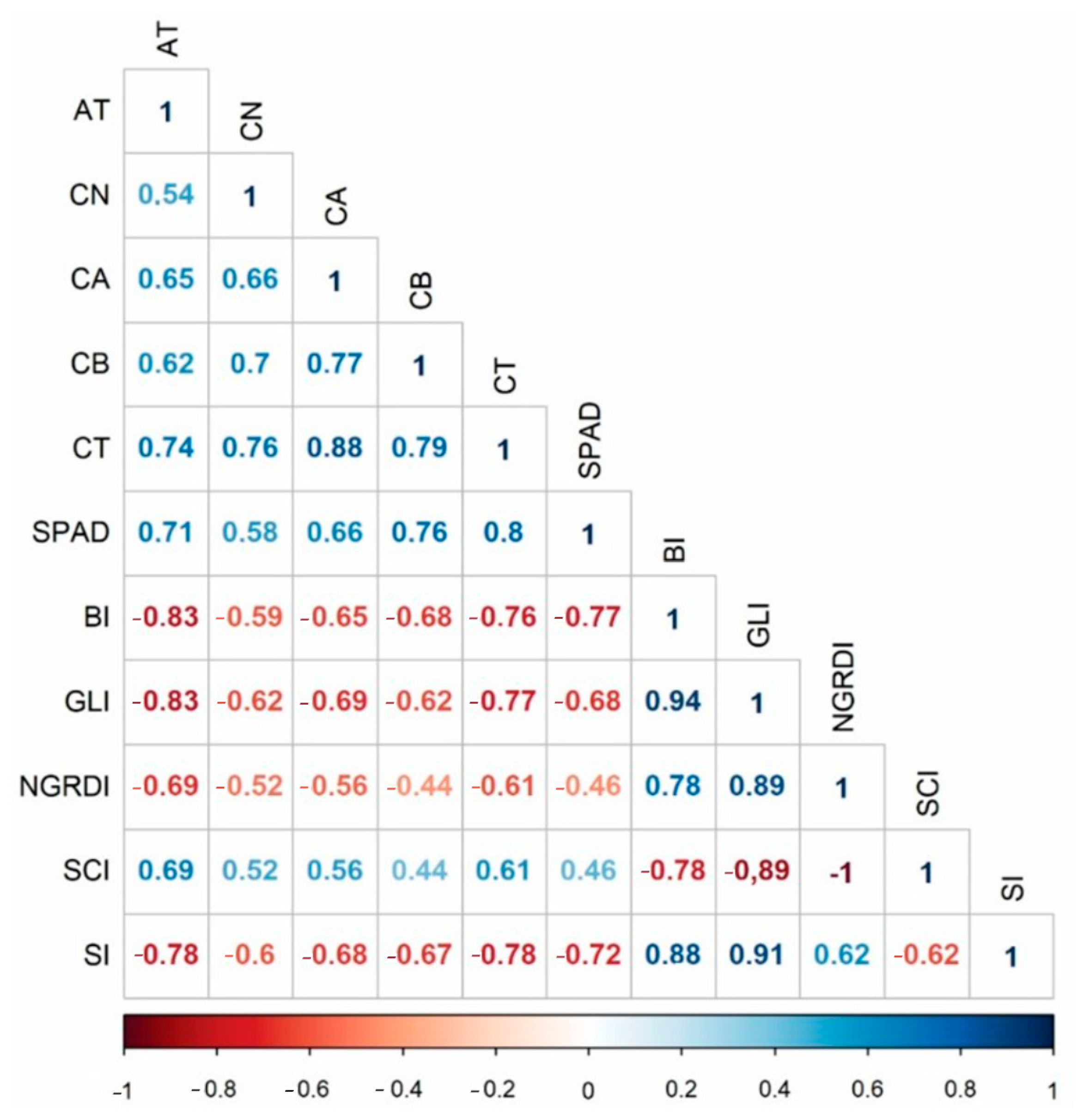
Strongly correlated characteristics allow indirect selection using the one with the simplest measurement [69,70]. Thus, to verify the possibility of selecting leaf pigment-rich genotypes of red and green lettuce from vegetation indices calculated in RPA images, the correlation matrix between the traits was calculated (Figure 7).
Leaf pigments in red and green lettuces were correlated with all indices evaluated (p ≤ 0.05), indicating that anthocyanins, carotenoids, and chlorophylls can be evaluated indirectly by vegetation indices obtained via remote sensing.
The correlations showed that as the BI, GLI, NGRDI, and SI increased, the content of leaf pigment decreased (Figure 7). However, the vegetation indices were expected to increase with increases in the contents of chlorophyll, the pigment responsible for green color. Additionally, a reduction in the spectral response in the green band was observed with increasing contents of carotenoids in red lettuce germplasm [20]. In an experiment with several plant species, it was concluded that green reflectance is reduced in plants that had high concentrations of anthocyanin [71]. Therefore, red lettuce genotypes may have influenced the results. Nevertheless, high correlations were observed between these indices and the pigments in green and red lettuces, which indicates that the models generated in this work can be applied to different types of lettuce.
Anthocyanin, the pigment that most contributed to genetic diversity, was highly correlated with the vegetation indices evaluated, mainly BI and GLI (both −83%). GLI also stood out with high correlations with carotenoids (−62%), chlorophyll a (−69%), chlorophyll b (−62%), and total chlorophyll (−77%).
High correlations were also observed between leaf pigments and the SPAD index. Several studies confirmed that SPAD indirectly expresses the contents of chlorophyll and carotenoid in the leaf [72,73,74]. However, although chlorophyll meter measurements can be used to evaluate leaf pigments, this method is still time-consuming and is subject to several technical problems in field evaluations.
It is worth pointing out that anthocyanins, carotenoids, and chlorophylls showed significant positive correlations among themselves. This indicates that, in red and green lettuce, the analysis of these leaf pigments can be carried out concurrently. After analyzing Figure 8, the similarity between the behavior of pigments quantified by the traditional method (laboratory) and that estimated by the high-performance phenotyping method (GLI vegetation index) becomes evident.
High-performance phenotyping made it possible to evaluate each plant individually with a reduction of time and cost, while in the traditional method, only the average content of each pigment was obtained for the plot. In addition, the technique that uses RPA images is non-destructive, which is important for the advancement of generation in breeding programs of this vegetable, as well as for the commercialization of plants in commercial crops.
Several studies have demonstrated the influences of environmental conditions, such as temperature, fertility, soil moisture, and mainly light, in addition to genetic factors, on the accumulation of phytopigments [75,76,77,78,79]. Thus, a lettuce cultivar may show different concentrations of these compounds depending on the environment in which it is grown. Given this and the efficiency of high-performance phenotyping in discriminating levels of leaf pigment contents, the use of this technique in large-scale crops is also proposed. Therefore, the tool can be useful for breeders in breeding programs aiming at biofortification, but also for horticulturalists and the public sector in order to monitor the levels of anthocyanins, carotenoids, and chlorophylls in crops using RPA.
To enable the quantification of leaf pigments by image, prediction models were generated (Table 2). The linear regression equations were fitted to the average response variable of the GLI vegetation index.
The GLI vegetation index based on RPA was a good predictor for estimating leaf pigment content in red and green lettuce. Regression analysis indicated that the carotenoid and total chlorophyll contents could be predicted using RPA-based GLI with only ±11% and 16% difference according to the root mean squared error, respectively. For anthocyanin and chlorophyll b, this difference was ±34% and 41%, respectively.

4. Conclusions

Despite the existence of a wide genetic variability of leaf colors in mini-lettuce, the vegetation indices showed strong power to estimate anthocyanin, carotenoids, and chlorophyll pigments.
The GLI vegetation index stood out, being a good predictor of leaf pigment contents in field experiments with mini-lettuce of different leaf colors.
To explore the efficiency of the GLI vegetation index in other studies, the R2 and RMSE values obtained for each pigment should be considered. In this study, the R2 and RMSE ranged from 0.39 to 0.75 and 0.6 to 134.4, respectively.
Additionally, it should be emphasized that the correlations between pigments and GLI were negative, ranging from moderate to strong (r = −0.83 to −0.62).
High-performance phenotyping obtained by remotely piloted aircraft can be used to characterize nutritional aspects in lettuce, either from a germplasm bank with wide genetic variability or in commercial crops.

Author Contributions

Conceptualization, G.M.M., A.C.S.S. and R.B.d.A.G.; methodology, A.A.C. and L.M.P.; software, A.A.C. and R.B.d.A.G.; validation, A.A.C. and L.M.P.; formal analysis, G.M.M., A.C.S.S., J.M.Q.L. and F.C.S.; investigation, A.A.C. and G.M.M.; resources, G.M.M., A.C.S.S. R.B.d.A.G. and J.M.Q.L.; data curation, G.M.M. and R.B.d.A.G.; writing —original draft preparation, A.A.C.; writing—review and editing, G.M.M., A.C.S.S., J.M.Q.L., F.C.S. and R.Y.Y.; visualization, A.C.S.S. and R.Y.Y.; supervision, G.M.M.; project administration, G.M.M. and A.C.S.S.; funding acquisition, G.M.M. and A.C.S.S. All authors have read and agreed to the published version of the manuscript.

Funding

This research was funded by the Brazilian National Council for Scientific and Technological Development (CNPq) Grant No 308824/2020-2, the Minas Gerais Research Foundation (FAPEMIG), the Coordination for the Improvement of Higher Education Personnel (CAPES), and the Federal University of Uberlândia (UFU).

Data Availability Statement

Not applicable.

Conflicts of Interest

The authors declare no conflict of interest.

References

  1. Cuenca, M.H.; Proaño, G.V.; Blankenship, J.; Cano-Gutierrez, C.; Chew, S.T.H.; Fracassi, P.; Keller, H.; Mannar, V.; Mastrilli, V.; Milewska, M.; et al. Building global nutrition policies in health care: Insights for tackling malnutrition from the academy of nutrition and dietetics 2019 global nutrition research and policy forum. J. Acad. Nutr. Diet. 2020, 120, 1407–1416. [Google Scholar] [CrossRef] [PubMed]
  2. Murray, R.D.; Kerr, K.W.; Brunton, C.; Williams, J.A.; Dewitt, T.; Wulf, K.L. A First Step Towards Eliminating Malnutrition: A Proposal for Universal Nutrition Screening in Pediatric Practice. Nutr. Diet. Suppl. 2021, 13, 17–24. [Google Scholar] [CrossRef]
  3. FAO; IFAD; UNICEF; WFP; WHO. The State of Food Security and Nutrition in the World 2019. Safeguarding against Economic Slowdowns and Downturns. Rome, FAO. 2019. Available online: http://www.fao.org/3/ca5162en/ca5162en.pdf (accessed on 20 September 2020).
  4. Ventura, D.D.F.L.; Ribeiro, H.; Giulio, G.M.D.; Jaime, P.C.; Nunes, J.; Bógus, C.M.; Waldman, E.A. Desafios da pandemia de COVID-19: Por uma agenda brasileira de pesquisa em saúde global e sustentabilidade. Cad. Saude Publica 2020, 36, e00040620. [Google Scholar] [CrossRef]
  5. Giordano, M.; El-Nakhel, C.; Pannico, A.; Kyriacou, M.C.; Stazi, S.R.; De Pascale, S.; Rouphael, Y. Iron biofortification of red and green pigmented lettuce in closed soilless cultivation impacts crop performance and modulates mineral and bioactive composition. Agronomy 2019, 9, 290. [Google Scholar] [CrossRef]
  6. Finkelstein, J.L.; Fothergill, A.; Hackl, L.S.; Haas, J.D.; Mehta, S. Iron biofortification interventions to improve iron status and functional outcomes. Proc. Nutr. Soc. 2019, 78, 197–207. [Google Scholar] [CrossRef]
  7. Lockyer, S.; White, A.; Buttriss, J.L. Biofortified crops for tackling micronutrient deficiencies—What impact are these having in developing countries and could they be of relevance within Europe? Nutr. Bull. 2018, 43, 319–357. [Google Scholar] [CrossRef]
  8. Almeida, H.J.; Dutra, A.F.; Cecílio Filho, A.B. Biofortificação de hortaliças e saúde global—Um enfoque para selênio, zinco, ferro e iodo. In Nutrição e Adubação de Hortaliças; Prado, R.M., Cecílio Filho, A.B., Eds.; FCAV/Unesp: Jaboticabal, Brazil, 2016; pp. 103–150. [Google Scholar]
  9. Camejo, D.; Frutos, A.; Mestre, T.C.; Piñero, M.C.; Rivero, R.M.; Martínez, V. Artificial light impacts the physical and nutritional quality of lettuce plants. Hortic. Environ. Biotechnol. 2020, 61, 69–82. [Google Scholar] [CrossRef]
  10. Sala, F.C.; Costa, C.P. Retrospectiva e tendência da alfacicultura brasileira. Hortic. Bras. 2012, 30, 187–194. [Google Scholar] [CrossRef]
  11. Clemens, S. How metal hyperaccumulating plants can advance Zn biofortification. Plant Soil 2017, 411, 111–120. [Google Scholar] [CrossRef]
  12. Zhang, Y.Z.; Xu, S.Z.; Cheng, Y.W.; Ya, H.Y.; Han, J.M. Transcriptome analysis and anthocyanin-related genes in red leaf lettuce. Genet. Mol. Res. 2016, 15, 10–4238. [Google Scholar] [CrossRef]
  13. Sala, F.C.; Costa, C.P. Melhoramento de alface. In Melhoramento de Hortaliças, 1st ed.; Nick, C., Borém, A., Eds.; Editora UFV: Viçosa, Brazil, 2016; pp. 95–126. [Google Scholar]
  14. Lopes, D.C.; Moura, L.O.; Steidle Neto, A.J.; Ferraz, L.C.L.; Carlos, L.A.; Martins, L.M. Spectral indices for non-destructive determination of lettuce pigments. Food Anal. Methods 2017, 10, 2807–2814. [Google Scholar] [CrossRef]
  15. Bendokas, V.; Skemiene, K.; Trumbeckaite, S.; Stanys, V.; Passamonti, S.; Borutaite, V.; Liobikas, J. Anthocyanins: From plant pigments to health benefits at mitochondrial level. Crit. Rev. Food Sci. Nutr. 2019, 60, 3352–3365. [Google Scholar] [CrossRef] [PubMed]
  16. Braga, A.R.C.; Murador, D.C.; Mesquita, L.M.S.; Rosso, V.V. Bioavailability of anthocyanins: Gaps in knowledge, challenges and future research. J. Food Compos. Anal. 2018, 68, 31–40. [Google Scholar] [CrossRef]
  17. Ocean, N.; Howley, P.; Ensor, J. Lettuce be happy: A longitudinal UK study on the relationship between fruit and vegetable consumption and well-being. Soc. Sci. Med. 2019, 222, 335–345. [Google Scholar] [CrossRef]
  18. Santana, C.V.S.; Almeida, A.C.; Turco, S.H.N. Produção de alface vermelha em ambientes sombreados na região do submédio São Francisco-BA. Rev. Verde 2009, 4, 1–6. [Google Scholar]
  19. Yang, X.; Wei, S.; Liu, B.; Guo, D.; Zheng, B.; Feng, L.; Liu, Y.; Tomás-Barberán, F.A.; Luo, L.; Huang, D. A novel integrated non-targeted metabolomic analysis reveals significant metabolite variations between different lettuce (Lactuca sativa L.) varieties. Hortic. Res. 2018, 5, 33. [Google Scholar] [CrossRef]
  20. Maciel, G.M.; Gallis, R.B.A.; Barbosa, R.L.; Pereira, L.M.; Siquieroli, A.C.S.; Peixoto, J.V.M. Image phenotyping of inbred red lettuce lines with genetic diversity regarding carotenoid levels. Int. J. Appl. Earth. Obs. Geoinf. 2019, 81, 154–160. [Google Scholar] [CrossRef]
  21. Guo, Y.; Yin, G.; Sun, H.; Wang, H.; Chen, S.; Senthilnath, J.; Wang, J.; Fu, Y. Scaling Effects on Chlorophyll Content Estimations with RGB Camera Mounted on a UAV Platform Using Machine-Learning Methods. Sensors 2020, 20, 5130. [Google Scholar] [CrossRef]
  22. Riccardi, M.; Mele, G.; Pulvento, C.; Lavini, A.; D’andria, R.; Jacobsen, S.E. Non-destructive evaluation of chlorophyll content in quinoa and amaranth leaves by simple and multiple regression analysis of RGB image components. Photosynth. Res. 2014, 120, 263–272. [Google Scholar] [CrossRef]
  23. Yadav, S.P.; Ibaraki, Y.I.; Gupta, S.D. Estimation of the chlorophyll content of micropropagated potato plants using RGB based image analysis. Plant Cell Tissue Organ Cult. 2010, 100, 183–188. [Google Scholar] [CrossRef]
  24. Garcia-Caparros, P.; Llanderal, A.; Lao, M.T. Correlations between R, G, and B values, pigment concentration, and nitrogen status in three ornamental potted plants. Agronomy 2023, 13, 177. [Google Scholar] [CrossRef]
  25. Radočaj, D.; Šiljeg, A.; Marinović, R.; Jurišić, M. State of major vegetation indices in precision agriculture studies indexed in web of science: A review. Agriculture 2023, 13, 707. [Google Scholar] [CrossRef]
  26. Wu, Q.; Zhang, Y.; Xie, M.; Zhao, Z.; Yang, L.; Liu, J.; Hou, D. Estimation of Fv/Fm in spring wheat using UAV-Based multispectral and RGB imagery with multiple machine learning methods. Agronomy 2023, 13, 1003. [Google Scholar] [CrossRef]
  27. Maciel, G.M.; Gallis, R.B.D.A.; Barbosa, R.L.; Pereira, L.M.; Siquieroli, A.C.S.; Peixoto, J.V.M. Image phenotyping of lettuce germplasm with genetically diverse carotenoid levels. Bragantia 2020, 79, 224–235. [Google Scholar] [CrossRef]
  28. Clemente, A.A.; Maciel, G.M.; Siquieroli, A.C.S.; Gallis, R.B.A.; Pereira, L.M.; Duarte, J.G. High-throughput phenotyping to detect anthocyanins, chlorophylls, and carotenoids in red lettuce germplasm. Int. J. Appl. Earth Obs. Geoinf. 2021, 103, 102533. [Google Scholar] [CrossRef]
  29. Maciel, G.M.; Siquieroli, A.C.S.; Gallis, R.B.A.; Pereira, L.M.; Sales, V.F. Programa de Computador BG α Biofort. 2019. Depositante: Universidade Federal de Uberlândia. BR512019002403-6. Deposit: 1 February 2019. Concession: 23 October 2019. Available online: https://busca.inpi.gov.br/pePI/servlet/ProgramaServletController (accessed on 10 March 2023).
  30. Sousa, C.S.D.; Bonetti, A.M.; Goulart Filho, L.R.; Machado, J.R.D.A.; Londe, L.N.; Baffi, M.A.; Ramos, R.G.; Vieira, C.U.; Kerr, W.E. Divergência genética entre genótipos de alface por meio de marcadores AFLP. Bragantia 2007, 66, 11–16. [Google Scholar] [CrossRef]
  31. Burggraaff, O.; Schmidt, N.; Zamorano, J.; Pauly, K.; Pascual, S.; Tapia, C.; Spyrakos, E.; Snik, F. Standardized spectral and radiometric calibration of consumer câmeras. Opt. Express 2019, 27, 19075–19101. [Google Scholar] [CrossRef]
  32. Francis, F.J. Analysis of anthocyanins. In Anthocyanins as Food Colors, 1st ed.; Markakis, P., Ed.; Academic Press: New York, NY, USA, 1982; pp. 182–208. [Google Scholar]
  33. Witham, F.H.; Blaydes, D.F.; Devlin, R.M. Experiments in Plant Physiology; Van Nostrand Reinhold: New York, NY, USA, 1971; 245p. [Google Scholar]
  34. Lichtenthaler, H.; Wellburn, A. Determinations of total carotenoids and chlorophylls a and b of leaf extracts in different solvents. Biochem. Soc. Trans. 1983, 603, 91–592. [Google Scholar] [CrossRef]
  35. Minolta. Chlorophyll meter SPAD-502. In Instruction Manual; Minolta Co.: Osaka, Japan, 1989. [Google Scholar]
  36. Richardson, A.J.; Wiegand, C.L. Distinguishing vegetation from soil background information. Photogramm. Eng. Remote Sens. 1977, 43, 1541–1552. [Google Scholar]
  37. Zarco-Tejada, P.J.; Berjón, A.; López-Lozano, R.; Miller, J.R.; Martín, P.; Cachorro, M.R.G.; de Frutos, A. Assessing vineyard condition with hyperspectral indices: Leaf and canopy reflectance simulation in a row-structured discontinuous canopy. Remote Sens. Environ. 2005, 99, 271–287. [Google Scholar] [CrossRef]
  38. Louhaichi, M.; Borman, M.M.; Johnson, D.E. Spatially located platform and aerial photography for documentation of grazing impacts on wheat. Geocarto Int. 2001, 16, 65–70. [Google Scholar] [CrossRef]
  39. Escadafal, R.; Belghith, A.; Bem, M.H. Indices spectraux pour la télédétection de la dégradation des milieux naturels en Tunisie aride. In Proceedings of the Actes du Sixième Symposium International. Mesures Physiques et Signatures Spectrales en Télédétection, Val d’Isère, France, 17–24 January 1994; pp. 7–21. [Google Scholar]
  40. Tucker, C. Red and Photographic Infrared Linear Combinations for Monitoring Vegetation. Remote Sens. Environ. 1979, 8, 127–150. [Google Scholar] [CrossRef]
  41. Mathieu, R.; Pouget, M.; Cervelle, B.; Escadafal, R. Relationships between satellite-based radiometric indices simulated using laboratory reflectance data and typic soil color of an arid environment. Remote Sens. Environ. 1998, 66, 17–28. [Google Scholar] [CrossRef]
  42. Gitelson, A.A.; Kaufman, Y.J.; Stark, R.; Rundquist, D. Novel algorithms for remote estimation of vegetation fraction. Remote Sens. Environ. 2002, 80, 76–87. [Google Scholar] [CrossRef]
  43. Hassanein, M.; Lari, Z.; El-Sheimy, N. A new vegetation segmentation approach for cropped fields based on threshold detection from hue histograms. Sensors 2018, 18, 1253. [Google Scholar] [CrossRef]
  44. R Core Team. R: A Language and Environment for Statistical Computing; R Foundation for Statistical Computing: Vienna, Austria, 2020; Available online: https://www.r-project.org/ (accessed on 10 January 2023).
  45. Matias, F.I.; Caraza-Harter, M.V.; Endelman, J.B. FIELDimageR: An R package to analyze orthomosaic images from agricultural field trials. Plant. Phenome J. 2020, 3, e20005. [Google Scholar] [CrossRef]
  46. Ferreira, E.B.; Cavalcanti, P.P.; Nogueira, D.A. ExpDes: Experimental Designs Package. R Package Version 1.1.2. 2013. Available online: http://CRAN.R-project.org/package=ExpDes (accessed on 10 January 2023).
  47. Singh, D. The relative importance of characters affecting genetic divergence. Indian J. Genet. Plant. Breed. 1981, 41, 237–245. [Google Scholar]
  48. Oksanen, J.; Blanchet, F.G.; Friendly, M.; Kindt, R.; Legendre, P.; McGlinn, D.; Minchin, P.R.; O’hara, R.B.; Simpson, G.L.; Solymos, P.; et al. Vegan: Community Ecology Package. R Package Version 2.5-6. 2019. Available online: https://CRAN.R-project.org/package=vegan (accessed on 10 January 2023).
  49. Charrad, M.; Ghazzali, N.; Boiteau, V.; Niknafs, A.; Charrad, M.M. Package “NbClust”. J. Stat. Softw. 2014, 61, 1–36. [Google Scholar]
  50. Da Silva, A.R.; Malafaia, G.; Menezes, I.P.P. Biotools: An R function to predict spatial gene diversity via an individual-based approach. Genet. Mol. Res. 2017, 16, gmr16029655. [Google Scholar] [CrossRef]
  51. Harrell, F.E.E., Jr.; Dupont, M.C. Package ‘hmisc’. R Package Version 4.3-0. 2019. Available online: https://CRAN.R-project.org/package=Hmisc (accessed on 10 January 2023).
  52. Alasalvar, C.; Grigor, J.M.; Zhang, D.; Quantick, P.C.; Shahidi, F. Comparison of volatiles, phenolics, sugars, antioxidant vitamins, and sensory quality of different colored carrot varieties. J. Agric. Food Chem. 2001, 49, 1410–1416. [Google Scholar] [CrossRef]
  53. Prior, R.L.; Cao, G.; Martin, A.; Sofic, E.; Mcewen, J.; O’brien, C.; Lischner, N.; Ehlenfeldt, M.; Kalt, W.; Krewer, G.; et al. Antioxidant capacity as influenced by total phenolic and anthocyanin content, maturity, and variety of Vaccinium species. J. Agric. Food Chem. 1998, 46, 2686–2693. [Google Scholar] [CrossRef]
  54. Yoo, K.S.; Bang, H.; Pike, L.; Patil, B.S.; Lee, E.J. Comparing carotene, anthocyanins, and terpenoid concentrations in selected carrot lines of different colors. Hortic. Environ. Biotechnol. 2020, 61, 385–393. [Google Scholar] [CrossRef]
  55. Da Silveira, A.J.; Finzi, R.R.; Neto, L.D.C.; Maciel, G.M.; Beloti, I.F.; Jacinto, A.C.P. Genetic dissimilarity between lettuce genotypes with different levels of carotenoids biofortification. Nativa 2019, 7, 656–660. [Google Scholar] [CrossRef]
  56. Llorach, R.; Martínez-Sánchez, A.; Tomás-Barberán, F.A.; Gil, M.I.; Ferreres, F. Characterisation of polyphenols and antioxidant properties of five lettuce varieties and escarole. Food Chem. 2008, 108, 1028–1038. [Google Scholar] [CrossRef] [PubMed]
  57. Sherwin, J.C.; Reacher, M.H.; Dean, W.H.; Ngondi, J. Epidemiology of vitamin A deficiency and xerophthalmia in at-risk populations. Trans. R. Soc. Trop. Med. Hyg. 2012, 106, 205–214. [Google Scholar] [CrossRef]
  58. Vijayageetha, M.; Jasmine, J.; Felicia, C.A.; Manjubala, D. Dark Adaptation—An Emerging Nutritional Problem. J. Nurs. Sci. Prac. Res. Adv. 2019, 1, 7–12. [Google Scholar] [CrossRef]
  59. Li, Y.; Cui, Y.; Lu, F.; Wang, X.; Liao, X.; Hu, X.; Zhang, Y. Beneficial effects of a chlorophyll-rich spinach extract supplementation on prevention of obesity and modulation of gut microbiota in high-fat diet-fed mice. J. Funct. Foods 2019, 60, 103436. [Google Scholar] [CrossRef]
  60. Ferruzzi, M.G.; Blakeslee, J. Digestion, absorption, and cancer preventative activity of dietary chlorophyll derivatives. Nutr. Res. 2007, 27, 1–12. [Google Scholar] [CrossRef]
  61. Queiroz, M.I.; Fernandes, A.S.; Deprá, M.C.; Jacob-Lopes, E.; Zepka, L.Q. Introductory Chapter: Chlorophyll Molecules and Their Technological Relevance Chlorophyll; IntechOpen: London, UK, 2017. [Google Scholar] [CrossRef]
  62. Wang, J.; Lu, W.; Tong, Y.; Yang, Q. Leaf morphology, photosynthetic performance, chlorophyll fluorescence, stomatal development of lettuce (Lactuca sativa L.) exposed to different ratios of red light to blue light. Front. Plant Sci. 2016, 7, 250. [Google Scholar] [CrossRef]
  63. Fontana, L.; Rossi, C.A.; Hubinger, S.Z.; Ferreira, M.D.; Spoto, M.H.; Sala, F.C.; Verruma-Bernardi, M.R. Physicochemical characterization and sensory evaluation of lettuce cultivated in three growing systems. Hortic. Bras. 2018, 36, 20–26. [Google Scholar] [CrossRef]
  64. Silva, E.; Ferreira, E.A.; Ferreira, M.R. Desempenho da alface americana sob a aplicação de adubos químico e orgânico. Cienc. Prax. 2016, 9, 21–24. Available online: https://revista.uemg.br/index.php/praxys/article/view/2627 (accessed on 15 February 2023).
  65. Sala, F.C. Melhoramento genético de alface. In Proceedings of the Congresso Brasileiro de Olericultura, Viçosa, Brazil, 25 July 2011. [Google Scholar]
  66. Sala, F.C.; Costa, C.P. “Brunela”: Cultivar de minialface crocante tropicalizada. In Proceedings of the Congresso Brasileiro de Olericultura, Palmas, Brazil, 28 July 2014. [Google Scholar]
  67. Milligan, G.W.; Cooper, M.C. An examination of procedures for determining the number of clusters in a data set. Psychometrika 1985, 50, 159–179. [Google Scholar] [CrossRef]
  68. Beniaich, A.; Silva, M.L.N.; Avalos, F.A.P.; de Menezes, M.D.; Cândido, B.M. Determination of vegetation cover index under different soil management systems of cover plants by using an unmanned aerial vehicle with an onboard digital photographic camera. Semin. Cienc. Agrar. 2019, 40, 49–66. [Google Scholar] [CrossRef]
  69. Nascimento-Júnior, I.; Moro, G.V.; Moro, F.V. Indirect selection of maize genotypes based on associations between root agronomic and anatomical characters. Chil. J. Agric. Res. 2018, 78, 39–47. [Google Scholar] [CrossRef]
  70. Reis, M.C.; Cardoso, D.B.O.; Júnior, E.S.; Gomes, B.C.; Pereira, L.T.G.; Gomes, D.A.; Sousa, L.B. Correlation among traits as criterion of cotton genotypes indirect selection. Genet. Mol. Res. 2017, 16, 3–12. [Google Scholar] [CrossRef]
  71. Falcioni, R.; Moriwaki, T.; Pattaro, M.; Furlanetto, R.H.; Nanni, M.R.; Antunes, W.C. High resolution leaf spectral signature as a tool for foliar pigment estimation displaying potential for species differentiation. J. Plant Physiol. 2020, 249, 153161. [Google Scholar] [CrossRef]
  72. Cassetari, L.S.; Gomes, M.S.; Santos, D.C.; Santiago, W.D.; Andrade, J.; Guimarães, A.C.; Souza, J.A.; Cardoso, M.G.; Maluf, W.R.; Gomes, L.A. β-Carotene and chlorophyll levels in cultivars and breeding lines of lettuce. Acta Hortic. 2015, 1083, 469–473. [Google Scholar] [CrossRef]
  73. Hlavinka, J.; Nauš, J.; Špundová, M. Anthocyanin contribution to chlorophyll meter readings and its correction. Photosynth. Res. 2013, 118, 277–295. [Google Scholar] [CrossRef]
  74. León, A.P.; Vina, S.Z.; Frezza, D.; Chaves, A.; Chiesa, A. Estimation of Chlorophyll Contents by Correlations between SPAD-502 Meter and Chroma Meter in Butterhead Lettuce. Commun. Soil Sci. Plant Anal. 2007, 38, 2877–2885. [Google Scholar] [CrossRef]
  75. Dong, T.; Han, R.; Yu, J.; Zhu, M.; Zhang, Y.; Gong, Y.; Li, Z. Anthocyanins accumulation and molecular analysis of correlated genes by metabolome and transcriptome in green and purple asparaguses (Asparagus officinalis L.). Food Chem. 2019, 271, 18–28. [Google Scholar] [CrossRef]
  76. Hichri, I.; Barrieu, F.; Bogs, J.; Kappel, C.; Delrot, S.; Lauvergeat, V. Recent advances in the transcriptional regulation of the flavonoid biosynthetic pathway. J. Exp. Bot. 2011, 62, 2465–2483. [Google Scholar] [CrossRef] [PubMed]
  77. Islam, M.Z.; Lee, Y.T.; Mele, M.A.; Choi, I.L.; Kang, H.M. The effect of phosphorus and root zone temperature on anthocyanin of red romaine lettuce. Agronomy 2019, 9, 47. [Google Scholar] [CrossRef]
  78. Mol, J.; Jenkins, G.; Schäfer, E.; Weiss, D.; Walbot, V. Signal perception, transduction, and gene expression involved in anthocyanin biosynthesis. Crit. Rev. Plant. Sci. 1996, 15, 525–557. [Google Scholar] [CrossRef]
  79. O’Connell, S. Miniature head lettuce yield and anthocyanin concentration under high tunnels and the field in georgia. HortTechnology 2021, 31, 53–63. [Google Scholar] [CrossRef]
Figure 1. Location of the experiment: municipality of Monte Carmelo, Minas Gerais, Brazil (A) and distribution of red and green mini-lettuce genotypes in the field. 1: Purpurita; 2: UFU-215#2#2; 3: UFU-215#3#2; 4: UFU-104#1#1; 5: UFU-215#6#5; 6: UFU-215#1#2; 7: UFU-215#7#3; 8: UFU MC MINIBIOFORT2; 9: UFU-215#10#2; 10: UFU-215#13#1; 11: UFU-66#4#2; 12: UFU-66#3#1; 13: UFU-66#8#1; 14: UFU-66#7; 15: Vitória (B).
Agronomy 13 01403 g001
Figure 2. Flowchart of the stages of acquisition and processing of images, quantification of leaf pigments, and data analysis in red and green mini-lettuce germplasm.
Agronomy 13 01403 g002
Figure 3. Segmentation of the mini-lettuce genotype vegetation. In the mask layer, value 0 refers to vegetation and value 1 refers to soil.
Agronomy 13 01403 g003
Figure 4. Contents of anthocyanin, carotenoid, chlorophyll a, chlorophyll b, and total chlorophyll of 15 genotypes of red and green mini-lettuce from the UFU breeding program. 1: Purpurita; 2: UFU-215#2#2; 3: UFU-215#3#2; 4: UFU-104#1#1; 5: UFU-215#6#5; 6: UFU-215#1#2; 7: UFU-215#7#3; 8: UFU MC MINIBIOFORT2; 9: UFU-215#10#2; 10: UFU-215#13#1; 11: UFU-66#4#2; 12: UFU-66#3#1; 13: UFU-66#8#1; 14: UFU-66#7; 15: Vitória. Bars not sharing similar letters differ from each other by the Scott-Knott test at a 0.05 significance level.
Agronomy 13 01403 g004
Figure 5. Dendrogram of genetic diversity among 15 mini-lettuce genotypes of red and green types, obtained by the average group linkage method—UPGMA. I, II, and III: clusters established.
Agronomy 13 01403 g005
Figure 6. Histograms of distribution and frequency of pixels found for the visible range and vegetation indices in 15 red and green mini-lettuce genotypes.
Agronomy 13 01403 g006
Figure 7. Correlation matrix (Pearson’s r) for leaf pigments, SPAD index and vegetation indices in red and green mini-lettuce. Significant correlations by Pearson’s test at 5% are presented in shades of blue (positive) and red (negative). AT = anthocyanin; CA = chlorophyll a; CB = chlorophyll b; CT = Total chlorophyll; CN = carotenoids; SPAD = SPAD index; BI = brightness index; GLI = green leaf index; NGRDI = normalized green/red difference index; SCI = soil color index; SI = spectral slope saturation index.
Agronomy 13 01403 g007
Figure 8. Maps of anthocyanin (AT), carotenoid (CN), chlorophyll a (CA), chlorophyll b (CB), and total chlorophyll (CT) contents quantified in the laboratory (A) and leaf pigment content from the GLI vegetation index of 15 mini-lettuce genotypes (B).
Agronomy 13 01403 g008
Table 1. Vegetation indices evaluated for phenotyping in red and green mini-lettuce genotypes of UFU.
Vegetation IndexEquation 1References
Brightness Index (BI)√((R2 + G2 + B2)/3)[36]
Blue Green Pigment Index (BGI)B/G[37]
Green Leaf Index (GLI)(2G − R − B)/(2G + R + B)[38]
Primary Colors Hue Index (HI)(2 × R − G − B)/(G − B)[39]
Overall Hue Index (HUE)atan (2 × (B − G − R)/30.5 × (G − R))[39]
Normalized Green/Red Difference Index (NGRDI)(G − R)(G + R)[40]
Soil Color Index (SCI)(R − G)/(R + G)[41]
Spectral Slope Saturation Index (SI)(R − B)/(R + B)[39]
Visible Atmospherically Resistant Index (VARI)(G − R)/(G + R − B)[42]
1 R = red band; G = green band; B = blue band.
Table 2. Regression models for estimating leaf pigments in red and green mini-lettuce from the GLI vegetation index.
Leaf Pigment (ug·g−1) ModelR2p-ValueRMSE
AnthocyaninY = −1205.23 GLI + 360.680.75***134.4
CarotenoidY = −2.888 GLI + 5.32510.43***0.6
Chlorophyll aY = −52.282 GLI + 62.670.48***26.4
Chlorophyll bY = −50.072 GLI + 37.9590.39***10.6
Total chlorophyllY = −143.23 GLI + 116.6660.65***27.0
*** p ≤ 0.001. R2 = Coefficient of determination; RMSE = Root Mean Squared Error.
Disclaimer/Publisher’s Note: The statements, opinions and data contained in all publications are solely those of the individual author(s) and contributor(s) and not of MDPI and/or the editor(s). MDPI and/or the editor(s) disclaim responsibility for any injury to people or property resulting from any ideas, methods, instructions or products referred to in the content.