Article

Extracellular DNA of Fusarium oxysporum f. sp. cubense as a Priming Agent for Inducing the Resistance of Banana Plantlets

1
School of Life Sciences and Technology, Institut Teknologi Bandung, Jl. Ganesha No. 10, Bandung 40132, Indonesia
2
School of Agriculture, Universidad de la República, Montevideo 11200, Uruguay
*
Author to whom correspondence should be addressed.
Agronomy 2023, 13(2), 441; https://doi.org/10.3390/agronomy13020441
Submission received: 22 December 2022 / Revised: 24 January 2023 / Accepted: 26 January 2023 / Published: 1 February 2023

Abstract

:
Fusarium wilt is one of the major causes of global losses in the banana industry. The application of extracellular DNA (eDNA) is explored as a natural fungicide. eDNA is categorized on the basis of the receiving cell’s perception, namely self and non-self. The application of self-eDNA in agriculture presents the potential for limiting the growth of pathogens, while non-self-eDNA, as a vaccine for plants. This study evaluated whether the eDNA from Fusarium oxysporum f. sp. cubense (Foc) could limit the growth of Foc itself (self-inhibition test) while increasing the resistance of banana plant (priming test). A self-inhibition test showed that the administration of 400 and 800 μg mL−1 eDNA inhibited Foc TR4 spore germination. A priming test was carried out on banana plantlets in the interaction medium containing eDNA Foc TR4 suspension with final concentrations of 40, 80, and 200 μg mL−1. After 24 h, the plantlets were infected with a suspension of 106 spore mL−1. Increased resistance was observed in plantlets treated with 200 μg mL−1 of Foc TR4 eDNA, as indicated by the decrease in leaf symptoms and rhizome discoloration. The accumulation of O2- and H2O2 was observed 24 h after priming as was a significant increase in the relative expression of CAT, PR1, and chitinase 1 genes on day 9 post-infection. In conclusion, eDNA Foc TR4 as a growth inhibitor of pathogen and a priming agent to the banana plantlets could be considered as a biofungicide candidate to induce the resistance of banana plants.

1. Introduction

Banana is the fourth most important food crop in the world after rice, wheat, and corn [1]. The demand has expanded recently, with global banana exports reaching 20.5 million tons in 2021 [2]. However, banana production is constrained by several diseases, one of the most notorious being fusarium, or Panama wilt disease, caused by the fungus Fusarium oxysporum f. sp. cubense (Foc). Among the Foc strains, Foc TR4 is found to be the most virulent. The disease has been confirmed in 17 countries, mostly in South and Southeast Asia [3], with the annual economic loss by Foc TR4 estimated at USD 121 million in Indonesia [4]. Foc TR4 infects plants up to the xylem vessel network and causes blockage in the xylem, resulting in leaf chlorosis, basal split, and finally, the death of the banana plants [5]. Due to the persistent nature and massive spreading of Foc TR4, it is predicted that the disease will reduce global production of Cavendish banana by 2200 tons by 2028 [3].
Treatment to limit Foc TR4 infections has been tested in several ways globally. The most basic is to control through eradication, destroying infected plants to avoid pathogen spreading to disease-free areas. However, the eradication approach has never been reported to be successful [6] nor has the use of chemically synthesized fungicides [3]. Biological control has also been carried out by applying non-pathogenic microbial competitors for Foc TR4 or by planting disease-free and disease-resistant banana seedlings [7]. However, these methods were not able to significantly reduce the impact of the fusarium wilt disease. Therefore, an integrated control is needed to overcome Foc TR4 in banana plants. The application of stress factors at an inadequate dose (eustress dose) to prime plant immune system and inhibiting Foc TR4 growth could be a promising alternative to explore.
The response of the plant immune system begins when the cell surface receptors and pattern recognition receptors (PRRs) recognize danger signals, exogenously in the form of microbe-/pathogen-associated molecular patterns (MAMPs/PAMPs) that indicate the presence of pathogenic microbes and endogenously in the form of DAMPs that indicate cell damage [8]. The recognition of MAMPs/PAMPs/DAMPs by the PRR triggers the pattern-triggered immunity (PTI) response [9], the innate immune system that includes the production of reactive oxygen species (ROS), activation of mitogen-activated protein kinases (MAPKs), calcium signaling, membrane potential depolarization (Ca2+ and K+ influx), plasma membrane phosphorylation, and activation of transcription factors. These transcription factors lead to the activation of defense responses, such as the production of defense-related hormones [10].
Previous studies [11,12,13,14] provide new insights into the application of randomly fragmented extracellular DNA (eDNA) for inhibiting microbial growth and priming plant immune systems. Self-DNA fragments (extracellular DNA derived from the organisms of one species) with a concentration of 800 μg mL−1 limited Trichoderma germination [12]. According to several authors, [12,15,16], self-DNA was perceived by an organism as DAMP, which then modulated ROS production and ultimately triggered cell cycle arrest [17]. Meanwhile, non-self-DNA fragments (extracellular DNA derived from another species of organism) were found to induce plant defense systems and were considered as PAMP/MAMP [10,11,14]. Further, the global transcript of A. thaliana was profoundly regulated at 16 h post-application of eDNA, including in genes related to hypersensitive response, biotic and abiotic stress [18].
In this study, we tested the potential of fragmented eDNA of Foc TR4 to enhance the resistance of banana plantlets, while at the same time limiting the growth of the fungus. We determined the disease index on the basis of the morphological feature and germination rate of the Foc TR4 spore. We also quantified the ROS generation (superoxide, hydrogen peroxide) and expression of antioxidant (superoxide dismutase and catalase), pathogenesis-related 1 (PR 1), and chitinase 1 genes. The results are discussed regarding the potential application of eDNA as a biofungicide to protect banana from fusarium wilt disease.

2. Materials and Methods

2.1. Preparation of Foc TR4 eDNA and Banana Plantlets

Fusarium oxysporum (Foc) TR4 was isolated from infected banana (Musa acuminata) cv. Bading kayu susu, a local cultivar from Bali Island, Indonesia. The confirmation of fungal race identity was made by microscopic observation on the basis of the presence of macroconidia, microconidia, and septate mycelia [19]. The presence of Foc TR4 specific ITS2 sequence was also confirmed [20]. Foc TR4 was grown in potato dextrose broth (HIMEDIA, Mumbai, India) at 25 °C for 14 days. Fungal DNA was isolated according to [21], with modification of sample incubation duration during cell lysis and dissolved in sterile deionized water up to the concentration of 1000 µg mL−1. Then, 30 mL of 1000 µg mL−1 DNA suspension was fragmented using an Ultrasonic Homogenizer Sonic Ruptor 4000 (Omni, Kennesaw, GA, USA) for 36 min with a pulse of 0.2 S and power of 30% [11].
Banana cv. Cavendish was obtained from the plant tissue culture laboratory of the School of Life Sciences and Technology, Institut Teknologi Bandung, ITB. The plantlets were propagated on an instant Murashige and Skoog (MS) [22] medium containing vitamins and gibberellic acid/GA3 (PhytoTechnology Laboratories, Lenexa, KS, USA) at a concentration of 0.5 µg mL−1. Following this, plantlets with 3-4 leaves were transferred to a liquid MS medium enriched with indole-3-butyric acid (PhytoTechnology Laboratories, Lenexa, KS, USA) at a concentration of 1 µg mL−1 to induce root propagation.

2.2. Self-Inhibition Test

Foc TR4 spore suspension was prepared from Foc TR4 culture inoculated onto potato dextrose agar (PDA) (Liofilchem, Roseto, Italy) then incubated for 7 days. The number of spores (microspores, macrospores, and chlamydospores) was counted with the help of lactophenol cotton blue solution and a haemocytometer. Foc TR4 spore suspension at a concentration of 1 × 10⁶ spores mL−1 was inoculated by spreading 100 µL of the suspension on PDA (Liofilchem, Roseto, Italy). Then, 100 µL of Foc TR4 DNA (or extracellular self-DNA) suspension with concentrations of 0, 80, 400, and 800 µg mL−1 was spread over the Foc TR4-inoculated PDA (replication: 5 plates for each concentration) [23]. The culture was then incubated at room temperature for 3 days and quantification of the self-inhibition effect was performed by counting the colony formation to determine the spore germination rate.

2.3. eDNA Treatment and Plantlet Resistance Test towards Foc TR4

Banana plantlets with 3–4 leaves were transferred to a liquid interaction medium (½MS, distilled water, pH 5.6–5.8) [24] and acclimatized for four days. The plantlets were then immersed in an interaction medium containing eDNA of Foc TR4 suspension with final concentrations of 0, 40, 80, and 200 µg mL−1 and 5 µM of salicylic acid (HIMEDIA, Mumbai, India) for 24 h, aseptically. Negative control was represented by treatment with 0 µg mL−1 of eDNA, while the treatment with salicylic acid with a final concentration of 5 µM served as a comparison to priming by chemical compounds [25]. There were 5 biological replicates for each treatment group. The plantlets were then incubated at room temperature for 1 day. Following this, 1 mL of Foc TR4 inoculum containing 1 × 10⁶ spores mL−1 was aseptically added to each treatment and control [26,27]. The Foc TR4-inoculated plantlets were then incubated at room temperature for 9 days.

2.4. Morphological Observation and Disease Index Determination

Morphological observations were carried out on day 9 post-infection (dpi) according to the scales of leaf severity index (LSI) and rhizome discoloration index (RDI) [28]. There are 5 scales of LSI, from scale 1 for plants with healthy green leaves to a scale of 5 for dead plants. There are 8 scales for the RDI, from scale 1 for rhizomes that did not change color to a scale of 8 for dead plants [28]. The disease index was then determined by incorporating the respective LSI and RDI values into Equation (1).
D i s e a s e   I n d e x = i   ×   n i N
Equation (1) is the disease index measurement formula (DSI) [28], where i is the respective score from the scale, ni is the number of plantlets receiving i score, and N is the total number of observed plantlets.

2.5. Detection of Superoxide and Hydrogen Peroxide in Plantlets

Superoxide detection in plantlet tissues was carried out by staining using nitroblue tetrazolium (NBT-HIMEDIA, Mumbai, India) and hydrogen peroxide detection, using 1,3′-diaminobenzidine (DAB, Sigma Aldrich, St. Louis, MO, USA). Detection was carried out in plantlets treated with 0 and 200 µg mL−1 eDNA and 5 µM salicylic acid on day 0, day 1 after priming, and day 9 after Foc TR4 infection [29]. The plantlets were then photographed, and the stained area was quantified using ImageJ software [30]. The proportion of ROS accumulation was calculated on the basis of the stained area over the total area observed of the plantlets.

2.6. Gene Expression Analysis

Gene expression analysis was carried out on 0 and 9 dpi to the 0 and 200 µg mL−1 eDNA-treated group. RNA extraction was executed from the stem (tuber), as explained by Cordeiro et al. [31]. Only samples with the A260/280 ratio of 2 and highly integrated bands of 28rRNA and 18rRNA were used in the subsequent analysis. cDNA synthesis was carried out by using the GoScript™ reverse transcription system (Promega, Madison, WI, USA) [31]. Confirmation of the cDNA synthesis result was carried out through PCR using GoTaq (Promega, Madison, WI, USA) and visualized in 2% agarose gel. The PCR was used to amplify PR 1 (EF055881.2), chitinase 1 (FJ040801.1), CAT (HM061079), and SOD (AF510071.1) as the target genes, and GAPDH (AY821550.1) as the housekeeping gene. The respective amplified fragments were then sent for sequencing to Macrogen Inc., Seoul, Republic of Korea. The specific primers for each gene were designed and are listed in Table 1, and validation results are in Appendix A Table A1.
Further, the quantitative PCR of target genes PR1, chitinase 1, CAT, and SOD was performed to measure the relative expression of the respective genes compared to GAPDH, with a cycle number of 40 and annealing temperature at 60 °C. The qPCR reaction was carried out with the Thunderbird SYBR qPCR mix (Toyobo, Osaka, Japan) and the CFX 96 qPCR machine (Biorad, Hercules, CA, USA). Ct value reading was performed through CFX manager software version 3.1. The Ct value was then used to calculate the relative expression value [32,33].

2.7. Statistical Analysis

The results were statistically analyzed using IBM SPSS Statistics for Windows version 20.0 [34]. The effect of the treatment on the self-inhibition test as well as NBT and DAB staining were determined by one-way analysis of variance (One Way-ANOVA) at a 95% confidence level. Further tests were carried out to determine the grouping of data using Duncan’s Multiple Range Test. Meanwhile, the differences in gene expression levels of the tested groups were determined by an independent t-test at a 95% confidence level.

3. Results

3.1. Exposure to Fragmented Self-DNA Limited Growth of Fusarium oxysporum (Foc) TR4

Fragmented Foc TR4 eDNA (Figure 1A) was used at different concentrations to test self-growth-inhibition. The self-growth-inhibition of Foc TR4 was quantified on the basis of colony-forming units on day 3 post-inoculation (Figure 1B, Table A2). The suppression of germination was significant in the medium containing 400 and 800 µg mL−1 fragmented extracellular DNA (eDNA) up to 300 colonies compared with control (0 µg mL−1), in a dose-dependent manner. The result of Pearson’s test also suggested a strong negative correlation (−0.817) between eDNA concentration and spore germination. This response was retained and the mycelium growth on day 7 post-inoculation was also suppressed on the PDA plate containing eDNA. The clear area closer to the middle of the agar plate indicated the least growth of mycelium (Figure 1C).

3.2. The Application of Foc TR4 eDNA Increases Resistance in Banana Plantlets

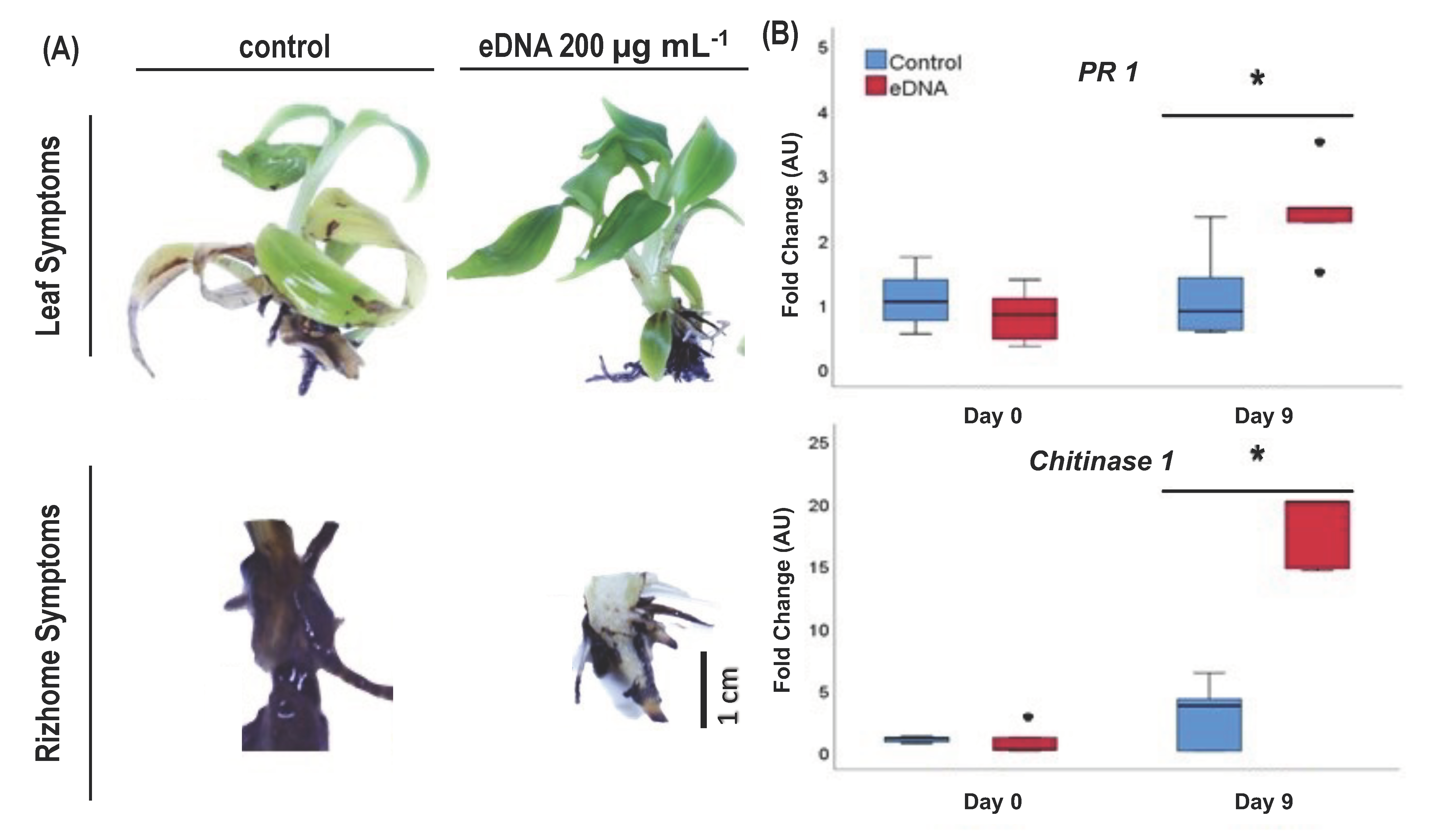
To determine the effect of eDNA of Foc TR4 on priming plant defense against fusarium wilt disease, the roots of Cavendish plantlets were immersed in eDNA Foc TR4 at a range of concentrations (40, 80, and 200 µg mL−1). Deionized water was used as a negative control and salicylic acid, a natural elicitor, as a positive control. After 9 days of infection, symptoms of fusarium wilt, such as leaf chlorosis and rhizome discoloration, were observed. Morphological observation showed a significant symptom difference documented at the highest eDNA concentration (200 µg mL−1), compared with the negative control (see Figure 2A). On the basis of the disease index [28], we found that Cavendish banana resistance increased from very susceptible (control) to tolerant when treated with 200 µg mL−1 eDNA (Table 2). Interestingly, the plants treated with salicylic acid (positive control) were more susceptible to Foc TR4 infection than those treated with 200 µg mL−1 eDNA.
Further, we investigated the relative expression of two genes related to the defense mechanism against biotic stress. Pathogenesis-related 1 (PR 1) and chitinase 1 genes have been documented to be involved in banana response to fungal infection [35,36,37,38]. In our study, eDNA of Foc TR4 caused a significant increase in the relative expression of PR 1 (ca. twofold) and chitinase 1 (ca. twentyfold) genes on 9 DPI (Figure 2B).

3.3. eDNA of Foc TR4 Modulated ROS (O2 and H2O2) Production and Expression of the Gene Coding for Antioxidant Enzymes in Banana Plantlets

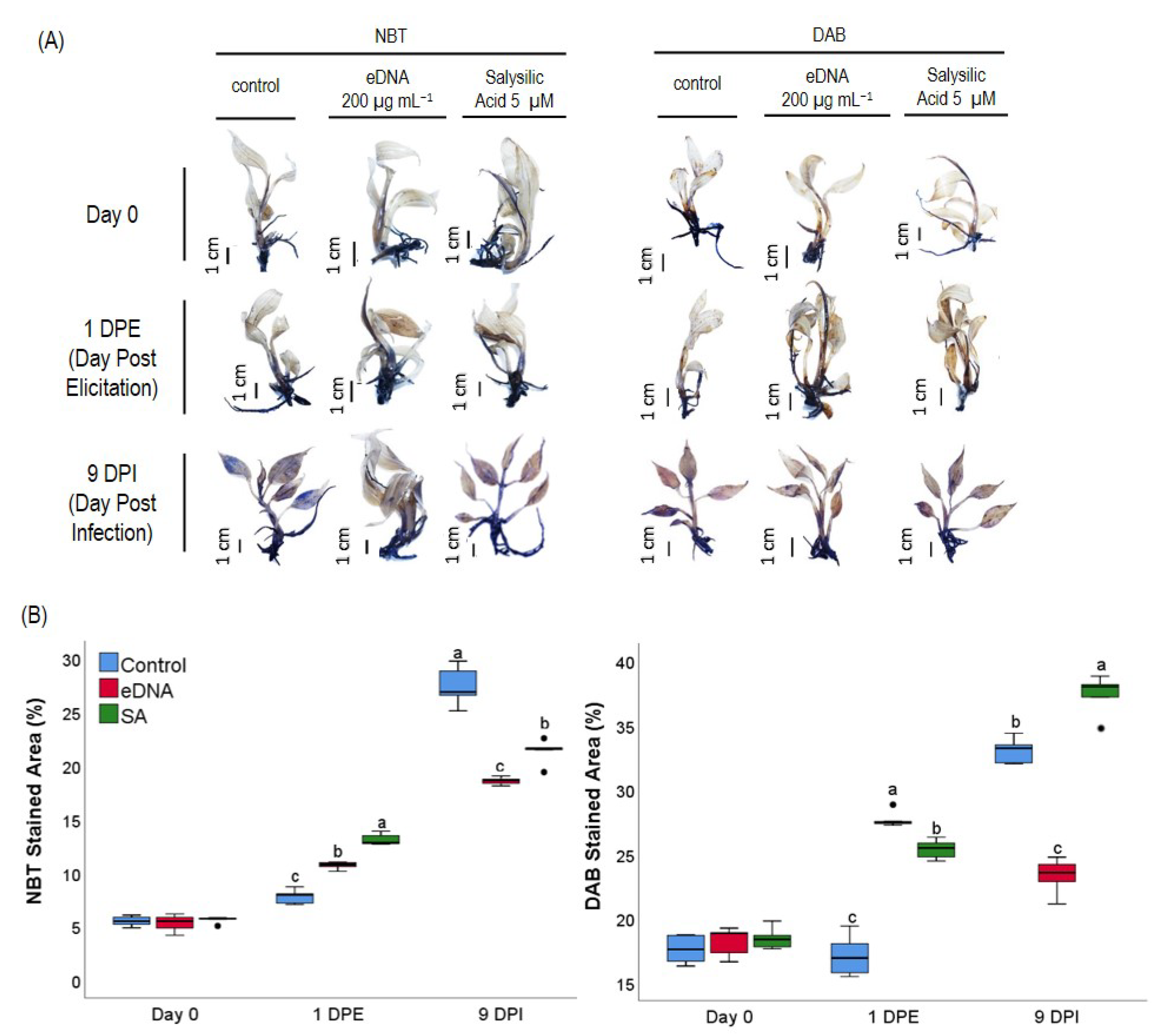
Plant–pathogen interaction often involves ROS as the messenger molecules. Hence, in this study, O2 and H2O2 were quantified on day 0, day 1 post priming (DPP), and day 9 post-inoculation (DPI) in the plantlets treated with water, 200 µg mL−1 eDNA of Foc TR4 and salicylic acid (Figure 3A). On 1 DPP or 24 h after receiving the priming treatment, O2 production was significantly higher in the eDNA-treated group than in the water-treated group but lower than in the salicylic acid treated group (Figure 3B). Meanwhile, the accumulation of H2O2 on the same day was the highest in the eDNA-treated group. On 9 DPI by Foc TR4, the eDNA-treated group had the lowest accumulation of both O2 and H2O2. These suggest that O2 and H2O2 were involved in the priming process by both Foc TR4 eDNA and salicylic acid.
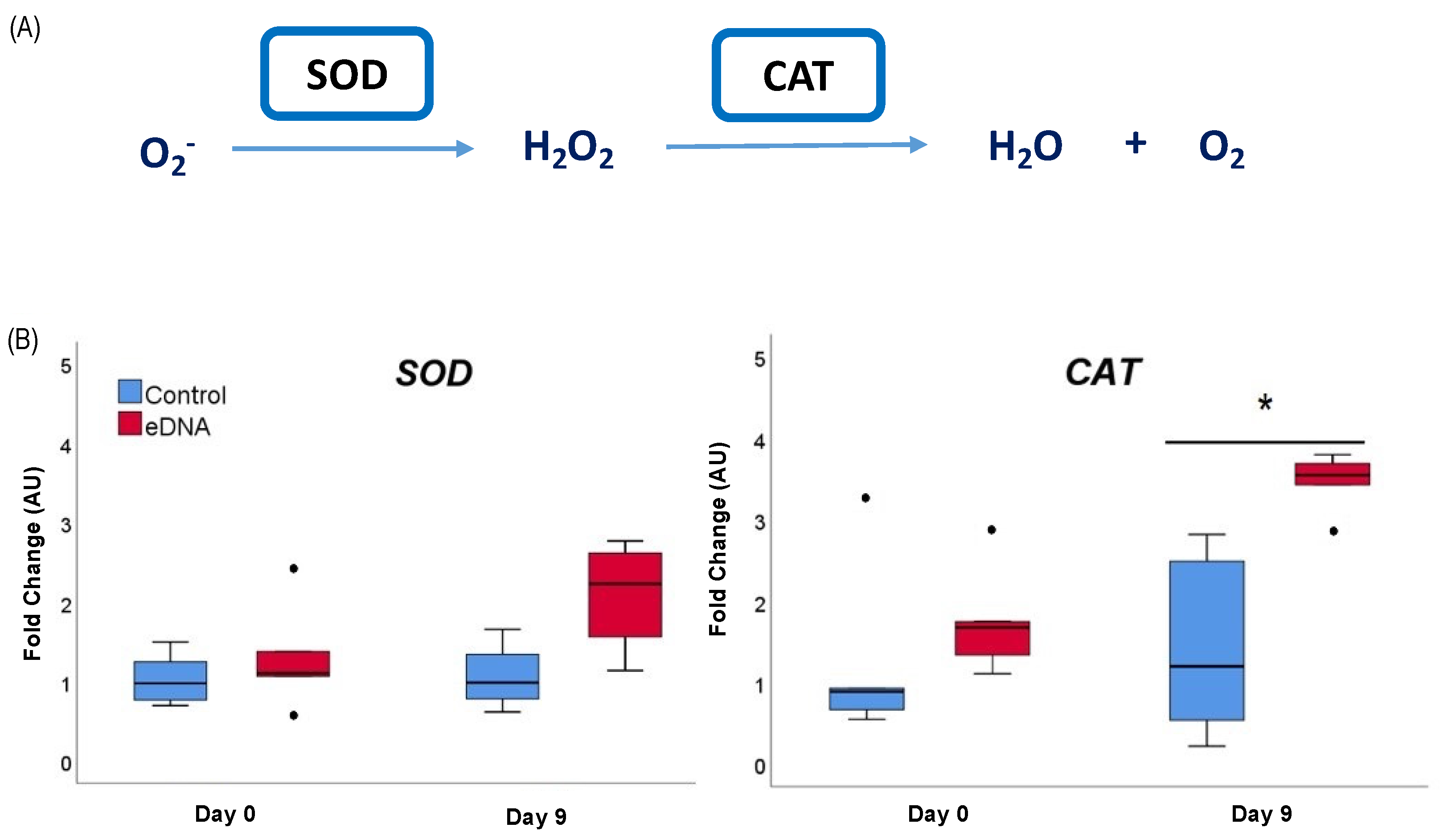
We then analyzed the expression of two genes coding for enzymes involved in ROS scavenging, superoxide dismutase (SOD) and catalase (CAT) (Figure 4A), to determine the effect of eDNA of Foc TR4 exposure on the expression of the antioxidant system in the plantlets. On 9 DPI, we found that eDNA of Foc TR4 significantly induced the expression of the CAT gene but not the SOD gene (Figure 4B).

4. Discussion

The capacity of an organism to recognize self from non-self is fundamental to its survival. This capacity is conserved across kingdoms [12,14,16], although the receptors responsible and the cascading processes of signaling are not yet fully understood. In highly evolved organisms, such as mammals, it was found that the TLR9 receptor enables cells to differentiate normal from cancerous ones [39] or placental from the fetal origin [40]. In phyto-pathogenesis, the capacity to fine-tune the response towards exposure to self- and non-self-extracellular-DNA (eDNA), a signal molecule, is explored to develop novel treatments for controlling a persistent global disease such as fusarium wilt in banana.
Recent research [12,15,41] demonstrated that growth inhibition is induced when an organism is exposed to self-eDNA, a species-specific mechanism. However, the inducing concentration is varied among species, and determining the lethal or most limiting concentration would be beneficial to developing an eDNA-based fungicide for Foc TR4. In this study, we tested the growth inhibition of Foc TR4 following the application of eDNA at 3 concentrations. Similar to Trichoderma harzianum [12], we found that the spore germination of Foc TR4 was highly suppressed upon application of 800 µg mL−1 eDNA. However, we suggest that the application of only half the concentration, i.e., 400 µg mL−1 eDNA, is also adequate to suppress Foc TR4 germination, as it was already reduced compared with the control (Figure 1).
To understand the mechanism by which Foc TR4 eDNA limits pathogen growth, we tested its priming capacity to improve Foc TR4 resistance in banana plantlets. Serrano-Jamaica et al. (2021) found that the foliar application of 100 µg mL−1 of cocktail eDNA from P. capsici, R. solani, and Foc decreased the mortality of chili plants following infection by all those fungi [11]. However, these authors applied a less concentrated spore of Foc (1 × 10⁵ spores mL−1) compared with our study (1 × 10⁶ spores mL−1). We, hence, increased the concentration of applied Foc eDNA such that the highest was 200 µg mL−1. We found that the application of 200 µg mL−1 was able to shift the disease index of Foc-infected banana plantlet from highly susceptible to tolerant, while the smaller concentrations only shifted the index to susceptible or moderately susceptible. Altogether, these studies confirm the priming role of exposing pathogen eDNA to plants and confirm that the applied concentration of eDNA determines the phenotypic responses upon infection.
Following the recognition of MAMPs, plants could fine-tune their responses regarding whether to develop systemic acquired resistance (SAR) or induced systemic resistance (ISR) [42]. SAR is triggered by pathogen infection and characterized by the elevated production of salicylic acid and expression of pathogenesis related (PR) genes, whereas ISR is activated in response to beneficial microbes (endosymbiont/antagonist) and mostly characterized by jasmonic acid or ethylene signaling and increased callose synthesis [43]. Furthermore, PR 1 and chitinase 1 genes are regarded as convenient molecular markers for SAR [35,44]. In this study, we also found that the application of Foc TR4 eDNA elevated the expression of PR 1 and chitinase 1 genes in the stem (tuber) of banana plantlets, suggesting the activation of SAR. Hence, the application of salicylic acid as a control treatment has been suitable for this study.
Previous studies suggested that PR 1 gene was involved in defense mechanisms of ripe banana [35], while chitinase 1 was involved in banana resistance to disease before ripening [35,36] upon being infected by Colletotrichum musae. In resistant cultivar (GCTCV-218), Foc TR4 infection increased PR 1 expression in the roots [38]. Similarly, in the plantlet roots, PR 1 was also highly expressed along with PR 3, NPR 1A, and NPR 1B, as a general response of fungal recognition, although the inoculated Foc was incompatible with the Cavendish banana [37]. These studies and ours agree that PR 1 and chitinase 1 are highly regulated in the process of recognition and defense towards fungal infection in several organs of banana plantlets and plants. Thus, our results also emphasize the plant’s capacity to fine-tune the response on the basis of the recognition of not only surface-related molecules of pathogens, such as flagellin, elongation factor Tu, fungal chitin, etc. [45], but also internal molecules that would normally be expelled only by dead microbes.
Signal cascading in plant cells often involves the regulation of ROS. Increasing the ROS concentration induces plant response by activating the mitogen-activated protein kinase (MAPK) cascade [46,47] and adjusting the intracellular redox state to regulate thiol-sensitive proteins with signaling function [48,49] to propagate the response. The response by ROS also induces the antioxidant capacity to control them and avoid an over-oxidation of cellular components [50,51,52]. The failure to increase ROS concentration might result in canceled signal propagation [53,54], while delayed activation of the antioxidant system could cause cell oxidative stress [55]. Interestingly, this pattern of ROS regulation was documented in plantlets treated with 200 µg mL−1 of Foc TR4 eDNA. The treatment increased O2 and H2O2 production following 24 h priming, which by 9 DPI was already readjusted, but not in plantlets treated with salicylic acid or water. The elevated expression of CAT on 9 DPI indicates the involvement of the enzymatic antioxidant system in scavenging H2O2. However, we were not able to document regulation of SOD gene in the eDNA-treated samples. This might be suggesting the role of non-enzymatic scavengers for quenching O2. Some secondary metabolites have been shown to also scavenge O2, for instance, phenolic compounds in Gallum verum var. asiaticum [56] and proline in Spinacia oleracea [57].
Noting the importance of ROS modulation, González-Bosch (2018) stated that plant resistance priming involves the activation of redox-sensitive genes, such as through histone modification. It is also known that the modulation of the redox state interacts with the signaling pathways mediated by salicylic acid, jasmonic acid, and ethylene [58,59]. Thus, we suggest that the smaller Foc TR4 eDNA concentration was required for priming resistance in the plantlets rather than repressing the Foc TR4 germination rate. This might be due to the signal propagation through ROS modulation in plant tissues.
Altogether, we suggest that the antifungal property of Foc TR4 eDNA is a combination of the self-inhibitory effect and the priming of the plant immune system. It is interesting to note that the eDNA concentration for repressing the Foc TR4 spore germination rate was much higher than for priming banana plantlets’ resistance upon infection.
In this study, signal propagation through ROS may play a role as a key factor that eventually leads to increased expression of pathogen-resistant (PR) 1 and chitinase 1 genes, and finally shifts the resistance of the banana plantlets from highly susceptible to tolerant. Our result also emphasizes the ability of plant cells to differentiate the recognized MAMPs—whether they belong to beneficial or pathogenic microbe groups—and to fine-tune the response accordingly (Figure 5).

5. Conclusions

We conclude that Foc TR4 eDNA exerts antifungal activity by a combination of the self-inhibitory effect on Foc TR4 and by priming the plant immune system. The priming effect involved the systemic acquired response, likely involving SA, and the early induction of ROS and enhanced antioxidant activity mediated by catalase at the later stages of infection. Overall, the application of exogenous pathogen’s DNA can be a strategy to reduce plant susceptibility to pathogens in banana. In the future, it will be interesting to explore the priming effect of combining pathogen fungal eDNA with other natural antifungal compounds, such as chitosan.

Author Contributions

Conceptualization, K.M. and R.R.E.; data curation, R.H.H.; funding acquisition, R.R.E.; investigation, R.H.H.; supervision, K.M., I. and R.R.E.; visualization, S.S.; writing—original draft, R.H.H.; writing—review and editing, K.M., S.S., T.F. and R.R.E. All authors have read and agreed to the published version of the manuscript.

Funding

This research was funded by Institut Teknologi Bandung through the Institute for Research and Community Services of ITB (LPPM-ITB) in P3MI 2021 grant to the Plant Sciences and Biotechnology Research Group.

Data Availability Statement

The data that support the findings of this study are available from the corresponding author upon reasonable request.

Acknowledgments

The authors would like to thank the Institute for Research and Community Services of ITB (LPPM-ITB) for the P3MI 2021 grant to the Plant Sciences and Biotechnology Research Group.

Conflicts of Interest

The authors declare no conflict of interest. The funders had no role in the design of the study; in the collection, analyses, or interpretation of data; in the writing of the manuscript; or in the decision to publish the results.

Appendix A

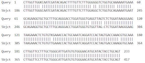
Table A1. The identity result of sequence of PCR products for primers that are used in this study.
NoAccession NumberGene NameScoreE-ValueIdent.
1EF055881.2Musa acuminata pathogenesis-related protein 1 (PR1) cds1852 × 10−4397%
Agronomy 13 00441 i001
2FJ040801.1Musa acuminata AAA Group chitinase I mRNA1276 × 10−2998%
Agronomy 13 00441 i002
3AF510071.1Musa acuminata superoxide dismutase mRNA. Partial cds38310−10796%
Agronomy 13 00441 i003
4HM061079.1Musa acuminata AAA Group catalase (CAT) mRNA partial cds1042 × 10−1898%
Agronomy 13 00441 i004
5AY821550.1Musa acuminata glyceraldehyde-3-phosphate dehydrogenase mRNA, partial cds14710−31100%
Agronomy 13 00441 i005
Table A2. Colony counts of Foc TR4 in the self-inhibition test. Different letters represent significantly different groups based on the results of the Duncan test.
TreatmentColony Count
Control635 ± 111.25 c
eDNA 80 µg mL−1557 ± 85 bc
eDNA 400 µg mL−1552 ± 48 b
eDNA 800 µg mL−1337 ± 47 a

References

  1. Nelson, S.C.; Ploetz, R.C.; Kepler, A.K.; Daniells, J. Species profiles for pacific island agroforestry Musa species (banana and plantain). Species Profiles Pac. Isl. Agrofor 2006, 2, 1–27. Available online: http://www.traditionaltree.org (accessed on 15 November 2019).
  2. FAO. Banana Market Review 2021; FAO: Rome, Italy, 2022; Volume 5. [Google Scholar]
  3. FAO. Food Outlook—Biannual Report on Global Food Markets; FAO: Rome, Italy, 2019; pp. 5–7. [Google Scholar]
  4. Aquino, A.P.; Bandoles, G.G.; Lim, V.A.A. R&D and Policy Directions for Effective Control of Fusarium Wilt Disease of Cavendish Banana in the Asia-Pacific Region. FFTC Agricultural Policy Platform (FFTC-AP). 2013. Available online: https://ap.fftc.org.tw/article/598 (accessed on 15 November 2019).
  5. Swarupa, V.; Ravishankar, K.V.; Rekha, A. Plant defense response against Fusarium oxysporum and strategies to develop tolerant genotypes in banana. Planta 2014, 239, 735–751. [Google Scholar] [CrossRef] [PubMed]
  6. Dita, M.; Barquero, M.; Heck, D.; Mizubuti, E.S.G.; Staver, C.P. Fusarium wilt of banana: Current knowledge on epidemiology and research needs toward sustainable disease management. Front. Plant Sci. 2018, 871, 1468. [Google Scholar] [CrossRef] [PubMed]
  7. Dinas Pertanian Kabupaten Buleleng. Gejala dan Cara Pengendalian Penyakit Layu Fusarium Pada Tanaman Pisang. 2019. Available online: https://distan.bulelengkab.go.id/informasi/detail/artikel/gejala-dan-cara-pengendalian-penyakit-layu-fusarium-pada-tanaman-pisang-36 (accessed on 15 November 2019).
  8. Malik, N.A.A.; Kumar, I.S.; Nadarajah, K. Elicitor and Receptor Molecules: Orchestrators of Plant Defense and Immunity. Int. J. Mol. Sci. 2020, 21, 963. [Google Scholar] [CrossRef] [PubMed]
  9. Ranf, S. Sensing of molecular patterns through cell surface immune receptors. Curr. Opin. Plant Biol. 2017, 38, 68–77. [Google Scholar] [CrossRef]
  10. Ferrusquía-Jiménez, N.I.; Chandrakasan, G.; Torres-Pacheco, I.; Rico-Garcia, E.; Feregrino-Perez, A.A.; Guevara-González, R.G. Extracellular DNA: A Relevant Plant Damage-Associated Molecular Pattern (DAMP) for Crop Protection Against Pests—A Review. J. Plant Growth Regul. 2020, 40, 451–463. [Google Scholar] [CrossRef]
  11. Serrano-Jamaica, L.M.; Villordo-Pineda, E.; González-Chavira, M.M.; Guevara-González, R.G.; Medina-Ramos, G. Effect of Fragmented DNA From Plant Pathogens on the Protection Against Wilt and Root Rot of Capsicum annuum L. Plants. Front. Plant Sci. 2021, 11, 2043. [Google Scholar] [CrossRef]
  12. Mazzoleni, S.; Cartenì, F.; Bonanomi, G.; Senatore, M.; Termolino, P.; Giannino, F.; Incerti, G.; Rietkerk, M.; Lanzotti, V.; Chiusano, M.L. Inhibitory effects of extracellular self-DNA: A general biological process? New Phytol. 2015, 206, 127–132. [Google Scholar] [CrossRef]
  13. Nagler, M.; Insam, H.; Pietramellara, G.; Ascher-Jenull, J. Extracellular DNA in natural environments: Features, relevance and applications. Appl. Microbiol. Biotechnol. 2018, 102, 6343–6356. [Google Scholar] [CrossRef]
  14. Yakushiji, S.; Ishiga, Y.; Inagaki, Y.; Toyoda, K.; Shiraishi, T.; Ichinose, Y. Bacterial DNA activates immunity in Arabidopsis thaliana. J. Gen. Plant Pathol. 2009, 75, 227–234. [Google Scholar] [CrossRef]
  15. Duran-Flores, D.; Heil, M. Extracellular self-DNA as a damage-associated molecular pattern (DAMP) that triggers self-specific immunity induction in plants. Brain. Behav. Immun. 2018, 72, 78–88. [Google Scholar] [CrossRef] [PubMed]
  16. Heil, M.; Vega-Muñoz, I. Nucleic Acid Sensing in Mammals and Plants: Facts and Caveats. In International Review of Cell and Molecular Biology; Elsevier Inc.: Amsterdam, The Netherlands, 2019; Volume 345, pp. 225–285. [Google Scholar] [CrossRef]
  17. Quintana-Rodriguez, E.; Duran-Flores, D.; Heil, M.; Camacho-Coronel, X. Damage-associated molecular patterns (DAMPs) as future plant vaccines that protect crops from pests. Sci. Hortic. 2018, 237, 207–220. [Google Scholar] [CrossRef]
  18. Chiusano, M.L.; Incerti, G.; Colantuono, C.; Termolino, P.; Palomba, E.; Monticolo, F.; Benvenuto, G.; Foscari, A.; Esposito, A.; Marti, L.; et al. Arabidopsis thaliana Response to Extracellular DNA: Self Versus Nonself Exposure. Plants 2021, 10, 1744. [Google Scholar] [CrossRef]
  19. Maryani, N.; Lombard, L.; Poerba, Y.S.; Subandiyah, S.; Crous, P.W.; Kema, G.H.J. Phylogeny and genetic diversity of the banana Fusarium wilt pathogen Fusarium oxysporum f. sp. cubense in the Indonesian centre of origin. Stud. Mycol. 2019, 92, 155–194. [Google Scholar] [CrossRef] [PubMed]
  20. Rizanti, M. Expression of PR1 and PR3 Resistance Genes in In Vitro Assay of Banana Plantlets Cultivars Kepok and Mas Infected with Fusarium oxysporum f.sp cubense Tropical Race 4 (Foc TR4); Bandung Institute of Technology: Bandung, Indonesia, 2018. [Google Scholar]
  21. González-Mendoza, D.; Argumedo-Delira, R.; Morales-Trejo, A.; Pulido-Herrera, A.; Cervantes-Díaz, L.; Grimaldo-Juarez, O.; Alarcón, A. A rapid method for isolation of total DNA from pathogenic filamentous plant fungi Isolation of total DNA from pathogenic filamentous plant fungi. Genet. Mol. Res. 2010, 9, 162–166. [Google Scholar] [CrossRef]
  22. Murashige, T.; Skoog, F. A Revised Medium for Rapid Growth and Bio Assays with Tobacco Tissue Cultures. Physiol. Plant. 1962, 15, 473–497. [Google Scholar] [CrossRef]
  23. Mazzoleni, S.; Bonanomi, G.; Incerti, G.; Chiusano, M.L.; Termolino, P.; Mingo, A.; Senatore, M.; Giannino, F.; Carten, F.; Rietkerk, M.; et al. Inhibitory and toxic effects of extracellular self-DNA in litter : A mechanism for negative plant—Soil feedbacks? New Phytol. 2015, 205, 1195–1210. [Google Scholar] [CrossRef]
  24. Al-Amin, M.D.; Karim, M.R.; Amin, M.R.; Rahman, S.M.A.N. In Vitro Micropropagation of Banana (Musa spp.). Bangladesh J. Agril. Res. 2009, 13, 645–659, ISSN 0258-7122. [Google Scholar]
  25. Emilda, D.; Sutanto, A.; Sukartini; Jumjunidang. Application of salicylic acid to induce disease resistance against fusarium wilt on banana. In Proceedings of the IOP Conference Series: Earth and Environmental Science, Changchun, China, 21–23 August 2020; Institute of Physics Publishing: Bristol, UK, 2020; Volume 468. [Google Scholar] [CrossRef]
  26. Purwati, R.D.; Harran, S. In Vitro Selection of Abaca for Resistance to Fusarium oxysporum f.sp cubense. HAYATI J. Biosci. 2007, 14, 65–70. [Google Scholar] [CrossRef]
  27. Subramaniam, S.; Maziah, M.; Sariah, M.; Puad, M.P.; Xavier, R. Bioassay method for testing Fusarium wilt disease tolerance in transgenic banana. Sci. Hortic. 2006, 108, 378–389. [Google Scholar] [CrossRef]
  28. Mak, C.; Mohamed, A.A.; Liew, K.W.; Ho, Y.W. Early screening technique for Fusarium wilt resistance in banana micropropagated plants. In Banana Improvement: Cellular, Molecular Biology, and Induced Mutations, Proceedings of The Bananas and Crop Improvement Congresses, Leuven, Belgium, 24–28 September 2001; Science Publishers, Inc.: Hauppauge, NY, USA, 2004; Volume 17, pp. 219–227. ISBN 1578083400. [Google Scholar]
  29. Liu, X.; Williams, C.E.; Nemacheck, J.A.; Wang, H.; Subramanyam, S.; Zheng, C.; Chen, M.S. Reactive oxygen species are involved in plant defense against a gall midge. Plant Physiol. 2010, 152, 985–999. [Google Scholar] [CrossRef] [PubMed]
  30. Matsunaga, T.M.; Ogawa, D.; Taguchi-shiobara, F.; Ishimoto, M.; Matsunaga, S.; Habu, Y. Direct quantitative evaluation of disease symptoms on living plant leaves growing under natural light. Breed. Sci. 2017, 67, 316–319. [Google Scholar] [CrossRef]
  31. Cordeiro, M.C.M.; Silva, M.S.; de Oliveira-Filho, E.C.; Miranda, Z.; Aquino, F.; Fragoso, R.; Almeida, J.; Andrade, L. Optimization of A Method of Total RNA Extraction From Brazilian Native Plants Rich in Polyphenols and Polysaccharides. In Proceedings of the Simposio Nacional Cerrado, ParlaMundi, Brazil, 12–17 October 2008; Volume 1, pp. 1–6. [Google Scholar] [CrossRef]
  32. Haimes, J.; Kelley, M. Demonstration of a ΔΔCq Calculation Method to Compute Relative Gene Expression from qPCR Data. Horiz. Tech Note 2018, 1, 1–4. [Google Scholar]
  33. Livak, K.J.; Schmittgen, T.D. Analysis of Relative Gene Expression Data Using Real-Time Quantitative PCR and the 2−ΔΔCT Method. Methods 2001, 25, 402–408. [Google Scholar] [CrossRef] [PubMed]
  34. IBM Corp. IBM SPSS Statistics for Windows, Version 20.0; IBM Corp.: Armonk, NY, USA, 2011. [Google Scholar]
  35. Tang, W.; Zhu, S.; Li, L.; Liu, D.; Irving, D.E. Differential expressions of PR1 and chitinase genes in harvested bananas during ripening, and in response to ethephon, benzothiadizole and methyl jasmonate. Postharvest Biol. Technol. 2010, 57, 86–91. [Google Scholar] [CrossRef]
  36. Ma, B.C.; Tang, W.L.; Ma, L.Y.; Li, L.L.; Zhang, L.B.; Zhu, S.J.; Zhuang, C.; Irving, D. The Role of Chitinase Gene Expression in the Defense of Harvested Banana Against Anthracnose Disease. J. Am. Soc. Hortic. Sci. 2009, 134, 379–386. [Google Scholar] [CrossRef]
  37. Wu, Y.; Yi, G.; Peng, X.; Huang, B.; Liu, E.; Zhang, J. Systemic acquired resistance in Cavendish banana induced by infection with an incompatible strain of Fusarium oxysporum f. sp. cubense. J. Plant Physiol. 2013, 170, 1039–1046. [Google Scholar] [CrossRef]
  38. Van Den Berg, N.; Berger, D.K.; Hein, I.; Birch, P.R.J.; Wingfield, M.J.; Viljoen, A. Tolerance in banana to Fusarium wilt is associated with early up-regulation of cell wall-strengthening genes in the roots. Mol. Plant Pathol. 2007, 8, 333–341. [Google Scholar] [CrossRef]
  39. Goldfarb, I.T.; Adeli, S.; Berk, T.; Phillippe, M. Fetal and Placental DNA Stimulation of TLR9: A Mechanism Possibly Contributing to the Pro-inflammatory Events During Parturition. Reprod. Sci. 2018, 25, 788–796. [Google Scholar] [CrossRef]
  40. Tuomela, J.; Sandholm, J.; Kaakinen, M.; Patel, A.; Kauppila, J.H.; Ilvesaro, J.; Chen, D.; Harris, K.W.; Graves, D.; Selander, K.S. DNA From Dead Cancer Cells Induces TLR9-Mediated Invasion and Inflammation In Living Cancer Cells. Breast Cancer Res. Treat. 2013, 142, 477. [Google Scholar] [CrossRef] [Green Version]
  41. Cartení, F.; Bonanomi, G.; Giannino, F.; Incerti, G.; Vincenot, C.E.; Chiusano, M.L.; Mazzoleni, S. Self-dna inhibitory effects: Underlying mechanisms and ecological implications. Plant Signal. Behav. 2016, 11, e1158381. [Google Scholar] [CrossRef]
  42. Pieterse, C.M.J.; Leon-Reyes, A.; Van Der Ent, S.; Van Wees, S.C.M. Networking by small-molecule hormones in plant immunity. Nat. Chem. Biol. 2009, 5, 308–316. [Google Scholar] [CrossRef]
  43. Pieterse, C.M.J.; Zamioudis, C.; Berendsen, R.L.; Weller, D.M.; Van Wees, S.C.M.; Bakker, P.A.H.M. Induced Systemic Resistance by Beneficial Microbes. Annu. Rev. Phytopathol. 2014, 52, 347–375. [Google Scholar] [CrossRef]
  44. Linthorst, H.J.M. Pathogenesis-related proteins of plants. Crit. Rev. Plant Sci. 1991, 10, 123–150. [Google Scholar] [CrossRef]
  45. Newman, M.A.; Sundelin, T.; Nielsen, J.T.; Erbs, G. MAMP (microbe-associated molecular pattern) triggered immunity in plants. Front. Plant Sci. 2013, 4, 139. [Google Scholar] [CrossRef]
  46. Xing, Y.; Jia, W.; Zhang, J. AtMKK1 mediates ABA-induced CAT1 expression and H2O2 production via AtMPK6-coupled signaling in Arabidopsis. Plant J. 2008, 54, 440–451. [Google Scholar] [CrossRef] [PubMed]
  47. Jammes, F.; Song, C.; Shin, D.; Munemasa, S.; Takeda, K.; Gu, D.; Cho, D.; Lee, S.; Giordo, R.; Sritubtim, S.; et al. MAP kinases MPK9 and MPK12 are preferentially expressed in guard cells and positively regulate ROS-mediated ABA signaling. Proc. Natl. Acad. Sci. USA 2009, 106, 20520–20525. [Google Scholar] [CrossRef]
  48. Rouhier, N. Plant glutaredoxins: Pivotal players in redox biology and iron–sulphur centre assembly. New Phytol. 2010, 186, 365–372. [Google Scholar] [CrossRef] [PubMed]
  49. Bashandy, T.; Guilleminot, J.; Vernoux, T.; Caparros-Ruiz, D.; Ljung, K.; Meyer, Y.; Reichheld, J.P. Interplay between the NADP-Linked Thioredoxin and Glutathione Systems in Arabidopsis Auxin Signaling. Plant Cell 2010, 22, 376. [Google Scholar] [CrossRef] [PubMed]
  50. Das, K.; Roychoudhury, A. Reactive oxygen species (ROS) and response of antioxidants as ROS-scavengers during environmental stress in plants. Front. Environ. Sci. 2014, 2, 53. [Google Scholar] [CrossRef] [Green Version]
  51. Signorelli, S.; Corpas, F.J.; Rodríguez-Ruiz, M.; Valderrama, R.; Barroso, J.B.; Borsani, O.; Monza, J. Drought stress triggers the accumulation of NO and SNOs in cortical cells of Lotus japonicus L. roots and the nitration of proteins with relevant metabolic function. Environ. Exp. Bot. 2019, 161, 228–241. [Google Scholar] [CrossRef]
  52. Dvořák, P.; Krasylenko, Y.; Zeiner, A.; Šamaj, J.; Takáč, T. Signaling Toward Reactive Oxygen Species-Scavenging Enzymes in Plants. Front. Plant Sci. 2021, 11, 2178. [Google Scholar] [CrossRef] [PubMed]
  53. González-Bosch, C. Priming plant resistance by activation of redox-sensitive genes. Free Radic. Biol. Med. 2018, 122, 171–180. [Google Scholar] [CrossRef] [PubMed]
  54. Lee, D.H.; Lal, N.K.; Lin, Z.J.D.; Ma, S.; Liu, J.; Castro, B.; Toruño, T.; Dinesh-Kumar, S.P.; Coaker, G. Regulation of reactive oxygen species during plant immunity through phosphorylation and ubiquitination of RBOHD. Nat. Commun. 2020, 11, 1838. [Google Scholar] [CrossRef] [PubMed]
  55. Considine, M.J.; Foyer, C.H. Stress effects on the reactive oxygen species-dependent regulation of plant growth and development. J. Exp. Bot. 2021, 72, 5795–5806. [Google Scholar] [CrossRef]
  56. Kim, D.K. Superoxide Quenching Activity of Phenolic Compounds from the Whole Plant of Galium verum var. asiaticum. Nat. Prod. Sci. 2011, 17, 261–266. [Google Scholar]
  57. Rehman, A.U.; Bashir, F.; Ayaydin, F.; Kóta, Z.; Páli, T.; Vass, I. Proline is a quencher of singlet oxygen and superoxide both in in vitro systems and isolated thylakoids. Physiol. Plant. 2021, 172, 7–18. [Google Scholar] [CrossRef]
  58. Grant, M.R.; Jones, J.D.G. Hormone (Dis)harmony Moulds Plant Health and Disease. Science 2009, 324, 750–752. [Google Scholar] [CrossRef]
  59. Caarls, L.; Pieterse, C.M.J.; Van Wees, S.C.M. How salicylic acid takes transcriptional control over jasmonic acid signaling. Front. Plant Sci. 2015, 6, 170. [Google Scholar] [CrossRef]
Figure 1. Self-inhibition test of Fusarium oxysporum (Foc) TR4. (A) Intact genomic and fragmented eDNA of Foc TR4, (B) quantification of Foc TR4 germination rate on day 3 post-inoculation, (C) limited mycelium growth of Foc TR4 upon culturing on PDA medium with the respective treatment of eDNA on day 7 post-inoculation. ANOVA test results showed a p-value < 0.05. Duncan’s test results are indicated by the asterisk and line on top of the boxes of the box plot.
Agronomy 13 00441 g001
Figure 2. Effect of eDNA application from Foc TR4 on the resistance of banana plantlets cv. Cavendish to Foc TR4 infection, determined by examining the morphology and relative expression of pathogenesis-related 1 (PR 1) and chitinase 1 genes. (A) Leaf and rhizome morphology of banana plantlet on day 8 after Foc TR4 infection. The observed symptoms of Foc TR 4 infection include leaves turning yellow and discolored rhizomes. (B) Relative expression of PR 1 and chitinase 1 genes compared with the GAPDH on day 9 post-infection. The asterisk represents a significantly different value based on independent t-test, while the black dots represent outlier data in each sampling point.
Agronomy 13 00441 g002
Figure 3. Detection of superoxide (O2) and hydrogen peroxide (H2O2) production in banana plantlets. Representative staining of tissue by nitroblue tetrazolium (NBT) and diaminobenzidine (DAB), respectively, to detect O2 and H2O2 (A). Quantification of the stained areas by NBT and DAB on day 0, day 1 post priming (DPP) by eDNA of Foc TR4, and day 9 post-infection (DPI) Foc TR4 (B). Statistically significant differences (Duncan’s test, p < 0.05) are indicated by different letters on top of the box plot corresponding to each treatment control (deionized water), eDNA of Foc TR4 (200 µg mL−1), and salicylic acid (5 µM). The black dots represent outlier data in each sampling point.
Agronomy 13 00441 g003
Figure 4. Effect of eDNA treatment from Foc TR4 on banana cv. Cavendish resistance to Foc TR4 infection by examining SOD and CAT gene relative expression: (A) SOD catalyzes the dismutation of O2 into H2O2 molecules and subsequently CAT will catalyze the breaking up of H2O2 into water and oxygen. (B) Relative expression of SOD and CAT genes to the GAPDH gene on 0 and 9 DPI by Foc TR4. The asterisk represents a significantly different value based on an independent t-test, comparing the two groups of treatment. The black dots represent outlier data in each sampling point.
Agronomy 13 00441 g004
Figure 5. Mechanisms of Foc TR4 DNA in shifting banana plants’ susceptibility towards tolerance through suppression of fungal spore germination and priming plant defense. Priming action involves a precise upsurging of ROS (O2 and H2O2) for signal cascading and quenching before reaching oxidative stress (elevated expression of catalase). Following this, systemic acquired resistance (SAR) is activated, as indicated by the upregulation of PR 1 and chitinase 1 genes.
Agronomy 13 00441 g005
Table 1. Sequence of primers used in this study.
No.Primer NamePrimer Sequence (5′ → 3′)Amplicon Length (bp)
1Pathogen Related Gene 1–F
Pathogen Related Gene 1–R
GCAGTACTACGACTACAACA
GTTGCAGATGATGAAGATGG
142
2Chitinase 1–F
Chitinase 1–R
CAAGAAGAAGAGGGAGATCG
GTTCTGTTCCTGGACGAAG
121
3Superoxide dismutase–F
Superoxide dismutase–R
ATCAACCACTCGATCTTCTG
GCCTCCAAAGAACAAAAGTC
113
4Catalase–F
Catalase–R
AGCAAACATCTGATACGGAG
GCGGACTTGACATGGTATAT
107
5Glyceraldehyde-3-Phosphate Dehydrogenase–F
Glyceraldehyde-3-Phosphate Dehydrogenase–R
TGTGGAGGAGGACTTGGTCT
CGTGAGCTGTAACCCCACTC
127
Table 2. The effect of eDNA application to the dynamic of susceptible–resistant category based on leaf and rhizome discoloration indices [28].
TreatmentLeaf Discoloration IndexRhizome Discoloration IndexCategory
Control3.80 ± 0.84 a7.00 ± 1.41 AHighly Susceptible
Salicylic Acid 5 µM2.20 ± 0.84 bc3.40 ± 0.55B CSusceptible
eDNA 40 µg mL−12.80 ± 0.45 b4.60 ± 1.52 BSusceptible
eDNA 80 µg mL−11.80 ± 0.84 bc3.80 ± 1.30 BCModerately Susceptible
eDNA 200 µg mL−11.60 ± 0.89 c2.60 ± 0.89 CTolerant
Data are mean ± standard deviation of 5 biological replications. ANOVA test results show a p-value < 0.05. Different letters (lowercase for leaf discoloration index, while upper case for rhizome discoloration index) represent significantly different groups based on the results of the Duncan test.
Disclaimer/Publisher’s Note: The statements, opinions and data contained in all publications are solely those of the individual author(s) and contributor(s) and not of MDPI and/or the editor(s). MDPI and/or the editor(s) disclaim responsibility for any injury to people or property resulting from any ideas, methods, instructions or products referred to in the content.