Article

Chorisia speciosa Extract Induces Systemic Resistance against Tomato Root Rot Disease Caused by Rhizoctonia solani

by
Said I. Behiry
1,*,
Seham A. Soliman
2,
Al-Naji A. Al-Mansori
3,
Abdulaziz A. Al-Askar
4,
Amr A. Arishi
5,
Mohsen Mohamed Elsharkawy
6,
Ahmed Abdelkhalek
2,* and
Ahmed A. Heflish
1
1
Agricultural Botany Department, Faculty of Agriculture (Saba Basha), Alexandria University, Alexandria 21531, Egypt
2
Plant Protection and Biomolecular Diagnosis Department, ALCRI, City of Scientific Research and Technological Applications, New Borg El Arab City, Alexandria 21934, Egypt
3
Botany Department, Faculty of Science, Derna University, Derna 99999, Libya
4
Department of Botany and Microbiology, College of Science, King Saud University, P.O. Box 2455, Riyadh 11491, Saudi Arabia
5
School of Molecular Sciences, The University of Western Australia, Perth, WA 6009, Australia
6
Department of Agricultural Botany, Faculty of Agriculture, Kafrelsheikh University, Kafr El-Sheikh 33516, Egypt
*
Authors to whom correspondence should be addressed.
Agronomy 2022, 12(10), 2309; https://doi.org/10.3390/agronomy12102309
Submission received: 21 August 2022 / Revised: 15 September 2022 / Accepted: 23 September 2022 / Published: 26 September 2022
(This article belongs to the Section Pest and Disease Management)

Abstract

:
Chemical pesticides and fungicides are used extensively, negatively affecting people’s health and the environment. Reducing synthetic pesticides and increasing the efficiency of sustainable food production using plant extracts as natural chemicals is a win–win. Here, we first describe and evaluate an ethanolic extract of Chorisia speciosa for its protective and curative activities against Rhizoctonia solani in greenhouse-grown tomato plants. The results showed that the mycelial growth of R. solani was completely suppressed in vitro by C. speciosa extract (10 µg/mL). Twenty days after fungal inoculation, the results demonstrated that using C. speciosa extract (10 µg/mL) in vivo significantly improved shoot and root growth parameters in protective and curative treatments. Further, the protective and curative treatments decreased the disease index by 26.67% and 53.33%, respectively. C. speciosa-treated tomato plants showed significantly increased antioxidant enzyme production (PPO, CAT, and SOD) and up-regulated PR-1, PR-2, PR-3, PAL, and CHS expression levels compared to untreated plants. According to HPLC examination, the most prevalent phenolic acids or flavonoid components quantities (µg/mL) noticed in C. speciosa extract were 7-OH-flavone (10.36), kaempferol (9.23), p-coumaric acid (8.65), ferulic acid (8.14), caffeic acid (7.59), gallic acid (6.33), and iso-ferulic (5.71). Our findings are the first to demonstrate that a C. speciosa extract can assist plants in combating fungal infestation. Therefore, the data imply that C. speciosa extract, as a natural and renewable product, could be adopted as a long-term approach for regulating plant fungus.

1. Introduction

Tomato (Lycopersicon esculentum L.) is one of the world’s most widely cultivated fruits and vegetables. It is a member of the Solanaceae family and includes the beneficial amino acids tryptophan and tomatin (a glycoalkaloid major component). Tomatoes are the world’s second-largest processed vegetable and the second-largest growing vegetable. FAOSTAT (2017) estimates global tomato production at 178.24 million US tons in 2017, with a value of USD 59 billion. Crop production and environmental stability are threatened by harmful pathogens [1]. Rhizoctonia, Verticillium, and Fusarium are soil-borne pathogens that cause different plant diseases [2]. Rhizoctonia solani is the causal agent of plant damping-off and root rot diseases, which have various symptoms that lead to tomato seedling death [3]. Finding new ways to stop plant diseases is difficult because physiological pathogen races have become more resistant to fungicides [4].
As a result, natural substances may be able to combat a wide variety of plant diseases without creating harmful environmental effects. Many plant extracts’ antimicrobial and antifungal activities have gained considerable popularity and scientific interest [5]. Plant chemicals, such as natural antifungal chemical compounds, are considered safer and more acceptable than synthetic counterparts [6]. Many plants’ antimycotic properties and extracts have been investigated [7]. Chorisia speciosa A. St. Hil (Syn. Ceiba speciosa) is a member of the family Bombacaceae, which contains about 28 genera and about 200 species [8]. These plants traditionally cure many health disorders, e.g., headaches and diabetes, and pathogens, such as Bacillus cereus, Staphylococcus aureus, Klebsiella pneumoniae, and Psuedomonas aeruginosa [9,10]. A few Chorisia species were subjected to phytochemical analyses that yielded a number of flavonoids, triterpenes, and carbohydrates [11]. Anti-inflammatory, hepatoprotective, cytotoxic, antioxidant, hypoglycemic, and antibacterial actions with high safety margins have been observed in various Chorisia species, according to biological studies [12,13,14,15].
Because of their safety, environmental friendliness, and public acceptance, plant extracts have been studied for plant diseases control [16,17]. The current study aimed to assess the growth antifungal activity of C. speciosa ethanol extract against R. solani and its protective and curative properties in vivo as this has not previously been completed. Screening and introducing potential plant extracts with promising antimicrobial properties for agricultural applications is critical. In addition, the levels of expression of defense-related genes, such as PR-1, PR-2, PR-3, PAL, and CHS, as well as tomato growth parameters, were measured to see how the extract affected them. In addition, the main phytochemical components of C. speciosa ethanolic extract were identified using HPLC analysis.

2. Materials and Methods

2.1. Isolation and Characterization of Fungal Isolate

Tomato-root-rot-disease-like signs were collected from an Egyptian field in El-Gharbia governorate. The symptomatic plant parts were cut and washed with tap water to remove the dust, kept in 2% sodium hypochlorite for 2 min, then rinsed in sterile water 2-3 times before drying in laminar flow. PDA medium was utilized in the isolation and cultivation processes. The hyphal tip technique was used to purify the fungus. The fungus was identified based on morphological and microscopic properties, according to Alexopoulos et al. [18]. Furthermore, the internal transcribed spacer (ITS) region was amplified by ITS1 and ITS4 primers to identify the fungal isolate at the molecular level (Table 1). Heflish et al. [19] previously described the PCR settings reaction as containing 0.4 μL of each primer, 12.5 μL of 2x Taq PCR Ready Mix, and 1 μL of DNA, and the molecular grade H2O was added to obtain a final volume of 25 μL. The ITS region was amplified using a TECHNE Prime PCR with the following settings: 94 °C for 5 min, then 40 cycles of 94 °C, 55 °C, and 72 °C for 40 s each, and a final elongation at 72 °C for 7 min. The PCR reaction was separated on an agarose gel, refined, and sequenced. The retrieved nucleotide sequences were aligned and compared to those of other related species using the MEGA X program and the Genbank database. The nucleotide sequence of the fungus was then deposited in the NCBI database entry portal to obtain an accession number.

2.2. Preparation of C. speciosa Extract

Chorisia speciosa plant leaves were collected from the Faculty of Agriculture, Saba Basha, Alexandria, Egypt and dried at room temperature (25 °C) for two weeks before being crushed to a fine powder with a grinding mill (Moulinex AR1044, Paris, France). 100 g of the powder was immersed in 200 mL of methanol for six days. The methanol mixture was put through a filter, and the extract that came out was then placed in a rotary vacuum evaporator to concentrate it [17].

2.3. The Antifungal Activity of C. speciosa Extract against Root Rot Fungus In Vitro

The C. speciosa extract was investigated for its effectiveness against the root rot pathogen using the food poisoning technique [20]. Different amounts of C. speciosa extract (1, 2, 4, 8, and 10 µg/mL) were mixed with PDA dishes and compared to fungicide (Rizolex 2 µg/mL) and the negative control (PDA-free medium from additives). Fungal round discs were cut from the edges of one-week cultures, seeded in the center of the treated pouring PDA Petri plates, and cultured for a week at room temperature. All the treatments are triplicated. The effectiveness of C. speciosa extract on fungal linear growth was determined as a percentage of growth inhibition ( % ) = [ ( T r T s ) / T r ] × 100 , where Tr is the fungal growth length control and Ts is the length of fungal hyphae growth in the treatment [21].

2.4. Greenhouse Experimental Design and Growth Parameters Assessment

A greenhouse experiment was conducted under controlled conditions (27 ± 3 °C, 80% relative humidity, 14 h of light, and 10 h of darkness) to investigate the efficacy of C. speciosa extract in controlling R. solani and boosting tomato development. Seedlings of the 023 variety of tomato, aged 20 days, were transplanted in sterile soil in each of the 20-cm pots. C. speciosa extract (10 µg/mL) was sprayed on the leaves of the transplanted plants a week after they were established. An R. solani inoculum was created by inoculating 250 g of pre-autoclaved, watered barley grains in a bag with a 0.5-cm-diameter plug of the fungus and incubating for 7 days at room temperature. The grain inoculum was then air-dried, crushed into a powder, and distributed as 10 g/kg of soil in pots, where it was to be applied to the plant root zone [22]. There were five independent sets of treatments. Treatments I and II involved spraying the extract on plants two days after (curative) or before (protective) inoculation with R. solani; treatment III involved only inoculating plants with R. solani, and treatment IV involved treating plants with C. speciosa extract. The plants were inoculated with potato dextrose broth in the control treatment. Each treatment has nine pots; each pot has three plants. After one month of planting, the plants were removed to evaluate root rot severity on a scale from 0 to 5 [23]. The data were expressed as a percentage of the disease index.
( D I ) % = [ S u m   o f   s c a l e   n u m b e r   o f   i n f e c t e d   p l a n t s m a x i m u m   s c a l e   r a t e   n u m b e r × t o t a l   p l a n t s   n u m b e r ] × 100
The effects of the C. speciosa extract on the height (cm), root length (cm), fresh and dry weight (g), and root fresh and dry weight (g) of the uprooted plants were also evaluated (g). Leaves were also obtained from each treatment to analyze the expression of defensive scheme genes and the total phenolic compounds. Twenty days after being inoculated with R. solani, plant leaves were picked.

2.5. Total Phenolic Content (TPC) in Tomato Plants

TPC levels in tomato extracts were evaluated using the Folin–Ciocalteu (FoC) procedure. 0.1 mL µL of 1 mg/mL tomato extract was added to 0.75 mL of Folin–Ciocalteu (aqueous diluted ten times). After five minutes at room temperature, 0.75 mL of CaCO3 was added, and the combination was carefully mixed. After 90 min, the mixture was measured at 725 nm with 6305 UV/Visible Spectrophotometer (Cole-Parmer Instrument Company, Vernon Hills, IL, USA). Gallic acid concentrations between 0.01 and 0.05 mg/mL were used to produce a calibration graph. TPC levels were calculated using the method provided by Velioglu et al. [24] to measure gallic acid content (µg GAE/100 g extract).

2.6. Oxidative Stress Markers

2.6.1. Malondialdehyde Assay (MDA)

Thiobarbituric acid (TBA) was used to evaluate the malondialdehyde level (MDA) in all treatments. Plant leaves were pulverized in 0.1% trichloroacetic acid (TCA) and spun down at 12,000× g for 30 min. Afterward, one milliliter of the obtained liquid was combined with a solution comprising TCA (40%):TBA (10%) in a water bath (95 °C, 30 min). The combination was quickly placed on ice to cool. The sample’s wavelength absorbance (WA) was measured at 600 nm, and the MDA concentration was represented as µM/g F.Wt. [25].

2.6.2. Hydrogen Peroxide Assay (H2O2)

Velikova et al. [26] determined the H2O2 concentration of tomato leaves. Little pieces of leaves were homogenized with 0.1 TCA (%) and spun with a centrifuge. The supernatant, KH2PO4 (10 mM, pH 7), and 1 M KI were combined in equal parts. The WA was calculated at 390 nm. The H2O2 amount is expressed as µM/g F.Wt. [26].

2.7. Antioxidant Enzymes

For all enzyme activity experiments, leaves were mashed in potassium phosphate buffer pH 7 and spun down with a centrifuge at 10,000× g for 10 min, with the supernatant used for further analysis. All enzymes are reported as µM/g F. Wt.

2.7.1. Polyphenol Oxidase (PPO)

Polyphenol oxidase (PPO) activity was determined using the Cho and Ahn [16] technique. 500 µL of enzyme extract was treated at 25 °C for 10 min with 1 mL of Tris-HCl and quinone (50 mM, pH 6). At 420 nm, all measurements were taken three times [27].

2.7.2. Catalase (CAT) Activity

CAT activity was quantified using the Cakmak and Marschner technique with slight adjustments by measuring the rate of disappearance of H2O2 [28]. 50 µL enzyme extract was treated with 1 mL of a K-phosphate buffer (25 mM, pH 7) and H2O2 combination (10 mM as final concentration). CAT activity was detected at 240 nm.

2.7.3. Superoxide Dismutase (SOD) Activity

The efficacy of SOD to prevent the photochemical reduction of nitro blue tetrazolium (NBT) was tested using a slightly modified Beauchamp and Fridovich method. KH2PO4 (50 mM, pH 7.8), EDTA (0.1 mM), L-methionine (10 mM), NBT (75 mM), and riboflavin (20 mM) were combined in a tube with 100 µL of enzyme extract. Before being placed in the dark, the tubes were kept at 25 °C for 15 min under two 15-watt fluorescent lamps. The WA at 560 nm was then measured [29].

2.8. Real-Time Quantitative Reverse Transcription PCR Analysis (RT-qPCR) of Defense-Related Genes

Further, 0.1 g of plant leaves was taken at 20 dpi and subjected to a manual whole RNA extraction using a customized guanidium isothiocyanate (GITC) technique [30]. The quality and quantity of the extracted RNA were evaluated using the 6305 UV/Visible Spectrophotometer (Cole-Parmer Instrument Company, Vernon Hills, IL, USA). By utilizing a reverse transcriptase (RT) enzyme, the cDNA from the extracted RNA was synthesized [31]; the conventional reverse transcriptase PCR was carried out in a Bio-Rad cycler (Bio-Rad Laboratories Inc. Hercules, CA, USA) at 45 °C for 1 h and then inactivated at 85 °C for 3 min. The template cDNA was reserved in a −40 refrigerator until used in quantitative real-time PCR.
Using quantitative polymerase chain reaction (qPCR), the relative transcription of three genes is involved in tomato disease (PR-1, PR-2, and PR-3) and two phenolic genes (PAL and CHS). The housekeeping β-actin gene was used to normalize the data obtained. Sequences of the oligonucleotides employed as primers in this study are presented in Table 1. Experiments were conducted three times for each biological sample. Q-PCR was performed using a Bio-Rad real-time cycler (Bio-Rad laboratories Inc., Hercules, CA, USA) and TB Green Advantage qPCR Premix (Takara Bio USA Inc., San Jose, CA, USA) [32]. The PCR reaction mixture was performed using 10 µL of TB green Advantage qPCR Premix, 0.4 µL of each for/rev primer, 2 µL of template cDNA, and 7.2 µL of dH2O for a total of 20 µL. The real-time PCR conditions were as follows: the initial denaturation step at 95 °C for 10 s, denaturation at 95 °C for 3 s, and 40 cycles of annealing/extension at 60 °C for 20 s. The relative transcription levels of all genes were measured according to the 2−ΔΔCT method [33].

2.9. Antioxidant Activity of C. speciosa Extract

The Shimada et al. [34] technique was applied to assess scavenging free radical efficiency. In 100 mL of methanol, 3.94 mg of DPPH (1,1-diphenyl-2-picrylhydrazyl) was diluted to 0.1 mL. 2 mL of diluted DPPH was combined with 6 mL of each concentration sample (1000, 500, 250, 125, and 62.5 μg/mL). The mix was vigorously stirred and held at 25 °C for 30 min. Each combination’s absorbance value (Va) was measured at 517 nm. The DPPH inhibition was computed as D % = [ ( D i D s ) / D i ] × 100 , where Ds is the sample Va and Di is the control Va reaction (which contains all reagents except the sample). The antioxidant activity was measured according to the 50% DPPH (IC50) scavenging calibration curve compared to butylated hydroxytoluene (BHT).

2.10. HPLC Analysis of Polyphenolic Components for the Obtained Plant Extract

Using an HPLC-Agilent 1260 with an Infinity II analytical quaternary pump (Santa Clara, CA, USA) and an Eclipse Plus C18 column with a particle size of 3.5 m, polyphenolic components of the C. speciosa plant extract were identified [35]. The following standards were bought from Merck KGaA in Darmstadt, Germany: catechin, pyrogallol, ellagic acid, kaempferol, ferulic acid, iso-ferulic acid, gallic acid, rutin, caffeic acid, naringenin, p-coumaric acid, hesperidin, 7-OH flavone, apigenin, and myricetin. The 14 common phenolic and flavonoid components were the basis for the analysis.

2.11. Statistical Analysis

The data were processed with the Costat program, and the means were compared with post hoc Tukey’s honest differences (HSD) at p ≤ 0.05. The standard deviation (SD) was displayed at the top of the column bar. Up-regulated genes indicated that the relative transcription values are more than 1, whereas down-regulated gene levels indicated less than 1.

3. Results and Discussion

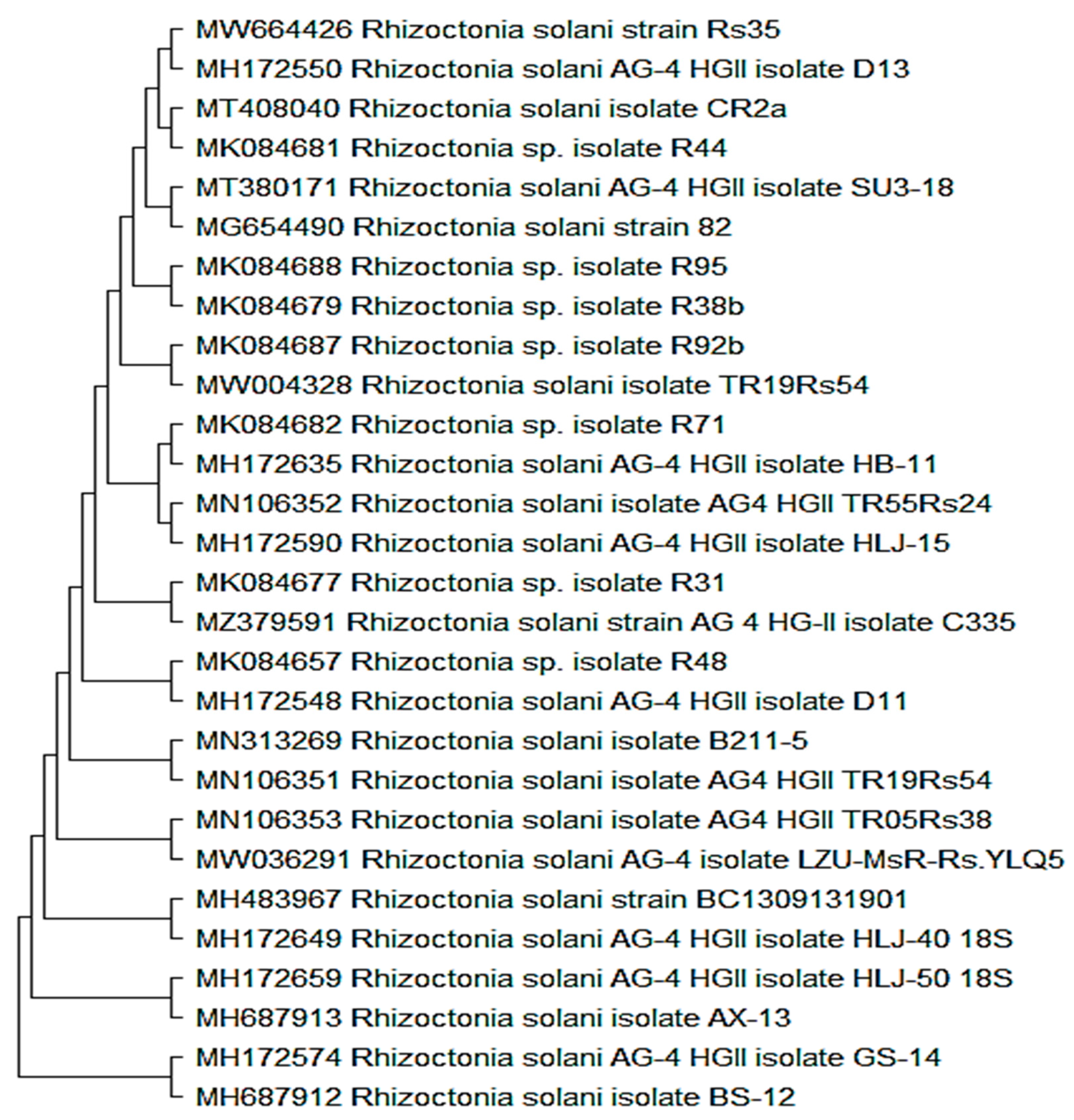
3.1. Rhizoctonia Solani Fungus Identification

The morphological examination of the isolated root rot fungus was congruent with Rhizoctonia genus features because it is an anamorphic hyphal septate pathogen that does not form asexual spores [19]. An amplified fragment of the ITS region was carried out and sequenced to verify the morphological identity of the Rhizoctonia isolate. The sequence of the amplified amplicon was retrieved and blasted in the NCBI-GeneBank database, where it was recognized as Rhizoctonia solani and accessioned with the number MW664426 and provided strain identification RS35. The matching of the obtained sequence of Rs35 with the other relative NCBI sequences revealed the genomic similarity closest to 100% with R. solani (MH172550 and MT408040) and an isolate of Rhizoctonia sp. (MK084681) Figure 1.

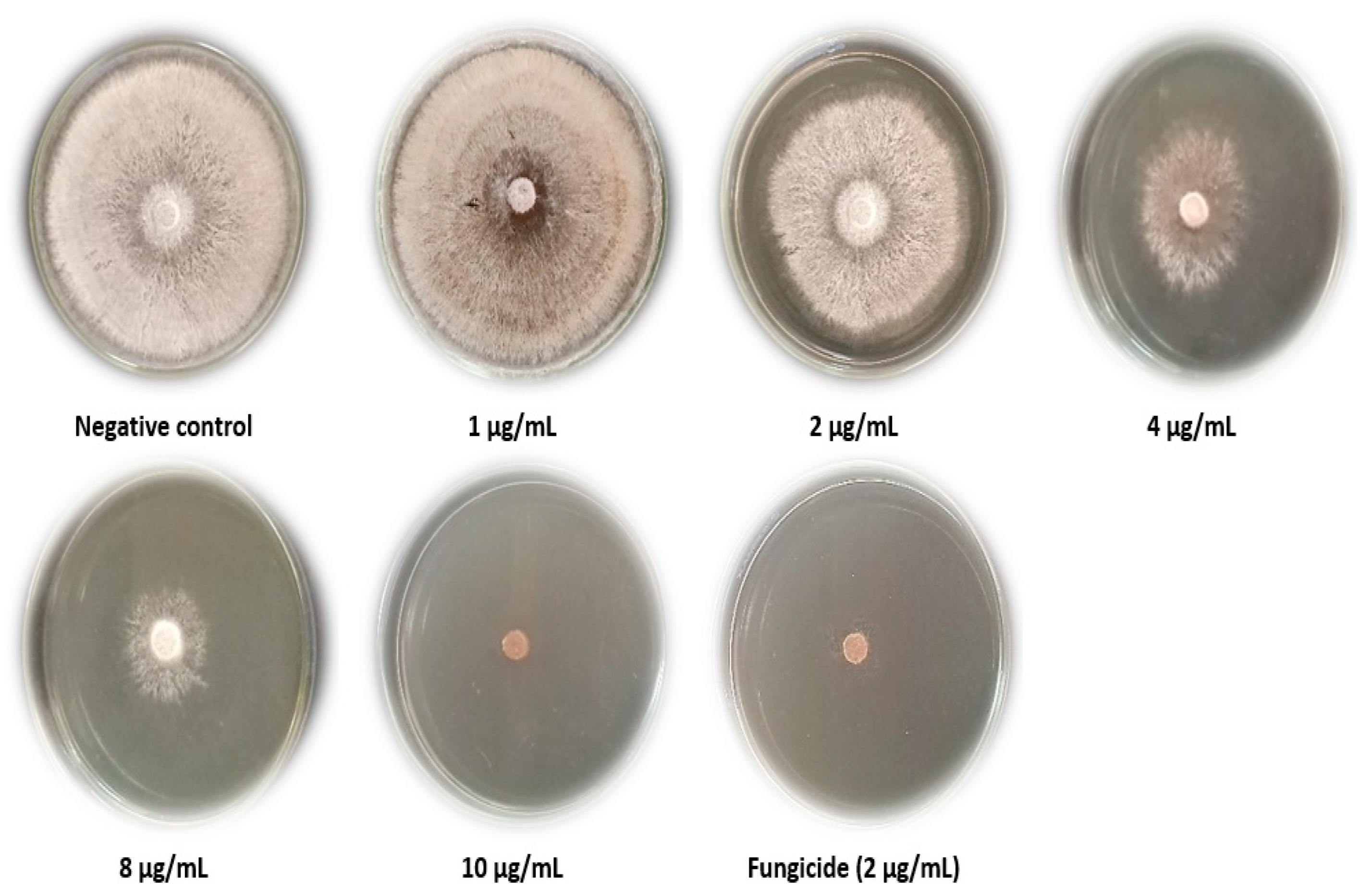
3.2. C. speciosa Extract inhibitory Effect In Vitro

Table 2 and Figure 2 show the growth inhibition of R. solani regarding the evaluated Chorisia speciosa extract. The fungal growth was reduced as the concentration of C. speciosa extract was increased from 1 to 10 µg/mL. The results showed that, at 10 µg/mL, C. speciosa extract inhibited R. solani radial growth completely. Simultaneously, there was no effect on R. solani hyphal development at 1 µg/mL when compared to the Rizolex fungicide (2 μg/mL). As reported before, different methods, including chemical fungicides and biological control agents, work well to minimize crop loss resulting from fungal infections [36,37].
In our previous study, the growth of Fusarium culmorum, Rhizoctonia solani, and Botrytis cinerea was suppressed by 38.5%, 64.4%, and 100%, respectively, when using the ethanol extract of Coccoloba uvifera at 30 µg/mL [38]. R. solani and F. culmorum growth were highly inhibited at a 30 µg/mL concentration of an n-hexane Eucalyptus camaldulensis extract [39]. Acacia saligna aqueous extract also stifled F. culmorum and R. solani fungal development [40]. It was found that the oil extracted from Chorisia speciosa leaves had varying degrees of inhibition against bacteria, including S. aureus, which showed the most sensitivity (25 mm), Escherichia coli showing intermediate activity (15 mm), and S. typhi showing no sensitivity at all at the concentration of 3.64 mg [41]. The ether and ethyl acetate fractions of the 70% ethyl alcohol extract of Chorisia insignis leaves had strong antimicrobial properties against Bacillus subtilis and B. cereus [42].

3.3. Disease Index of R. solani Pathogen in Response to C. speciosa Extract

Under greenhouse conditions, the extract of C. speciosa was evaluated on R. solani to determine whether it promoted or suppressed the development of the fungal pathogen. Root discoloration browning symptoms and their severity (measured on a scale from 0 to 5) were used to calculate the disease index (DI%) for each treatment. Compared to the control treatment, C. speciosa extract significantly decreased the DI %. The DI % was reduced by 26.67% in the protective and 53.33% in the curative treatments. DI% was 93.33% in the Rhizoctonia treatment, and no disease symptoms were observed in the control and plant extract treatments (0%). In our investigation, C. speciosa extract resulted in a statistically significant decrease in disease symptoms. Similarly, Al-Askar and Rashad [43] found that clove extract reduced the occurrence of Pea root-rot infection in a greenhouse trial. Both clove extract at 4% concentration and the fungicide significantly reduced disease occurrence and increased the percentage of surviving plants (from 40% to 48%). Numerous research studies have compared the antimicrobial properties of organic and water extracts and realized that the alcoholic is better than the water extracts [44,45,46,47,48].

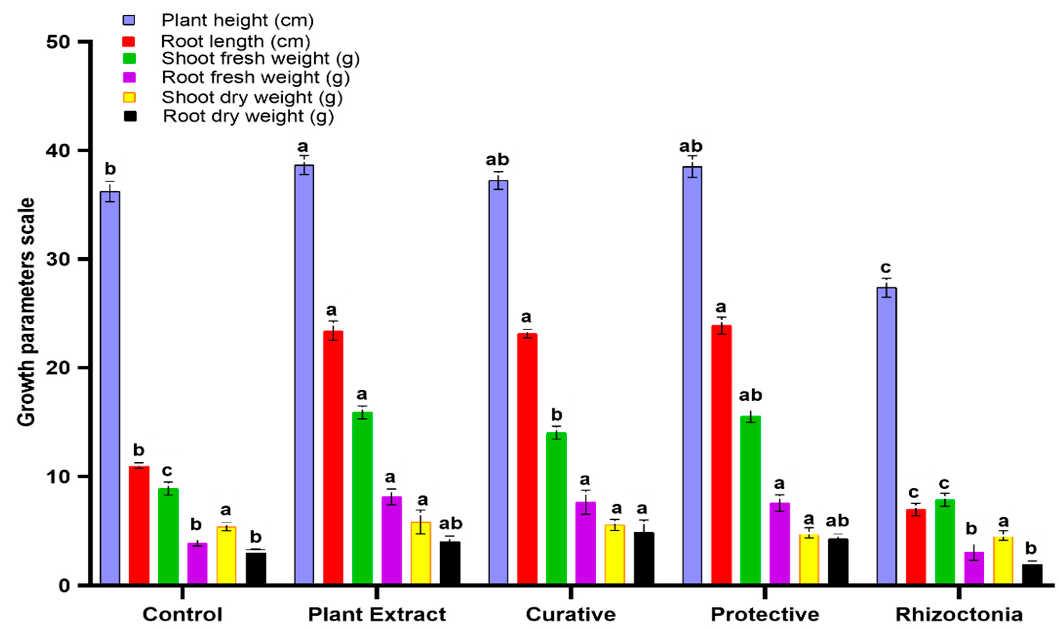
3.4. In Vivo Effect of C. speciosa Extract on Tomato Plants

Tomato plant growth was significantly increased (p ≤ 0.05) in a pot trail when Chorisia speciosa extract was used in either protective or curative treatments (Figure 3). In addition, the treatment with C. speciosa extracts considerably affected plant length. The C. speciosa extract treatment resulted in a height of 38.66 cm, preceded by the protective treatment with 38.51 cm. Root length raised with the C. speciosa extract treatment, but not much more so than in the protective or curative treatments, and quite different from the control. Treatment with C. speciosa resulted in greater fresh shoot weight (15.88 g) and fresh root weight (8.14 g) than other treatments. Dry shoot weights were also measured and found to show no statistically significant differences between treatments and the control. The C. speciosa extract treatment affected the root dry weight compared to the control (Figure 3). Different studies found that using Moringa oleifera leaves extract improved tomato and bean growth and yield [49,50], as well as growth characteristics and photosynthesis in Eruca vesicaria [51]. The development and biomass of maize roots were boosted by blueberry, red grape, and hawthorn leaf extracts [52]. Many researchers have demonstrated that bioactive compounds, such as terpenoids, flavonoids, alkaloids, and polyphenols, are functional as promoters or antagonists of plant development [53,54,55].

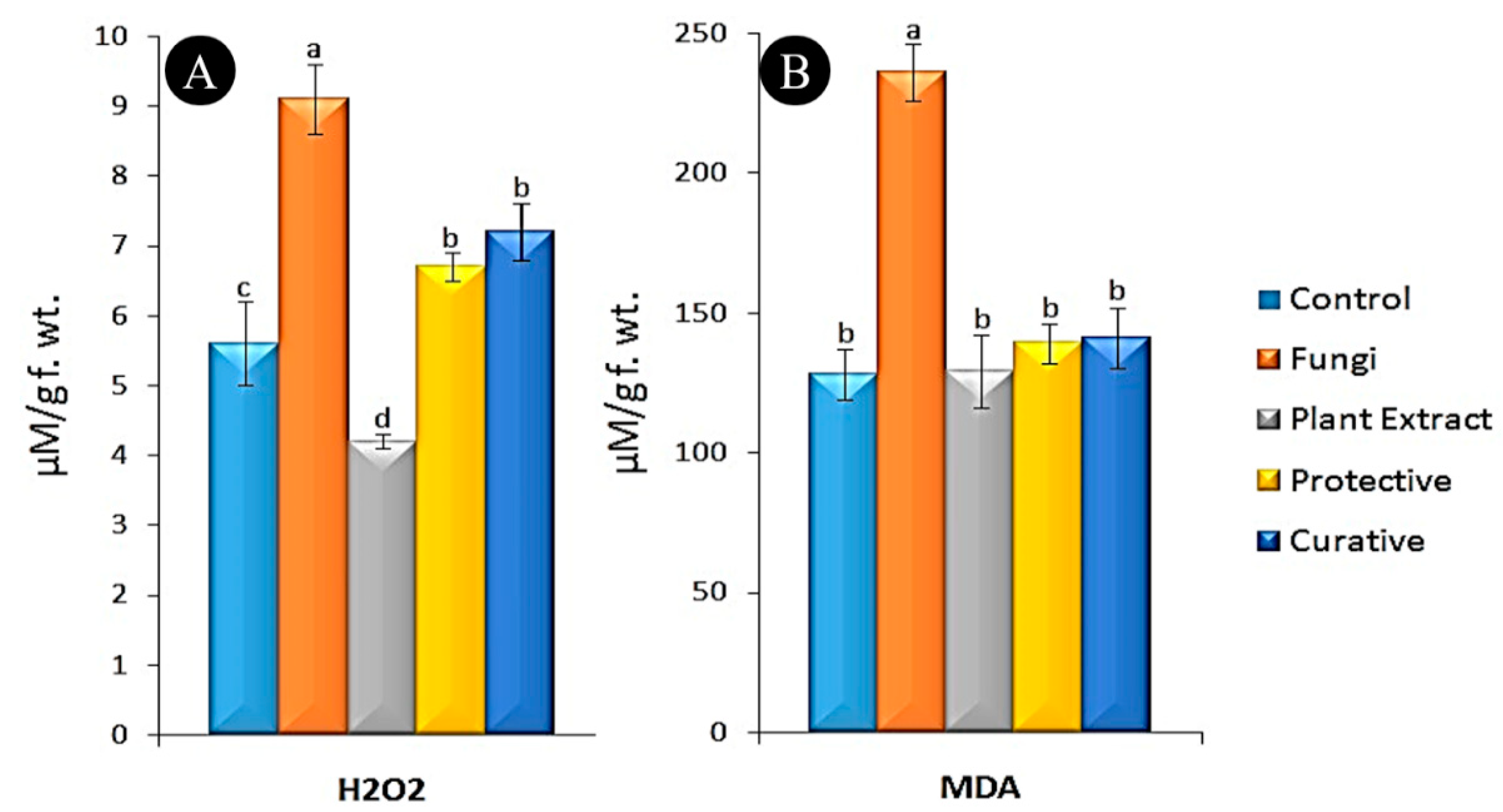
3.5. Oxidative Stress Markers Assay

Rhizoctonia solani fungi increased the quick buildup of H2O2 in infected plants by 1.6 times at 20 dpi (fungi, 9.1 µM/g f. wt.) compared to the non-inoculated control (control, 5.6 µM/g f. wt.). In general, the H2O2 content in all treatments was significantly higher than in the control group, except for the plant extract treatment in which the level was expressed down (4.2 µM/g f. wt.). The H2O2 levels in protective and curative treatments (Figure 4A) were non-significantly increased by 1.19–1.29 times compared to the control (6.7 and 7.2 µM/g f. wt., respectively). Compared to uninoculated controls (128 µM/g f. wt.), the fungal treatment had 1.84 times higher MDA levels (Figure 4B). On the other hand, MDA buildup was shown to be minimally raised in protective and curative treatments (139 and 141, respectively). Lipid peroxidation levels in the protective, curative, and control treatments did not significantly change with the plant extract treatment. According to the data found in our study, the fungal infection was responsible for a substantial rise in the amount of lipid peroxidation that MDA measured. Similar findings were reported previously in different plant fungal infections: Refs. [56,57] reported similar findings in bananas infected with Fusarium oxysporum. Mondal et al. [58] reported that cellular oxidation was prevented in infected tomato leaves due to the enhanced biosynthesis of peroxidases, which regulate the highest MDA and H2O2 levels. Enhanced levels of MDA are indicative of severe oxidative stress in plants. When pathogens attack plants, MDA levels may be a superb indicator of membrane damage [59]. In the context of plant–pathogen interactions, H2O2 has several functions. It is a systemic acquired resistance (SAR) signal molecule that controls the defense machinery system, inhibits the transmission of pathogens, and lignifies cell walls [60,61,62].

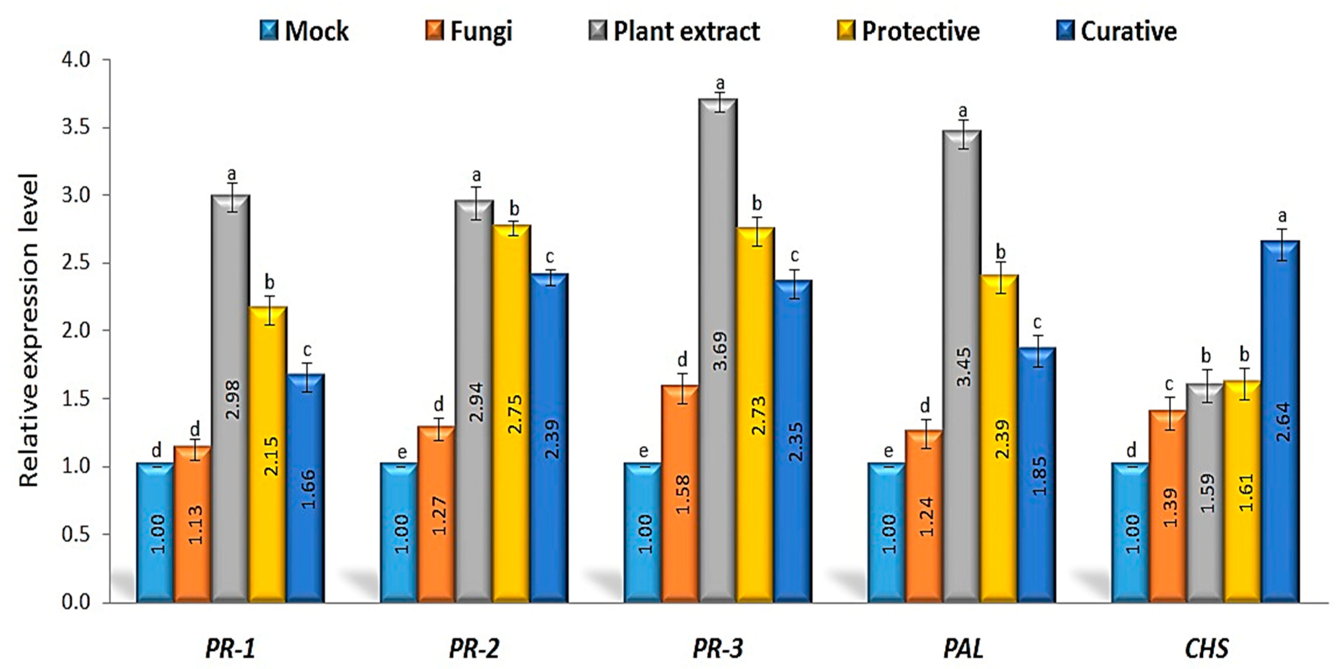
3.6. Defense-Related Genes’ Relative Expression Levels

The relative expression levels of four defense-related genes (PR-1, PR-2, PR-3, PAL, and CHS) were studied at 20 days post-inoculation. The qPCR results confirmed C. speciosa’s antifungal efficacy by showing an impressive upregulation of defense genes within the treated plants (Figure 5). It has been known for over a couple of decades that PR-1 is an important regulator of SAR and may act as a signal for early responses of plant defense [63]. SA induction in response to pathogenic organisms is related to the buildup and activation of PR-1, an SA biomarker signal gene [32,64]. All treatments, including extract, showed up-regulation of PR-1 compared to controls, while non-treated plants showed down-regulation. The extract treatment alone was observed to have the best average transcript levels (2.98-fold), followed by the protective (2.15-fold) and curative treatments (1.66-fold). Elevated PR transcription promotes resistance to microbial infections, which agrees with previous reports [65,66]. Therefore, it is feasible that C. speciosa can alter the plant’s responses, boost resistance, and stop R. solani from silencing defensive genes. Inhibition of fungal cell proliferation is caused by sterol-binding PR-1 proteins [67], while PR2-1,3-glucanases and PR-3-chitinases cause lysis of the fungus wall. In all C. speciosa extract treatments, PR-2 upregulation rose (Figure 5). Relative expression level reports showed that curative treatment was 2.94-fold higher than the control, with protective treatment reporting a 2.75-fold increase. A 1.27-fold decrease in regulatory activity was observed between the control plants and those exposed to the fungus (Figure 5). Multiple studies have shown that SAR inducers, particularly SA, activate the PR-2 protein family, which then participates in various physiological plant defense activities [68]. Plants given an extract of C. speciosa may have shown an increase in PR-2 activity due to the presence of fungal metabolites [69]. The current study suggests that microbe elicitors increase PR-2 mRNA in plants [70]. Enhanced PR-2 activity in the plant cell has been demonstrated to improve the number of oligosaccharides generated, stimulating defense mechanisms.
When comparing treated and control plants (mock plants), PR-3 relative expression levels were significantly higher in all cases (Figure 5). Treatment with plant extract alone resulted in the greatest transcriptional level (3.69-fold), followed by protective treatment (2.73-fold), curative treatment (2.35-fold), and mock plants (1.58-fold). Chitinase, an enzyme that catalyzes the breakdown of chitin and protects plants from fungal attacks, is encoded by the PR-3 gene [17]. In this study, the PR-3 gene was induced in tomato plants in response to fungal infection, C. speciosa, and curative and protective applications. The results demonstrate that PR-3 is a gene that works to improve plant tolerance to fungal invasions. When C. speciosa is applied, numerous defense genes are activated in plant leaves, including PR-3, which results in greater resistance to pathogens [19]. All treatments increased CHS gene transcription levels when comparing treated and mock plants. CHS converts p-coumaroyl CoA to naringenin chalcones [71,72] and is thus a necessary first step in the plant flavonoid biosynthetic pathway. The curative application showed the highest transcript levels (2.64-fold) compared to the other treatments (protective, plant extract, and mock plants; Figure 5). After treating with C. speciosa extract, curative, and protective treatments [17,73], CHS was found to be most activated, which is needed for flavonoid formation. Extensive flavonoids with broad antifungal effects against a variety of plant diseases have been observed to be produced by CHS over-transcription in prior studies [74,75]. For this reason, using C. speciosa on tomato plants as a protective or curative application may lead to greater production of flavonoid molecules. Thus, we hypothesize that the C. speciosa extract includes elicitor compounds that can trigger SAR and enhance plant resistance to fungal infection. In many cases of host–pathogen interactions, the primed state is characterized by the subsequent activation of defense-related PR genes upon pathogen attack [76,77]. This is also the case in inducible systemic resistance (ISR) against hemitrophic and necrotrophic diseases, as shown by the priming of defense-related genes by Harpophora oryzae in the rice–Magnaporthe oryzae interaction [65] and by B. subtilis-induced related proteins in tomato confronted by Pectobacterium carotovorum [66]. However, C. speciosa can potentially be employed as a biocontrol agent to combat R. solani infections. However, further studies, such as the fractionated compounds from the extract, are required for field applications in the future.

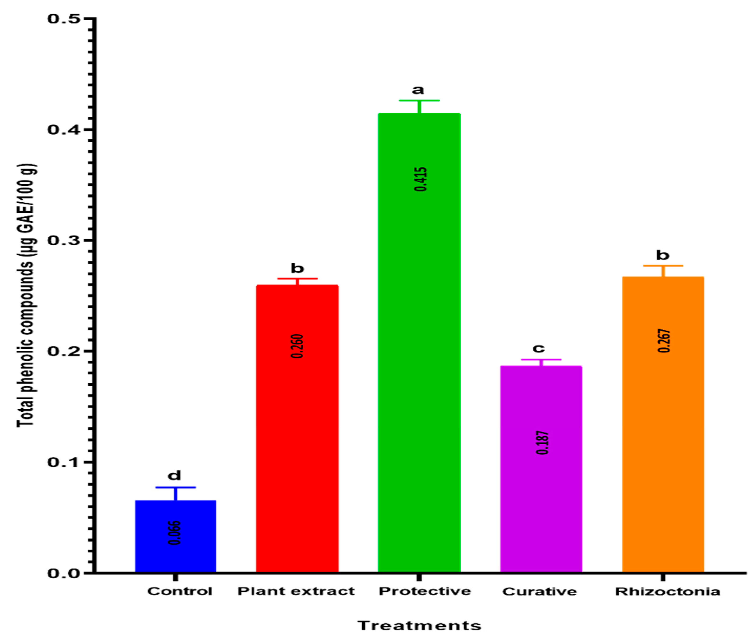
3.7. TPC Accumulation

The accumulation of polyphenolic compounds in plants treated with plant extract is associated with a robust biochemical defense against pathogenic diseases. About 6.3 times more phenolics were present in the protective treatment than in the control, whereas C. speciosa extract and R. solani treatments produced 3.9 and 4.0 times the phenolic content, respectively (Figure 6). Several investigations [78,79,80] found that infections with fungi or bacteria significantly increased total phenolic content. In addition, the curative treatment had the highest phenolic content, nearly 2.8-fold higher than control plants, despite being lower than all treatments except the control (0.066 g GAE/100 g). Therefore, the phenolic compound chemicals that build in plants treated with plant extracts may act as proton donors, protecting root cells from oxidative damage caused by microbial infection [81]. Tomato plants would be protected against plant diseases if their TPC increased as a plant response to the administration of plant extract in protective treatment only. According to Mikulic-Petkovsek et al. [82], these TPC variations result from biosynthesis phenylpropanoid pathway regulation.

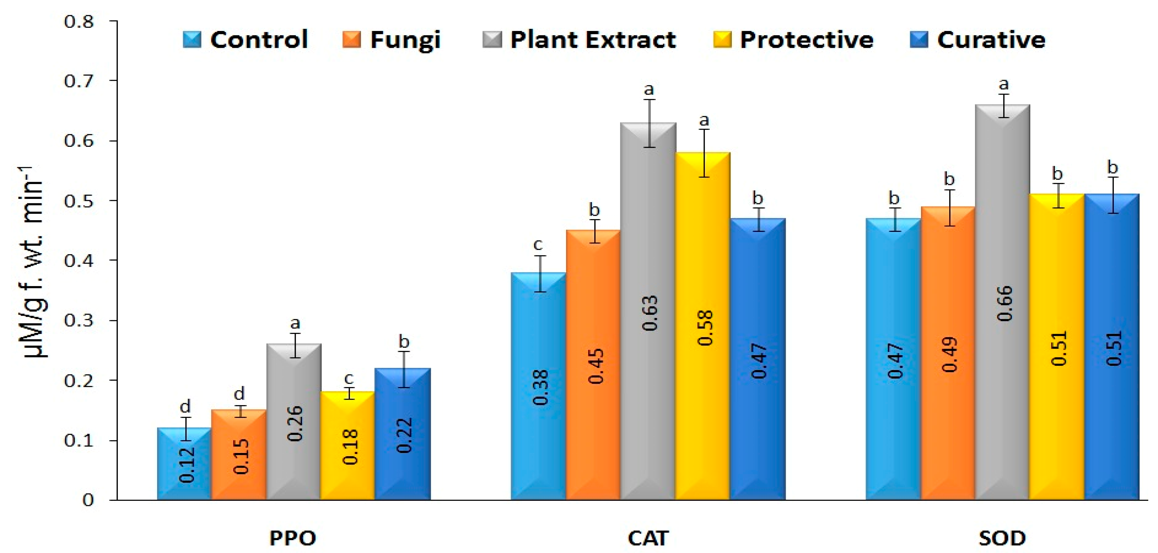
3.8. Antioxidant Enzymes Activity

The current study found that inoculating tomatoes with R. solani significantly increased the levels of three antioxidant enzymes (PPO, CAT, and SOD). Antioxidant enzyme PPO activity was 1.25-fold higher in the fungal treatment (0.15 µM/g f.wt.) compared to the control (0.12 µM/g f.wt.), but this difference was not statistically significant. The enzyme value was raised 2.16 times over the control after treatment with the plant extract. Enzyme levels in preventive and curative treatments (0.18 and 0.22 µM/g f.wt., respectively) differed significantly from the control (Figure 7). PPO’s consumption of reactive oxygen species (ROS) as a substrate has also been linked to the production of lignin within the cell wall, which acts as a protective border against the spread of plant diseases [83]. Figure 7 shows that accumulated CAT was 0.63 M/g f.wt. greater after treatment with plant extract compared to the control treatment. The accumulation rates for the preventative and curative treatments were drastically different. However, when comparing the fungal treatment (0.45 µM/g f.wt.) to the control treatment (0.38 µM/g f.wt.), there was a statistically significant increase in CAT activity levels. It has been established that CAT plays a crucial role in shielding plant cells from the damage generated by reactive oxygen species (ROS) under plant stress [84]. SOD activity did not increase substantially in any of the treatments’ leaf tissues except for the plant extract. In tomato plants, the extract alone increased enzyme activity by 1.29-fold compared to the control (0.66 µM/g f.wt). The SOD enzyme has been shown to prevent pathogen penetration by fortifying cell walls [85], a role that has been previously reported. Our findings indicate that pre- or post-fungal inoculation treatment with C. speciosa extract significantly alters tomato plants’ PPO, CAT, and SOD activities. Based on their findings, Sobhy et al. [56] conclude that the extract has a high phenolic content and a potent ability to quench radicals. C. speciosa extract has a wide variety of phenolic and flavonoid components with antioxidant properties, which may account for the lack of antioxidant enzyme activity observed in the treatments. In contrast, increasing CAT levels can reduce H2O2-induced oxidative stress and mimic H2O2 signaling in disease progression [86]. Therefore, foliar application of C. speciosa extract before fungal inoculation may be an option for reducing the negative consequences of fungal infections. Our results suggest that C. speciosa extract enhances defense and detoxification systems, leading to more rapid and effective responses to fungal inoculation. Moreover, flavonoids, phenolics, PPO, CAT, and SOD activities may play an important role in tomato survival when exposed to fungal stress. By improving growth parameters, boosting antioxidant status, and modulating gene expression in fungal-stressed plants, foliar application of C. speciosa extract before R. solani inoculation greatly protected tomato plants from fungal infection problems. Thus, this research may have inspired the development of a new extract–host–pathogen system, albeit the underlying mechanism of how this would work is now unknown. More research is needed in the future to understand the changes to the proteome in extract-primed tomato.

3.9. Characterization of C. speciosa Extract

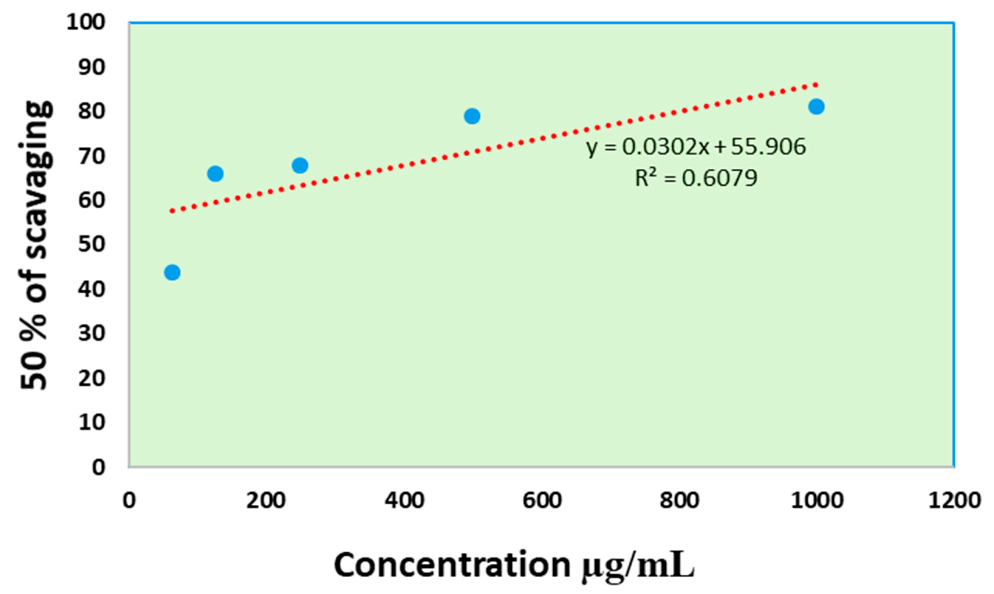
3.9.1. Antioxidant Activity

The ability of C. speciosa extract to scavenge free radicals was measured using the DPPH (1,1-diphenyl-2-picrylhydrazyl) technique. As shown in Figure 8, the extract’s antioxidant activity value (IC50) was 93.46 µg/mL compared with butylated hydroxytoluene (BHT, 5.04 µg/mL). Our findings demonstrated that Chorisia species possessed antioxidant properties consistent with previous research. Few studies showed high antioxidant activity for different fractions of Chorisia speciosa leaves and stem ethanol extract [87]. In addition, Refaat et al. [14] showed that different plant parts’ ethyl acetate, aqueous, and chloroform fractions of both C. speciosa and C. chodatii plants exhibited the most extraordinary free radical scavenging properties and the highest polyphenol contents.

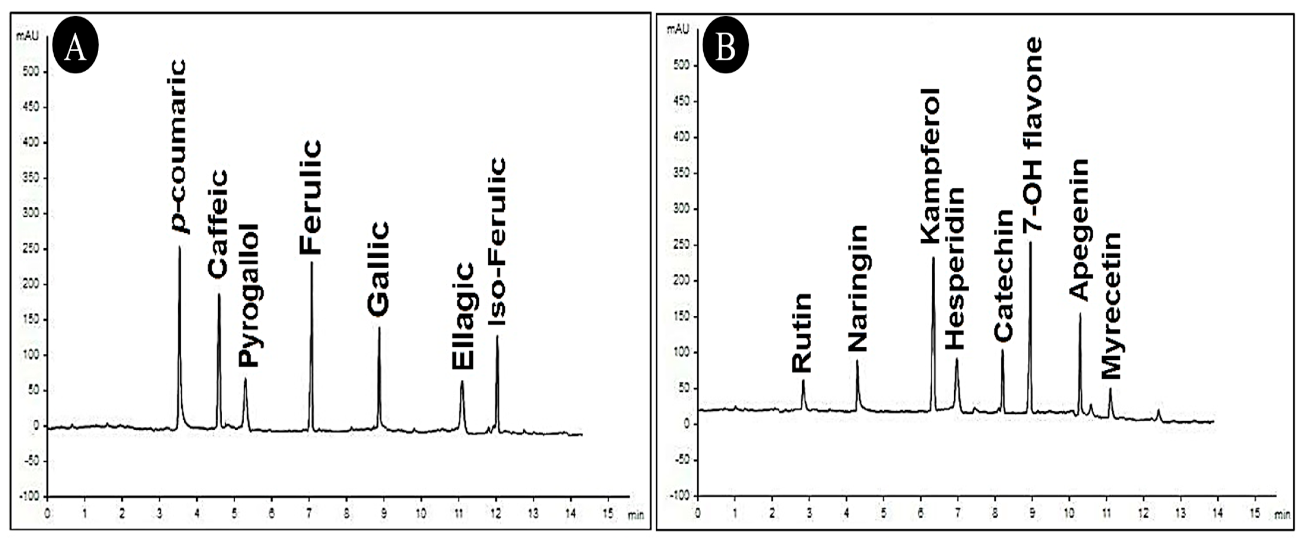
3.9.2. HPLC of C. speciosa Extract

HPLC was used to analyze the C. speciosa extract and revealed a list of chemical compounds in the graphs shown in Figure 9 and Table 3. The main compounds were determined as µg/mL: 7-OH-flavone (10.36), kaempferol (9.23), p-coumaric acid (8.65), ferulic acid (8.14), caffeic acid (7.59), gallic acid (6.33), iso-ferulic (5.71), apigenin (5.47), catechin (3.51) and naringenin (3.14), pyrogallol, ellagic acid, hesperidin, and myricetin. Many plants used for medical purposes for many years contain flavonoids and polyphenols. Ferulic acid, quercetin, ellagic acid, chlorogenic acid, catechins, gallic acid, caffeic acid, and myricetin are all forms of polyphenols that have antibacterial, antiviral, and antioxidant properties [88,89]. Our data suggest that polyphenolic molecules serve critical functions in SAR as elicitor compounds. The antimicrobial activity of C. insignis ethyl acetate extract may be due to flavonoids and kaempferol derivatives, as previously reported by El Sawi et al. [90]. Previous studies have identified various phenolic compounds in medicinal plants, many of which contribute to the plants’ antioxidant and antibacterial activities [91,92].

4. Conclusions

Applying 10 µg/mL of a C. speciosa extract to the leaves of tomato plants increased their growth, induced systemic resistance, and reduced the incidence of root rot disease caused by the R. solani RS 35 pathogen. Our results showed that C. speciosa-treated plants had a lower disease index than control plants, and the expression levels of PR-3, PAL, and CHS genes had increased significantly at 20 dpi. The HPLC examination of the C. speciosa extract showed that the main compounds were 7-OH-flavone, kaempferol, p-coumaric acid, ferulic acid, caffeic acid, gallic acid, iso-ferulic, apigenin, catechin, naringenin, pyrogallol, ellagic acid, hesperidin, and myricetin. Our findings indicate that curative treatment is the most effective way of minimizing root rot. Consequently, the evaluated extract could be a suitable source for the production of antifungal agents to combat plant diseases. However, further studies are needed for open field applications.

Author Contributions

Conceptualization, S.I.B. and A.A.; methodology, A.-N.A.A.-M., S.A.S., A.A. and S.I.B.; software, S.I.B., M.M.E. and A.A.; validation, M.M.E., S.A.S. and A.A.A.; formal analysis, A.A.H., M.M.E. and S.I.B.; investigation, A.A. and M.M.E.; resources, A.A.H.; data curation, M.M.E.; writing—original draft preparation, S.I.B. and A.A.; writing—review and editing, S.I.B. and A.A.; visualization, A.A.A.; supervision, A.A.A.-A.; project administration, A.A.A.; funding acquisition, A.A.A.-A. All authors have read and agreed to the published version of the manuscript.

Funding

This research was financially supported by the Researchers Supporting Project number (RSP2022R505), King Saud University, Riyadh, Saudi Arabia.

Institutional Review Board Statement

Not applicable.

Informed Consent Statement

Not applicable.

Data Availability Statement

Not applicable.

Acknowledgments

The authors would like to extend their appreciation to the Researchers Supporting Project number (RSP2022R505), King Saud University, Riyadh, Saudi Arabia.

Conflicts of Interest

The authors declare no conflict of interest.

References

  1. Sabuquillo, P.; De Cal, A.; Melgarejo, P. Biocontrol of tomato wilt by Penicillium oxalicum formulations in different crop conditions. Biol. Control 2006, 37, 256–265. [Google Scholar] [CrossRef]
  2. Kuprashvili, T.D. The use of phytoncides for seed treatment. Zas. Karantin Rast. 1996, 5, 31. [Google Scholar]
  3. Gondal, A.S.; Rauf, A.; Naz, F. Anastomosis Groups of Rhizoctonia solani associated with tomato foot rot in Pothohar Region of Pakistan. Sci. Rep. 2019, 9, 3910. [Google Scholar] [CrossRef] [PubMed]
  4. Abdelkhalek, A.; Behiry, S.I.; Al-Askar, A.A. Bacillus velezensis PEA1 Inhibits Fusarium oxysporum Growth and Induces Systemic Resistance to Cucumber Mosaic Virus. Agronomy 2020, 10, 1312. [Google Scholar] [CrossRef]
  5. Parveen, S.; Wani, A.H.; Bhat, M.Y.; Malik, A.R.; Koka, J.A.; Ashraf, N. Antimycotic potential of some phytoextracts on some pathogenic fungi. J. Biopestic. 2017, 10, 60–65. [Google Scholar]
  6. Nazzaro, F.; Fratianni, F.; Coppola, R.; Feo, V. De Essential oils and antifungal activity. Pharmaceuticals 2017, 10, 86. [Google Scholar] [CrossRef] [PubMed]
  7. Shuping, D.S.S.; Eloff, J.N. The use of plants to protect plants and food against fungal pathogens: A review. African J. Tradit. Complement. Altern. Med. 2017, 14, 120–127. [Google Scholar] [CrossRef] [PubMed]
  8. Joly, A.B. Botany: An Introduction to Plant Taxonomy; National Publishing Company: São Paulo, Brazil, 1991; Volume 10, p. 462. [Google Scholar]
  9. Adjanohoun, E.J.; et Technique, A.D.C.C.; Ahyi, A.M.R.; Ake Assi, L.; Baniakina, J.; Chibon, P. Médecine Traditionnelle et Pharmacopée: Contribution aux Études Ethnobotaniques et Floristiques en République Populaire du Congo; Agence de Cooperation Culturelle et Technique: Paris, France, 1988. [Google Scholar]
  10. Khan, A.; Asadsaeed, M.; Chaudhary, M.A.; Ahmad, Q.; Ansari, F. Antimicrobial, anti-inflammatory and antipyretic activity of Chorisia speciosa leaves (bombacaceae). Inter. J. Biol. Pharm. Allied Scien. 2015, 4, 6826–6838. [Google Scholar]
  11. Refaat, J.; Desoky, S.Y.; Ramadan, M.A.; Kamel, M.S. Bombacaceae: A phytochemical review. Pharm. Biol. 2013, 51, 100–130. [Google Scholar] [CrossRef]
  12. Hafez, S.S.; Ghani, A.; Afaf, E.; El Shazly, A.M. Pharmacognostical and antibacterial studies of chorisia speciosa st. Hil. flower [Bomhacaceae]. Mansoura J. Pharm. Sci. 2003, 19, 40–59. [Google Scholar]
  13. Ashmawy, A.M.; Azab, S.S.; Eldahshan, O.A. Effects of Chorisia crispiflora ethyl acetate extract on P21 and NF-κB in breast cancer cells. J. Am. Sci. 2012, 8, 965–972. [Google Scholar]
  14. Refaat, J.; Yehia Desoukey, S.; Ramadan, M.A.; Kamel, M.S.; Han, J.; Isoda, H. Comparative polyphenol contents, free radical scavenging properties and effects on adipogenesis of Chorisia Chodatii and Chorisia Speciosa. J. Med. Herbs 2015, 5, 193–207. [Google Scholar]
  15. El-Alfy, T.S.; El-Sawi, S.A.; Sleem, A.; Moawad, D.M. Investigation of flavonoidal content and biological activities of Chorisia insignis Hbk. leaves. Aust. J. Basic Appl. Sci. 2010, 4, 1334–1348. [Google Scholar]
  16. Youssef, N.H.; Qari, S.H.; Matar, S.; Hamad, N.A.; Dessoky, E.S.; Elshaer, M.M.; Sobhy, S.; Abdelkhalek, A.; Zakaria, H.M.; Heflish, A.A. Licorice, Doum, and Banana Peel Extracts Inhibit Aspergillus flavus Growth and Suppress Metabolic Pathway of Aflatoxin B1 Production. Agronomy 2021, 11, 1587. [Google Scholar] [CrossRef]
  17. Abdelkhalek, A.; Al-Askar, A.A.; Alsubaie, M.M.; Behiry, S.I. First Report of Protective Activity of Paronychia argentea Extract against Tobacco Mosaic Virus Infection. Plants 2021, 10, 2435. [Google Scholar] [CrossRef] [PubMed]
  18. Alexopoulos, C.J.; Mims, C.W.; Blackwell, M. Introductory Mycology; John Wiley and Sons: Hoboken, NJ, USA, 1996; ISBN 0471522295. [Google Scholar]
  19. Heflish, A.A.; Abdelkhalek, A.; Al-Askar, A.A.; Behiry, S.I. Protective and Curative Effects of Trichoderma asperelloides Ta41 on Tomato Root Rot Caused by Rhizoctonia solani Rs33. Agronomy 2021, 11, 1162. [Google Scholar] [CrossRef]
  20. Kumar, A.; Shukla, R.; Singh, P.; Prasad, C.S.; Dubey, N.K. Assessment of Thymus vulgaris L. essential oil as a safe botanical preservative against post harvest fungal infestation of food commodities. Innov. Food Sci. Emerg. Technol. 2008, 9, 575–580. [Google Scholar] [CrossRef]
  21. Dissanayake, M. Inhibitory effect of selected medicinal plant extracts on phytopathogenic fungus Fusarium oxysporum (Nectriaceae) Schlecht. Emend. Snyder and Hansen. Annu. Res. Rev. Biol. 2014, 4, 133–142. [Google Scholar] [CrossRef]
  22. Montealegre, J.; Valderrama, L.; Sánchez, S.; Herrera, R.; Besoain, X.; Pérez, L.M. Biological control of Rhizoctonia solani in tomatoes with Trichoderma harzianum mutants. Electron. J. Biotechnol. 2010, 13, 1–2. [Google Scholar] [CrossRef]
  23. Abdeljalil, N.O.-B.; Vallance, J.; Gerbore, J.; Bruez, E.; Martins, G.; Rey, P.; Daami-Remadi, M. Biocontrol of Rhizoctonia root rot in tomato and enhancement of plant growth using rhizobacteria naturally associated to tomato. J. Plant Pathol. Microbiol. 2016, 7, 1–8. [Google Scholar] [CrossRef]
  24. Velioglu, Y.; Mazza, G.; Gao, L.; Oomah, B.D. Antioxidant activity and total phenolics in selected fruits, vegetables, and grain products. J. Agric. Food Chem. 1998, 46, 4113–4117. [Google Scholar] [CrossRef]
  25. Heath, R.L.; Packer, L. Photoperoxidation in isolated chloroplasts: I. Kinetics and stoichiometry of fatty acid peroxidation. Arch. Biochem. Biophys. 1968, 125, 189–198. [Google Scholar] [CrossRef]
  26. Velikova, V.; Yordanov, I.; Edreva, A. Oxidative stress and some antioxidant systems in acid rain-treated bean plants: Protective role of exogenous polyamines. Plant Sci. 2000, 151, 59–66. [Google Scholar] [CrossRef]
  27. CHO, Y.K.; AHN, H.Y.E.K. Purification and characterization of polyphenol oxidase from potato: II. Inhibition and catalytic mechanism. J. Food Biochem. 1999, 23, 593–605. [Google Scholar] [CrossRef]
  28. Cakmak, I.; Marschner, H. Magnesium deficiency and high light intensity enhance activities of superoxide dismutase, ascorbate peroxidase, and glutathione reductase in bean leaves. Plant Physiol. 1992, 98, 1222–1227. [Google Scholar] [CrossRef]
  29. Beauchamp, C.; Fridovich, I. Superoxide dismutase: Improved assays and an assay applicable to acrylamide gels. Anal. Biochem. 1971, 44, 276–287. [Google Scholar] [CrossRef]
  30. Abdelkhalek, A.; Sanan-Mishra, N. Differential expression profiles of tomato miRNAs induced by tobacco mosaic virus. J. Agric. Sci. Technol. 2019, 21, 475–485. [Google Scholar]
  31. AbdEl-Rahim, W.M.; Khalil, W.K.B.; Eshak, M.G. Evaluation of the gene expression changes in Nile tilapia (Oreochromis niloticus) as affected by the bio-removal of toxic textile dyes from aqueous solution in small-scale bioreactor. Environmentalist 2010, 30, 242–253. [Google Scholar] [CrossRef]
  32. Abo-Zaid, G.; Abdelkhalek, A.; Matar, S.; Darwish, M.; Abdel-Gayed, M. Application of Bio-Friendly Formulations of Chitinase-Producing Streptomyces cellulosae Actino 48 for Controlling Peanut Soil-Borne Diseases Caused by Sclerotium rolfsii. J. Fungi 2021, 7, 167. [Google Scholar] [CrossRef] [PubMed]
  33. Livak, K.J.; Schmittgen, T.D. Analysis of Relative Gene Expression Data Using Real-Time Quantitative PCR and the 2ΔΔCT Method. Methods 2001, 25, 402–408. [Google Scholar] [CrossRef]
  34. Shimada, K.; Fujikawa, K.; Yahara, K.; Nakamura, T. Antioxidative properties of xanthan on the autoxidation of soybean oil in cyclodextrin emulsion. J. Agric. Food Chem. 1992, 40, 945–948. [Google Scholar] [CrossRef]
  35. Abdelkhalek, A.; Király, L.; Al-Mansori, A.-N.A.; Younes, H.A.; Zeid, A.; Elsharkawy, M.M.; Behiry, S.I. Defense Responses and Metabolic Changes Involving Phenylpropanoid Pathway and PR Genes in Squash (Cucurbita pepo L.) following Cucumber mosaic virus Infection. Plants 2022, 11, 1908. [Google Scholar] [CrossRef]
  36. Singh, R.P.; Singh, P.K.; Rutkoski, J.; Hodson, D.P.; He, X.; Jørgensen, L.N.; Hovmøller, M.S.; Huerta-Espino, J. Disease impact on wheat yield potential and prospects of genetic control. Annu. Rev. Phytopathol. 2016, 54, 303–322. [Google Scholar] [CrossRef]
  37. Hof, H. Critical annotations to the use of azole antifungals for plant protection. Antimicrob. Agents Chemother. 2001, 45, 2987–2990. [Google Scholar] [CrossRef]
  38. Ashmawy, N.A.; Salem, M.Z.M.; El Shanhorey, N.; Al-Huqail, A.; Ali, H.M.; Behiry, S.I. Eco-friendly wood-biofungicidal and antibacterial activities of various Coccoloba uvifera L. leaf extracts: HPLC analysis of phenolic and flavonoid compounds. BioResources 2020, 15, 4165–4187. [Google Scholar] [CrossRef]
  39. Salem, M.Z.M.; Behiry, S.I.; EL-Hefny, M. Inhibition of Fusarium culmorum, Penicillium chrysogenum and Rhizoctonia solani by n-hexane extracts of three plant species as a wood-treated oil fungicide. J. Appl. Microbiol. 2019, 126, 1683–1699. [Google Scholar] [CrossRef]
  40. Al-Huqail, A.A.; Behiry, S.I.; Salem, M.Z.M.; Ali, H.M.; Siddiqui, M.H.; Salem, A.Z.M. Antifungal, antibacterial, and antioxidant activities of Acacia saligna (Labill.) HL Wendl. flower extract: HPLC analysis of phenolic and flavonoid compounds. Molecules 2019, 24, 700. [Google Scholar] [CrossRef]
  41. Kausar, F.; Intisar, A.; Din, M.I.; Aamir, A.; Hussain, T.; Aziz, P.; Mutahir, Z.; Fareed, S.; Samreen, B.; Sadaqat, K. Volatile Composition and Antibacterial Activity of Leaves of Chorisia speciosa. J. Mex. Chem. Soc. 2020, 64, 339–348. [Google Scholar] [CrossRef]
  42. El Sawi, S.A.M.; Hanafy, D.M.M.M.; El Alfy, T.S.M.A. Composition of the non-polar extracts and antimicrobial activity of Chorisia insignis HBK. leaves. Asian Pacific J. Trop. Dis. 2014, 4, 473–479. [Google Scholar] [CrossRef]
  43. Al-Askar, A.A.; Rashad, Y.M. Efficacy of some plant extracts against Rhizoctonia solani on pea. J. Plant Prot. Res. 2010, 50, 239–243. [Google Scholar] [CrossRef]
  44. Jat, J.G.; Agalave, H.R. Fungitoxic properties of some leaf extracts against oilseed-borne fungi. Sci. Res. Rep. 2013, 3, 210–215. [Google Scholar]
  45. Ambikapathy, V.; Gomathi, S.; Panneerselvam, A. Effect of antifungal activity of some medicinal plants against Pythium debaryanum (Hesse). Asian J. Plant Sci. Res. 2011, 1, 131–134. [Google Scholar]
  46. Behbahani, B.A.; Shahidi, F.; Yazdi, F.T.; Mohebbi, M. Antifungal effect of aqueous and ethanolic mangrove plant extract on pathogenic fungus “in vitro”. Int. J. Agron. Plant Prod. 2013, 4, 1652–1658. [Google Scholar]
  47. Ashraf, Z.; Muhammad, A.; Imran, M.; Tareq, A.H. In vitro antibacterial and antifungal activity of methanol, chloroform and aqueous extracts of Origanum vulgare and their comparative analysis. Int. J. Org. Chem. 2011, 1, 257–261. [Google Scholar] [CrossRef]
  48. Moorthy, K.K.; Subramaniam, P.; Senguttuvan, J. In vitro antifungal activity of various extracts of leaf and stem parts of Solena amplexicaulis (Lam.) Gandhi. Int. J. Pharm. Pharm. Sci. 2013, 5, 745–747. [Google Scholar]
  49. Culver, M.; Fanuel, T.; Chiteka, A.Z. Effect of moringa extract on growth and yield of tomato. Greener J. Agric. Sci. 2012, 2, 207–211. [Google Scholar]
  50. Mvumi, C.; Tagwira, F.; Chiteka, A.Z. Effect of moringa extract on growth and yield of maize and common beans. Greener J. Agric. Sci. 2013, 3, 55–62. [Google Scholar] [CrossRef]
  51. Abdalla, M.M. Boosting the growth of rocket plants in response to the application of Moringa oleifera extracts as a biostimulant. Life Sci. J. 2014, 11, 1113–1121. [Google Scholar]
  52. Ertani, A.; Pizzeghello, D.; Francioso, O.; Tinti, A.; Nardi, S. Biological activity of vegetal extracts containing phenols on plant metabolism. Molecules 2016, 21, 205. [Google Scholar] [CrossRef]
  53. Biradar, S.R.; Rachetti, B.D. Extraction of some secondary metabolites & thin layer chromatography from different parts of Centella asiatica L.(URB). Am. J. Life Sci. 2013, 1, 243–247. [Google Scholar]
  54. James, J.; Dubery, I. Identification and quantification of triterpenoid centelloids in Centella asiatica (L.) Urban by densitometric TLC. JPC-J. Planar Chromatogr. TLC 2011, 24, 82–87. [Google Scholar] [CrossRef]
  55. Singh, D.; Singh, P.; Gupta, A.; Solanki, S.; Sharma, E.; Nema, R. Qualitative estimation of the presence of bioactive compound in Centella asiatica: An important medicinal plant. Int. J. Life Sci. Med. Sci. 2012, 2, 4–7. [Google Scholar]
  56. Sobhy, S.E.; Abo-Kassem, E.-E.M.; Sewelam, N.A.; Hafez, E.E.; Aseel, D.G.; Saad-Allah, K.M. Pre-soaking in Weed Extracts is a Reasonable Approach to Mitigate Fusarium graminearum Infection in Wheat. J. Plant Growth Regul. 2021, 41, 2261–2278. [Google Scholar] [CrossRef]
  57. Anthony, K.K.; George, D.S.; Baldev Singh, H.K.; Fung, S.M.; Santhirasegaram, V.; Razali, Z.; Somasundram, C. Reactive oxygen species activity and antioxidant properties of Fusarium infected bananas. J. Phytopathol. 2017, 165, 213–222. [Google Scholar] [CrossRef]
  58. Mondal, S.; Phadke, R.R.; Badigannavar, A.M. Genetic variability for total phenolics, flavonoids and antioxidant activity of testaless seeds of a peanut recombinant inbred line population and identification of their controlling QTLs. Euphytica 2015, 204, 311–321. [Google Scholar] [CrossRef]
  59. Loreto, F.; Velikova, V. Isoprene produced by leaves protects the photosynthetic apparatus against ozone damage, quenches ozone products, and reduces lipid peroxidation of cellular membranes. Plant Physiol. 2001, 127, 1781–1787. [Google Scholar] [CrossRef]
  60. Kuźniak, E.; Urbanek, H. The involvement of hydrogen peroxide in plant responses to stresses. Acta Physiol. Plant 2000, 22, 195–203. [Google Scholar] [CrossRef]
  61. Grene, R. Oxidative stress and acclimation mechanisms in plants. In The Arabidopsis Book; American Society of Plant Biologists: Rockville, MD, USA, 2002; Volume 1. [Google Scholar]
  62. Taheri, P.; Kakooee, T. Reactive oxygen species accumulation and homeostasis are involved in plant immunity to an opportunistic fungal pathogen. J. Plant Physiol. 2017, 216, 152–163. [Google Scholar] [CrossRef] [PubMed]
  63. Hoegen, E.; Strömberg, A.; Pihlgren, U.; Kombrink, E. Primary structure and tissue-specific expression of the pathogenesis-related protein PR-1b in potato. Mol. Plant Pathol. 2002, 3, 329–345. [Google Scholar] [CrossRef] [PubMed]
  64. D’Maris Amick Dempsey, A.C.; Vlot, M.C.W.; Daniel, F.K.; Dempsey, D.A.; Vlot, A.C.; Wildermuth, M.C.; Klessig, D.F.; D’Maris Amick Dempsey, A.C.; Vlot, M.C.W.; Daniel, F.K.; et al. Salicylic acid biosynthesis and metabolism. Arab. Book/Am. Soc. Plant Biol. 2011, 9, e0156. [Google Scholar] [CrossRef]
  65. Su, Z.-Z.; Mao, L.-J.; Li, N.; Feng, X.-X.; Yuan, Z.-L.; Wang, L.-W.; Lin, F.-C.; Zhang, C.-L. Evidence for biotrophic lifestyle and biocontrol potential of dark septate endophyte Harpophora oryzae to rice blast disease. PLoS ONE 2013, 8, e61332. [Google Scholar] [CrossRef] [PubMed]
  66. Chandrasekaran, M.; Chun, S.C. Expression of PR-protein genes and induction of defense-related enzymes by Bacillus subtilis CBR05 in tomato (Solanum lycopersicum) plants challenged with Erwinia carotovora subsp. carotovora. Biosci. Biotechnol. Biochem. 2016, 80, 2277–2283. [Google Scholar] [CrossRef] [PubMed]
  67. Schneiter, R.; Di Pietro, A. The CAP protein superfamily: Function in sterol export and fungal virulence. Biomol. Concepts 2013, 4, 519–525. [Google Scholar] [CrossRef]
  68. Abdelkhalek, A. Expression of tomato pathogenesis related genes in response to Tobacco mosaic virus. JAPS J. Anim. Plant Sci. 2019, 29, 1596–1602. [Google Scholar]
  69. Druzhinina, I.S.; Seidl-Seiboth, V.; Herrera-Estrella, A.; Horwitz, B.A.; Kenerley, C.M.; Monte, E.; Mukherjee, P.K.; Zeilinger, S.; Grigoriev, I.V.; Kubicek, C.P. Trichoderma: The genomics of opportunistic success. Nat. Rev. Microbiol. 2011, 9, 749–759. [Google Scholar] [CrossRef]
  70. Roylawar, P.; Panda, S.; Kamble, A. Comparative analysis of BABA and Piriformospora indica mediated priming of defence-related genes in tomato against early blight. Physiol. Mol. Plant Pathol. 2015, 91, 88–95. [Google Scholar] [CrossRef]
  71. Abdelkhalek, A.; Dessoky, E.S.; Hafez, E. Polyphenolic genes expression pattern and their role in viral resistance in tomato plant infected with Tobacco mosaic virus. Biosci. Res. 2018, 15, 3349–3356. [Google Scholar]
  72. Abdelkhalek, A.; Salem, M.Z.M.; Hafez, E.; Behiry, S.I.; Qari, S.H. The Phytochemical, Antifungal, and First Report of the Antiviral Properties of Egyptian Haplophyllum tuberculatum Extract. Biology 2020, 9, 248. [Google Scholar] [CrossRef] [PubMed]
  73. Marais, J.P.J.; Deavours, B.; Dixon, R.A.; Ferreira, D. The stereochemistry of flavonoids. In The Science of Flavonoids; Springer: Amsterdam, The Netherlands, 2006; pp. 1–46. [Google Scholar]
  74. Abdelkhalek, A.; Al-Askar, A.A.; Behiry, S.I. Bacillus licheniformis strain POT1 mediated polyphenol biosynthetic pathways genes activation and systemic resistance in potato plants against Alfalfa mosaic virus. Sci. Rep. 2020, 10, 16120. [Google Scholar] [CrossRef] [PubMed]
  75. Martínez, G.; Regente, M.; Jacobi, S.; Del Rio, M.; Pinedo, M.; de la Canal, L. Chlorogenic acid is a fungicide active against phytopathogenic fungi. Pestic. Biochem. Physiol. 2017, 140, 30–35. [Google Scholar] [CrossRef] [PubMed]
  76. Ahn, I.-P.; Lee, S.-W.; Suh, S.-C. Rhizobacteria-induced priming in Arabidopsis is dependent on ethylene, jasmonic acid, and NPR1. Mol. Plant-Microbe Interact. 2007, 20, 759–768. [Google Scholar] [CrossRef] [PubMed]
  77. Niu, D.-D.; Liu, H.-X.; Jiang, C.-H.; Wang, Y.-P.; Wang, Q.-Y.; Jin, H.-L.; Guo, J.-H. The plant growth–promoting rhizobacterium Bacillus cereus AR156 induces systemic resistance in Arabidopsis thaliana by simultaneously activating salicylate-and jasmonate/ethylene-dependent signaling pathways. Mol. Plant-Microbe Interact. 2011, 24, 533–542. [Google Scholar] [CrossRef] [PubMed]
  78. Martin, N.; Vesentini, D.; Rego, C.; Monteiro, S.; Oliveira, H.; Ferreira, R.B. Phaeomoniella chlamydospora infection induces changes in phenolic compounds content in Vitis vinifera. Phytopathol. Mediterr. 2009, 48, 101–116. [Google Scholar]
  79. Petkovsek, M.M.; Slatnar, A.; Stampar, F.; Veberic, R. Phenolic compounds in apple leaves after infection with apple scab. Biol. Plant 2011, 55, 725–730. [Google Scholar] [CrossRef]
  80. Rusjan, D.; Veberič, R.; Mikulič-Petkovšek, M. The response of phenolic compounds in grapes of the variety ‘Chardonnay’(Vitis vinifera L.) to the infection by phytoplasma Bois noir. Eur. J. Plant Pathol. 2012, 133, 965–974. [Google Scholar] [CrossRef]
  81. Singh, B.N.; Singh, A.; Singh, S.P.; Singh, H.B. Trichoderma harzianum-mediated reprogramming of oxidative stress response in root apoplast of sunflower enhances defence against Rhizoctonia solani. Eur. J. Plant Pathol. 2011, 131, 121–134. [Google Scholar] [CrossRef]
  82. Mikulic-Petkovsek, M.; Schmitzer, V.; Stampar, F.; Veberic, R.; Koron, D. Changes in phenolic content induced by infection with Didymella applanata and Leptosphaeria coniothyrium, the causal agents of raspberry spur and cane blight. Plant Pathol. 2014, 63, 185–192. [Google Scholar] [CrossRef]
  83. Mohammadi, M.; Kazemi, H. Changes in peroxidase and polyphenol oxidase activities in susceptible and resistant wheat heads inoculated with Fusarium graminearum and induced resistance. Plant Sci. 2002, 162, 491–498. [Google Scholar] [CrossRef]
  84. Garg, N.; Manchanda, G. ROS generation in plants: Boon or bane? Plant Biosyst. 2009, 143, 81–96. [Google Scholar] [CrossRef]
  85. Zhang, P.; Zhou, M.P.; Zhang, X.; Huo, Y.; Ma, H.X. Change of defensive-related enzyme in wheat crown rot seedlings infected by Fusarium graminearum. Cereal Res. Commun. 2013, 41, 431–439. [Google Scholar] [CrossRef]
  86. Blackman, L.M.; Hardham, A.R. Regulation of catalase activity and gene expression during Phytophthora nicotianae development and infection of tobacco. Mol. Plant Pathol. 2008, 9, 495–510. [Google Scholar] [CrossRef] [PubMed]
  87. Nasr, E.M.; Assaf, M.H.; Darwish, F.M.; Ramadan, M.A. Phytochemical and biological study of Chorisia speciosa A. St. Hil. cultivated in Egypt. J. Pharmacogn. Pharmacol 2018, 7, 649–656. [Google Scholar]
  88. Mani, J.S.; Johnson, J.B.; Steel, J.C.; Broszczak, D.A.; Neilsen, P.M.; Walsh, K.B.; Naiker, M. Natural product-derived phytochemicals as potential agents against coronaviruses: A review. Virus Res. 2020, 284, 197989. [Google Scholar] [CrossRef] [PubMed]
  89. Shaygannia, E.; Bahmani, M.; Zamanzad, B.; Rafieian-Kopaei, M. A review study on Punica granatum L. J. Evid. Based Complement. Altern. Med. 2016, 21, 221–227. [Google Scholar] [CrossRef]
  90. El Sawi, S.; Moawad, D.; El Alfy, S. Activity of Chorisia insignis HBK. against larynx carcinoma and chemical investigation of its polar extracts. J. Appl. Sci. Res. 2012, 8, 5564–5571. [Google Scholar]
  91. Youssef, N.H.; Qari, S.H.; Behiry, S.I.; Dessoky, E.S.; El-Hallous, E.I.; Elshaer, M.M.; Kordy, A.; Maresca, V.; Abdelkhalek, A.; Heflish, A.A. Antimycotoxigenic Activity of Beetroot Extracts against Altenaria alternata Mycotoxins on Potato Crop. Appl. Sci. 2021, 11, 4239. [Google Scholar] [CrossRef]
  92. Mohamed, A.A.; Behiry, S.I.; Ali, H.M.; EL-Hefny, M.; Salem, M.Z.M.; Ashmawy, N.A. Phytochemical compounds of branches from P. halepensis oily liquid extract and S. terebinthifolius essential oil and their potential antifungal activity. Processes 2020, 8, 330. [Google Scholar] [CrossRef] [Green Version]
Figure 1. The evolutionary dendrogram displays the similarity of Rhizoctonia solani Rs35 (MW664426) to other R. solani nucleotide ITS sequences.
Agronomy 12 02309 g001
Figure 2. Antifungal activity of different concentrations of Chorisia speciosa extract against Rhizoctonia solani fungus compared with negative control and fungicide (Rizolex).
Agronomy 12 02309 g002
Figure 3. Effect of treatment with Chorisia speciosa extract on growth parameters of tomato plants. The different letters on top of the columns mean the data values were significantly different at p ≤ 0.05.
Agronomy 12 02309 g003
Figure 4. H2O2 activities (A) and MDA content (B) are expressed in different treatments as µM/g f.wt. The different letters on top of the columns mean the data values were significantly different at p ≤ 0.05.
Agronomy 12 02309 g004
Figure 5. Relative expression levels of five genes: PR-1, PR-2, PR-3, PAL, and CHS at 20 dpi. The different letters on top of the columns mean the data values were significantly different in each gene at p ≤ 0.05.
Agronomy 12 02309 g005
Figure 6. Total phenolic compounds of all treatments used in this study. The different letters on top of the columns mean the data values were significantly different at p ≤ 0.05.
Agronomy 12 02309 g006
Figure 7. PPO, CAT, and SOD enzyme activities under the studied different treatments. The post hoc test analysis of each antioxidant enzyme was conducted separately. The different letters on top of the columns of each antioxidant enzyme mean the data values were significantly different at p ≤ 0.05.
Agronomy 12 02309 g007
Figure 8. The scavenging percentage of 50% inhibition of DPPH (IC50) of the Chorisia speciosa extract.
Agronomy 12 02309 g008
Figure 9. HPLC chromatograms of the phenolic (A) and flavonoid (B) chromatograms identified in Chorisia speciosa extract.
Agronomy 12 02309 g009
Table 1. Oligonucleotide primers used in this study.
GeneAbbreviationNucleotide Sequences
Internal Transcribed SpacerITSITS1-TCCGTAGGTGAACCTGCGG
ITS4-TCCTCCGCTTATTGATATGC
Pathogenesis-related protein-1PR-1For-GTTCCTCCTTGCCACCTTC
Rev-TATGCACCCCCAGCATAGTT
EndoglucanasePR-2For-TATAGCCGTTGGAAACGAAG
Rev-CAACTTGCCATCACATTCTG
ChitinasePR-3For-ATGGAGCATTGTGCCCTAAC
Rev-TCCTACCAACATCACCACCA
Phenylalanine ammonia-lyasePALFor-GTTATGCTCTTAGAACGTCGCCC
Rev-CCGTGTAATGCCTTGTTTCTTGA
Chalcone SynthaseCHSFor-CACCGTGGAGGAGTATCGTAAGGC
Rev-TGATCAACACAGTTGGAAGGCG
β-actinβ-actinFor-TGGCATACAAAGACAGGACAGCCT
Rev-ACTCAATCCCAAGGCCAACAGAGA
Table 2. In vitro growth inhibition (%) of R. solani in response to Chorisia speciosa extract. The different letters mean the data values were significantly different at p ≤ 0.05.
Treatment (µg/mL)Growth Inhibition %
Negative control00.00 ± 0.00 e
100.00 ± 0.00 e
240.37 ± 0.29 d
489.77 ± 0.12 c
892.43 ± 0.15 b
10100.00 ± 0.00 a
Fungicide (Rizolex, 2 µg/mL)100.00 ± 0.00 a
Table 3. Polyphenolic compounds identified in Chorisia speciosa extract.
CompoundsRetention Time (min.)Amount (µg/mL)
p-Coumaric acid3.58.65
Caffeic acid4.87.59
Pyrogallol5.21.22
Ferulic acid7.08.14
Gallic acid9.06.33
Ellagic acid11.01.18
Iso-Ferulic12.05.71
Rutin3.00.88
Naringenin4.13.14
Kaempferol6.19.23
Hesperidin7.02.89
Catechin8.13.51
7-OH flavone9.010.36
Apigenin10.15.47
Myricetin11.00.79
Publisher’s Note: MDPI stays neutral with regard to jurisdictional claims in published maps and institutional affiliations.

