Article

Pits of Date Palm: Bioactive Composition, Antibacterial Activity and Antimutagenicity Potentials

1
Department of Clinical Laboratory Sciences, College of Applied Medical Sciences, Jouf University, Sakaka 72388, Saudi Arabia
2
Desert Research Centre, Department of Medicinal and Aromatic Plants, Cairo 11753, Egypt
3
Department of Medical Microbiology and Parasitology, Faculty of Medicine, King Abdulaziz University, Jeddah 21589, Saudi Arabia
4
Hematology/Pediatric Oncology, Yousef Abdulatif Jameel Scientific Chair of Prophetic Medicine Application, Faculty of Medicine, King Abdulaziz University, Jeddah 21589, Saudi Arabia
5
Basic Sciences Department, Higher Technological Institute, Beni-Suef University, Beni Suef 62521, Egypt
6
Department of Biological Sciences, National University of Medical Sciences, Rawalpindi 46000, Pakistan
7
Department of Botany and Microbiology, Faculty of Science, Beni-Suef University, Beni Suef 62511, Egypt
*
Authors to whom correspondence should be addressed.
Agronomy 2022, 12(1), 54; https://doi.org/10.3390/agronomy12010054
Submission received: 12 November 2021 / Revised: 22 December 2021 / Accepted: 23 December 2021 / Published: 27 December 2021
(This article belongs to the Special Issue Bioactivity of Natural Products from Raw Horticultural Crops)

Abstract

:
Palm date pits as a source of bioactive chemicals have received only a limited amount of attention. This study was conducted to determine the bioactive components of Ajwa date pits, as well as their antibacterial and antimutagenic activities. Therefore, chromatographic techniques were used to extract and isolate the phenolic compounds in date pits, which were further elucidated by using spectroscopic techniques (FTIR and NMR) for identification of most abundant bioactive metabolites. Data showed that the methanolic extract of date pits contained high levels of total phenols (17.38 mg GA/g) and flavonoids (5.324 mg QE/g). The HPLC analysis of date pits’ extract showed the presence of six phenolic compounds. Interestingly, there was a significant amount of gallic acid (11.85 mg GA/gFW), which was isolated via chromatography (thin layer chromatography) and identified by spectroscopic investigation (FTIR, MS, 1H, and 13C NMR). An inhibitory effect on Gram-positive and Gram-negative bacteria was observed for date pit gallic acid purified. S. aureus cell viability was completely inhibited at 200 µg/mL of acid for 6 h. Moreover, gallic acid was found to have a significant antimutagenic activity against Salmonella typhimurium at all tested concentrations, with a decrease in percent mutagenicity from 52 to 32 in the case of test substance TA98, and from 39 to 15 for test substance TA100 in terms of percent antimutagenicity. Gallic acid at a dose of 3000 g/plate had the greatest antimutagenic effect on S. typhimurium TA98, with the lowest toxicity. Overall, the bioactive properties of date pits’ extracts were investigated, with a particular attention to their chemical composition, biological activity, and pharmaceutical applications.

1. Introduction

The Middle East has been cultivating date palms (Phoenix dactylifera L., Palmae) for at least 6000 years [1]. Dates are a staple diet for indigenous people because of their high carbohydrate content [2]. The edible section of the plant (pericarp) is found inside the pit, which is mostly discarded as a by-product [3]. According to Hussein et al. [4], date pit mass fluctuates between 10% and 15% of total date–fruit mass, with a crude oil content averaging around 10%. As well as being rich in nutritive substances (including proteins and fibers), date pits also include bioactive compounds and polyphenols, which are important for human health. However, only little is known about the chemical composition of pits and their health benefits, despite the fact that they contain a high concentration of natural substances.
Using date pits as a source of ingredients for foods, extracting bioactive chemicals with health benefits, purifying water, and producing biomass have shown promising applications [5]. For example, date pits could be employed as an antioxidant supplement in nutraceutical, pharmaceutical, and therapeutic industries [6]. As demonstrated by Waly et al. [7], date pits inhibited the cytotoxicity of azoxymethane-induced colon cancer in rats, and had a capacity to restore the normal functional condition of the poisoned liver, as well as protection of the liver from subsequent carbon tetrachloride hepatotoxicity in rats [8]. On the other hand, date pits have antibacterial and antimutagenic properties that are still unknown. Previous studies have investigated the antimicrobial activity of date pits’ ethanolic extract, but it was found to have only poor antibacterial efficacy against a variety of bacteria [9,10].
Gallic acid is an antioxidant phenolic compound [11,12] isolated from plants and could be used for medical purposes [13,14]. Many researches have demonstrated the antioxidant, antimicrobial, and antifungal properties of gallic acid [13,15,16]. Additionally, medicinal activities such as anti-inflammatory and anticancer activities were also identified [17].
Therefore, this study was designed to investigate the phenolic content and bioactive properties of Ajwa date pits. For this purpose, different chromatographic and spectroscopic techniques (TLC, HPLC, FTIR, and NMR) were employed for extraction, isolation, and identification of the most bioactive phenolic compound in Ajwa date pits. Furthermore, the antibacterial and antimutagenic activities of such compounds were investigated. We hypothesize that date pits could be a rich source of phenolic substances, particularly gallic acid, with promising antibacterial and antimutagenic properties.

2. Materials and Methods

2.1. Chemicals

All chemicals and solvents were obtained from Sigma-Aldrich Co., Ltd. (St. Louis, France).

2.2. Date Material and Extraction Method

A farm in Saudi Arabia’s Madinah region provided the Ajwa date fruit samples used in this study. It took five palms to produce one kg of fruit, which was then combined in a bag. The fruit was in the “Tamar” or ripened stage when it was harvested, then it was identified through the farmer’s local knowledge. All studies were conducted on uniformly-sized, fully ripe fruits that had not been damaged or infected with insects or fungi. When the samples were transferred to the lab, they were sealed in polyethylene bags and kept at 20 °C until they could be analyzed. Manually separated from meat, pits were cleaned in a drying cabinet (Unitemp) at 40 °C and dried for 18 h before being stored at room temperature. One hundred grams of dried date pits were crushed and extracted with 80% methanol at room temperature while being agitated. Using Whatman No. 2 filter paper, the extract was dried in a drying cabinet (Unitemp) at 40 °C for 48 h before being stored in a desiccator at room temperature and filtered through Whatman No. 2 filter paper (Whatman International Limited, Kent, England). The date pits’ extract remained stable at room temperature for several months after it was first made available. The extract’s yield was around 30% of the date pits’ total weight.

2.3. Estimation of Total Flavonoids

The aluminum chloride calorimetric method was used to evaluate the total flavonoid content in a plant’s methanolic extract [18]. This was done by mixing 0.50 mL of the methanolic extract with 1.50 mL of distilled water, 0.50 mL of 10% aluminum chloride, and 0.10 mL of 1 M potassium acetate in total. The final concentration was 2.80 mL of distilled water. This combination was left for 30 min to sit at room temperature. A 415 nm UV spectrophotometer was used to test the reaction mixture’s absorbance. Flavonoids were measured in mg of quercetin equivalent (QE) per gram fresh weight (FW) of extract using a standard curve produced in 80% methanol.

2.4. Determination of Total Phenolics

Folin’s calorimetric Ciocalteu’s method was used to determine the total phenolic content [19]. This reaction took up to 5 min to complete using 0.25 mL methanolic extract added to 2.25 mL distilled water and 0.25 mL Folin–Ciocâlteu’s reagent for 90 min, the mixture was held at room temperature and was neutralized with 2.50 mL of 7% sodium carbonate (w/v) in the dark. A UV spectrophotometer was used to measure the absorbance of the ensuing blue color at 765 nm. The results were expressed in mg of gallic acid equivalent (GAE) per gram of FW of extract and quantified using a standard gallic acid curve produced in 80% methanol.

2.5. HPLC

The water-soluble extract was refluxed in 1.5 M HCl for 90 min prior to HPLC analysis, whereas the water-insoluble extracts were evaporated in a nitrogen stream and reconstituted in methanol before the reflux was carried out. The individual flavonoids and phenolic acids were detected by homogenizing 50 mg of samples in acetone–water (4:1) for 24 h. Thereafter, phenolic compounds have been analyzed by using HPLC (SCL-10A vp, Shimadzu Corporation, Kyoto, Japan), equipped with a Lichrosorb Si-60, 7 µm, 3 × 150 mm column, diode array detector; UV-visible spectra were recorded online from 250 to 600 nm). The mobile phase was water:formic acid (90:10 (v/v)), and acetonitrile:water:formic acid (85:10:5 (v/v/v)) at the flow rate of 1 mL/min and oven temperature 30 °C. The elution with linear gradients was from 5% to 30% B in 40 min, from 30% to 50% B in 20 min, and from 50% to 80% B in 10 min. The detection of each compound’s concentration was done using a calibration curve of the corresponding standard [20]. About 13 standards were used with high purity and bought from Sigma-Aldrich, i.e., syringic acid, gallic acid, ferulic acid, p-coumaric acid, vanillic acid, caffeic acid, ellagic acid, apigenin, quercetrin, luteolin, naringenin, myricetin, quinol, pyrogallol, and quercetin. The used phenolic and flavonoid standards are commonly present in plants. For each compound, the peak heights in the internal standard and reference solutions were used to calculate concentrations.

2.6. Extraction, Fractionation, and Isolation of Bioactive Compound

A Soxhlet apparatus was employed in which 10 g of sample was placed into a thimble with 300 mL of solvent composition (95% ethanol) in a 500 mL round-bottom flask. Extraction was carried out for up to 6 h. The extract was then filtered with a filter paper (Whatman No. 1) and the filtrate was concentrated under vacuum using a rotary evaporator (BUCHI, Essen, Germany). The dried ethanolic extract was eluted by using gradients of heptane:ethanol (9:1 to 0:1) as a mobile phase to yield 7 fractions depending upon TLC analysis (Merck KGaA, Darmstadt, Germany). The abundant fraction was evaporated under reduced pressure at 45 °C to remove the solvent and then subjected to column chromatography (Merck KGaA, Germany) on a silica gel column (Sigma-Aldrich Labor Chemikalien, Germany). The mobile phase (dichloromethane:water) with gradient (10:0 to 3:10) was chosen for elution depending upon TLC analyses (Merck KGaA, Germany) to give 5 sub-fractions (F1-1 to F1-5). Sub-fraction 2 (F5-2) (3.14 g) showed the highest activity by using coleoptiles’ bioassay. TLC analyses of F1-3 showed the presence of a mixture of three compounds, whereas one of them was very low, the major one was purified by subjecting to Sephadex LH-20 column (Sephadex LH-20, Merck, Germany) using n-BuOH saturated with H2O for elution, and 104.5 mg of the purified compound was produced. The preliminary bioactivity test of the isolated pure compound indicated its phyotoxicity.

2.7. Identification of the Pure Compound

To identify the isolated compound, FTIR spectra (Fourier transform infrared spectrometer (VERTEX 70 FT-IR) within the wave number ranged from 400-600 cm−1 at room temperature in ATR discs was used. Moreover, 1H and 13C NMR spectra were applied on a Bruker Avance-400 spectrometer (400 MHz), using acetone-d6 as a solvent and tetramethylsilane (TMS) as an internal standard, and the chemical shifts reported in d (ppm) units relative to TMS signal and coupling constants (J) in Hz. A complete attribution was performed on the basis of 2D-experiment (heteronuclear multiple bond correlation, HMBC).

2.8. Antimicrobial Tests

2.8.1. Microbial Strains

The microorganisms used in this study included 3 Gram-positive bacteria: Streptococcus sp (ATCC25975), Serratia marcescens (ATCC99006), and Staphylococcus aureus (ATCC 19701), and 4 Gram-negative bacteria: Escherichia coli (ATCC29998), Klebsiella pneumonia (ATCC13883), Proteus vulgaris (ATCC8427), and Pseudomonas aeruginosa (ATCC10145), as well as a yeast Candida albicans (ATCC90028). One loopful of each tested organism was suspended in 3 mL 0.9% NaCl solution separately. The microbial strains used in this work (other than ATCC strains) were isolated from human beings and belong to the microbiological laboratory collection of the Department of Clinical Laboratory Sciences, College of Applied Medical Sciences, Jouf University, KSA. Nutrient agar (for bacterial strains) and YEA media (for fungi) were inoculated with this suspension of the respective organism and poured into a sterile petri dish.

2.8.2. Disc-Diffusion Assay

The extracts were dissolved in dimethylsulfoxide (DMSO) to a final concentration of 30 mg/mL and sterilized by filtration in 0.45 µm Millipore filters. Antimicrobial tests were then carried out by disc-diffusion method [21] using 100 μL of suspension containing 108 cfu/mL of bacteria and 106 cfu/mL of yeast spread on nutrient agar (NA) and Sabouraud dextrose agar (SDA), respectively. The discs (6 mm in diameter) were impregnated with 5 mg/disc and placed on the inoculated agar. Negative controls were prepared using the same solvent employed to dissolve extract. Amoxicillin (30 μg/disc), gentimycin (30 μg/disc), and streptomycin (30 μg/disc) were used as positive reference standards to determine the sensitivity of one strain/isolate in each microbial species tested. The inoculated plates were incubated at 37 °C for 24 h for clinical bacterial strains, and for 48 h for yeast isolates. Antimicrobial activity was evaluated by measuring the zone of inhibition against the tested organisms.

2.8.3. Micro-Well Dilution Assay of MIC and MBC

In the disc-diffusion assay, the bacterial strains that were sensitive to extract were studied for their minimal inhibitory concentration (MIC) values. Strains of bacteria were inoculated with suspensions that had been adjusted to a 0.5 McFarland standard turbidity after being grown in broth for 12 h. It was diluted to the highest concentration (5 mg/mL) before testing, and serial two-fold dilutions in a concentration range from 0.1 to 5 mg/mL were then made in 10 mL sterile test tubes with nutrient broth in the concentration range. Micro-well dilution was used to measure the extract’s antimicrobial activity against previously isolated strains of bacteria. Briefly, 95 mL of nutrient broth and 5 mL of inocula were dispensed into each well of the 96-well plates. Into the first wells, a 100 mL aliquot of the 50 g/mL stock solutions of the compounds first prepared was added. In six separate wells, 100 mL of each of their serial dilutions was added. This well served as a negative control, containing 5 mL of inocula on each strip and 195 mL of nutrient broth without compound. Each well had a final volume of 200 mL. A sterile plate sealer was used to protect the plate from contamination. Each well’s contents were mixed for 20 s on a plate shaker at 300 rpm before being incubated for 24 h at the appropriate temperature. Samples from clear wells were plated on nutrient agar medium in order to determine the microbial growth. The extract used in this study was put through its paces twice, once against humans and once against bacteria and fungi. For the purposes of inhibiting microorganism growth, the MIC was established as the concentration at which the compounds had the greatest effect. Mostly, MBC could be used as an extension of the MIC, which quantifies the lowest concentration at which a viable microorganism can be found in the culture.

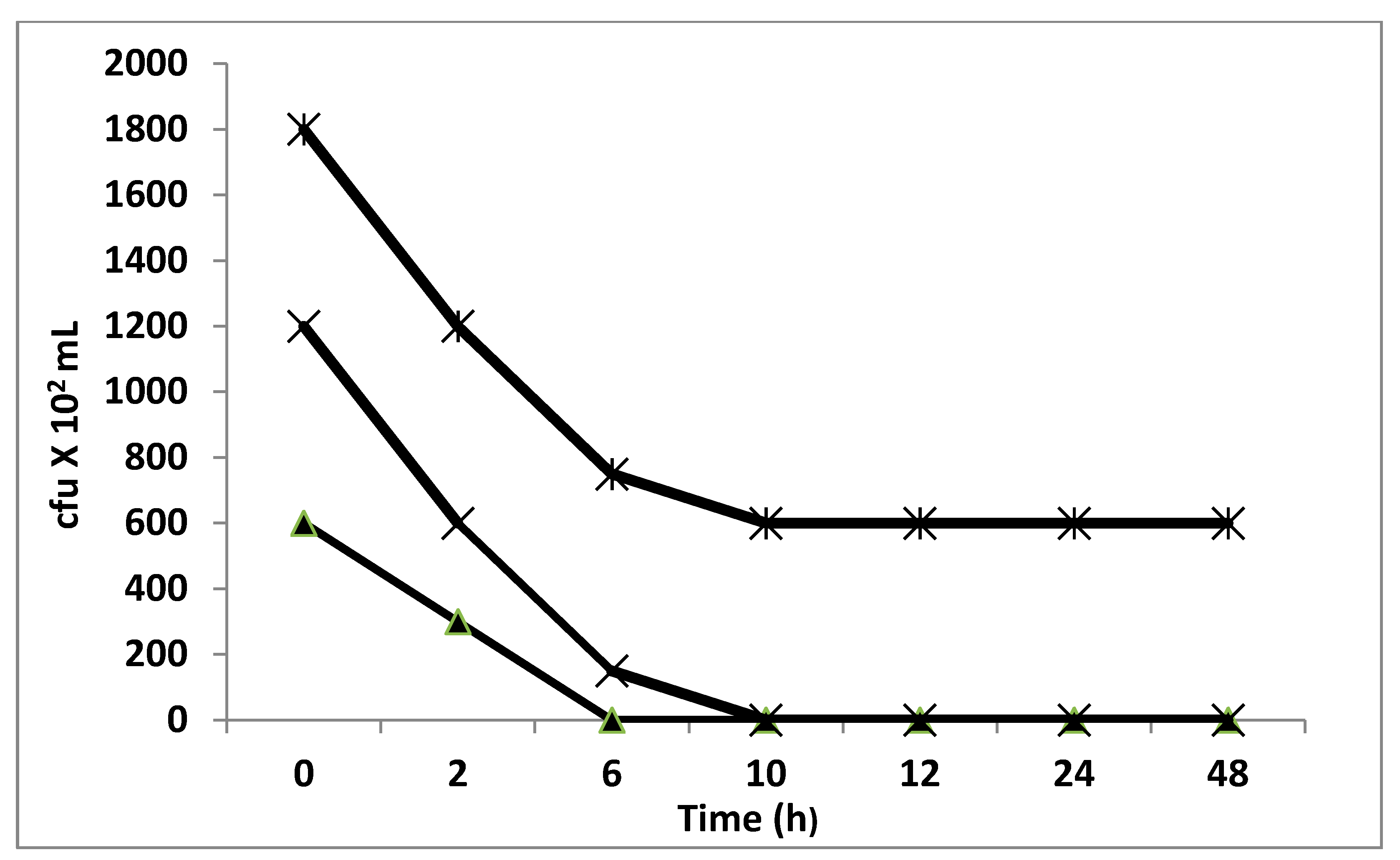
2.8.4. Time–Kill Studies

Time–kill studies were performed for Staphylococcus aureus in McCartney bottles using a method based on the European Standard quantitative suspension test (Selim, 2012) [21]. An initial inoculum of 5 × 10⁸ c.f.u. mL−1 was prepared as previously described for each isolate for use in time–kill studies by diluting an actively growing culture in mannitol salt broth with the inoculum used for each isolate verified by a total viable count. Samples (1 mL) of the initial inoculum were then added to 9 mL sterile water containing either tamer extract and Tween 80 (test) or Tween 80 only (control). The final concentrations of tamer extract and Tween 80 were 5 and 0.5%, respectively. The McCartney bottles for all isolates were shaken (100 rpm) at 37 °C, samples (1 mL) were taken in triplicate at 0, 1, 2, and 3 h, and serial tenfold dilutions were made and plated on mannitol salt agar (Oxoid, UK). The total viable count was determined after overnight incubation at 37 °C.

2.9. Antimutagenicity Assay (Ames Test)

Antimutagenicity of gallic acid was checked by using Ames assay as proposed by [22]. In this study, histidine requiring strains of Salmonella typhimurium, i.e., TA98 and TA100, were used and the experiments were carried out with (+S9 mix) and without metabolic activation (–S9 mix) system. Mutagen 4-nitro-o-phenylene diamine (NPD) and NaN3 were used for TA98 and TA100 in experiments as positive controls. Fresh minimal agar medium, top agar, and bacterial culture (density of 1–2x 10⁹ CFU/mL) was used in all the experiments. Dimethyl sulfoxide (DMSO) was used as a solvent for preparation of different concentrations of gallic acid (100–2500 µg/0.1 mL). Mutagens were used in nontoxic concentrations, i.e., NPD (20 µg/0.1 mL), 2-AF (2.5 µg/0.1 mL), and NaN3 (20 µg/0.1 mL). The spontaneous reversion frequency of TA98 and TA100 was also found for each experiment. Toxicity of gallic acid was checked against all the used mutagens. The experiments were conducted in co-incubation and pre-incubation modes. All the experiments were conducted in triplicate and percentage of inhibition of mutagenic activity was calculated as follows:
The inhibitory activity of gallic acid was expressed as:
Inhibitory activity (%) = [(a − b)/(a − c)]× 100
where ‘a’ is the number of histidine revertants induced by mutagen alone (positive control), ‘b’ is the number of histidine revertants induced by mutagen in presence of gallic acid, and ‘c’ is the number of histidine revertants induced in presence of gallic acid alone and solvent (negative control).

2.10. Statistical Analysis

Data were subjected to one-way analysis of variance for means of comparison, and significant differences were calculated according to Duncan’s multiple range test. Data were reported as means ± standard error of the means. Differences at p < 0.05 were considered statistically significant. SPSS (version 11.0) was used to perform the statistical analysis.

3. Results and Discussion

3.1. Determination of Total Phenolic and Flavonoid Contents of Date Pits

Date pits’ phenolic and flavonoid contents have received very little research attention [1,3,5]. Consequently, it is important to determine the levels of phenolics and flavonoids in pits of date palm Phoenix dactylifera L. methanolic extract. Phenolic acids are present in plants as a type of secondary metabolites that are found both free and bound. It was discovered that all plant extracts contained hydroxybenzoates, hydroxyl-cinnamates, and ferulic acid in the bound phenolic fraction. Phenolics are well-known for their antioxidant and anti-inflammatory properties [23,24,25,26,27].
Date pits’ phenolic and flavonoid extract was improved using methanol as a solvent for extraction (Table 1, Figure 1). The results showed that the total phenolic content was 24.84 mg GA/gFW, which was higher than the total flavonoid content, (only 5.324 mg QE/gDW). The common phenolic was identified using a phenolic profile (Shimadzu liquid chromatograph system (LC 10ADVP)). Gallic acid (11.85 mg mg/g) was the most abundant phenolic compound in the extract. Meanwhile, caffeic acid content was the lowest (at 0.05 mg/gDW). Flavonoids include flavonols and flavones. Flavonols were found in higher concentrations (0.198 to 2.986 mg quercetin equivalent/g DW) than flavones (0.198 to 1.52 mg quercetin equivalent/g DW).

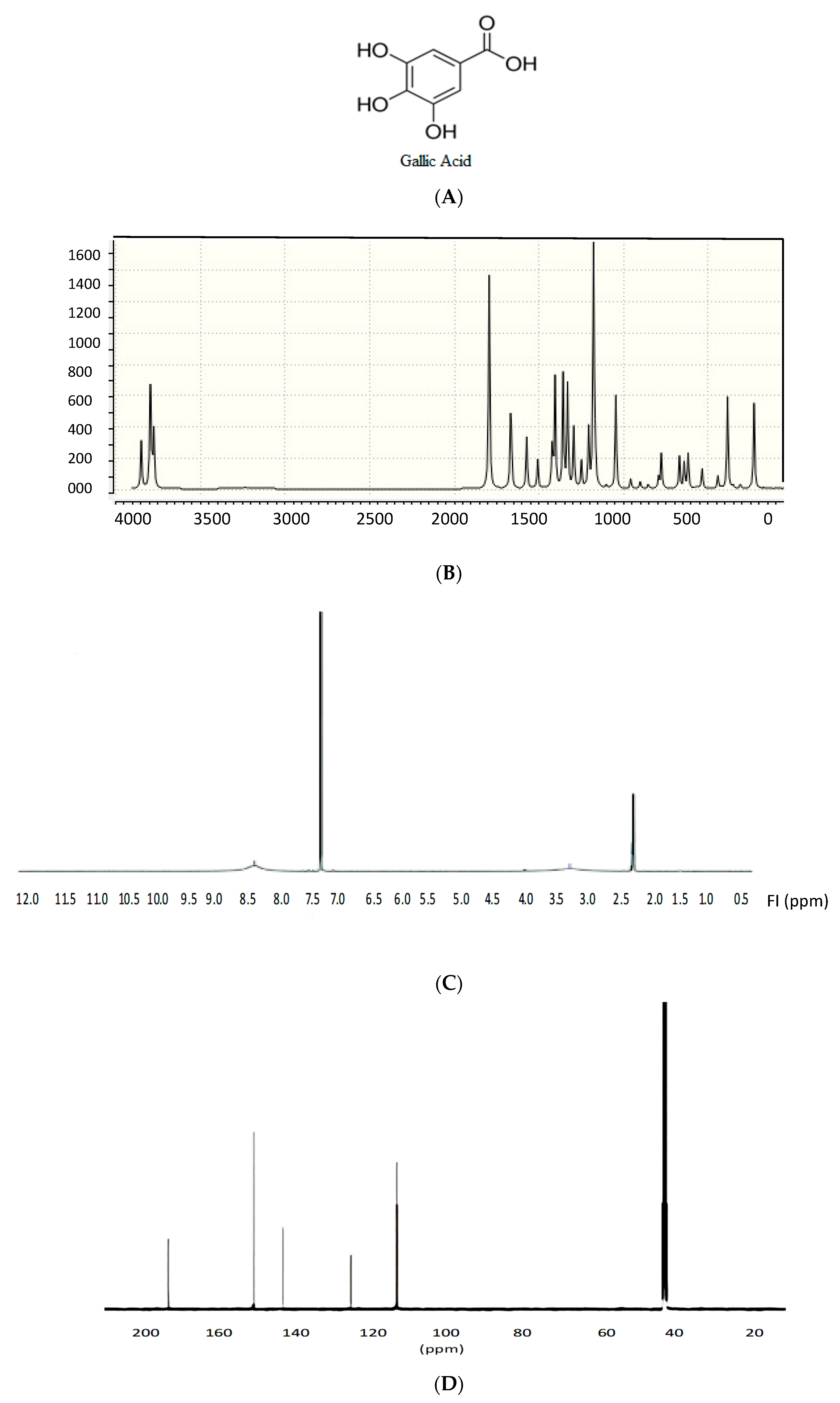
3.2. Extraction, Identification, and Elucidation of the Most Active Compound

Due to the abundance of gallic acid in the methanolic extract of date pits, chromatographic, and spectroscopic techniques were able to detect it. One-ringed phenolic acid gallic acid has a straightforward structure. Some green algae and several types of angiosperms contain it (Waterman and Mole, 1994) [15]. Previous reports have demonstrated the antioxidant, anti-inflammatory, and anticancer properties of gallic acid [17]. Moreover, the antibacterial and antifungal properties of gallic acid have been well demonstrated [13,16,28,29]. Gallic acid was extracted by using TLC. Ciocalteu’s spot on the TLC chromatogram made with hexane–ethyl acetate–acetic acid (2:1:0.3 v/v/v) showed a distinctive blue color when using Folin’s reagent, indicating that the active fraction contained a phenolic compound. A preliminary identification was made using the extract’s Rf value and retention time, which were compared to reference standards. Finally, spectral data from FTIR MS, and 1H and 13C NMR revealed the structure of the fraction and identified it as gallic acid (Figure 2). The spectroscopic characterization of gallic acid (3,4,5-Trihydroxybenzoic acid) is a white solid with melting point 260 c and its molar mass is 170.12 gm/mol, molecular formula C7H6O5. Theoretical FTIR analysis of gallic acid at umax/cm2 showed distinct peaks at 3600–3100 (combination between OH carboxylic and phenolic), 1712 (C=O carboxylic), 1600, 1430 (C=C aromatic) and 1134 (C–O) (Figure 2B). Chromatographic and spectroscopic studies revealed gallic acid to be the most active and potent phenolic compound in the active fraction. The compound in F1-3 was identified based on 1H and 13C NMR data analysis and with comparison with the previous literature values. Accordingly, 1H NMR (CDCl3) were δ = 1.8–2.1 (s, 3H phenolic protons), 6.9–7.2 (2, 2H, aromatic protons), and 9.8–10 (s, 2H, carboxlic protons) (Figure 2C). 13C NMR results: 75 MHz, DMSO-d6,δ, ppm) showed 5 distinct peaks for gallic acid at (110, 123, 138, 147 aromatic carbons of benzene ring) and (170 C=O carboxlic group) (Figure 2D).

3.3. Antimicrobial Activity of Date Pits

There has been an enormous improvement to health-related quality since the discovery of antibiotics as weapons against bacterial illnesses. Drug-resistant bacteria and side effects from commonly used antibiotics have made them less effective, putting the benefits to human health in jeopardy [30]. For this reason, it was critical to look into less resistant bioactive molecules. Antibacterial compounds with novel mechanisms of action may be found in higher plant natural products [31]. Several studies have linked the antibacterial activity of plant extracts to their phenolic compounds [32,33]. Phenolics’ antimicrobial effect may be explained by the inhibition of bacterial growth, while these compounds could adsorb to cell membranes, by interactions with enzymes and effectors, or by deprivation of substrates and metal ions by these substances [34]. The structural diversity of date pit extract’s phenolics therefore might influence their antimicrobial potential, as we can assume. Phenolic acids were tested against four pathogenic bacterial and fungal strains for their activity and mechanism of action.
Reliable and repeatable results obtained using the traditional disk-diffusion technique showed a significant antibacterial activity for gallic acid isolated from date pits (Table 2). As a result, it can be concluded that microorganisms have variable intrinsic tolerances, and the type and mixture of phenolic chemicals found in date pits are responsible for their susceptibility to gallic acid treatment. As an antibiotic, gallic acid is most effective against Staphylococcus aureus and the least effective against Candida albicans. Meanwhile, it is completely inactive against the other bacteria. According to our findings, gallic acid found in date pits had a more inhibitory effect on Gram-positive bacteria than Gram-negative bacteria. A possible antibacterial effect on the viability of Staphylococcus aureus strains was found in experiments using gallic acid at concentrations of 100 and 200 µg/mL. S. aureus cell viability was completely inhibited at 200 µg/mL of acid for 6 h and at 100 µg/mL of acid for 12 h (Figure 3). Antimicrobial activity of phenolic compounds is mediated by various mechanisms, including cytoplasmic membrane instability and permeabilization and enzyme inhibition by oxidized products. Such suppression might be caused by a reaction with sulfhydryl groups or nonspecific interactions with proteins, such as the generation of reactive quinones that react with amino acids and proteins ([35]. In addition, phenols can stop bacteria, both Gram-negative and Gram-positive, from production of nucleic acids [36].
As a result, the extracts had antibacterial and bactericidal properties, with MIC and MBC values that ranged from 250 to 500 µg/mL, with the MIC and MBC values being equal (Table 2). As bacterial biochemical markers, MIC, MBC, and suppression of cell viability have all been studied. S. aureus was found to be more vulnerable to gallic acid with a MIC of 250 µg/mL. This strain and also Streptococcus sp. had higher sensitivity to gallic acid. Gram-positive bacteria were more sensitive to phenolic acids than Gram-negative bacteria. To inhibit Streptococcus mutans and Streptococcus sobrinus, various phenolic compounds (tea flavonoids) have been used in studies ranging from 500 µg/mL to 1000 µg/mL. According to study [37], MIC values for hydroxybenzoic, hydroxycinnamic, and methoxy-benzoic acids were also reported by Sanchez-Maldonado et al. [38] for three Gram-positive bacteria (Lactobacillus cereus, Lactobacillus paracasei, and Lactobacillus paracasei).
Phenolic acids have been shown to affect bacterial cell surface physicochemical properties, with gallic acid, in particular, changing the bacteria’s hydrophobicity. Phenolic acids have the potential to alter bacterial cells’ polar, apolar, and electron acceptor components [30]. Treatment with gallic acid increased the ability of Gram-positive bacteria to accept electrons while decreasing it in Gram-negative bacteria, indicating that gallic acid is an electrophilic product that interacts with bacterial surface components. Gallic acid is phenolic acids’ antibacterial activity and was also found to be correlated with the amount of dissociated acid present. Phenoxemic acid is thought to permeate the cell membrane passively due to its lipophilic nature. This disrupts the cell membrane’s structure and may acidify the cytoplasm, leading to denaturation of proteins [39]. Date pit extract may be useful in fighting staphylococcal infection if used in this way. Date pit extract, which has a strong inhibitory effect on S. aureus cell viability, is the source of inspiration for this theory.

3.4. Antimutagenicity of Isolated Gallic Acid from Date Pits

Based on its potential antibacterial activity, purified gallic acid was tested for antimutagenic action with the Ames test against the indirect mutagen cyclophosphamide. Second-stage inhibitors, such as bioantimutagens, are thought to keep mutagens from attacking DNA [40]. Using the co-incubation method, the bioantimutagenic properties of phytochemicals were investigated [41]. Two S. typhimurium strains, TA98 and TA100, were used to determine the antimutagenic efficacy of gallic acid against 4-NPD and NaN3 (Table 3). Under all applied concentrations of gallic acid, the Ames test showed that it had a significant antimutagenic activity (p ≤ 0.01). When used at a concentration of 3000 µg/plate of S. typhimurium TA98, gallic acid was found to have a better antimutagenic efficacy. The mutagenicity of TA98 decreased from 52 to 39, and that of TA100 decreased from 39 to 15 for the two bacterial strains examined. The antimutagens that prevent mutation include desmutagens and bioantimutagens. According to Ames et al. [42], a chemical is classified as a mutagen if it can produce at least two times as many revertants as spontaneous revertants. Gallic acid’s potent radical scavenger properties may have contributed to the antimutagenic activity observed in the Ames test, according to recent findings. Negi et al. [43] stated that a substance has a minimal antimutagenic activity if its inhibition percentage is less than 25%, moderate activity if the inhibition percentage is between 25% and 40%, and a strong antimutagenicity if the inhibition percentage is greater than 40%. This shows gallic acid has significant antimutagenic activity when used at the highest doses tested (3000 µg/plate), which reduces indirect acting mutagen cyclophosphamide-induced mutations in two test strains by 45% and 36%, respectively (when S9 is present). In this assay, it was discovered that cyclophosphamide-resistant gallic acid extract from date pits had a strong antimutagenicity.

4. Conclusion

Phenolic components were abundant in the methanolic extract of date pits. In particular, gallic acid has been isolated and identified from date pits’ extract to test its bioactivity. Gallic acid has shown antibacterial activity against microbial infections, in addition to its strong antimutagenic activity. This study could support the use of all parts of date palm, particularly the pits, in order to enhance their food functional and health-promoting values. The pits have proven to be a rich source of gallic acid, which could strengthen their applications in nutraceutical and pharmaceutical industries. Our study also has demonstrated that the antibacterial and antimutagenic properties, as well as phenolics, can be used to find new antimicrobial compounds.

Author Contributions

Conceptualization, S.S., S.K.A.J.; methodology, S.S., T.A.-s., M.A.-M., M.S.A., M.W., M.H.A., H.A., N.A., H.S.M. and M.H.A.; validation, S.S., T.A.-s., M.A.-M., M.S.A., M.W., M.H.A., H.A., N.A., H.S.M. and M.H.A.; formal analysis, S.S., S.K.A.J., M.W., H.S.M. and H.A.; investigation, S.S., T.A.-s., M.A.-M., M.S.A., M.W., M.H.A., H.A., N.A., and M.H.A.; data curation, S.S., T.A.-s., M.A.-M., M.S.A., M.W., M.H.A., H.A., N.A., H.S.M. and M.H.A.; writing—original draft preparation, S.S., T.A.-s., M.A.-M., M.S.A., M.W., M.H.A., H.A., N.A., and M.H.A.; writing—review and editing, S.S., S.K.A.J., M.W., H.S.M. and H.A.; visualization, S.S., S.K.A.J., M.W., and H.A.; supervision, S.S., M.W. and S.K.A.J.; project administration, S.S. All authors have read and agreed to the published version of the manuscript.

Funding

This work was funded by the Deanship of Scientific Research at Jouf University under grant number DSR-2021-01-0349.

Data Availability Statement

Not applicable.

Conflicts of Interest

The authors declare that there is no conflict of interests regarding the publication of this paper.

References

  1. Al-Farsi, M.; Lee, C.Y. Optimization of phenolics and dietary fiber extraction from date seeds. Food Chem. 2008, 108, 977–985. [Google Scholar] [CrossRef] [PubMed]
  2. Al-Shahib, W.; Marshal, R.J. The fruit of the date palm: It’s possible use as the best food for the future. Food Sci. Nutr. 2003, 54, 247–259. [Google Scholar] [CrossRef] [PubMed]
  3. Besbes, S.; Blecker, C.; Deroanne, C.; Drira, N.; Attia, H. Date seeds Chemical composition and characteristic profiles of the lipid fraction. Food Chem. 2004, 84, 577–584. [Google Scholar] [CrossRef]
  4. Hussein, A.S.; Alhadrami, G.A.; Khalil, Y.H. The use of dates and date pits in broiler starter and finisher diets. Biores. Technol. 1998, 66, 219–223. [Google Scholar] [CrossRef]
  5. Hossain, M.Z.; Waly, M.I.; Singh, V.; Sequeira, V.; Rahman, M.S. Chemical Composition of Date-Pits and Its Potential for Developing Value-Added Product—A Review. Polish J. Food Nutrit. Sci. 2014, 4, 215–226. [Google Scholar] [CrossRef]
  6. Al–Farsi, M.; Alasalvar, C.; Al–Abid, M.; Al–Shoaily, K.; Al–Amry, M.; Al–Rawahy, F. Compositional and functional characteristics of dates, syrups and their by–products. Food Chem. 2007, 104, 943–947. [Google Scholar] [CrossRef]
  7. Waly, M.I.; Al–Ghafri, B.R.; Guizani, N.; Rahman, M.S. Phytonutrients effect of date pit extract against azoxymethane—Induced oxidative stress in rat colon. Asian Pacific J. Cancer Prev. 2015, 16, 3473–3477. [Google Scholar] [CrossRef] [PubMed] [Green Version]
  8. Al-Qarawi, A.A.; Mousa, H.M.; Ali, B.E.H.; Abdel-Rahman, H.; El-Mougy, A.A. Protective effect of extracts from dates (Phoenix dactylifera L.) on carbon tetrachloride—Induced hepatotoxicity in rats. Int. J. Appl. Res. Vet. Med. 2004, 2, 176–180. [Google Scholar]
  9. Mossa, J.S.; Hifnawy, M.S.; Mekkawi, A.G. Phytochemical and biological investigations on date seeds (Phoenix dactylifera L.) produced in Saudi Arabia. Arab. Gulf J. Sci Res. 1986, 4, 495–507. [Google Scholar]
  10. Saddiq, A.A.; Bawazir, A.E. Antimicrobial activity of date palm (Phoenix dactylifera) pits extracts and its role in reducing the side effect of methyl prednisolone on some neurotransmitter content in the brain. In Proceedings of the IV International Date Palm Conference, Abu Dhabi, UAE, 15 March 2010. [Google Scholar]
  11. Rajan, V.K.; Muraleedharan, K. A computational investigation on the structure, global parameters and antioxidant capacity of a polyphenol, Gallic acid. Food Chem. 2017, 220, 93–99. [Google Scholar] [CrossRef]
  12. Bai, J.; Zhang, Y.; Tang, C.; Hou, Y.; Ai, X.; Chen, X.; Zhang, Y.; Wang, X.; Meng, X. Gallic acid: Pharmacological activities and molecular mechanisms involved in inflammation-related diseases. Biomed. Pharm. 2021, 133, 110985. [Google Scholar] [CrossRef]
  13. Chanwitheesuk, A.; Teerawutgulrag, A.; Kilburn, J.D.; Rakariyatham, N. Antimicrobial gallic acid from Caesalpinia mimosoides Lamk. Food Chem. 2007, 100, 1044–1048. [Google Scholar] [CrossRef]
  14. Alu’datt, M.H.; Rababah, T.; Alhamad, M.N.; Al-Mahasneh, M.A.; Almajwal, A.; Gammoh, S.; Ereifej, K.; Johargy, A.; Alli, I. A review of phenolic compounds in oil-bearing plants: Distribution, identification and occurrence of phenolic compounds. Food Chem. 2017, 218, 99–106. [Google Scholar] [CrossRef] [PubMed]
  15. Waterman, P.G.; Mole, S. Method in Ecology: Analysis of Phenolic Plant Metabolites; Blackwell Scientific Publishers: London, UK, 1994. [Google Scholar]
  16. Panizzi, L.; Caponi, C.; Catalano, S.; Cioni, P.L.; Morelli, I. In vitro antimicrobial activity of extracts and isolated constituents of Rubus ulmifolius. J. Ethnopharmacol. 2002, 79, 165–168. [Google Scholar] [CrossRef]
  17. Kroes, B.H.; van den Berg, A.J.; Quarles van Ufford, H.C.; van Dijk, H.; Labadie, R.P. Anti-inflammatory activity of gallic acid. Planta Med. 1992, 58, 499–504. [Google Scholar] [CrossRef] [PubMed]
  18. Chang, C.; Yang, M.; Wen, H. Estimation of total flavonoid content in propolis by two complementary colorimetric methods. J. Food Drug Anal. 2002, 10, 178–182. [Google Scholar]
  19. Velioglu, Y.S.; Mazza, G.; Gao, L.; Oomah, B.D. Antioxidant activity and total phenolics in selected fruits, vegetables and grain products. J. Agric. Food Chem. 1998, 46, 4113–4117. [Google Scholar] [CrossRef]
  20. Okla, M.K.; Abdel-Mawgoud, M.; Alamri, S.A.; Abbas, Z.K.; Al-Qahtani, W.H.; Al-Qahtani, S.M.; Al-Harbi, N.A.; Hassan, A.H.; Selim, S.; Alruhaili, M.H.; et al. Developmental Stages-Specific Response of Anise Plants to Laser-Induced Growth, Nutrients Accumulation, and Essential Oil Metabolism. Plants 2021, 10, 2591. [Google Scholar] [CrossRef]
  21. Selim, S.A.; El Alfy, S.; Al-Ruwaili, M.; Abdo, A.; Al Jaouni, S. Susceptibility of imipenem-resistant Pseudomonas aeruginosa to flavonoid glycosides of date palm (Phoenix dactylifera L.) tamar Growing in Al Madinah, Saudi Arabia. Afr. J. Biotech. 2012, 11, 416–422. [Google Scholar] [CrossRef]
  22. Maron, D.M.; Ames, B.N. Revised method for Salmonella mutagenicity test. Mutation Res. 1983, 113, 173–215. [Google Scholar] [CrossRef]
  23. Chen, H.J.; Ho, C.T. Antioxidant activities of caffeic acid and its related hydroxycinnamic acid compounds. J. Agri. Food Chem. 1997, 45, 2374–2378. [Google Scholar] [CrossRef]
  24. Cai, Y.Z.; Sun, M.; Xing, J.; Luo, Q.; Corke, H. Structure-radical scavenging activity relationship of phenolic compounds from traditional Chinese medicinal plants. Life Sci. 2006, 78, 2872–2888. [Google Scholar] [CrossRef] [PubMed]
  25. Chen, H.M.; Wu, Y.C.; Chia, Y.C.; Chang, F.R.; Hsu, H.K.; Hsieh, Y.C.; Chen, C.C.; Yuan, S.S. Gallic acid, a major component of Toona sinensis leaf extracts, contains a ROS mediated anticancer activity in human prostate cancer cells. Cancer Lett. 2009, 286, 161–171. [Google Scholar] [CrossRef]
  26. Steenkamp, V.; Nkwane, O.; van Tonder, J.; Dinsmore, A.; Gulumian, M. Evaluation of the phenolic and flavonoid contents and radical scavenging activity of three southern African medicinal plants. Afr. J. Pharm. Pharmacol. 2013, 7, 703–709. [Google Scholar]
  27. Tunon, M.J.; Garcia-Mediavilla, M.V.; Sanchez-Campos, S.; Gonzales-Gallego, J. Potential of flavonoids as anti-inflammatory: Modulation of proinflammatory gene expression and signal transduction pathways. Curr. Drug Metab. 2009, 10, 256–271. [Google Scholar] [CrossRef] [PubMed]
  28. Akiyama, H.; Fujii, K.; Yamasaki, O.; Oono, T.; Iwatsuki, K. Antibacterial action of several tannins against Staphylococcus aureus. J. Antimicrob. Chemother. 2001, 4, 487–491. [Google Scholar] [CrossRef] [PubMed] [Green Version]
  29. Penna, C.; Marino, S.; Vivot, E.; Cruañes, M.C.; de D Muñoz, J.; Cruañes, J.; Ferraro, G.; Gutkind, G.; Martino, V. Antimicrobial activity of Argentine plants used in the treatment of infectious diseases: Isolation of active compounds from Sebastiania brasiliensis. J. Ethnopharm. 2001, 77, 37–40. [Google Scholar] [CrossRef]
  30. Bhalodia Nayan, R.; Shukla, V.J. Antibacterial and antifungal activities from leaf extracts of Cassia Fistula L.: An Ethnomedicinal plant. J. Adv. Pharm. Technol. Res. 2011, 2, 104–109. [Google Scholar] [CrossRef] [PubMed]
  31. Cowan, M.M. Plant Products as Antimicrobial Agents. Clin. Microbiology Rev. 1999, 12, 564–582. [Google Scholar] [CrossRef] [Green Version]
  32. Amensour, M.; Sendra, E.; Abrini, J.; Bouhdid, S.; Pérez-Alvarez, J.A.; Fernández-López, J. Total phenolic content and antioxidant activity of myrtle (Myrtus communis) extracts. Nat. Prod. Commun. 2009, 4, 819–824. [Google Scholar] [CrossRef] [PubMed] [Green Version]
  33. Ahmed, D.; Khan, M.M.; Saeed, R. Comparative Analysis of Phenolics, Flavonoids, and Antioxidant and Antibacterial Potential of Methanolic, Hexanic and Aqueous Extracts from Adiantum caudatum Leaves. Antioxidants 2015, 4, 394–409. [Google Scholar] [CrossRef]
  34. Fukumoto, L.R.; Maza, G. Assessing antioxidant and prooxidant activities of phenolic compounds. J. Agric. Food Chem. 2000, 48, 3597–3604. [Google Scholar] [CrossRef] [PubMed]
  35. Puupponen-Pimia, R.; Nohynek, L.; Alakomi, H.L.; Oksman-Caldentey, K.M. Bioactive berry compounds novel tools against human pathogens. Appl. Microbiol. Biotechnol. 2005, 67, 8–18. [Google Scholar] [CrossRef] [PubMed]
  36. Cushnie, T.P.T.; Lamb, A.J. Antimicrobial activity of flavonoids. Int. J. Antimicrob. Agents 2005, 26, 343–356. [Google Scholar] [CrossRef] [PubMed]
  37. Wang, H.; Provan, G.J.; Helliwell, K. Tea flavonoids: Their functions, utilization and analysis. Trends Food Sci. Technol. 2000, 11, 152–160. [Google Scholar] [CrossRef]
  38. Sanchez-Maldonado, A.F.; Schieber, A.; Ganzle, M.G. Structure-function relationships of the antibacterial activity of phenolic acids and their metabolism by lactic acid bacteria. J. Appl. Microbiol. 2011, 111, 1176–1184. [Google Scholar] [CrossRef]
  39. Campos, F.M.; Couto, J.A.; Figueiredo, A.R.; Tóth, I.V.; Rangel, A.O.S.S.; Hogg, T.A. Cell membrane damage induced by phenolic acids on wine lactic acid bacteria. Int. J. Food Microbiol. 2009, 135, 144–151. [Google Scholar] [CrossRef] [PubMed]
  40. Ramel, C.; Alekperov, B.N.; Ames, T.; Kada, L.W. Wattenberg, Inhibitors of mutagenesis and their relevance to carcinogenesis. Mutat. Res. 1986, 168, 47–65. [Google Scholar] [CrossRef]
  41. Hung, Y.H.; Wang, Y.J.; Chou, C.C. Antimutagenic activity of Aspergillus awamori-fermented black soyabean response to stimulated digestive juice treatments and its antimutagenic mechanisms. LWT-Food Sci. Technol. 2009, 42, 56–62. [Google Scholar] [CrossRef]
  42. Ames, B.N.; McCann, J.; Yamasaki, E. Methods for detecting carcinogens and mutagens with the Salmonella/mammalian microsome mutagenicity test. Mutat. Res. 1975, 31, 347–364. [Google Scholar] [CrossRef]
  43. Negi, P.S.; Jayaprakasha, G.K.; Jena, B.S. Antioxidant and antimutagenic activities of pomegranate peel extracts. Food Chem. 2003, 80, 393–397. [Google Scholar] [CrossRef]
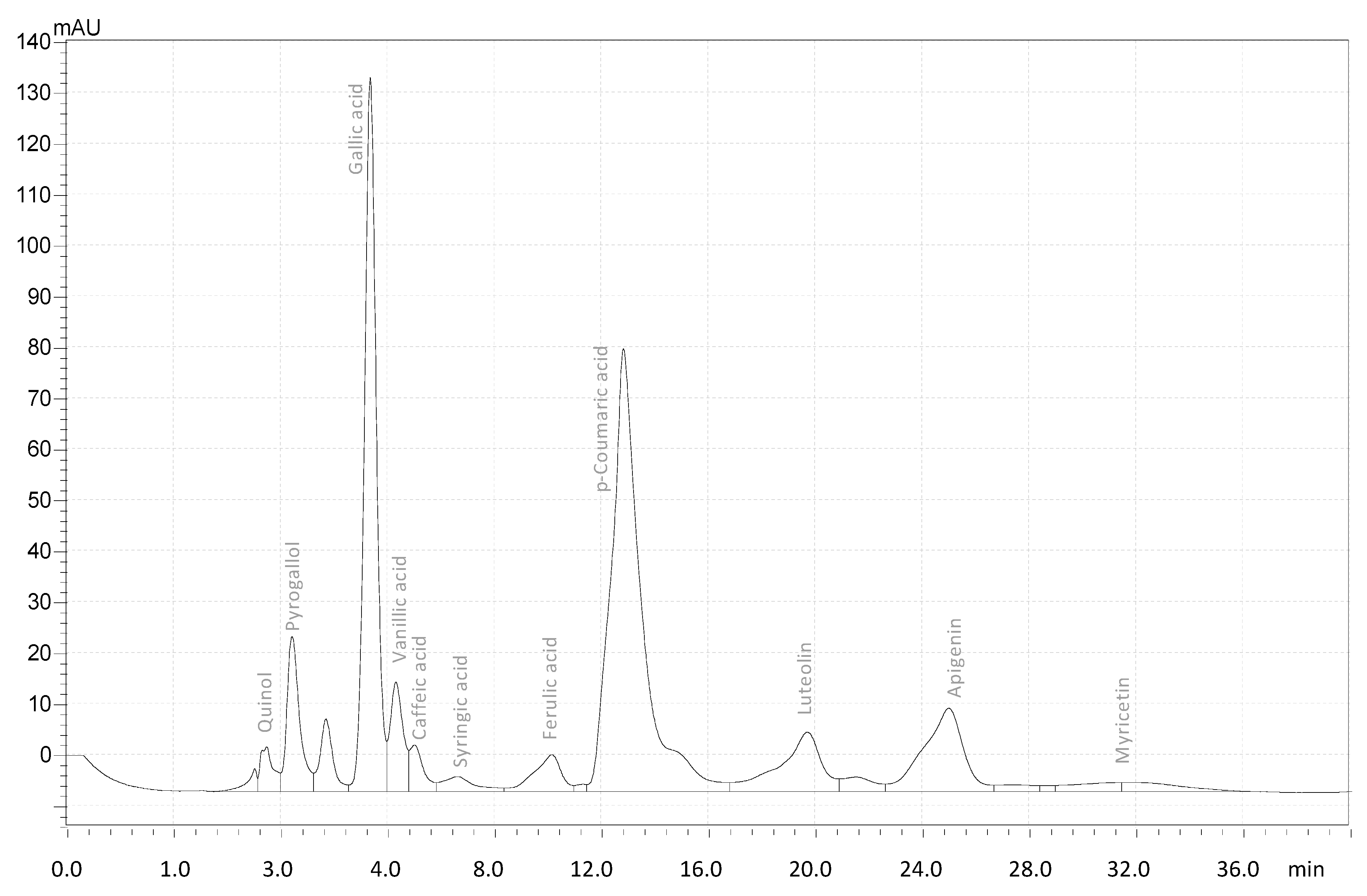
Figure 1. HPLC chromatogram of phenolic and flavonoid compounds in Ajwa date pits’ methanolic extract.
Agronomy 12 00054 g001
Figure 2. (A) Structure; (B) FTIR analysis; (C) 1HNMR; (D) 13C NMR, analysis of gallic acid.
Agronomy 12 00054 g002
Figure 3. The viability of Staphylococcus aureus in the presence of varying concentrations of gallic acid extracted from Ajwa date pits (0, 100, and 200 µg/mL). The average of three replicates (mean ± standard deviation) is used. Student’s t-test was used to assess statistically significant differences between samples, and p ≤ 0.05 was considered significant.
Agronomy 12 00054 g003
Table 1. HPLC analysis of phenolic and flavonoid compounds in Ajwa date pits’ methanolic extract (mg/100 g DW).
CompoundsConcentration
Hydroxybenzoic acids
Syringic acid0.62 ± 0.015
Gallic acid
Pyrogallol
Quinol
11.85 ± 0.25
1.870 ± 0.12
0.354 ± 0.04
Hydroxycinnamic acids
Ferulic acid3.6 ± 0.78
p-Coumaric acid5.58 ± 1.68
Vanillic acid0.92 ± 0.75
Caffeic acid0.05 ± 0.0023
Total Phenolic a24.84 ± 0.19
Flavones
Apigenin0.198 ± 0.90
Luteolin1.52 ± 0.13
Flavonols
Myricetin0.65 ± 0.007
Quercetin2.986 ± 0.135
Total Flavonoid b5.324 ± 0.069
a mg gallic acid equivalent (GAE)/g DW. b mg quercetin equivalent/g DW. Values are means of three biological replicates.
Table 2. Antimicrobial activity of gallic acid (100 μg/disc) extracted from Ajwa date pits.
MicroorganismsIZD aMICMBC
Streptococcus sp13500500
Staphylococcus aureus22250250
Serratia marcescens
Escherichia coli11500500
Klebsiella pneumoniae11500500
Proteus vulgaris
Pseudomonas aeruginosa
Candida albicans10500500
a Inhibition zone in diameter (mm) around the discs impregnated with 10 μg of gallic acid. (–) No antibacterial activity; MIC (minimum inhibitory concentration), and MBC (minimum bactericidal concentration). The average of three replicates is used.
Table 3. Antimutagenic potential of gallic acid extracted from Ajwa date pits against S. typhimurium TA98 and TA100 bacterial strains.
Compound Concentration (μg/mL)Inhibition % of Revertants
TA98TA100
Gallic acid30005239
17004327
7003821
5003215
4-NPD (3 μg/mL) and NaN3 (8 μg/mL) were used as positive controls for S. typhimurium TA98 and TA100 strains, respectively. Data shown are mean ± SD of experiments with triplicate plates/concentration/experiment. 4-NPD and NaN3did not show any inhibition of revertants.
Publisher’s Note: MDPI stays neutral with regard to jurisdictional claims in published maps and institutional affiliations.

