Article

Phylogenetic Analyses of Rhizobia Isolated from Nodules of Lupinus angustifolius in Northern Tunisia Reveal Devosia sp. as a New Microsymbiont of Lupin Species

1
Laboratory of Biodiversity and Valorization of Arid Areas Bioresources, BVBAA, Faculty of Sciences, University of Gabès, Erriadh 6072, Tunisia
2
Institute of Agricultural Sciences, ICA-CSIC, 28006 Madrid, Spain
3
Centre for Plant Biotechnology and Genomics, CBGP, UPM-INIA, Pozuelo de Alarcón, 28223 Madrid, Spain
*
Author to whom correspondence should be addressed.
Agronomy 2021, 11(8), 1510; https://doi.org/10.3390/agronomy11081510
Submission received: 16 June 2021 / Revised: 19 July 2021 / Accepted: 26 July 2021 / Published: 29 July 2021
(This article belongs to the Special Issue Analysis of the Genetic Diversity of Crops and Associated Microbiota)

Abstract

:
Thirty-two bacterial isolates were obtained from root nodules of Lupinus angustifolius growing in Northern Tunisia. Phylogenetic analyses based on recA and gyrB partial gene sequences grouped the strains into six clusters: four clusters belonged to the genus Bradyrhizobium (22 isolates), one to Microvirga (8 isolates) and one to Devosia (2 isolates), a genus that has not been previously reported to nodulate lupin. Representative strains of each group were further characterized. Multi-Locus Sequence Analysis (MLSA) based on recA and glnII gene sequences separated the strains within the genus Bradyrhizobium into four divergent clusters related to B. canariense, B. liaoningense, B. lupini, and B. algeriense, respectively. The latter might constitute a new Bradyrhizobium species. The strains in the Microvirga cluster showed high identity with M. tunisiensis. The Devosia isolates might also represent a new species within this genus. An additional phylogenetic analysis based on the symbiotic gene nodC affiliated the strains to symbiovars genistearum, mediterranense, and to a possibly new symbiovar. These results altogether contributed to the existing knowledge on the genetic diversity of lupin-nodulating microsymbionts and revealed a likely new, fast-growing, salt-tolerant rhizobial species within the genus Devosia as a potentially useful inoculant in agricultural practices or landscape restoration.

1. Introduction

The Leguminosae or Fabaceae family includes more than 19,700 species, many of which are agronomically important [1]. The Genisteae tribe is presently considered to include 618 species in 25 genera [2]. Lupinus is considered the largest Genisteae genus in terms of species number with around 270 species [3,4]. This genus may be divided into two groups, the first of which is centered in the Mediterranean Basin and the Canary Islands [5,6] and comprises 13 Old World species with smooth and rough seeds [3,7]. The second group includes the New World species [8]. The New World species number ranges from 200 to 400 located in the Americas. Lupins are able to colonize very different environments and diverse ecosystems [9], including marginal impoverished or contaminated soils [10]. L. angustifolius, commonly named narrow leaf lupin and blue lupin, grows wild in agricultural lands as well as in the wetlands of coastal plains in the Northern region of Tunisia.
Lupins, partly due to their effective nitrogen-fixing symbiosis with different soil bacteria (collectively called rhizobia), have been grown since antiquity as an important pulse crop and as a green manure. Lupinus species have been studied from various perspectives as indicators of phosphorus and nitrogen in soils poor in nutrients, because of their dual efficacy in fixing atmospheric nitrogen and solubilizing soil phosphates [11], phytoremediation of polluted soils [12,13] and as medicinal plants [14].The high protein content of lupin seeds is similar to that of soybean, and lupin cultivation has been suggested as an alternative to soybean in Europe [15], as new sources are needed to meet the increasing demand for plant protein [16].
Rhizobia belong to the Alpha and Betaproteobacteria [17]. Legume-rhizobium symbioses provide a net input of nitrogen into the ecosystems. The great diversity of lupin species reflects a vast diversity of their nodulating rhizobia. Lupinus species are mainly nodulated by slow-growing bacteria that belong to the genus Bradyrhizobium. The major species are B. canariense, B. japonicum [18,19,20,21], B. elkanii [22], and B. valentinum [23]. Other fast-growing rhizobia species, such as Ochrobactrum lupini [24], Mesorhizobium loti [25], Phylobacterium trifolii [26] and Microvirga ssp. [27,28,29] can also effectively nodulate different Lupinus spp. Recently, several microsymbionts isolated from L. albus growing in Tunisian soils were identified as species within the genera Agrobacterium, Rhizobium and Neorhizobium [30].
The genus Devosia was defined is a result of the reclassification of Pseudomonas riboflavin as Devosia riboflavina [31]. Devosia bacteria are aerobic, Gram-negative, rod-shaped or oval and they do not form spores. Twenty-one validated species have been identified within the genus (http://www.bacterio.net/devosia.html, accessed on 16 July 2020). Some Devosia species have been reported to nodulate legumes [32,33,34]. We have previously reported that L. angustifolius from Tunisia was nodulated by new lineages within the Bradyrhizobium and Microvirga genera [35], but, to the best of our knowledge, there are no reports on Devosia symbiotic nitrogen-fixing bacteria that nodulate Lupinus species.
The main objective of the present paper was to further investigate and characterize the diversity of the rhizobia that nodulate L. angustifolius growing in different sites in Northern Tunisia.

2. Materials and Methods

2.1. Sampling Sites, Isolation of Bacterial Strains and Culture Conditions

Effective nodules were collected from L. angustifolius roots growing wild or in soil collected from six different locations in Northern Tunisia: Mraissa, Takelsa, Borj Hfaied, Hammamet, Sejnane and Tabarka. The ecological and climatic characterization of the sampling sites is indicated in Table 1.
Nodules were surface-sterilized with 95% ethanol for 1 min, 25% sodium hypochlorite for 3 min, then rinsed several with sterile distilled water; they were individually crushed on sterile plates, and a loopful of nodule material was streaked onto yeast-mannitol agar (YMA) [36]. Plates were then incubated aerobically at 28 °C and colonies could be visualized after 4 to 10 days.
Single colonies were streaked on fresh YMA plates. Pure isolates were maintained on YMA plates at 4 °C for temporary preservation, or stored in 20% (w/v) glycerol suspensions at −80 °C. The isolates used in this study are listed in Table 1.

2.2. Nodulation and Cross-Inoculation Test

The ability of all purified isolates to nodulate their original host was assessed. The isolates were also tested for nodulation on L. luteus, L. micranthus, L. mariae-josephae, Retama sphaerocarpa, Vigna unguiculata and Glycine max. Seed germination and plant inoculation were carried out as previously described [37]. Plants were grown under bacteriologically controlled conditions in a greenhouse for 3 to 8 weeks, depending on the legume host, with a 16.0/8.0 h light/dark photoperiod at 25/23 °C, and they were watered with sterile Jensen’s liquid medium once a week. Three uninoculated Leonard jars per legume were included as negative controls. Nodules were examined for number and inside color. Three replicates of each strain were used in the nodulation tests.

2.3. DNA Isolation, PCR Amplifications and Sequencing

Bacterial DNA was obtained by the alkaline lysis method [38]. PCR was performed in a 25 µL volume containing DNA (5–10 ng), 2.5 µL of 10× PCR buffer containing magnesium chloride (Roche Applied Science), 10 µM of each primer, 10 mM of each dNTP, 1 µL DMSO and 1 U of Taq DNA polymerase (Roche Applied Science, Mannheim, Germany).
PCR amplicons of genes coding for DNA recombination and repair protein RecA (recA), DNA gyrase subunit B (gyrB), 16S rRNA (rrs), chaperone protein DnaK (dnaK), glutamine synthetase 2 (glnII), and N-acetylglucosaminyl transferase (nodC) were obtained using previously described primers and conditions [37,39]. PCR amplifications of recA, 16S rRNA, glnII and nodC genes were performed and amplicons were sequenced as previously reported [37]. nifH was amplified by using the appropriate primers with PCR conditions previously described [34].
glnII appeared to be absent in the genomes of Microvirga spp., as it could not be retrieved from data banks or from any sequenced Microvirga genomes. Hence, dnaK was used, and the phylogenetic tree for this group of strains was assembled using gyrB and dnaK concatenated sequences.
Amplification products were purified from the unincorporated primers and (deoxynucleosidetriphosphates (dNTPs) with the NucleoSpin® Extract II (Macherey-Nagel, Dueren, Germany) or by gel electrophoresis followed by band purification with the same kit, if necessary. Sequencing was performed externally at Secugen (Madrid, Spain) and STAB Vida (Lisbon, Portugal).

2.4. Phenotypic Characterization

Cell morphology and size after growth on YMA agar plates at 28 °C for 10 days were visualized. The growth temperature range (20–40 °C) was tested on YMA plates at pH 7.0 for 10 days. pH tolerance was determined over the pH range 4.0–12.0 at one unit intervals. The resistance to various antibiotics (ampicillin: 100 µg/mL, gentamicin: 30 µg/mL, tetracycline: 5 µg/mL, spectinomycin: 50 µg/mL, kanamycin: 50 µg/mL, streptomycin: 10 µg/mL and chloramphenicol: 10µg/mL), as well as salt tolerance (0–5%, w/v), were assessed on YMA plates at pH = 7.0 after 10 days at 28 °C. All phenotypic analyses were performed in triplicate.

2.5. Phylogenetic Analyses

We used a polyphasic approach including genetic analyses by sequencing of housekeeping (rrs, recA, glnII, gyrB and dnaK) and symbiotic (nodC and nifH) genes. Sequences were compared with those in GenBank using BLASTN (http://www.ncbi.nlm.nih.gov/blast, accessed on 6 March 2021) and the EzBiocloud Database (https://www.ezbiocloud.net/, accessed on 6 March 2021), and aligned with CLUSTALW.
Phylogenetic and molecular evolutionary analyses were conducted using the MEGA7.0 software [40]. The maximum likelihood (ML) and neighbor-joining (NJ) statistical methods and the Kimura two-parameter model were used for gene and for multilocus sequence analysis (MLSA) (combined sequences of glnII and recA; gyrB and dnaK). ML and NJ analyses provided similar results. NJ phylogenetic trees are shown in Figures. Phylogenetic trees were bootstrapped with 1000 bootstrap replications. Accession numbers are shown in the tree figures and were obtained after deposit in the Genbank sequence database.

3. Results

3.1. Isolation and Symbiotic Efficiency of L. angustifolius-Nodulating Bacteria from Northern Tunisia

A total of 32 rhizobial bacteria were isolated from L. angustifolius nodules growing naturally in six different areas in Northern Tunisia (Table 1). These isolates were tested for re-nodulation and efficient nitrogen-fixing activity by re-inoculating L. angustifolius seedlings and growing the plants under bacteriologically axenic conditions. All isolates were able to nodulate their original host, as corroborated by the formation of nodules that were pink inside, which is indicative of active nodules. The nodule number ranged from 3 to 10 per plant. Indirect nitrogen fixation effectiveness could be assessed by the dark green color of the leaves compared to uninoculated yellowish control plant leaves, and by visual observation of pink-red color due to the leghemoglobin presence inside the nodules.

3.2. Phylogenetic Analysis Based on recA and gyrB Genes

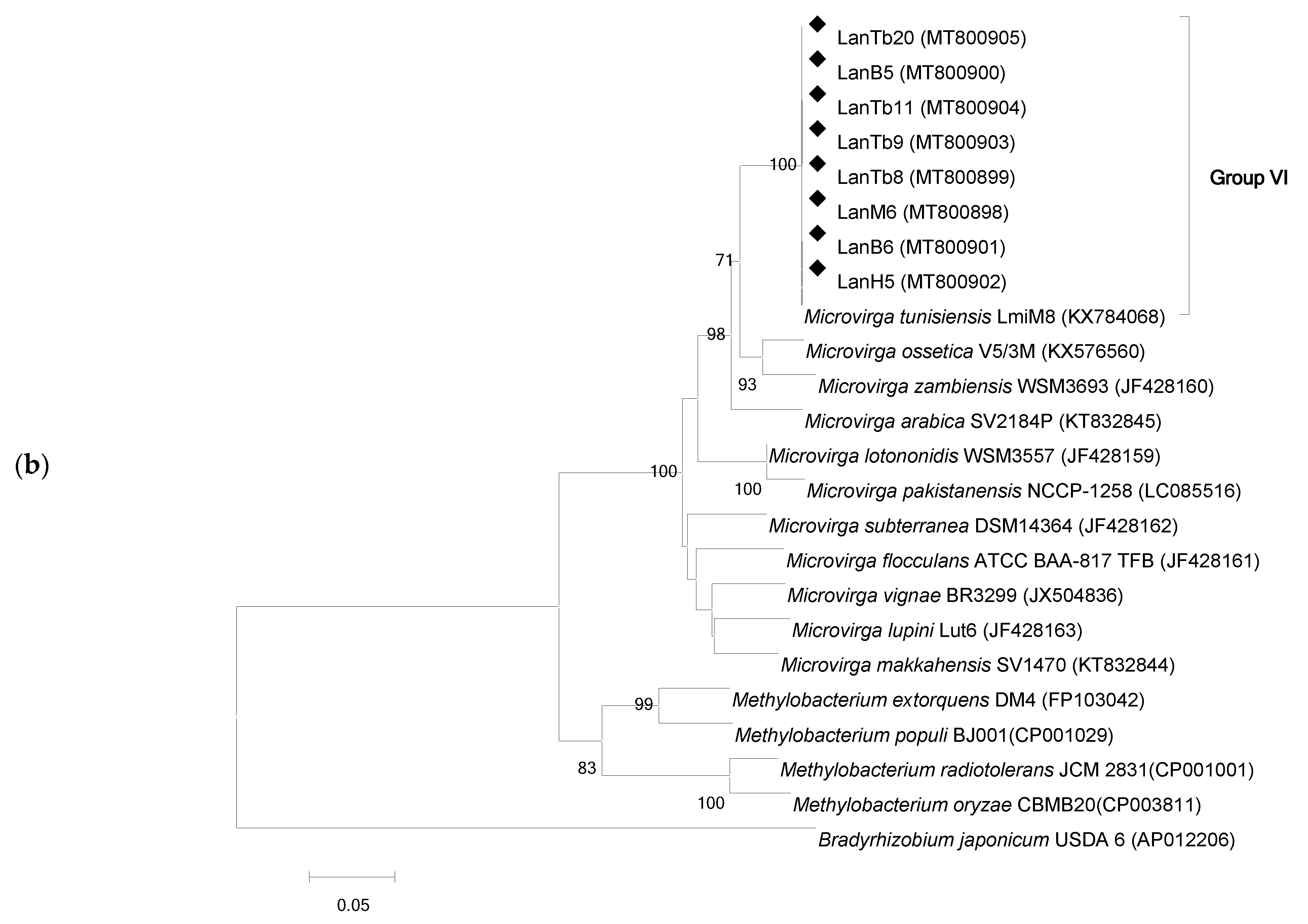
The recA gene was amplified and sequenced for all isolates. The sequences were compared with those available in GenBank. As shown in the recA phylogenetic tree topology (Figure 1a), L. angustifolius strains are grouped in six different clades, four within the Bradyrhizobium genus (22 strains) and one with Devosia (2 strains). A sixth group, based on gyrB gene phylogeny, formed by Microvirga (8 strains) is shown in Figure 1b. The bradyrhizobia subgroups contained 17 strains affiliated to B. lupini, 2 strains to B. canariense, 1 to B. liaoningense (86%) and 2 strains to B. algeriense. At least one representative isolate of each pattern was selected for further phylogenetic analyses.
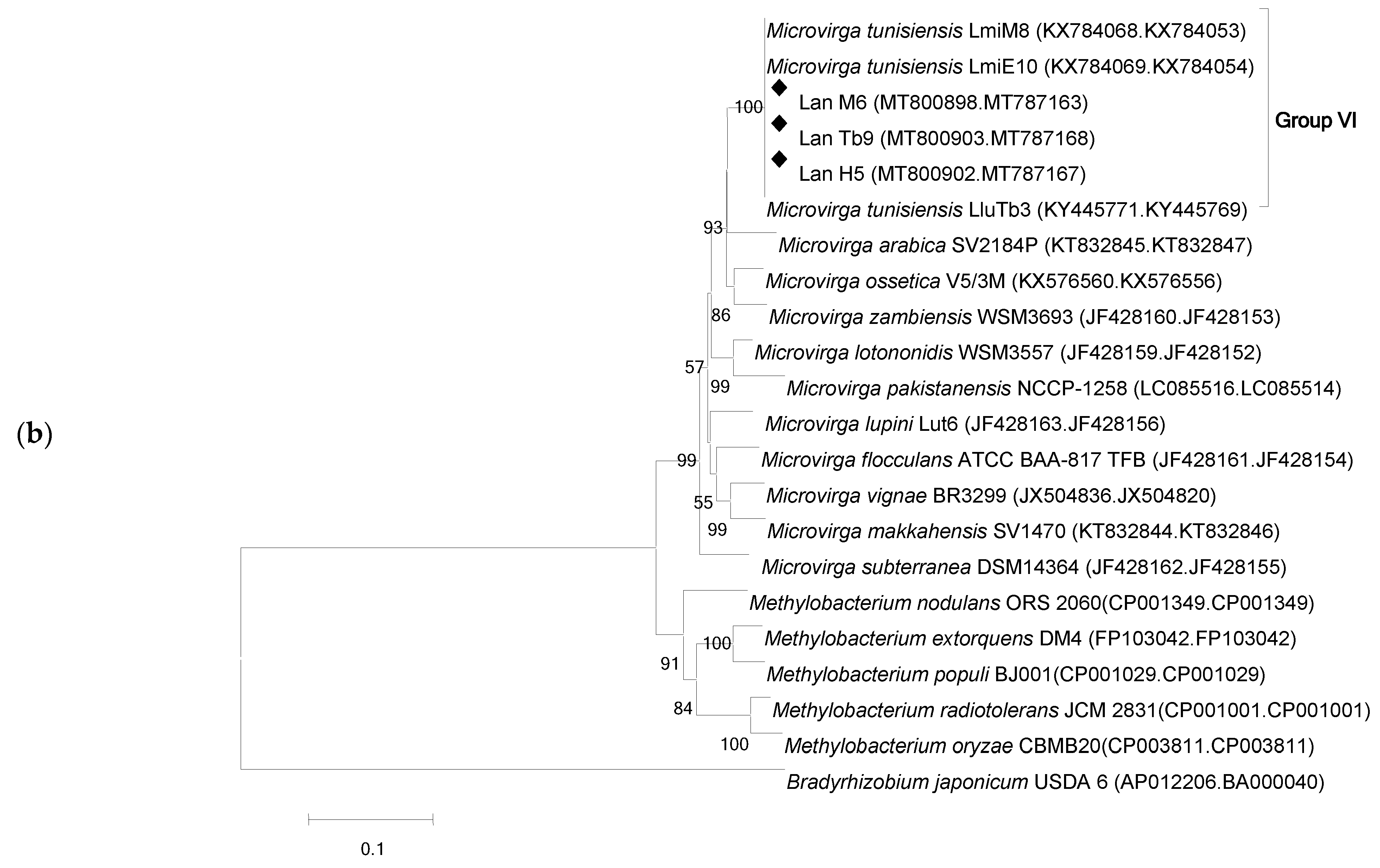
In order to define a more phylogenetically robust position of the strains, an analysis based on the concatenation of relevant housekeeping genes was performed. recA and glnII were used for Bradyrhizobium strains as they have been proven to be appropriate in phylogenetic studies of this genus [23,37]. glnII could not identified in any Microvirga strains draft genome sequences. Thus, dnaK was used instead, and the phylogenetic tree for this group of strains was made using gyrB and dnaK concatenated sequences. The trees again distributed L. agustifolius nodulating strains in six distinct groups, four (I, II, III and IV) contained all the bradyrhizobia, one (V) grouped the Devosia strain and one (VI) comprised the Microvirga strains (Figure 2a,b).
Strains LanT1, LanTb3, LanM5 and LanTb18 clustered with B. lupini USDA3051.LanS2 and LanTb17 grouped with B. canariense BTA-1 and B. liaoningense LMG18230, respectively. LanH1 occupied a position between B. valentinum LmjM3 and B. algeriense RST89. LanTb5 was located in a taxonomic position separate from the Bradyrhizobium spp., which suggested that it might constitute a new species under a different genus. The phylogenetic tree based on the concatenated gyrB and dnaK sequences (Figure 2b) revealed that strains in Group VI (LanM6, LanTb9 and LanH5) grouped with M. tunisiensis LmiM8T with almost 100% identity.

3.3. 16S rRNA Gene Phylogeny

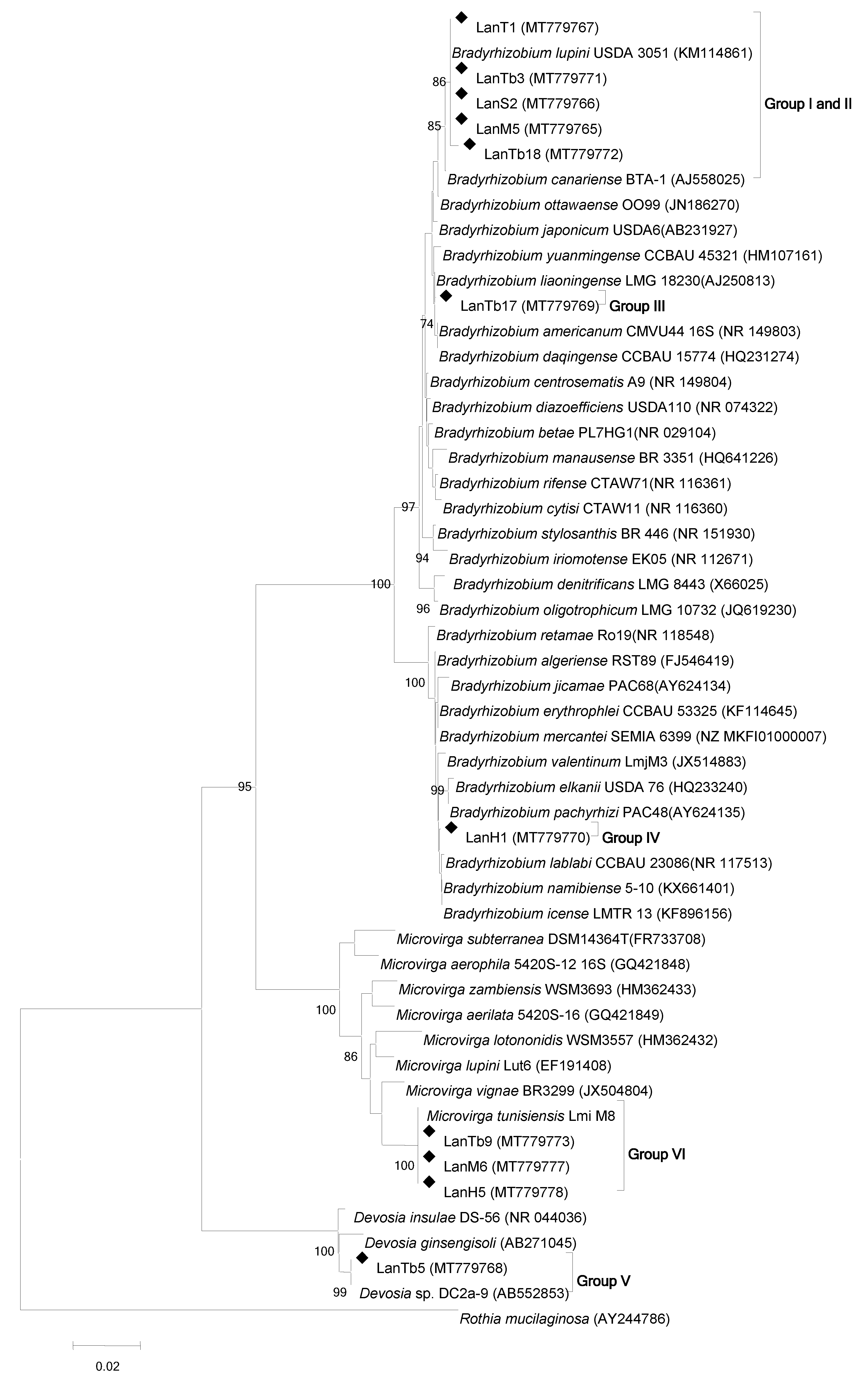
Phylogenetic analysis of the nearly full length sequences of 16S rRNA gene of 11 representative strains showed five large groups (Figure 3). Groups I and II of the analysis based on housekeeping genes form a single group when analyzed based on the rrs gene. This group contained five strains (LanTb18, LanM5, LanS2, LanT1, and LanTb3) grouped with B. canariense BTA-1 and B. lupini USDA 3051. The rest of the groups were similarly distributed. LanTb17 grouped with B. liaoningense LMG18230. Strain LanH1 occupied a separate position in the extra-slow bradyrhizobial clade formed by B. valentinum LmjM3, B. pachyrhizi PAC48 and B. lablabi CCBAU 23086, and might constitute a new species in the Bradyrhizobium genus. Three fast growing strains (LanM6, LanH5 and LanTb9) were identical to Microvirga tunisiensis LmiM8,also isolated in Northern Tunisia from L. micranthus [37]. The 16S rRNA sequence of LanTb5 shared 99% identity with Devosia sp. Dc2a-9 isolated from algal-bacterial consortia (https://www.ncbi.nlm.nih.gov/nuccore/300068687, accessed on 10 March 2021). Altogether, the phylogenetic analyses revealed several Bradyrhizobium, Microvirga and Devosia species, which had not been previously described, as microsymbionts of L. angustifolius.

3.4. NodC Gene Phylogeny

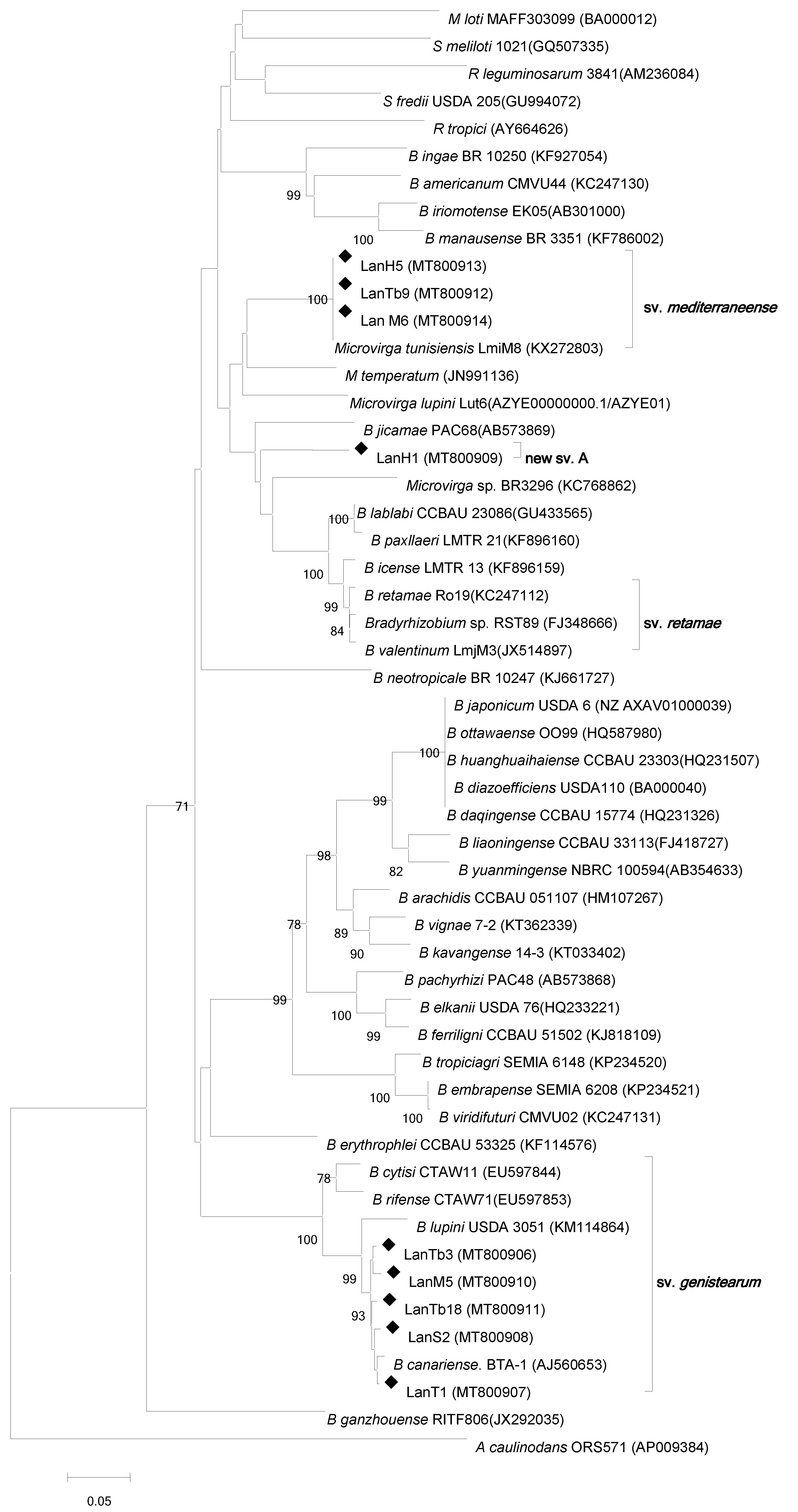
Partial nodC sequences were obtained from only six bradyrhizobial strains. Five were in groups I and II defined in Figure 1 and lined up with symbiovar genistearum formed by strains able to nodulate legumes in the Genisteae tribe, such as B. lupini, B. rifense, B. cytisi and B. canariense type strains (Figure 4). LanH1, in group IV of Figure 1, might correspond to a new symbiovar designated new sv. A, which is distant from other Bradyrhizobium symbiovars and close to symbiovar retamae, defined with strains nodulating Retama spp., and L. mariae-josephae [41]. nodC sequences from Microvirga strains (LanM6, LanTb9 LanH5) were identical to that of Microvirga tunisiensis LmiM8 belonging to the new mediterraneense symbiovar (Figure 4) [42]. The LmiM8 strain was an effective endosymbiont isolated from L. micranthus.
Amplification of nodC and nodA could not be obtained for strains LanTb17 and LanTb5. nifH gene sequencing affiliated LanTb17 (MZ343378) to Bradyrhizobium japonicum CCBAU43129 with 97% identity. LanTb5 (MZ369158) nifH gene was aligned to Rhizobium tropici CIAT 899 with 78% identity. These results might indicate that these novel L. angustifolius microsymbionts might have acquired their nif genes from phylogenetically distant sources through lateral transfer.

3.5. Host Range of Rhizobial Strains Isolated from L. angustifolius Nodules

The symbiotic specificity of L. angustifolius-nodulating rhizobia was investigated in plant cross-inoculation assays under axenic conditions. The selected strains were tested for nodulation with their original host and six additional legumes (L. luteus, L. micranthus, L. mariae-josephae, Retama sphaerocarpa, Vigna unguiculata and Glycine max) (Table 2). All tested strains were able to nodulate L. angustifolius, L. luteus and L. micranthus,with the exception of LanTb17, which only nodulated its original host (Table 2). LanT1, LanM5, LanTb3, LanTb18 and LanS2 strains formed white nodules with V. unguiculata. Microvirga sp. strains induced the formation of oversized non-effective nodules in L. mariae josephae roots. LanS2, Tb3, M5 and LanTb5 were unable to form root nodules in L. mariae josephae roots. LanH1 affiliated to extra-slow-growing Bradyrhizobium species, such as B. valentinum,which was originally isolated from L. mariae josephae, and formed effective nodules with L. mariae josephae. All tested strains were unable to form nodules on soybean, but formed white nodules on R. sphaerocarpa roots (Table 2). Overall, the nodulation analyses revealed that L. angustifolius endosymbionts present a diverse host range and symbiotic specificity.

3.6. Phenotypic Analyses of Narrow-Leaf Lupin-Nodulating Strains

The rhizobial isolates presented a broad biodiversity in their growth characteristics. Ten isolates could be classified as fast-growing (Devosia and Microvirga) and twenty-two of them as slow-growing rhizobia (Bradyrhizobium). The phenotypic traits of the 11representative strains are summarized in Table 3. All strains were able to grow in optimal growth conditions (28 °C, pH 6.8 and <0.01% NaCl). Cells were rod shaped, Gram negative and aerobic. The Bradyrhizobium isolates were slow growers with mean generation times >15 h in YMB. Colonies produced in YMA were translucent, gummy and circular. They presented convex smooth margins, and their sizes were 2–3 mm in diameter after 6–7 days in YMA medium at 28 °C. LmiH1 was an extra-slow grower (generation time in YMB > 30 h) that produced punctiform, non-mucoid colonies (<1 mm) after 10 to 11 days in YMA plates. Fast growers showed mean generation times of 4–7 h in TY. They also grew well on YMA plates. After 8 days at 28 °C, Microvirga colonies were 2–3 mm in diameter, convex, smooth, mucilaginous pink, and circular. The effect of temperature, salinity, pH and some antibiotics, used to quantify and qualify stress tolerance, showed a degree of heterogeneity (Table 3). All the Microvirga strains grew well between 20 and 37 °C, at pH 4–12, and on media containing 0.1; 0.5 and 1% salt. These strains were very sensitive to all antibiotics used (kanamycin (50 µg/mL), ampicillin (100 µg/mL), gentamicin (30 µg/mL), spectinomycin (50 µg/mL), tetracycline (5 µg/mL), streptomycin (10 µg/mL). The Bradyrhizobium strains grew at temperatures between 20 and 28 °C, at pH range 4 to 11 and on media containing 0.1% salt, with only strains LanS2, LanTb17 and LanH1 growing at temperatures between 20 and 37 °C. These strains were sensitive to kanamycin, gentamicin, spectinomycin, and streptomycin and were less resistant to ampicillin and tetracycline, except for strain LanTb17, which showed a good resistance to kanamycin, ampicillin and chloramphenicol. The Devosia LanTb5 strain grew well between 20 and 28 °C, at pH 4–11, and tolerated 1% salt added to the medium. This strain was sensitive to ampicillin, gentamicin, tetracycline, and chloramphenicol. LanTb5 could grow in presence of spectinomycin, kanamycin and streptomycin.

4. Discussion

In this study, the isolation, identification and phenotypic characterization of rhizobial bacteria isolated from effective root nodules of L. angustifolius plants growing in Northern Tunisia is reported. All isolated strains were effective endosymbionts of their original host, as evidenced by the formation of red–pink nodules and the leaves’ dark green color. Nodule numbers ranged from 3 to 10 per plant
Phylogenetic analyses revealed high genetic diversity levels among the L. angustifolius nodule isolates. Previous reports also described considerable genetic diversity among rhizobia that nodulate Lupinus by using diverse methodological approaches, such as M13 random amplified polymorphic DNA-PCR (RAPD-PCR) [20,21,23]. High diversity was shown by phylogenetic, symbiotic and phenotypic analyses. Some rhizobia assigned to the Bradyrhizobium genus were slow-growing bacteria, while some isolates were fast-growing and were assigned to the Microvirga and, unexpectedly, for the first time to our knowledge, to the Devosia genus. It is not unusual that legumes nodulated by slow-growing Bradyrhizobium species are also efficiently nodulated by different fast-growing rhizobial species [43,44].
The housekeeping genes glnII/recA and gyrB/dnaK were used because of their proven performance as molecular markers, either individually or in combination, for assessing the evolutionary genetics of Bradyrhizobium and Microvirga species, as described in previous works [28,45]. Phylogenetic analyses based on the housekeeping genes glnII/recA and gyrB/dnaK congruently placed some tested isolates in a new strongly supported clade within the Bradyrhizobium (Group III) and Devosia (Group V) genera. The concatenated tree of the representative strain showed that four strains affiliated with B. lupini USDA3051, one strain grouped with B. canariense BTA-1, one strain clustered together with B. liaoningense LMG18230, one strain occupied a separate position between B. valentinum LmjM3 and B. algeriense RST89, and could constitute a new species within the genus Bradyrhizobium, Three strains were included in the Microvirga genus and, surprisingly, one strain was related to the genus Devosia, never described before as a lupin microsymbiont. The concatenated phylogenetic trees constructed confirmed the results obtained by the single sequence of glnII, recA, gyrB and dnaK genes. The distribution of some strains separated from the existing type species might be indicative of novel species. Nonetheless, further polyphasic studies to precisely characterize and classify them are required.
The genus Devosia has been reported to establish nitrogen-fixing symbiosis with the aquatic legume Neptunia natans growing in India [34,46]. To our knowledge, no Devosia strain has previously been described as a lupin symbiont. In this study, two strains (LanTb4 and LanTb5) closely clustered with Devosia species. These isolates were clearly separated from other Devosia spp. and might constitute a new species under this genus. In previous works, Lupinus spp. has been reported to be nodulated by fast-growing bacteria affiliated to Mesorhizobium, Allorhizobium, Rhizobium and Sinorhizobium [24,27,30,47,48,49]. However, in general, annual lupins are nodulated by slow-growing Bradyrhizobium sp. rhizobia [21,50,51,52]. This is in agreement with our results, as approximately 60% of our isolates belonged to different Bradyrhizobium species.
The phylogeny of the important symbiotic gene nodC is well correlated with legume host range but independent of the rhizobial taxonomic position [53,54,55,56]. The nodC gene has been used as a molecular marker related to host specificity and range, and used to define Bradyrhizobium symbiovars [41,45,57]. The nodC tree revealed that the majority Bradyrhizobium strains clearly grouped with sequences withinthe genistearum symbiovar, which has been previously described for strains that nodulate species in the Genisteae tribe, including rhizobia nodulating Cytisus, Lupinus and Retama in Europe, America and Africa [41,45,58]. The sequences of Microvirga strains LanM6 and LanH5 grouped with sequences characteristic of the mediterraneense symbiovar. This type of nodC sequence was previously described for M. tunisiensis LmiM8 nodulating L. micranthus [42]. The remaining Bradyrhizobium-type representative isolate, LanH1, might correspond to a new symbiovar, as it was placed distant from other Bradyrhizobium symbiovars defined to date. Sanger chromatograms for nifH sequences of LanTb5 strain displayed sequence ambiguities. This may be explained by the presence of more than one copy [59].
Phenotypic tests have been used to study the diversity of rhizobial bacteria and to assess their tolerance under stress conditions [60,61]. The first macroscopic test showed that the morphology of the bradyrizobial strains in the present work was similar to those previously reported [23,60]. The colonies formed by the Microvirga strains were pinkish in color, similar to those of M. lupini [28] and M. vigna [39]. pH, salinity and extreme temperatures are abiotic stresses for nodulation of agricultural legumes in Mediterranean areas. The isolates exhibited differences in their response to abiotic stress (Table 3). All isolates grew at pH 4, in agreement with reports of other strains isolated from Lupinus spp. showing resistance to acid soils [62]. Most strains were also tolerant to alkaline conditions (pH 11), as previously reported for rhizobia that nodulate Prosopis juliflora growing in alkaline soil [63]. All strains were sensitive to salinity, as they were not able to grow when NaCl concentration was higher than 0.5%. Conversely, Microvirga strains were able to grow in with the presence of 1% sodium chloride. These results are also corroborated by those reported for Microvirga vignae [39]. Slow-growing rhizobia in the Bradyrhizobium genus have been reported to be more sensitive to salt stress than fast-growing rhizobia isolated from the same soils [61,64]. All strains in the present study grew at the optimum temperature of 28 °C, in contrast with a previous report that stated that the optimum temperature for M. lupini growth was 39 °C [28]. The effect of temperature, salinity and pH, used to qualify and quantify stresses tolerance, showed a degree of heterogeneity even when they were isolated from the same site (Table 3).
In conclusion, the isolates in this work showed a wide phenotypic and genotypic diversity. Significant heterogeneity among lupin endosymbionts was unraveled based on phylogenetic analysis using housekeeping and symbiotic gene sequences, and on their legume-host ranges. The study revealed the presence of rhizobia belonging to the three divergent genera Bradyrhizobium, Microvirga and Devosia. To our knowledge, this is the first report on the Alphaproteobacterial genus Devosia as a nodule-forming species in Lupinus spp. A practical aspect of the ability of lupin species to trap diverse rhizobial genotypes is that they might display important differences in their capacities to fix nitrogen depending on edaphoclimatic conditions [65,66]. The potentially new Bradyrhizobium and Devosia species reported here require further taxonomic characterization for confirmation and formal description. Finally, this work provides an array of native rhizobia that might have practical application as biofertilizers and/or inoculants with native legumes, including shrubs, in the restoration or revegetation of arid and semiarid Mediterranean areas.

Author Contributions

Conceptualization, A.M., L.R., J.I., J.M.P., M.M. and J.J.P.; methodology, A.M.; validation, A.M., L.R. and J.J.P.; formal analysis, A.M.; investigation, A.M.; resources, L.R., J.I., J.M.P., M.M. and J.J.P.; data curation, A.M.; writing—original draft preparation, A.M.; writing—review and editing, A.M., L.R., J.I., J.M.P., M.M. and J.J.P.; visualization, A.M. and J.J.P.; supervision, L.R., M.M. and J.J.P.; project administration, L.R., M.M. and J.J.P.; funding acquisition, J.M.P., M.M. and J.J.P. All authors have read and agreed to the published version of the manuscript.

Funding

This research was funded by the Ministry of High Education and Research Development, Tunisia, and by the Agencia Estatal de Investigación, AEI, Spain, grant numbers AGL2017-88381-R and RTI2018-094985-B-100. The APC was funded in part by CSIC Open Access Publication Support Initiative through its Unit of Information Resources for Research (URICI).

Data Availability Statement

All sequences are available from the GenBank database.

Conflicts of Interest

The authors declare no conflict of interest.

References

  1. Allen, E.K.; Allen, O.N. The Leguminosae: A Source Book of Characteristics, Uses and Nodulation; University of Wisconsin Press: Madison, WI, USA, 1981; p. 812. [Google Scholar]
  2. Cardoso, D.; Pennington, R.T.; de Queiroz, L.P.; Boatwright, J.S.; Van Wyk, B.E.; Wojciechowski, M.F.; Lavin, M. Reconstructing the deep-branching relationships of the papilionoid legumes. S. Afr. J. Bot. 2013, 89, 58–79. [Google Scholar] [CrossRef] [Green Version]
  3. Ainouché, A.K.; Bayer, R.J. Phylogenetic relationships in Lupinus (Fabaceae: Papilionoideae) based on internal transcribed spacer sequences (ITS) of nuclear ribosomal DNA. Am. J. Bot. 1999, 86, 590–607. [Google Scholar] [CrossRef]
  4. Drummond, C.S.; Eastwood, R.J.; Miotto, S.T.S.; Hughes, C.E. Multiple continental radiations and correlates of diversification in Lupinus (Leguminosae): Testing for key innovation within complete taxón sampling. Syst. Biol. 2012, 61, 443–460. [Google Scholar] [CrossRef] [Green Version]
  5. Cristofolini, G.; Chiapella, L.F. Origin and diversification of Genisteae (Fabaceae): A serosystematic purview. Webbia 1984, 38, 105–122. [Google Scholar] [CrossRef]
  6. Cristofolini, G. The biodiversity of the “Leguminosae-Genisteae” and its genesis. Lagascalia 1997, 19, 121–128. [Google Scholar]
  7. Mahé, F.; Pascual, H.; Coriton, O.; Huteau, V.; Navarro Perris, A.; Misset, M.T.; Aınouche, A. New data and phylogenetic placement of the enigmatic Old World lupin: Lupinus mariae-josephae H. Pascual. Genet. Res. Crop Evol. 2011, 58, 101–114. [Google Scholar] [CrossRef]
  8. Naganowska, B.; Wolko, B.S.; Liwinska, E.; Kaczmarek, Z.; Schifino-Wittmann, M.T. 2C DNA variation and relationships among New World species of the genus Lupinus (Fabaceae). Plant Syst. Evol. 2006, 256, 147–157. [Google Scholar] [CrossRef]
  9. Sprent, J.I. Nodulation in Legumes; Royal Botanic Gardens, Kew: London, UK, 2001; p. 146. [Google Scholar]
  10. Coba de la Peña, T.; Pueyo, J.J. Legumes in the reclamation of marginal soils, from cultivar and inoculant selection to transgenic approaches. Agron. Sustain. Dev. 2012, 32, 65–91. [Google Scholar] [CrossRef] [Green Version]
  11. Pueyo, J.J.; Quiñones, M.A.; Coba de la Peña, T.; Fedorova, E.E.; Lucas, M.M. Nitrogen and phosphorus interplay in lupin root nodules and cluster roots. Front. Plant Sci. 2021, 12, 644218. [Google Scholar] [CrossRef]
  12. Falkengren-Grerup, U.; Schottelndreier, M. Vascular plants as indicators of nitrogen enrichment in soils. Plant Ecol. 2004, 172, 51–62. [Google Scholar] [CrossRef]
  13. Bahmanyar, M.A.; Ranjbar, G.A. The role of potassium in improving growth índices and increasing amount of grain nutrient elements of wheat cultivars. J. Appl. Sci. 2008, 8, 1280–1285. [Google Scholar] [CrossRef] [Green Version]
  14. Leporatti, M.L.; Ghedira, K. Comparative analysis of medicinal plants used in traditional medicine in Italy and Tunisia. J. Ethnobiol. Ethnomed. 2009, 5, 31. [Google Scholar] [CrossRef] [Green Version]
  15. Lucas, M.M.; Stoddard, F.L.; Annicchiarico, P.; Frías, J.; Martínez-Villaluenga, C.; Sussmann, D.; Duranti, M.; Seger, A.; Zander, P.M.; Pueyo, J.J. The future of lupin as a protein crop in Europe. Front. Plant Sci. 2015, 6, 705. [Google Scholar] [CrossRef] [PubMed]
  16. De Ron, A.M.; Sparvoli, F.; Pueyo, J.J.; Bazile, D. Editorial: Protein crops: Food and feed for the future. Front. Plant Sci. 2017, 8, 105. [Google Scholar] [CrossRef] [Green Version]
  17. Andrews, M.; Andrews, M.E. Specificity in legume-rhizobia symbioses. Int. J. Mol. Sci. 2017, 18, 705. [Google Scholar] [CrossRef] [PubMed] [Green Version]
  18. Jarabo-Lorenzo, A.; Perez-Galdona, R.; Donate-Correa, J.; Rivas, R.; Velazquez, E.; Hernandez, M.; Temprano, F.; Martinez-Molina, E.; Ruiz-Argueso, T.; Leon-Barrios, M. Genetic diversity of bradyrhizobial populations from diverse geographic origins that nodulate Lupinus spp. and Ornithopus spp. Syst. Appl. Microbiol. 2003, 26, 611–623. [Google Scholar] [CrossRef] [PubMed]
  19. Stepkowski, T.; Zak, M.; Moulin, L.; Kroliczak, J.; Golinska, B.; Narozna, D.; Safronova, V.I.; Madrzak, C.J. Bradyrhizobium canariense and Bradyrhizobium japonicum are the two dominant rhizobium species in root nodules of lupin and serradella plants growing in Europe. Syst. Appl. Microbiol. 2011, 34, 368–375. [Google Scholar] [CrossRef]
  20. Velázquez, E.; Valverde, A.; Rivas, R.; Gomis, V.; Peix, A.; Gantois, I.; Igual, J.M.; Leon-Barrios, M.; Willems, A.; Mateos, P.F.; et al. Strains nodulating Lupinus albus on different continents belong to several new chromosomal and symbiotic lineages within Bradyrhizobium. Antonie Leeuwenhoek 2010, 97, 363–376. [Google Scholar] [CrossRef] [PubMed] [Green Version]
  21. Mellal, H.; Yacine, B.; Boukaous, L.; Khouni, S.; Benguedouar, A.; Castellano-Hinojosa, A.; Bedmar, E.J. Phylogenetic diversity of Bradyrhizobium strains isolated from root nodules of Lupinus angustifolius grown wild in the North East of Algeria. Syst. Appl. Microbiol. 2019, 42, 397–402. [Google Scholar] [CrossRef]
  22. Granada, C.E.; Beneduzi, A.; Lisboa, B.B.; Turchetto-Zolet, A.C.; Vargas, L.K.; Passaglia, L.M. Multilocus sequence analysis reveals taxonomic differences among Bradyrhizobium sp. symbionts of Lupinus albescens plants growing in arenized and non-arenized areas. Syst. Appl. Microbiol. 2015, 38, 323–329. [Google Scholar] [CrossRef]
  23. Durán, D.; Rey, L.; Sánchez-Cañizares, C.; Navarro, A.; Imperial, J.; Ruiz-Argüeso, T. Genetic diversity of indigenous rhizobial symbionts of the Lupinus mariae-josephae endemism from alkaline-limed soils within its area of distribution in Eastern Spain. Syst. Appl. Microbiol. 2012, 36, 128–136. [Google Scholar] [CrossRef]
  24. Trujillo, M.E.; Willems, A.; Abril, A.; Planchuelo, A.M.; Rivas, R.; Ludena, D.; Mateos, P.F.; Martinez-Molina, E.; Velázquez, E. Nodulation of Lupinus albus by strains of Ochrobactrum lupini sp. nov. Appl. Environ. Microbiol. 2004, 71, 1318–1327. [Google Scholar] [CrossRef] [Green Version]
  25. González-Sama, A.; Lucas, M.M.; De Felipe, M.R.; Pueyo, J.J. An unusual infection mechanism and nodule morphogenesis in white lupin (Lupinus albus). New Phytol. 2004, 163, 371–380. [Google Scholar] [CrossRef] [PubMed] [Green Version]
  26. Valverde, A.; Velázquez, E.; Fernández-Santos, F.; Vizcaíno, N.; Rivas, R.; Mateos, P.F.; Martínez-Molina, E.; Igual, J.M.; Willems, A. Phyllobacterium trifolii sp. nov., nodulating Trifolium and Lupinus in Spanish soils. Int. J. Syst. Evol. Microbiol. 2005, 55, 1985–1989. [Google Scholar] [CrossRef] [Green Version]
  27. El-Idrissi, M.M.; Aujjar, N.; Belabed, A.; Dessaux, Y.; Filali-Maltouf, A. Characterization of rhizobia isolated from carob tree (Ceratonia siliqua). J. Appl. Bacteriol. 1996, 80, 165–173. [Google Scholar] [CrossRef]
  28. Ardley, J.K.; Parker, M.A.; De Meyer, S.E.; Trengove, R.D.; O’Hara, G.W.; Reeve, W.G.; Yates, R.J.; Dilworth, M.J.; Willems, A.; Howieson, J.G. Microvirga lupini sp. nov., Microvirga lotononidis sp. nov. and Microvirga zambiensis sp. nov are alphaproteobacterial root-nodule bacteria that specifically nodulate and fix nitrogen with geographically and taxonomically separate legume hosts. Int. J. Syst. Evol. Microbiol. 2012, 62, 2579–2588. [Google Scholar] [PubMed]
  29. Msaddak, A.; Rejili, M.; Duran, D.; Rey, L.; Mars, M.; Palacios, J.; Ruiz-Argueso, T.; Imperial, J. Microvirga tunisiensis sp. nov., a root nodule symbiotic bacterium isolated from Lupinus micranthus and L. luteus grown in Northern Tunisia. Syst. Appl. Microbiol. 2019, 42, 6. [Google Scholar] [CrossRef]
  30. Tounsi-Hammami, S.; Le Roux, C.; Dhane-Fitouri, S.; De Lajudie, P.; Duponnois, R.; Ben Jeddi, F. Genetic diversity of rhizobia associated with root nodules of white lupin (Lupinus albus L.) in Tunisian calcareous soils. Syst. Appl. Microbiol. 2019, 42, 448–456. [Google Scholar] [CrossRef] [PubMed]
  31. Nakagawa, Y.; Sakane, T.; Yokota, A. Transfer of “Pseudomonas riboflavin” (Foster 1944), a gram-negative, motile rod with long-chain 3-hydroxy fatty acids, to Devosia riboflavin gen. nov., sp. nov., nom. rev. Int. J. Syst. Bacteriol. 1996, 46, 16–22. [Google Scholar] [CrossRef] [Green Version]
  32. Hoque, M.S.; Broadhurst, L.M.; Thrall, P.H. Genetic characterization of root-nodule bacteria associated with Acacia salicina and A. stenophylla (Mimosaceae) across south-eastern Australia. Int. J. Syst. Evol. Microbiol. 2011, 61, 299–309. [Google Scholar] [CrossRef] [Green Version]
  33. Bautista, V.V.; Monsalud, R.G.; Yokota, A. Devosia yakushimensis sp nov., isolated from root nodules of Pueraria lobata (Willd.) Ohwi. Int. J. Syst. Evol. Microbiol 2010, 60, 627–632. [Google Scholar] [CrossRef] [PubMed]
  34. Rivas, R.; Velazquez, E.; Willems, A.; Vizcaino, N.; Subba-Rao, N.S.; Mateos, P.F.; Gillis, M.; Dazzo, F.B.; Martinez-Molina, E. A new species of Devosia that forms a unique nitrogen-fixing root-nodule symbiosis with the aquatic legume Neptunia natans (L.f.) Druce. Appl. Environ. Microbiol. 2002, 68, 5217–5222. [Google Scholar] [CrossRef] [Green Version]
  35. Rejili, M.; Msaddak, A.; Filali, I.; Benabderrahim, M.A.; Mars, M.; Marın, M. New chromosomal lineages within Microvirga and Bradyrhizobium genera nodulate Lupinus angustifolius growing on different Tunisian soils. FEMS Microbiol. Ecol. 2019, 95, fiz118. [Google Scholar] [CrossRef]
  36. Vincent, J.M. A Manual for the Practical Study of Root Nodule Bacteria; International Biological Programme, Number 15; Blackwell Scientific: Oxford, UK, 1970. [Google Scholar]
  37. Msaddak, A.; Durán, D.; Rejili, M.; Mars, M.; Ruiz-Argüeso, T.; Imperial, J.; Palacios, J.; Rey, L. Diverse bacteria affiliated with the genera Microvirga, Phyllobacterium and Bradyrhizobium nodulate Lupinus micranthus growing in soils of Northern Tunisia. Appl. Environ. Microbiol. 2017, 83, 6. [Google Scholar] [CrossRef] [PubMed] [Green Version]
  38. Baele, M.; Baele, P.; Vaneechoutte, M.; Storms, V.; Butaye, P.; Devriese, L.A.; Verschraegen, G.; Gillis, M.; Haesebrouck, F. Application of tRNA intergenic spacer PCR for identification of Enterococcus species. J. Clin. Microbiol. 2000, 38, 4201–4207. [Google Scholar] [CrossRef] [Green Version]
  39. Radl, V.; Simões-Araújo, J.L.; Leite, J.; Passos, S.R.; Martins, L.M.V.; Xavier, G.R.; Rumjanek, N.G.; Baldani, J.I.; Zilli, J.E. Microvirga vignae sp. nov., a root nodule symbiotic bacterium isolated from cowpea grown in semi-arid Brazil. Int. J. Syst. Evol. Microbiol. 2014, 64, 725–730. [Google Scholar] [CrossRef] [PubMed]
  40. Kumar, S.; Stecher, G.; Tamura, K. MEGA7: Molecular evolutionary genetics analysis version 7.0 for bigger datasets. Mol. Biol. Evol. 2016, 33, 1870–1874. [Google Scholar] [CrossRef] [Green Version]
  41. Guerrouj, K.; Ruíz-Díez, B.; Chahboune, R.; Ramírez-Bahena, M.H.; Abdelmoumen, H.; Quinones, M.A.; ElIdrissi, M.M.; Velázquez, E.; Fernández-Pascual, M.; Bedmar, E.J.; et al. Definition of a novel symbiovar (sv. retamae) within Bradyrhizobium retamae sp. nov., nodulating Retama sphaerocarpa and Retama monosperma. Syst. Appl. Microbiol. 2013, 36, 218–223. [Google Scholar] [PubMed]
  42. Msaddak, A.; Rejili, M.; Duran, D.; Rey, L.; Palacios, J.M.; Imperial, J.; Ruiz-Argueso, T.; Mars, M. Definition of two new symbiovars, sv. lupini and sv. mediterranense, within the genera Bradyrhizobium and Phyllobacterium efficiently nodulating Lupinus micranthus in Tunisia. Syst. Appl. Microbiol. 2018, 41, 487–493. [Google Scholar] [CrossRef]
  43. Chamber, M.A.; Iruthayathas, E.E. Nodulation and nitrogen fixation by fast- and slow-growing rhizobia strains of soybean on several temperate and tropical legumes. Plant Soil 1988, 112, 239–245. [Google Scholar] [CrossRef]
  44. El-Akhal, M.R.; Rincón, A.; Arenal, F.; Lucas, M.M.; El Mourabit, N.; Barrijal, S.; Pueyo, J.J. Genetic diversity and symbiotic efficiency of rhizobial isolates obtained from nodules of Arachis hypogaea in Northwestern Morocco. Soil Biol. Biochem. 2008, 40, 2911–2914. [Google Scholar] [CrossRef]
  45. Vinuesa, P.; León-Barrios, M.; Silva, C.; Willems, A.; Jarabo-Lorenzo, A.; Pérez-Galdona, R.; Werner, D.; Martínez-Romero, E. Bradyrhizobium canariense sp. nov., an acid-tolerant endosymbiont that nodulates endemic genistoid legumes (Papilionoideae: Genisteae) from the Canary Islands, along with Bradyrhizobium japonicum bv. genistearum, Bradyrhizobium genospecies alpha and Bradyrhizobium genospecies beta. Int. J. Syst. Evol. Microbiol 2005, 55, 569–575. [Google Scholar]
  46. Subba-Rao, N.S.; Mateos, P.F.; Baker, D.; Pankratz, H.S.; Palma, J.; Dazzo, F.B.; Sprent, J.I. The unique root-nodule symbiosis between Rhizobium and the aquatic legume, Neptunia natans (L. f.) Druce. Planta 1995, 196, 311–320. [Google Scholar] [CrossRef]
  47. Miller, M.S.; Pepper, I.L. Survival of a fast-growing strain of lupin rhizobia in Sonoran Desert soils. Soil Biol. Biochem. 1988, 20, 323–327. [Google Scholar] [CrossRef]
  48. Pudelko, K.; Madrzak, J. Populations of Rhizobium and Bradyrhizobium nodulating Lupinus in Poland. In Abstracts of Biological Fixation for Ecology and Sustainable Agriculture, 2nd ed.; European Nitrogen Conference and NATO Advanced Research Workshop: Poznan, Poland, 1996; p. 156, Scientific Publishers OWN. [Google Scholar]
  49. Andam, C.P.; Parker, M.A. A novel Alpha-Proteobacterial root nodule symbiont associated with Lupinus texensis. Appl. Environ. Microbiol. 2007, 73, 5687–5691. [Google Scholar] [CrossRef] [Green Version]
  50. Barrera, L.L.; Trujillo, M.E.; Goodfellow, M.; Garcia, F.J.; Hernandez-Lucas, I.; Davila, G.; van Berkum, P.; Martinez-Romero, E. Biodiversity of bradyrhizobia nodulating Lupinus spp. Int. J. Syst. Bacteriol. 1997, 47, 1086–1091. [Google Scholar] [CrossRef]
  51. Fernández-Pascual, M.; Pueyo, J.J.; de Felipe, M.R.; Golvano, M.P.; Lucas, M.M. Singular features of the Bradyrhizobium Lupinus symbiosis. Dyn. Soil Dyn. Plant. 2007, 1, 1–16. [Google Scholar]
  52. Ryan-Salter, T.P.; Black, A.D.; Andrews, M.; Moot, D.J. Identification and effectiveness of rhizobial strains that nodulate Lupinus polyphyllus. Proc N. Z. Grassl. Assoc. 2014, 76, 61–66. [Google Scholar] [CrossRef]
  53. Ueda, T.; Suga, Y.; Yahiro, N.; Matsuguchi, T. Phylogeny of SymPlasmids of rhizobia by PCR-based sequencing of a nodC segment. J. Bacteriol. 1995, 177, 468–472. [Google Scholar] [CrossRef] [Green Version]
  54. Laguerre, G.; Nour, S.M.; Macheret, V.; Sanjuan, J.J.; Drouin, P.; Amarger, N. Classification of rhizobia based on nodC and nifH gene analysis reveals a close phylogenetic relationship among Phaseolus vulgaris symbionts. Microbiology 2001, 147, 981–993. [Google Scholar] [CrossRef] [Green Version]
  55. Rincón, A.; Arenal, F.; González, I.; Manrique, E.; Lucas, M.M.; Pueyo, J.J. Diversity of rhizobial bacteria isolated from nodules of the gypsophyte Ononis tridentata L. growing in Spanish soils. Microb. Ecol. 2008, 56, 223–233. [Google Scholar]
  56. Cordero, I.; Ruiz-Díez, B.; de la Peña, T.C.; Balaguer, L.; Lucas, M.M.; Rincón, A.; Pueyo, J.J. Rhizobial diversity, symbiotic effectiveness and structure of nodules of Vachellia macracantha. Soil Biol. Biochem. 2016, 96, 39–54. [Google Scholar] [CrossRef]
  57. Rogel, M.A.; Ormeno-Orrillo, E.; Martinez Romero, E. Symbiovars in rhizobia reflect bacterial adaptation to legumes. Syst. Appl. Microbiol. 2011, 34, 96–104. [Google Scholar] [CrossRef]
  58. Sánchez-Canizares, C.; Rey, L.; Durán, D.; Temprano, F.; Sánchez-Jiménez, P.; Navarro, A.; Polajnar, M.; Imperial, J.; Ruiz-Argüeso, T. Endosymbiotic bacteria nodulating a new endemic lupine Lupinus mariae-josephi from alkaline soils in Eastern Spain represent a new lineage within the Bradyrhizobium genus. Syst. Appl. Microbiol. 2011, 34, 207–215. [Google Scholar] [CrossRef] [Green Version]
  59. Barcellos, F.G.; Menna, P.; da Silva Batista, J.S.; Hungria, M. Evidence of horizontal transfer of symbiotic genes from a Bradyrhizobium japonicum inoculant strain to indigenous diazotrophs Sinorhizobium (Ensifer) fredii and Bradyrhizobium elkanii in a Brazilian Savannah soil. Appl. Environ. Microbiol. 2007, 73, 2635–2643. [Google Scholar] [CrossRef] [Green Version]
  60. Ruiz-Díez, B.; Fajardo, S.; Puertas-Mejía, M.A.; de Felipe, R.; Fernández-Pascual, M. Stress tolerance, genetic analysis and symbiotic properties of root-nodulating bacteria isolated from Mediterranean leguminous shrubs in Central Spain. Arch. Microbiol. 2009, 191, 35–46. [Google Scholar] [CrossRef]
  61. El-Akhal, M.R.; Rincon, A.; El Mourabit, N.; Pueyo, J.J.; Barrijal, S. Phenotypic and genotypic characterizations of rhizobia isolated from root nodules of peanut (Arachis hypogaea L.) grown in Moroccan soils. J. Basic Microbiol. 2009, 49, 415–425. [Google Scholar] [CrossRef] [PubMed]
  62. Howieson, J.G.; Fillery, I.R.P.; Legocki, A.B.; Sikorski, M.M.; Stepkowski, T.; Minchin, F.R.; Dilworth, M.J. Nodulation, nitrogen fixation and nitrogen balance. In Lupins as Crop Plants: Biology, Production and Utilization; Gladstones, J.S., Atkins, C.A., Hamblin, J., Eds.; CAB International: Wallingford, UK, 1998; pp. 149–180. [Google Scholar]
  63. Kulkarni, S.; Nautiyal, C.S. Characterization of high tolerant rhizobia isolated from Prosopis juliflora grown in alkaline soil. J. Gen. Appl. Microbiol. 1999, 45, 213–220. [Google Scholar] [CrossRef] [PubMed] [Green Version]
  64. El-Akhal, M.R.; Rincon, A.; Coba de la Pena, T.; Lucas, M.M.; El Mourabit, N.; Barrijal, S.; Pueyo, J.J. Effects of salt stress and rhizobial inoculation on growth and nitrogen fixation of three peanut cultivars. Plant Biol. 2013, 15, 415–421. [Google Scholar] [CrossRef] [PubMed] [Green Version]
  65. Gault, R.R.; Corbin, E.J.; Boundy, K.A.; Brockwell, J. Nodulation studies on legumes exotic to Australia: Lupinus and Ornithopus spp. Aust. J. Exp. Agric. 1986, 26, 37–48. [Google Scholar] [CrossRef]
  66. Kozhemyakov, A.P.; Cheremisov, B.M.; Kurlovich, B.S.; Tikhonovich, I.A.; Kartuzova, L.T.; Tchetkova, S.A.; Emeljanenko, T.A. Evaluation of the biological nitrogen fixing ability of lupin (Lupinus L.). Plant Genet. Resour. Newsl. 2000, 123, 66–77. [Google Scholar]
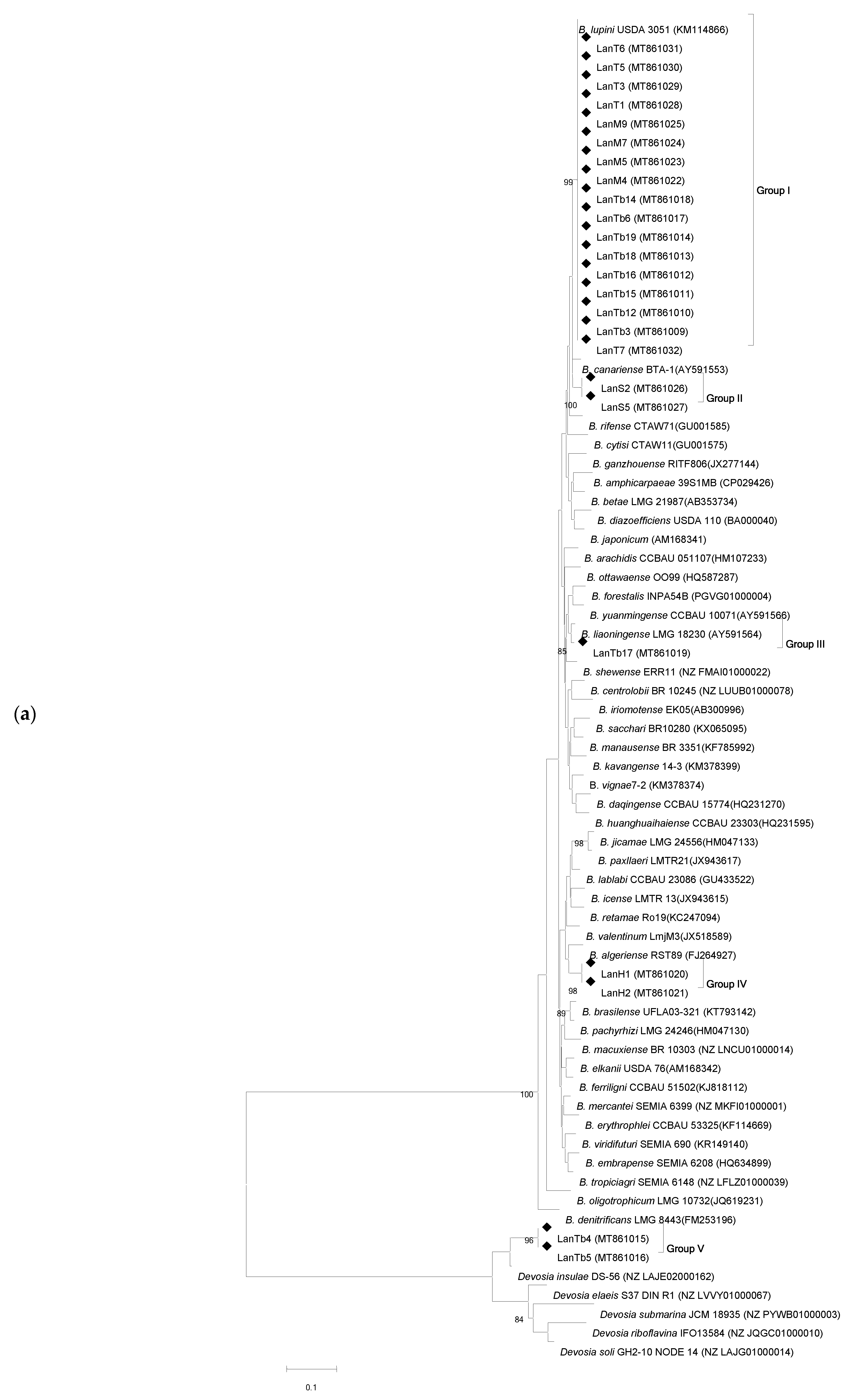
Figure 1. Neighbor-joining phylogenetic trees based on housekeeping recA (a) and gyrB (b) gene sequences of Tunisian L. angustifolius-nodulating isolates in this study and reference strains. Black diamonds (◆) indicate Tunisian strains. Bootstrap values (above 70%) using 1000 replicates are indicated at branching points. Accession numbers are shown in brackets. Bars: 0.1 and 0.05 estimated substitutions, respectively.
Agronomy 11 01510 g001aAgronomy 11 01510 g001b
Figure 2. Neighbor-joining phylogenetic trees of Tunisian L. angustifolius (Lan) microsymbionts and reference strains based on concatenated gene sequences. (a) Phylogeny of the Bradyrhizobium isolates based on recA and glnII sequences. (b) Phylogeny of the Microvirga isolates based on gyrB and dnaK sequences. Filled diamonds: Tunisian Lan isolates obtained in this work. Bootstrap values calculated for 1000 replications and those greater than 50% are indicated at the internodes. GenBank accession numbers are shown in brackets.
Agronomy 11 01510 g002aAgronomy 11 01510 g002b
Figure 3. Neighbor-joining phylogenetic tree based on 16S rRNA sequences of Tunisian isolates and reference strains. Black diamonds (◆) indicate Tunisian strains. Bootstrap values using 1000 replicates are indicated at branching points. Bar 0.02 estimated substitutions.
Agronomy 11 01510 g003
Figure 4. Neighbor-joining phylogenetic tree based on nodC sequences (620 bp) of Tunisian L. angustifolius endosymbionts and reference strains: Tunisian Lan (◆) isolates obtained in this work. Bootstrap values calculated for 1000 replications and those greater than 70% are indicated at the internodes. Accession numbers from GenBank and NCBI databases are shown in brackets. Bar: 0.05 estimated substitutions. M., Mesorhizobium; B., S., Sinorhizobium, Bradyrhizobium, R., Rhizobium.
Agronomy 11 01510 g004
Table 1. Designations and geographical origins of L. angustifolius endosymbionts in Northern Tunisia.
SiteNumber of
Isolates
Isolate
Code
Isolate
Code Number
Soil pHEnvironmentBioclimatic
Stage
Soil
Texture
Takelsa5LanT1, 3, 5, 6, 77.5Area with orange treessemi-arid superiorsandy
Mraissa5LanM4, 5, 6, 7, 98.0Area planted with cerealssemi-arid superiorsandy
Borj Hfaiedh2LanB5, 68.5Area with olive treessemi-arid superiorsandy
Hammamet3LanH1, 2, 59.0Semiurban area, next to the seasemi-arid superiorsandy
Sejnane2LanS2, 57.5Semiurban areahumidclay
Tabarka15LanTb3, 4, 5, 6, 8, 9, 11, 12, 14, 15, 16, 17, 18, 19, 206.5Area planted with cerealshumidsandy
Table 2. Legume host-range analysis of representative rhizobial strains isolated from L. angustifolius nodules.
Host Plant
L. angustifoliusL. luteusL. micranthusL. mariae-josephaeRetama sphaerocarpaVigna unguiculataGlycine max
StrainPG
LanT1I++++W+W+W-
LanM5I+++-+W+W-
LanTb3I+++-+W+W-
LanTb18I++++W+W+W-
LanS2II+++-+W+W-
LanTb17III+------
LanH1IV+++++--
LanTb5V+++-+W--
LanM6VI++++W+W--
LanH5VI++++W+W--
LanTb9VI++++W+W--
PG: Phylogenetic groups identified in Figure 1. Color of nodules: (+) red, (+W) white, (-) no nodules.
Table 3. Phenotypic characteristics of representative strains isolated from L. angustifolius growing in Northern Tunisia.
StrainPGGT
(h)
T Range (°C)pH RangeSalt Tolerance
(NaCl %)
Antibiotics Resistance
0.10.512AmpGenTetSpeKanStrChl
LanT1I1020–284–11+---+-+---+
LanM5I920–284–11+---+-+---+
LanTb3I9.520–284–11+---+-+---+
LanTb18I8.720–284–11+---+-+---+
LanS2II9.320–374–11++/------/+----/+
LanTb17III820–374–12++/---+-+-/++-+
LanH1IV1820–374–11++/------/+----/+
LanTb5V420–284–11+++/- - ---+++-
LanM6VI4.620–374–12+++/---------
LanH5VI4.620–374–12+++/---------
LanTb9VI4.620–374–12+++/---------
PG: Phylogenetic groups identified in Figure 1. GT: Generation Time-, no growth; +/-, weak growth (10–30% compared to the control in YMA); +, good growth (similar to the control) Amp: ampicillin (100 μg/mL); Gen: gentamicin (30 μg/mL); Tet: tetracycline (5 μg/mL); Spe: spectinomycin (50 μg/mL); Kan: kanamycin (50 μg/mL), Str: streptomycin (10 μg/mL).and Chl: chloramphenicol (10 µg/mL). Values represent the average of two experiments with three replicates each time.
Publisher’s Note: MDPI stays neutral with regard to jurisdictional claims in published maps and institutional affiliations.