Article

Phenotypic, Genetic, and Epigenetic Variation among Diverse Sweet Cherry Gene Pools

by
Evangelia V. Avramidou
1,†,
Theodoros Moysiadis
2,3,†,
Ioannis Ganopoulos
3,
Michail Michailidis
4,
Christos Kissoudis
5,
Dimitrios Valasiadis
4,
Konstantinos Kazantzis
3,
Eirini Tsaroucha
6,
Athanasios Tsaftaris
5,
Athanassios Molassiotis
4,
Filippos A. Aravanopoulos
7,* and
Aliki Xanthopoulou
3,4,*
1
Laboratory of Forest Genetics and Biotechnology, Institute of Mediterranean Forest Ecosystems, Athens, ELGO-DEMETER, Terma Alkmanos, Ilisia, 11528 Athens, Greece
2
Department of Computer Science, School of Sciences and Engineering, University of Nicosia, Nicosia 2417, Cyprus
3
Institute of Plant Breeding and Genetic Resources, ELGO-DEMETER, Thermi, 570001 Thessaloniki, Greece
4
Laboratory of Pomology, Department of Agriculture, Aristotle University of Thessaloniki, 54124 Thessaloniki, Greece
5
Perrotis College, American Farm School, GR-57001 Thessaloniki, Greece
6
Max Planck Institute for Plant Breeding Research, Department of Comparative Development and Genetics, Carl-von-Linné-Weg 10, 50829 Cologne, Germany
7
Laboratory of Forest Genetics & Tree Breeding, Faculty of Agriculture, Forestry & Environmental Science, Aristotle University of Thessaloniki, 54124 Thessaloniki, Greece
*
Authors to whom correspondence should be addressed.
These authors contributed equally to this work.
Agronomy 2021, 11(4), 680; https://doi.org/10.3390/agronomy11040680
Submission received: 26 February 2021 / Revised: 19 March 2021 / Accepted: 31 March 2021 / Published: 2 April 2021

Abstract

:
Sweet cherry germplasm contains a high variety of phenotypes which are associated with fruit size and shape as well as sugar content, etc. High phenotypic variation can be a result of genetic or epigenetic diversity that may interact through time. Recent studies have provided evidence that besides allelic variation, epiallelic variation can establish new heritable phenotypes. Herein we conducted a genetic and an epigenetic study (using amplified fragment length polymorphism (AFLP) and methylation-sensitive amplified polymorphism (MSAP) markers, respectively), accompanied by phenotypic traits correlation analysis in sweet cherry gene pools. The mean genetic diversity was greater than the epigenetic diversity (hgen = 0.193; hepi = 0.185), while no significant relationship was found between genetic and epigenetic distance according to a Mantel test. Furthermore, according to correlation analyses our results provided evidence that epigenetic diversity in predefined populations of sweet cherry had a stronger impact on phenotypic traits than their rich genetic diversity.

1. Introduction

Sweet cherry (Prunus avium L.) consists of phenotypes with high variability in fruit size and shape, sugar content, flowering time, pathogen resistance, and a variety of other traits [1,2,3,4,5,6]. Prunus avium cultivars originated from the Black and Caspian Seas, and the species currently presents a trans-European distribution as a result of the interplay between extant genetic variation and the genetic basis of adaptation [7]. The key to the evolution of sweet cherry breeding is therefore the in-depth study of genetic diversity and genes associated with traits of interest. In Greece, a large number of traditional local landraces are still highly varied, despite the fact that many are considered to have already been lost [7]. Moreover, many modern international varieties have a narrow genetic bottleneck, as reported by [8]. A variety of studies have been conducted in Greece using Simple Sequence Repeat (SSR) molecular markers and concern the analysis of the genetic basis of modern local cherry varieties. The main aim of the sweet cherry breeding program in Greece (‘Cherry4Breed’) was the development of new high-quality sweet cherry cultivars with high consumer appeal and self-compatible traits. Recently, the authors of [9], using whole genome resequencing (WGRS) analysis, found that a wild progenitor of Greek sweet cherry cultivars showed the highest genetic diversity. On the other hand, the mechanisms of epigenetic regulation in cherries have been poorly studied. To date only one such study has been published with regard to Greek wild cherry populations [10], investigating the possible coupling between epigenetic and genetic variation.
In eukaryotic organisms, gene regulation through genetic and epigenetic processes is of utmost importance for determining the phenotype [11,12,13,14]. Epigenetic changes such as DNA methylation [15] histone modifications [16], histone variants [17], and small RNAs [18] can present transgenerational inheritance. The ever-changing environmental conditions necessitate a more extensive study of epigenome variability [19,20,21]. Epigenetic regulation can be inherited, exerting influence on development, adaptation, and phenotype. Therefore, integrating the study of epigenetic variability and epigenome into breeding programs is of paramount importance.
Herein, in order to investigate epigenetic indices of diversity and structure as well as the relationships between epigenetic variants, genetic variation, and leaf traits, we used the amplified fragment length polymorphism (AFLP) and methylation-sensitive amplified polymorphism (MSAP) techniques along with correlation analysis statistics. Most of the leaf traits evaluated are very important for both sweet cherry breeding and cultivar registration in the test guidelines suggested by International Union for the Protection of New Varieties of Plants (UPOV). Our results may help breeders to understand and use epigenetic variation in order to accelerate the improvement of Prunus avium germplasm.

2. Materials and Methods

2.1. Plant Material

Twenty-two cultivated sweet cherry genotypes were selected (Table 1) from the Greek Fruit Gene Bank collection in Naousa (Institute of Plant Breeding and Genetic Resources- H.A.O. ELGO DEMETER), Greece, to represent the total diversity of Greek sweet cherry cultivars. The 2 elite wild cherry genotypes were obtained from the Wild Cherry Gene Bank, located at the Xyloupolis Forest Nursery of the Hellenic Forest Service in Greece. The 22 sweet cherry accessions are traditional Greek cultivars. We predefined 4 groups: ‘Breeding line’, ‘Landrace’, ‘Modern cultivar’, and ‘Wild’ (Table 1). Moreover, in the ‘Wild’ group we analyzed 1 genotype from Prunus mahaleb as a wild accession and also as outgroup in our study. Genomic DNA was isolated from a pool of young leaf samples of 5 plants per genotype using the NucleoSpin Plant II kit (Macherey-Nagel).

2.2. Leaf Phenotyping and Image Analysis

Plant images were obtained using a Scanalyzer PL semi-automated platform imaging system (LemnaTec GmbH, Aachem, Germany). The resolution of the digital camera (Baster AG, Ahresburg, Germany) was 1628 × 1236 pixels, with light in the visible spectrum in RGB (400–700 nm) with a pixel of 4.4 × 4.4 μm being used. Each plant was imaged individually in 3 (0°, 90°, and 180°) plane orientations. Plants were monitored over the mature leaf stage.
Plant pixel-projected leaf areas from the side view images were used to calculate the so-called ‘digital biomass’, which is an estimate of the plant volume and senescence. Data retrieved from the imaging platform were processed through an analysis Software (LemnaGrid) was specifically adjusted to obtain values for the phenotypic traits obtained from the images of each plant (Figure 1). The analysis was performed using a nearest-neighbor color classification, resulting in a binary image. Thereafter, geometric measurements of the object were taken, including caliper length (the longest dimension of the canopy when viewed from above), convex hull area (cm2), minimum enclosing area, and compactness calculated as the square of plant border length divided by the projected side or top area.
The caliper length, convex hull area, minimum enclosing area, and compactness of each population were assessed using the coefficient of variation (CV = 100 × standard deviation/mean).

2.3. AFLP Analysis

For the AFLP procedure, total genomic DNA (200 ng) was digested with 4 U of EcoRI and MseI for 3 h at 37 °C. Digested DNA fragments and EcoRI and MseI adapters were ligated with T4 DNA ligase (New England Biolabs) for 3 h at 26 °C. The resulting DNA was used as the primary template DNA in the AFLP analysis. A primer pair based on the sequences of the EcoRI and MseI adapters (Table 2) with one additional selective nucleotide at the 3’ end (EcoRI + A and MseI + C) was used for the first PCR step (pre-amplification). Pre-amplification PCR was performed in a total volume of 20 μL containing 1X Kapa Taq Buffer, 0.2 mM of each dNTP, 2.5 mM MgCl2, 30 ng of each primer EcoRI + A, MseI + C, 1 U Taq DNA polymerase (Kapa Biosystems), and 5 μL of diluted fragments (from the digestion and ligation reaction). Cycling was carried out on a BioRad thermocycler with a 95 °C hold for 30 s followed by 32 cycles of 95 °C for 30 s, 56 °C for 30 s and 72 °C for 1 min and subsequently followed by a final hold at 72 °C for 10 min. A 5-μL aliquot of the reaction was electrophoresed on agarose to verify amplification; the remaining 15 μL were diluted 5-fold with TE. Selective amplifications were carried out in 10-μL total volumes consisting of 3 μL of diluted pre-selective template and using the same reaction conditions as for pre-selective amplification but using 30 ng of an MseI primer and 5 ng of an EcoRI primer per reaction. Sixteen selective amplifications were performed on a BioRad thermocycler with the following program: an initial cycle of 95 °C for 30 s, 65 °C for 30 s, and 72 °C for 1 min; then 12 cycles of 95 °C for 30 s with an annealing temp starting at 65 °C for 30 s, but decreasing by 0.75 °C for each cycle, and then 72 °C for 1 min; and finally, 23 cycles of 94 °C for 30 s, 56 °C for 30 s, and 72 °C for 1 min, with a final hold at 72 °C for 30 min. Pre-selective and selective primers for the AFLP procedure are described in Table 2. Replicate analyses were conducted once, employing the same DNA extractions for AFLP and MSAP analysis.

2.4. MSAP Analysis

MSAP or methylation-sensitive amplified polymorphism is a modification of the standard AFLP technique. EcoRI is used as the rare cutter and the methylation-sensitive restriction enzymes HpaII and MspI as the frequent cutters due to the fact that the latter represent a pair of isoschizomers recognizing the same target sequence 5′- CCGG -3′, but they have differential sensitivity to methylation at the inner or outer cytosine. For the MSAP procedure, from each sample 200 ng of genomic DNA was digested with 4 U of EcoRI and 3 U of HpaII, and 200 ng was treated with 4 U of EcoRI and 3 U of MspI. The digestion was carried out at 37 °C for 3 h. The resulting DNA fragments and the EcoRI and HpaII/MspI adapters (Table 2) were ligated at 25 °C for 3 h using 400 U/μL of T4 DNA ligase (New England Biolabs). After that period the samples were subjected to a heat shock treatment for 10 min at 65 °C to end the ligation reaction. A primer pair based on the sequences of the EcoRI and HpaII/MspI adapters (Table 2) with one additional selective nucleotide at the 3′ end (EcoRI + A and HpaII/MspI + T) was used for the pre-selective PCR step. Pre-amplification PCR was performed in a total volume of 20 μL containing 1 × Kapa Taq Buffer, 0.4 U dNTPmix (10 mM), 2.5 mM MgCl2, 30 ng of each primer EcoRI + A, HpaII/MspI + C, 1 U Taq DNA polymerase (Kapa Biosystems), and 5 μL of diluted fragments (from the digestion and ligation reaction). The cycling program was the same as for AFLP procedure. Twelve selective amplifications were performed following same procedure as for AFLP above. The pre-selective and selective primers used are given in Table 2.

2.5. Scoring of AFLP Markers

AFLP product mixtures were denatured in formamide at 94 °C for 2 min and electrophoretically separated using an ABI Prism 3730 xl automated fluorescence sequencer (Applied Biosystems). The size of detected fragments was determined by the Genemapper v4.0 program using an internal standard (GS 500 LIZ, Applied Biosystems). Fragments ranging from 150 to 500 bases in size were counted and further analyzed in order to reduce the impact of potential size homoplasy [22].
Analysis of the AFLP data was carried out by employing POPGENE version 1.32 [23] using the option for dominant diploid markers. GenAlEx 6.502b [24] was used to determine the genetic structure at hierarchical levels (analysis of molecular variance, AMOVA), to reveal band patterns and frequencies, and to plot grouping of individuals in the principal coordinates analysis (PCoA).
A cluster analysis using an unweighted pair-group method with arithmetic averaging (UPGMA) [25] was performed using the POPGENE 1.32 software [23]. Tests for statistical significance were based on 9999 random permutations, followed by sequential Bonferroni correction.

2.6. Scoring of MSAP Markers

Only reproducible fragments ranging from 150 to 500 bases were counted and further analyzed in order to reduce the impact of potential size homoplasy [22]. For MSAP analyses, comparison of the banding patterns of EcoRI/HpaII and EcoRI/MspI reactions resulted in 4 conditions of a particular fragment—I: fragments present in both profiles (1/1), indicating an unmethylated state; II: fragments present only in EcoRI/MspI profiles (0/1), indicating hemi- or fully methylated CG sites; III: fragments present only in EcoRI/HpaII profiles (1/0), indicating hemimethylated CHG sites; and IV: absence of fragments in both profiles (0/0), representing an uninformative state caused either by different types of methylation, or due to restriction site polymorphism [26]. To separate unmethylated and methylated fragments and to test for the particular impact of the methylated conditions II and III, we used the ‘Mixed-Scoring 2′ approach [27].
Epigenetic diversity within populations was quantified using the R script MSAP_calc.r [27] as: (1) number of total and private bands (polymorphic subepiloci), (2) percentage of polymorphic subepiloci (Pepi), and (3) mean Shannon’s information index (Iepi). GenAlEx 6.502b [24] was employed to compute haploid gene diversity (h) within populations. GenAlEx was also used to conduct analysis of molecular variance (AMOVA)—separately for each subepilocus class—in order to study the variation of CCGG methylation states (epiloci) among the 4 populations. Separate principal coordinates analyses (PCoA) were employed to assess differentiation among populations based on MSAP data according to different subepilocus classes (h, m, and u epiloci). The similarity between matrices based on different marker systems (MSAP and AFLP) was calculated using the standardized Mantel coefficient [28]. The same test was used to evaluate the similarity between geographic and genetic, and between geographic and epigenetic distances, respectively.

2.7. Correlations Between Inter-Population Genetic, Epigenetic and Phenotypic Distance

Correlation and regression analyses were used to assess the relationships between intra-population epigenetic and genetic diversity, between intra-population epigenetic diversity and phenotypic variation, and between intra-population genetic diversity and phenotypic variation. Particularly, in each case the linear and the polynomial models of second order (quadratic) were evaluated. In order to opt for the best fit, analysis of variance was used to compare the polynomial model of second order (complete) and the respective linear (nested) models, resulting in the model of choice. For each chosen model, 90% confidence intervals were also developed. Correlation and regression analyses were performed in R 3.6.3. The packages that have been used for analysis with R were “made4” [29], “corrplot” [30], and “ggplot2” [31].

3. Results

3.1. AFLP Genetic Diversity

Sixteen primer combinations revealed 542 markers, of which 51.34% were polymorphic (Table 3). The percentage of polymorphic loci ranged from 37.08 % for the ‘Wild’ predefined population to 67.53% for the ‘Breeding line’ population. Nei’s gene diversity (hgen) ranged from 0.156 for the ‘Landrace’ population to 0.218 for the ‘Breeding line’ predefined population and Shannon’s diversity index (Igen) ranged from 0.234 for ‘Landrace’ population to 0.335 for ‘Breeding line’ population (Table 3).
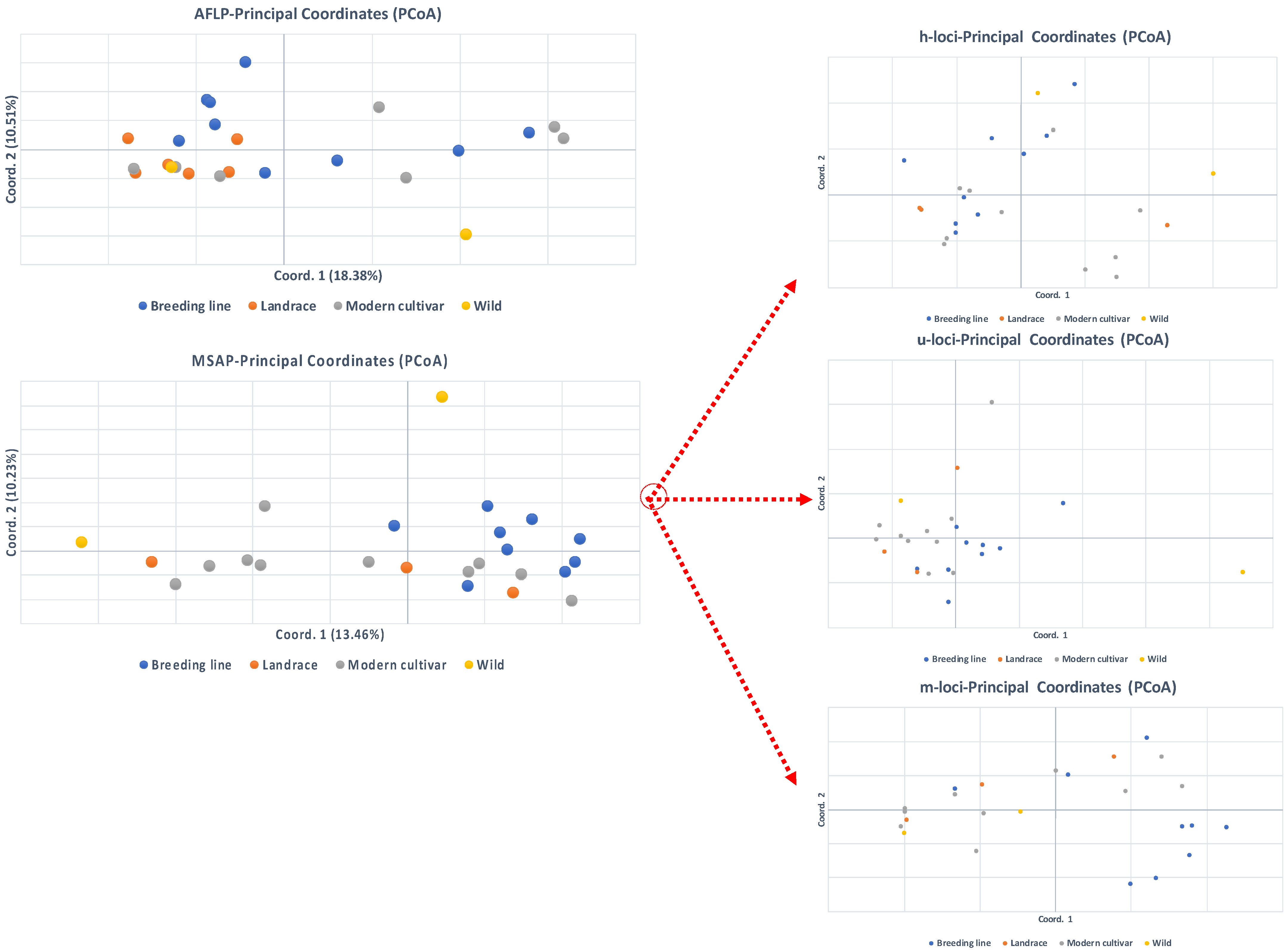
The analysis of molecular variance (AMOVA) showed that approximately 96% of the total genetic variation was partitioned within populations, while among population differentiation was 4% (Table 4). Furthermore, PCoA explained 28.89% of the total variation in two-dimensional multivariate space (Figure 2). Furthermore, the technical error rate from replicate analysis was 2% for AFLP replicate analysis.

3.2. MSAP Epigenetic Diversity

Fourteen primer combinations produced 283 markers, of which 102 were m-methylated (36%), 98 were h-methylated (35%), and 83 were u-methylated markers (unmethylated/uninformative; 29%). Among populations, the percentage of polymorphic markers (PLPepi) ranged from 35.69% for ‘Landrace’ to 66.08% for the ‘Modern cultivar’ population (Table 3). Nei’s diversity index (hepi) ranged from 0.159 for the ‘Breeding line’ population to 0.204 for the ‘Modern cultivar’ population, and Shannon’s diversity index (Iepi) ranged from 0.227 for the ‘Landrace’ population to 0.282 for the ‘Wild’ population (Table 3).
The analysis of molecular variance (AMOVA) showed that approximately 91% of the total epigenetic variation was partitioned within populations, while among population differentiation was 9% (Table 4). At the multivariate space, principal coordinate analysis of epigenetic distances revealed varying population differentiation patterns among all subepiloci (Figure 2) and explained 23.68% of the total variation in two-dimensional multivariate space. When a separate principal coordinates analysis was conducted for u-, m-, and h- epiloci, the absence of population differentiation was indicated. The first two axes respectively explained the percentages of the total variation: 28.46% for u-, 33.24% for m-, and 29.98% for h-subepiloci. Lastly, the technical error rate for MSAP replicate analysis was also low, presenting an average of 2.2%.

3.3. Correlations Between Intra-Population Genetic, Epigenetic and Phenotypic Variation

We found a strong concave-down relationship between intra-population genetic diversity and epigenetic diversity (Figure 3; for hepi: R2 = 0.998, and for Iepi: R2 = 0.975, Figure 3b,c), suggesting that in these cases epigenetic diversity assumed its highest values at the intermediate values of genetic diversity, and decreased at lower and higher values. The correlation for PLPepi was positive linear (R2 = 0.547, Figure 3a), suggesting that epigenetic diversity was in concordance with genetic diversity.
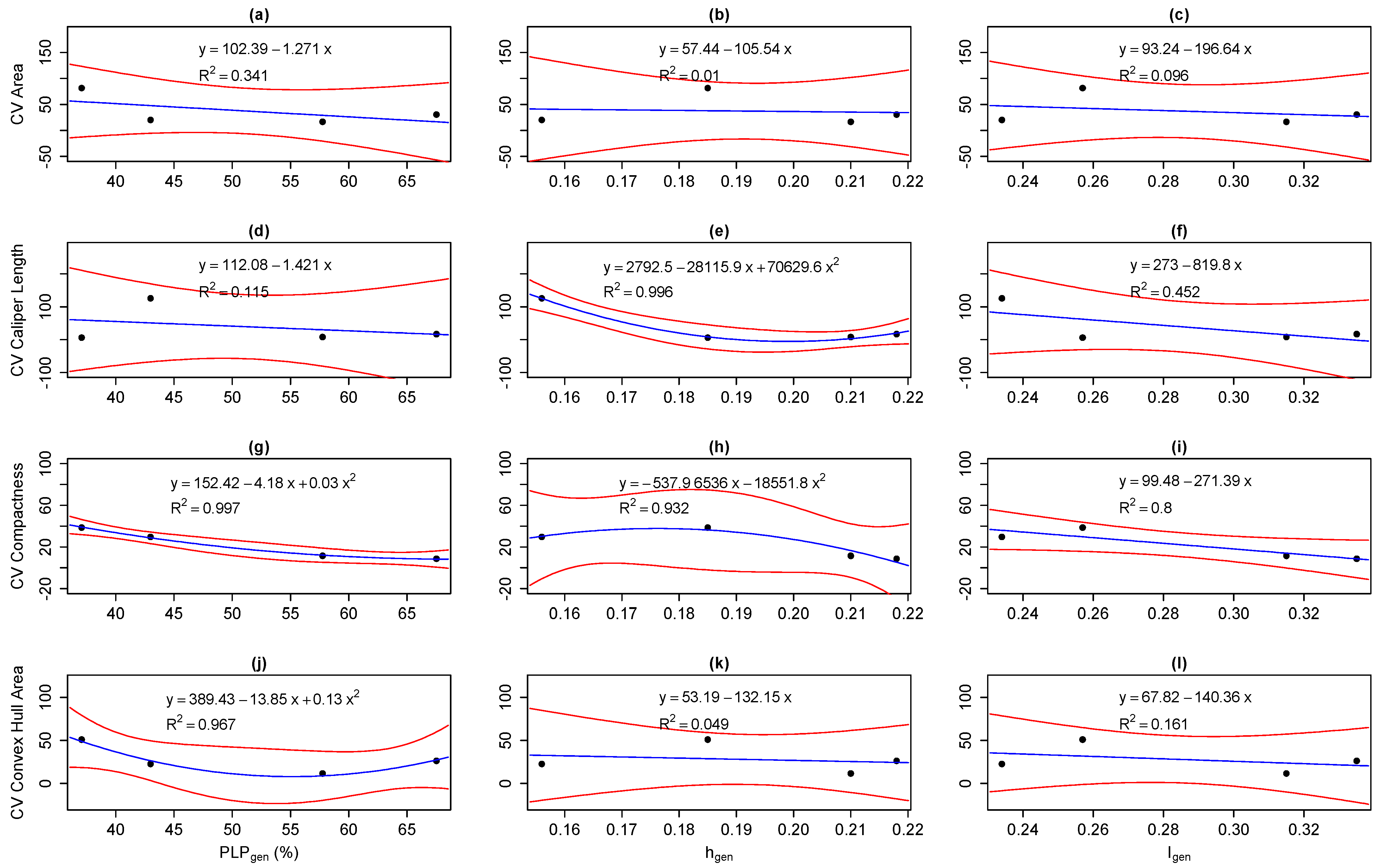
Furthermore, we found strong correlations between intra-population genetic diversity and phenotypic variation for: (1) CV caliper length with hgen (R2 = 0.996, Figure 4e), (2) CV compactness with the percentage of polymorphic loci (PLPgen) and hgen (R2 = 0.997 and R2 = 0.932, Figure 4g,h, respectively), and (3) CV convex hull area with PLPgen (R2 = 0.967, Figure 4j).
Assessing the relationship between intra-population epigenetic diversity and phenotypic variation, we found a strong concave-up correlation for: (1) CV caliper length with Iepi (R2 = 0.998, Figure 5f) and (2) CV compactness with hepi (R2 = 0.971, Figure 5h), as well as strong linear correlations for: (1) CV area with hepi (R2 = 0.548, Figure 5b), (2) CV caliper length with hepi (R2 = 0.551, Figure 5e), and (3) CV compactness with PLPepi (R2 = 0.678, Figure 5g), and weaker linear correlations for: (1) CV caliper length with PLPepi (R2 = 0.410, Figure 5d), (2) CV convex hull area with PLPepi (R2 = 0.359, Figure 5j), and (3) CV convex hull area with hepi (R2 = 0.315, Figure 5k).

3.4. Correlations Between Inter-Population Genetic, Epigenetic, and Phenotypic Distance

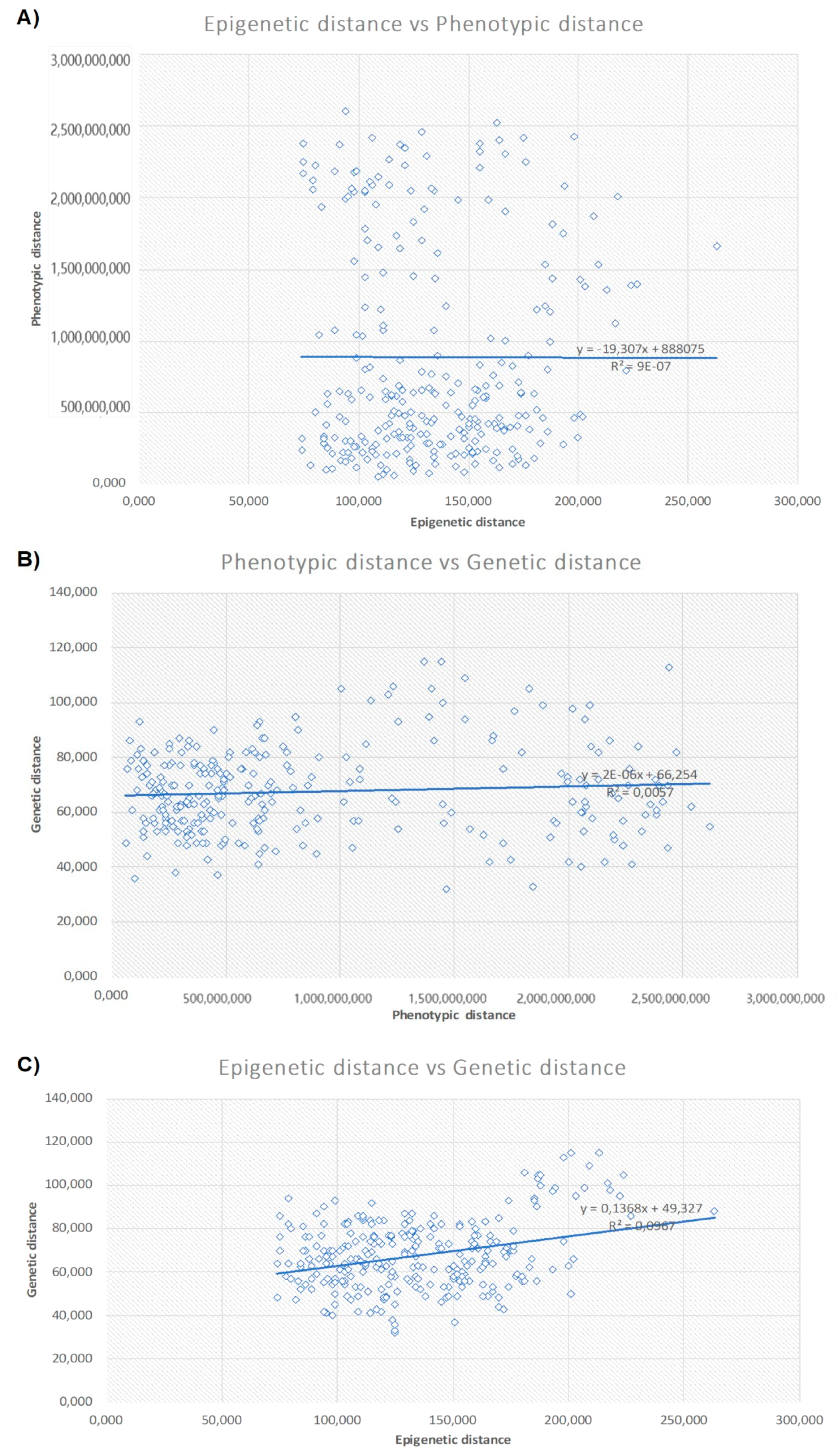
A Mantel test (Figure 6) revealed no correlation between inter-population phenotypic and epigenetic distance (R2 = 9.10–7, P = 0.610). Similarly, inter-population genetic distance had no correlation neither with phenotypic differentiation (R2 = 0.067, P = 0.3) or epigenetic differentiation (R2 = 0.096, P = 0.02).

4. Discussion

Recent studies in many plant species report that epigenetic effects can be heritable and can affect plant growth, adaptation, and phenotypic plasticity [15,32]. Therefore, it is important to incorporate epigenetic variation into plant breeding programs in order to comprehensively employ the total available variation from the genome and epigenome [33,34,35].
In our study we used the predefined ‘Breeding line’, ‘Landrace’, ‘Modern cultivar’ and ‘Wild’ sweet cherry populations in order to assess the total diversity of Greek sweet cherry cultivars with the main aim of estimating correlations between genetic, epigenetic, and phenotypic traits.
Results showed that genetic diversity was high (mean value hgen = 0.193) and the highest value was 0.218 for the ‘Breeding line’ population, which is a confirmation of high genetic diversity for sweet cherry cultivars. This was also reported in [9] after whole genome resequencing (WGRS) analysis of the same material. Epigenetic diversity (hepi) presented a mean value of 0.185 for all populations, while the ‘Wild’ population had the maximum value of 0.282. This value is numerically higher compared to the previous wild populations which were studied in [10], where the respective mean value was 0.108. Furthermore, overall mean genetic diversity was higher compared to epigenetic diversity for Greek sweet cherry cultivars, a result which is in agreement with the Greek wild cherry populations studied with MSAP and ISSR markers in [5]. On the contrary, in a study of 96 accessions of Prunus mume [33], genetic diversity was reported as being lower than epigenetic diversity. Limited studies have been reported for congruent genetic and epigenetic analyses in other species. For example, Wang [34] reported higher epigenetic diversity for Hydrocotyle vulgaris L., which was introduced to China and is cultivated as a clonal herb.

4.1. Correlations Between Epigenetic and Genetic Variation

The relationship between genetic and epigenetic variation in populations is still in debate according to various studies [26,36]. The central issue is “whether epigenetic variation is completely uncoupled from genetic variation”, suggesting that population-specific selection could act on both genetic and epigenetic variation independently. If we accept this scenario, then we have to consider that this is probably a result of random epigenetic drift—dramatic spontaneous alterations of epigenetic marks occurring during mitotic propagation or epimutations [10,37].
The second scenario suggests that epigenetic variation is at least in part a downstream, subsidiary effect of genetic variation, and various studies support this thesis [26,38,39]. In our study a significant concave-down correlation was found between genetic and epigenetic gene diversity (h) and Shannon´s information index (R2 = 0.998 and 0.975, respectively), and a linear correlation for PLP (R2 = 0.547), indicating that epigenetic and genetic variations strongly interact. Herrera and Bazaga [39] also found that epigenetic differentiation for Viola cazorlensis populations was correlated with adaptive genetic divergence, which suggests that differences at the epigenome could arise by variable selection.
Furthermore, according to the Mantel test no significant relationship was found between the genetic and epigenetic distance of predefined populations of sweet cherry. A similar result of no concordance between genetic and epigenetic distances was also found for Greek Prunus avium wild populations in [10].

4.2. Correlations of Phenotypic Variation with Genetic and Epigenetic Variation

In order to understand and estimate the proportion of phenotypic diversity in predefined populations that is explained by genetic and epigenetic variation, we conducted a series of regression analyses. According to various genetic mapping studies, genetic variation can explain phenotypic variation, but the ‘missing heritability’ problem still exists [40,41]. The problem lies in the fact that not all of the genetic variance in the phenotype can be accounted for by variance in the genome [40]. On the other hand, numerous studies have shown that epimutations are a faster force of evolution; they can have a strong impact on adaptation and can provide higher phenotypic variance [42,43].
In the Greek sweet cherry gene pool, a high degree of phenotypic variation exists [4]. We herein report strong intra-population correlations between genetic and phenotypic traits. Other studies have also shown associations between AFLP genetic markers and phenotypic traits [44].
Interestingly, our results provide evidence that epigenetic variance generally contributes more to phenotypic variation than genetic variance. We found that epigenetic indices (hepi, PLPepi) were correlated with a higher number of phenotypic traits than genetic indices. Epigenetic indices either presented strong concave-up (two phenotypic traits), strong linear (three phenotypic traits), or weaker linear correlations (three phenotypic traits). Ma [33] also showed that leaf length, width, and area were positively correlated with methylation levels. In Zea mays cultivars, Roy [45] reported a significant amount of epigenetic variation between them. Moreover, the authors of [42] found that methylation changes rather than DNA sequence changes in a floral symmetry gene explained the phenotypic change in natural populations of Linaria Vulgaris.

5. Conclusions

Data acquisition, in the era of studying genetic/epigenetic variation and explaining phenotypic diversity in populations and cultivars, is of paramount importance. Herein, we showed that epigenetic variance in predefined populations of sweet cherry possibly has a stronger impact on phenotypic traits than their rich genetic diversity. This first insight could provide a significant baseline in order to support further research focused on targeting more epigenetic markers (through bisulfite sequencing) that could serve for breeding programs. This study has elucidated the notable role of epigenetic diversity in shaping phenotypic variance, and therefore it justifies the need to reveal possible epigenetic differentiation associated with different environmental conditions with greater resolution.

Author Contributions

Conceptualization, I.G. and A.X.; methodology, T.M.; software, T.M.; validation, E.V.A., M.M., E.T., D.V., and A.X., investigation, A.X. and I.G.; resources, I.G., C.K., A.T., and A.M.; data curation, T.M.; writing—original draft preparation, E.V.A., T.M., A.X., F.A.A., and I.G.; writing—review and editing, E.V.A., T.M., C.K., A.T., K.K., M.M., A.T., E.T., and D.V.; visualization, A.X.; supervision, A.X. and F.A.A.; project administration, A.X. and F.A.A.; funding acquisition, A.X. All authors have read and agreed to the published version of the manuscript.

Funding

“This project has received funding from the Hellenic Foundation for Research and Innovation (HFRI) and the General Secretariat for Research and Technology (GSRT), under grant agreement No. 148.

Institutional Review Board Statement

Not applicable.

Informed Consent Statement

Not applicable.

Conflicts of Interest

The authors declare no conflict of interest.

References

  1. Zambounis, A.; Ganopoulos, I.; Avramidou, E.; Aravanopoulos, F.A.; Tsaftaris, A.; Madesis, P. Evidence of extensive positive selection acting on cherry (Prunus avium L.) resistance gene analogs (RGAs). Aust. J. Crop Sci. 2016, 10, 1324. [Google Scholar] [CrossRef]
  2. Jarausch, W.; Eyquard, J.; Mazy, K.; Lansac, M.; Dosba, F. High level of resistance of sweet cherry (Prunus avium L.) towards European stone fruit yellows phytoplasmas. Adv. Hortic. Sci. 1999, 13, 108–112. [Google Scholar]
  3. Castede, S.; Campoy, J.A.; Le Dantec, L.; Quero-Garcia, J.; Barreneche, T.; Wenden, B.; Dirlewanger, E. Mapping of candidate genes involved in bud dormancy and flowering time in sweet cherry (Prunus avium). PLoS ONE 2015, 10, e0143250. [Google Scholar] [CrossRef]
  4. Ganopoulos, I.; Farsakoglou, A.-M.; Aravanopoulos, F.; Molassiotis, A.; Michailidis, M.; Malliarou, E.; Avramidou, E.; Tsaftaris, A.; Osanthanunkul, M.; Madesis, P. Towards sweet cherry (Prunus avium L.) breeding: Phenotyping evaluation of newly developed hybrids. Euphytica 2018, 214, 99. [Google Scholar] [CrossRef]
  5. Schüller, E.; Fernández, F.F.; Antanaviciute, L.; Anhalt-Brüderl, U.; Spornberger, A.; Forneck, A. Autochthonous Austrian Varieties of Prunus avium L. Represent a Regional Gene Pool, Assessed Using SSR and AFLP Markers. Genes 2021, 12, 322. [Google Scholar] [CrossRef]
  6. El Baji, M.; Hanine, H.; En-Nahli, S.; Socias I Company, R.; Kodad, O. Morphological and Pomological Characteristics of Sweet Cherry (Prunus Avium L.) Grown In-situ under South Mediterranean Climate in Morocco. Int. J. Fruit Sci. 2021, 1–14. [Google Scholar] [CrossRef]
  7. Ganopoulos, I.; Aravanopoulos, F.A.; Argiriou, A.; Kalivas, A.; Tsaftaris, A. Is the genetic diversity of small scattered forest tree populations at the southern limits of their range more prone to stochastic events? A wild cherry case study by microsatellite-based markers. Tree Genet. Genomes 2011, 7, 1299–1313. [Google Scholar] [CrossRef]
  8. Campoy, J.A.; Lerigoleur-Balsemin, E.; Christmann, H.; Beauvieux, R.; Girollet, N.; Quero-García, J.; Dirlewanger, E.; Barreneche, T. Genetic diversity, linkage disequilibrium, population structure and construction of a core collection of Prunus avium L. landraces and bred cultivars. BMC Plant Biol. 2016, 16, 1–15. [Google Scholar] [CrossRef] [PubMed] [Green Version]
  9. Xanthopoulou, A.; Manioudaki, M.; Bazakos, C.; Kissoudis, C.; Farsakoglou, A.-M.; Karagiannis, E.; Michailidis, M.; Polychroniadou, C.; Zambounis, A.; Kazantzis, K. Whole genome re-sequencing of sweet cherry (Prunus avium L.) yields insights into genomic diversity of a fruit species. Hortic. Res. 2020, 7, 1–14. [Google Scholar] [CrossRef]
  10. Avramidou, E.V.; Ganopoulos, I.V.; Doulis, A.G.; Tsaftaris, A.S.; Aravanopoulos, F.A. Beyond population genetics: Natural epigenetic variation in wild cherry (Prunus avium). Tree Genet. Genomes 2015, 11, 95. [Google Scholar] [CrossRef]
  11. Heyn, H.; Moran, S.; Hernando-Herraez, I.; Sayols, S.; Gomez, A.; Sandoval, J.; Monk, D.; Hata, K.; Marques-Bonet, T.; Wang, L. DNA methylation contributes to natural human variation. Genome Res. 2013, 23, 1363–1372. [Google Scholar] [CrossRef] [Green Version]
  12. Massicotte, R.; Whitelaw, E.; Angers, B. DNA methylation: A source of random variation in natural populations. Epigenetics 2011, 6, 421–427. [Google Scholar] [CrossRef] [PubMed] [Green Version]
  13. Mirouze, M.; Paszkowski, J. Epigenetic contribution to stress adaptation in plants. Curr. Opin. Plant Biol. 2011, 14, 267–274. [Google Scholar] [CrossRef] [PubMed]
  14. Richards, C.L.; Bossdorf, O.; Verhoeven, K.J. Understanding natural epigenetic variation. New Phytol. 2010, 187, 562–564. [Google Scholar] [CrossRef] [PubMed]
  15. Dubrovina, A.; Kiselev, K. Age-associated alterations in the somatic mutation and DNA methylation levels in plants. Plant Biol. 2016, 18, 185–196. [Google Scholar] [CrossRef]
  16. Thorstensen, T.; Grini, P.E.; Aalen, R.B. SET domain proteins in plant development. Biochim. Biophys. Acta (BBA) Gene Regul. Mech. 2011, 1809, 407–420. [Google Scholar] [CrossRef]
  17. Chen, X.; Hu, Y.; Zhou, D.-X. Epigenetic gene regulation by plant Jumonji group of histone demethylase. Biochim. Biophys. Acta (BBA)-Gene Regul. Mech. 2011, 1809, 421–426. [Google Scholar] [CrossRef] [PubMed]
  18. Heo, J.B.; Lee, Y.-S.; Sung, S. Epigenetic regulation by long noncoding RNAs in plants. Chromosome Res. 2013, 21, 685–693. [Google Scholar] [CrossRef] [PubMed] [Green Version]
  19. Feng, S.; Jacobsen, S.E.; Reik, W. Epigenetic reprogramming in plant and animal development. Science 2010, 330, 622–627. [Google Scholar] [CrossRef] [PubMed] [Green Version]
  20. Feng, S.; Jacobsen, S.E. Epigenetic modifications in plants: An evolutionary perspective. Curr. Opin. Plant Biol. 2011, 14, 179–186. [Google Scholar] [CrossRef] [Green Version]
  21. Schmitz, R.J.; Ecker, J.R. Epigenetic and epigenomic variation in Arabidopsis thaliana. Trends Plant Sci. 2012, 17, 149–154. [Google Scholar] [CrossRef] [Green Version]
  22. Vekemans, X.; Beauwens, T.; Lemaire, M.; Roldán-Ruiz, I. Data from amplified fragment length polymorphism (AFLP) markers show indication of size homoplasy and of a relationship between degree of homoplasy and fragment size. Mol. Ecol. 2002, 11, 139–151. [Google Scholar] [CrossRef]
  23. Yeh, F.; Yang, R.; Boyle, T.; Ye, Z.; Xiyan, J.; Yang, R.; Boyle, T. PopGene32, Microsoft Windows-Based Freeware for Population Genetic Analysis; Version 1.32; Molecular Biology and Biotechnology Centre, University of Alberta: Edmonton, Alberta, 2000. [Google Scholar]
  24. Peakall, R.; Smouse, P.E. GENALEX 6: Genetic analysis in Excel. Population genetic software for teaching and research. Mol. Ecol. Notes 2006, 6, 288–295. [Google Scholar] [CrossRef]
  25. Sneath, P.H.; Sokal, R.R. Numerical Taxonomy: The Principles and Practice of Numerical Classification. Trans. Am. Microsc. Soc. 1974, 93, 303–304. [Google Scholar]
  26. Schulz, B.; Eckstein, R.L.; Durka, W. Epigenetic variation reflects dynamic habitat conditions in a rare floodplain herb. Mol. Ecol. 2014, 23, 3523–3537. [Google Scholar] [CrossRef] [PubMed]
  27. Schulz, B.; Eckstein, R.L.; Durka, W. Scoring and analysis of methylation-sensitive amplification polymorphisms for epigenetic population studies. Mol. Ecol. Resour. 2013, 13, 642–653. [Google Scholar] [CrossRef] [PubMed] [Green Version]
  28. Mantel, N. The detection of disease clustering and a generalized regression approach. Cancer Res. 1967, 27, 209–220. [Google Scholar]
  29. Culhane, A.C.; Thioulouse, J.; Perrière, G.; Higgins, D.G. MADE4: An R package for multivariate analysis of gene expression data. Bioinformatics 2005, 21, 2789–2790. [Google Scholar] [CrossRef] [PubMed] [Green Version]
  30. Wei, T.; Simko, V.; Levy, M.; Xie, Y.; Jin, Y.; Zemla, J. Package ‘corrplot’. Statistician 2017, 56, e24. [Google Scholar]
  31. Wickham, H. ggplot2. Wiley Interdiscip. Rev. Comput. Stat. 2011, 3, 180–185. [Google Scholar] [CrossRef]
  32. He, S.; Xu, W.; Li, F.; Wang, Y.; Liu, A. Intraspecific DNA methylation polymorphism in the non-edible oilseed plant castor bean. Plant Divers. 2017, 39, 300–307. [Google Scholar] [CrossRef]
  33. Ma, K.; Sun, L.; Cheng, T.; Pan, H.; Wang, J.; Zhang, Q. Epigenetic variance, performing cooperative structure with genetics, is associated with leaf shape traits in widely distributed populations of ornamental tree Prunus mume. Front. Plant Sci. 2018, 9, 41. [Google Scholar] [CrossRef] [PubMed] [Green Version]
  34. Wang, M.-Z.; Li, H.-L.; Li, J.-M.; Yu, F.-H. Correlations between genetic, epigenetic and phenotypic variation of an introduced clonal herb. Heredity 2020, 124, 146–155. [Google Scholar] [CrossRef] [PubMed]
  35. Xu, J.; House, M.; Booker, H.; Tanino, K. Exploiting epigenetic variation for plant breeding. CAB Rev. 2018, 13, 1–11. [Google Scholar] [CrossRef]
  36. Richards, E.J. Inherited epigenetic variation—revisiting soft inheritance. Nat. Rev. Genet. 2006, 7, 395–401. [Google Scholar] [CrossRef]
  37. Richards, E.J. Population epigenetics. Curr. Opin. Genet. Dev. 2008, 18, 221–226. [Google Scholar] [CrossRef]
  38. Dubin, M.J.; Zhang, P.; Meng, D.; Remigereau, M.-S.; Osborne, E.J.; Casale, F.P.; Drewe, P.; Kahles, A.; Jean, G.; Vilhjalmsson, B. DNA methylation in Arabidopsis has a genetic basis and shows evidence of local adaptation. eLife 2015, 4, e05255. [Google Scholar] [CrossRef]
  39. Herrera, C.M.; Bazaga, P. Epigenetic differentiation and relationship to adaptive genetic divergence in discrete populations of the violet Viola cazorlensis. New Phytol. 2010, 187, 867–876. [Google Scholar] [CrossRef]
  40. Banta, J.A.; Richards, C.L. Quantitative epigenetics and evolution. Heredity 2018, 121, 210–224. [Google Scholar] [CrossRef]
  41. Slatkin, M. Epigenetic inheritance and the missing heritability problem. Genetics 2009, 182, 845–850. [Google Scholar] [CrossRef] [Green Version]
  42. Cubas, P.; Vincent, C.; Coen, E. An epigenetic mutation responsible for natural variation in floral symmetry. Nature 1999, 401, 157–161. [Google Scholar] [CrossRef] [PubMed]
  43. Jablonka, E.; Raz, G. Transgenerational epigenetic inheritance: Prevalence, mechanisms, and implications for the study of heredity and evolution. Q. Rev. Biol. 2009, 84, 131–176. [Google Scholar] [CrossRef] [PubMed] [Green Version]
  44. Medrano, M.; Herrera, C.M.; Bazaga, P. Epigenetic variation predicts regional and local intraspecific functional diversity in a perennial herb. Mol. Ecol. 2014, 23, 4926–4938. [Google Scholar] [CrossRef] [PubMed]
  45. Roy, N.; Choi, J.-Y.; Lim, M.-J.; Lee, S.-I.; Choi, H.-J.; Kim, N.-S. Genetic and epigenetic diversity among dent, waxy, and sweet corns. Genes Genom. 2015, 37, 865–874. [Google Scholar] [CrossRef]
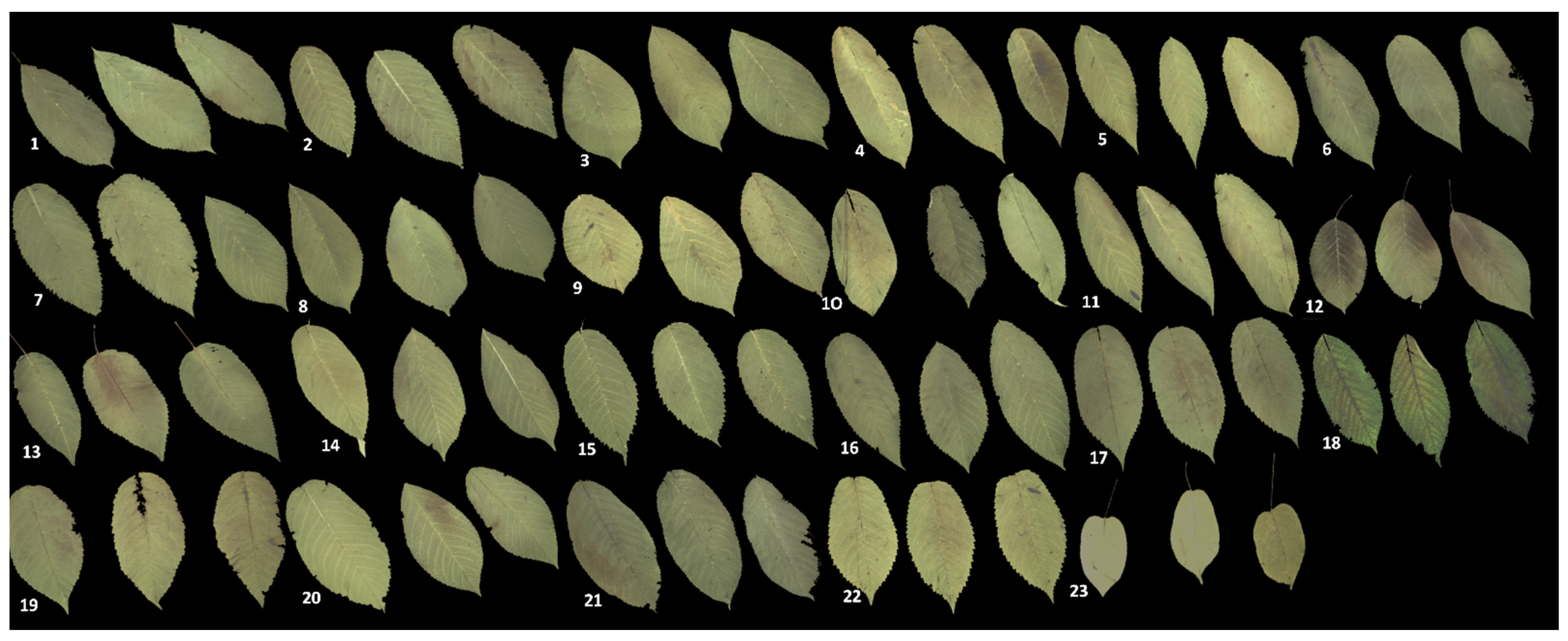
Figure 1. Images acquired from representative sweet cherry accessions (3 leaves per accession) at the mature leaf stage. Numbers 1–23 indicate the names of accession: (1) Bakirtzeika, (2) BxS5, (3) BxS14, (4) BxS17, (5) BxS19, (6) BxS21, (7) BxS22, (8) BxS33, (9) Chalkidos Anonimo, (10) Chiou, (11) HGXS11, (12) Lemonidi, (13) Mieza, (14) Petrokeraso Tragano Achaias, (15) Proimo Kolindrou, (16) Tragana Edessis-Naousis, (17) Tragana Edessis, (18) Tragana Rodohoriou, (19) Tsolakeika, (20) Vasiliadi, (21) TrXAg1, (22) Wild cherry, (23) Prunus mahaleb.
Agronomy 11 00680 g001
Figure 2. Principal coordinates analyses (PCoA) of genetic (AFLP) and epigenetic distances (MSAP) of four predefined populations of Prunus avium. Epigenetic data were partitioned into the three distinct methylation types, that is, unmethylated (u), CHG-hemimethylated (h) subepiloci, and CG-methylated (m).
Agronomy 11 00680 g002
Figure 3. Relationships between intra-population genetic diversity and epigenetic diversity (n = 4). The dots represent the observed mean values per population, the blue lines represent the model fit, and the red lines correspond to the lower and upper limits of the 90% confidence intervals. The fitted model and the corresponding R2 are also displayed (ac).
Agronomy 11 00680 g003
Figure 4. Relationships between intra-population genetic diversity and phenotypic variation of pre-defined sweet cherry populations (n = 4). The dots represent the observed mean values per population, the blue lines represent the model fit, and the red lines correspond to the lower and upper limits of the 90% confidence intervals. The fitted model and the corresponding R2 are also displayed (al).
Agronomy 11 00680 g004
Figure 5. Relationships between intra-population epigenetic diversity and phenotypic variation of sweet cherry pre-defined populations (n = 4). The dots represent the observed mean values per population, the blue lines represent the model fit, and the red lines correspond to the lower and upper limits of the 90% confidence intervals. The fitted model and the corresponding R2 are also displayed (al).
Agronomy 11 00680 g005
Figure 6. Mantel test for the correlation (A) between inter- population epigenetic and phenotypic differentiation, (B) between inter-population genetic distance and phenotypic differentiation, and (C) between inter-population epigenetic and genetic differentiation.
Agronomy 11 00680 g006
Table 1. Leaf parameters for 24 sweet cherry accessions.
CultivarPredefined PopulationCommon NameSurface AreaCaliper LengthCompactnessConvex Hull Area
BxS5Breeding line-994,3452.187.9030.5401,841,280
BxS33Breeding line-456,8431.264.4130.543841,147
HGxS11Breeding line-1,293,4342.314.3750.6142,105,830
BxS21Breeding line-1,642,6722.395.0990.6772,424,134
BxS14Breeding line-1,421,8522.214.0820.6462,200,543
BxS19Breeding line-1,169,2722.341.4920.5802,015,571
BxS17Breeding line-1,163,3902.030.1770.6101,904,195
PtrTrAchBreeding linePetrokeraso Tragano Achaias897,8611.965.0730.5801,545,661
HGxS30Breeding line 990,9531.929.4650.6791,457,352
ChalkAnLandraceChalkidos Anonimo1,211,6132.042.6990.5902,051,944
ChiLandraceChiou959,9941.941.1650.5461,756,523
MzLandraceMieza1,334,0362.177.8940.6302,116,484
PrKldLandraceProimo Kolindrou1,235,7472.180.3970.5982,065,893
TrRdLandraceTragana Rodohoriou1,192,4882.144.0860.5852,036,052
BxS22Landrace-1,443,1982347.440.7062,042,288
AgLdLandraceAgiorgitiko Lilantiou970,7052012.020.6021,611,516
LmndModern cultivarLemonidi1,227,2532.396.7110.5102,404,142
TrEdsModern cultivarTragana Edes-sis1,564,1112.282.9690.6662,345,886
TrEdsNModern cultivarTragana Edessis-Naousis1,166,6682.049.9080.6341,839,019
VasModern cultivarVasiliadi1,595,1012.245.9070.7102,245,480
TsolModern cultivarTsolakeika1,103,8781.965.5680.5711,931,870
BakModern cultivarBakirtzeika1,184,1772.363.9330.6111,935,432
MhlWildPrunus mahaleb345,5251.892.6870.359960,569
WildWild-1,285,4262.066.5770.6292,041,743
Table 2. Adapters and primers used for the amplified fragment length polymorphism (AFLP) and methylation-sensitive amplified polymorphism (MSAP) analysis.
5′ to 3′ Sequence
EcoRI adapterCTCGTAGACTGCGTACC
AATTGGTACGCAGTC
MseI adapterGACGATGAGTCCTGAG
TACTCAGGACTCAT
HpaII/MspI adapterGACGATGAGTCTCGAT
CGATCGAGACTCAT
Pre-selective EcoRI primerGACTGCGTACCAATTC-A
Pre-selective MseI primerGATGAGTCCTGAGTAA-C
Pre-selective HpaII/MspI primerATGAGTCTCGATCGG-A
Selective EcoRI primersGACTGCGTACCAATTC+ATG (FAM)
GACTGCGTACCAATTC+ACT (HEX)
GACTGCGTACCAATTC+AAC (ROX)
GACTGCGTACCAATTC+AAG (TAMRA)
Selective MseI primerGATGAGTCCTGAGTAA-CAA
GATGAGTCCTGAGTAA-CAC
GATGAGTCCTGAGTAA-CGT
GATGAGTCCTGAGTAA-CTC
Selective HpaII/MspI primerATGAGTCTCGATCGGATC
ATGAGTCTCGATCGGACT
ATGAGTCTCGATCGGAAT
Table 3. Intra-population (A) genetic and (B) epigenetic variation. PLP (%): percentage of polymorphic loci (PLPgen) and epiloci (PLPepi). h: Nei’s genetic diversity (hgen) and epigenetic diversity (hepi). I: Shannon’s information index of genetic diversity (Igen) and epigenetic diversity (Iepi). CV: coefficient of variation.
A. Genetic VariationB. Epigenetic Variation
Pre-Defined PopulationPLPgen (%)hgenIgenPLPepi (%)hepiIepi
Breeding line67.530.2180.33554.060.1740.268
Landrace42.990.1560.23435.690.1590.227
Modern cultivar57.750.2100.31566.080.2040.317
Wild37.080.1850.25740.640.2300.282
C. Phenotypic Variation
CV AreaCV Caliper LengthCV CompactnessCV Convex Hull Area
Breeding line30.42716.6818.59826.147
Landrace20.202125.84129.64822.562
Modern cultivar16.4657.81611.43711.490
Wild81.4996.21138.57850.927
Table 4. Analysis of molecular variance (AMOVA) table for genetic (AFLP) and epigenetic (MSAP) data.
AFLP Summary AMOVA TableMSAP Summary AMOVA Table
SourcedfSSMSEst. Var.dfSSMSEst. Var.%
Among populations3256,88585,62837763142,44447,48129559%
Within populations201,286,69864,33564,33520636,05631,80331,80391%
Total231,543,583 68,11123778,500 34,758100%
StatValueP (rand ≥ data)StatValueP (rand ≥ data)
PhiPT0.0550.037 0.0850.001
Publisher’s Note: MDPI stays neutral with regard to jurisdictional claims in published maps and institutional affiliations.