Article

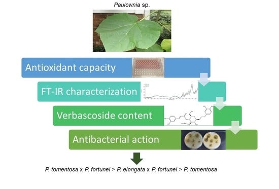
Antioxidant Activity, Polyphenolic Profiles and Antibacterial Properties of Leaf Extract of Various Paulownia spp. Clones

1
Department of Chemistry and Food Toxicology, Institute of Food Technology and Nutrition, University of Rzeszów, Ćwiklinskiej 1a, 35-601 Rzeszów, Poland
2
Department of Bioenergy, Food Analysis and Microbiology, Institute of Food Technology and Nutrition, University of Rzeszow, Zelwerowicza 4, 35-601 Rzeszow, Poland
3
Department of Plant Physiology and Biotechnology, Institute of Agricultural Sciences, Land Management and Environmental Protection, University of Rzeszów, Ćwiklinskiej 2, 35-601 Rzeszów, Poland
*
Author to whom correspondence should be addressed.
Agronomy 2021, 11(10), 2001; https://doi.org/10.3390/agronomy11102001
Submission received: 15 September 2021 / Revised: 29 September 2021 / Accepted: 1 October 2021 / Published: 2 October 2021

Abstract

:
Paulownia spp. are widely distributed ornamental trees with leaves abundant in secondary metabolites of high medicinal potential. Eighteen breeding clones of Paulownia spp. were tested in terms of their antioxidant activity and total polyphenolic contents. The 50% ethanolic extracts (2 g/30 mL) of leaves and petioles were compared in the screening step. Eight paulownia clones were selected for detailed analyses including HPTLC polyphenolic profile, verbascoside content and antibacterial activity against five bacteria species (S. aureus, B. cereus, E. coli, Y. enterocolitica, S. enterica). The species-specific differences in terms of antioxidant activity correlated with phenolic compounds were found mainly in the case of leaf blade extracts, the highest for P. tomentosa × P. fortunei and the lowest for P. elongata × P. fortunei clones. The P. tomentosa clones varied greatly in this regard. In the HPTLC polyphenolic profile, the occurrence of some polyphenols was proved and the specific verbascoside content was quantified (70 to 225 mg/g DW). The P. tomentosa × P. fortunei hybrids had the highest inhibitory activity, mainly against Gram-positive bacteria, whereas only slight inhibition of S. aureus growth was observed for P. elongata × P. fortunei clones. The obtained results indicate diverse suitability of paulownia clones as a source of active ingredients.

Graphical Abstract

1. Introduction

Paulownias (Paulownia spp.) are fast-growing deciduous trees planted mostly for their valuable wood and for ornamental purposes. They are known under many other names, among others: princess tree, royal tree, kiri tree or phoenix tree [1]. The genus Paulownia includes 17 species that naturally occur and grow in central China [2]. They are widely distributed throughout China, Korea and Japan [3]. Nowadays, having risen in popularity, paulownias are grown in many countries including North America, Australia, western and south Europe [4,5,6,7]. In the United States, the princess tree occurs in a variety of disturbed, high-light environments such as forest gaps and edges, stream banks and scoured riparian areas, steep rocky slopes, roadsides, fence-rows and vacant lots [8]. Most popular species are P. tomentosa, P. fortunei, P. elongata and P. catalpifolia [1,2]. Due to excessive spread, it was classified as an invasive species that threatens the native flora in the list of European and world invasive species [9,10,11]. However, paulownia species are not on such lists in Poland. In general, P. tomentosa is considered the most invasive species among others due to its higher resistance to low temperatures [12]. Moreover, hybrid species have theoretically reduced fertility, so they produce less fruit and seeds, which limits their spread.
Paulownia is a genus with increasing economic importance and has a very wide range of utilization [7]. Due to their tolerance to heavy metals, fast growth rate and well developed root system paulownias are used for phytoremediation, reclamation and reforestation of contaminated and devastated areas [2,13,14,15,16]. Paulownias—the C4-photosynthesis plants—belong to the fastest growing trees in the world [2,17]. They yield a higher biomass than other woody crops used for renewable energy purposes [18,19]. Paulownias have high relevance in the timber market [3,17,20]. Their leaves can be used as fertilizer or forage stuff. Their potential use as animal feed was also investigated [21]. The flowers are a source of nectar for honey bees [7,22,23].
Paulownias are considered as medicinal plants [4,7,24]. Their preparations can be used against gonorrhea and bruises, can stimulate hair growth, and prevent their graying [25]. Various preparations made of bark, fruit, xylem, or leaves are used in traditional Chinese medicine against various diseases (hemorrhoid, inflammatory bronchitis, gonorrhea, parotitis, asthma, erysipelas, bacteriologic diarrhea, bronchopneumonia, enteritis, high blood pleasure, and tonsillitis) [26]. Numerous phytochemical studies confirm that paulownia species produce many secondary metabolites, the most important of which are polyphenolic compounds with strong antioxidant properties [7,19,27]. Pharmacological studies have also reported antioxidant, anti-inflammatory, and antimicrobial activity against Staphylococcus aureus and Pseudomonas aeruginosa [28,29]. However, the chemical composition and biological properties of paulownia species still remain unexplored. Only a few compounds have been identified, mainly in flowers, including: phenolic glycosides, furofuran lignanes, furanoquinones, iridoides and flavonoids [27,30,31]. Among the secondary metabolites, phenylethanoids and phenylpropanoids occupy an important place, and their most abundant representative is verbascoside (acteoside), as well as its isomers and derivatives [27,32].
Due to their noteworthy characteristics, paulownias are being introduced in Poland. Based on the authors’ observation, the most promoted are two inter-specific hybrids: Spanish ‘Oxytree’ (P. elongata × P. fortunei, clone in vitro 112) and Chinese seed strain ‘Shan Tong’ (P. tomentosa × P. fortunei). Many Polish nurseries sell also seedlings of Paulownia tomentosa. As fast growing trees, which forming huge leaves (even 50–100 cm in width), paulownias are an efficient source of biomass, and probably secondary metabolites. It is a well-known fact that the content of active compounds is genotype/cultivar-dependent in many species. Such a problem was not thoroughly studied in the case of different paulownia cultivars and breeding clones.
As a part of our scientific efforts, we have attempted to find some paulownia clones that are characterized by high antioxidant and antibacterial active compounds in leaf extracts with their potential use in pharmacy and as animal feed additives. The differences in chemical composition were studied using Attenuated Total Reflectance Fourier-transform infrared (ATR FT-IR) spectroscopy and High Performance Thin Layer Chromatography (HPTLC), which are both simple and useful analytical methods, still rarely used in plant material characterization.

2. Materials and Methods

2.1. Plant Material

Different Paulownia spp. cultivars and breeding clones in the juvenile (or pseudo-juvenile) stage, obtained from single seedlings or plants, vegetatively propagated in vitro, were studied (Table 1). There were seven inter-specific hybrids (P. elongata × P. fortunei—2 and P. tomentosa × P. fortunei—5), including three cultivars (‘Oxytree’, ‘Cotevisa 2’, ‘Shan Tong’), and eleven P. tomentosa clones of different origin. Five sister clones derived from specimens or seedlings of fastest growing Polish seed strain ‘LuP’ after field or in vitro selection. One tetraploid (2n = 4x, ‘4C S9’) clone was also studied. All plants were micropropagated in Department of Plant Physiology and Biotechnology (University of Rzeszow, Poland) following standard procedure for this genus [33]. The plants were grown on the field paulownia collection established in Central Poland (Mazovia; 51°43′51.4″ N 21°45′54.5″ E) in July 2018. The fully expanded, healthy-looking leaves were collected from the top of several-months-old strong shoots (rods) in October 2020. The paulownia leaf bulk samples were dried at a room temperature, without exposure to sunlight and grounded in a laboratory mill (MMK-06M, MPM, Milanówek, Poland), separately leaf blades and petioles.

2.2. Experimental Design

The first step of the study was the screening of all 18 tested paulownia clones originating from the Plant Biotechnology Laboratory “Aeropolis” collection, in terms of antioxidant activity as well as total polyphenols and flavonoids content. The leaf blades and petioles were analyzed separately. Based on the obtained results, the 8 clones were selected for detailed analysis (step 2), including 4 with the highest and 4 clones with the lowest antioxidant activity. In this step of the study, dried extracts of leaf blades only were obtained and characterized by FT-IR and HPTLC methods. Finally, the antibacterial activity of leaf extracts was checked.

2.3. Extracts Preparation

2.3.1. Crude Extract

Two grams of pulverized dried plant material was extracted with 30 mL of aqueous ethanol (50%, v/v) for 20 min in ultrasonic bath (Sonic-10, Polsonic, Warsaw, Poland). The crude extracts were then filtered through filter paper and stored in a freezer until further analyses. The extracts were used for antioxidant assays.

2.3.2. Freeze-Dried Extract

An aliquot of 10 mL of each crude extract was subjected to ethanol evaporation using a rotational vacuum concentrator (RVC 2–18 CDPlus, Martin Christ, Osterode am Harz, Germany). The residue was freeze-dried using Alpha 1–2 LD plus lyophilizer (Martin Christ, Osterode am Harz, Germany) to obtain a dry extract used in the second step of the experiment.

2.4. Antioxidant Activity Determination

2.4.1. FRAP Assay

The FRAP assay (Ferric Reducing Antioxidant Power) was carried out according to Bertoncelj et al. [34]. The FRAP reagent contained 2.5 mL of a 10 mM 2,4,6-tripyridyltriazine (TPTZ) solution in 40 mM HCl, 2.5 mL of 20 mM FeCl3 and 25 mL of 0.3 M acetate buffer (pH 3.6). Briefly, to 0.02 mL of plant extract, 0.18 mL FRAP reagent was added and the absorbance of the reaction mixture was measured spectrophotometrically using a microplate reader (EPOCH 2) against blank at 593 nm after 10 min incubation at 37 °C. The results were expressed as μmol of Trolox (TE) equivalents per gram of plant dry mass (μmol TE/g) based on calibration curve (y = 0.026x, R2 = 0.9989), prepared Trolox in range 15–200 nmol per sample.

2.4.2. DPPH Test

2,2-diphenyl-1-picrylhydrazyl radical (DPPH) inhibition was measured according to assay described by Blois [35] with minor modifications. The aliquot of 0.02 mL plant extract was mixed with 0.18 mL of DPPH radical methanolic solution (0.1 mM) and kept in the dark for 60 min. After incubation, the absorbance of tested and control samples was measured at 517 nm against methanol using microplate reader (EPOCH 2). The reduction of DPPH radical was calculated using following equation:
DPPH% = [(Ao − As)/Ao] × 100
where Ao is the absorbance of control, and As is the absorbance of tested samples. The results were calculated for Trolox equivalents using a calibration curve in the range 0.5–6 μmol per sample (y = 15.553x, R2 = 0.9970).

2.4.3. ABTS Test

The antioxidant capacity was determined using the ABTS radical monocation test (2,2′-azino-bis(3-ethylbenzothiazoline-6-sulfonic acid) according to the method described by Re et al. [36] with slight modifications. ABTS solution (19.5 mg ABTS in 7.00 mL of distilled water) was mixed with 3.3 mg of potassium persulfate and stored in the dark for 24 h. Before use, it was diluted with phosphate buffer (0.1 M, pH = 7.4) to obtain an absorbance value at 734 nm of about 0.7. Then, 0.02 mL of each diluted extract was mixed with 0.18 mL of diluted ABTS radical solution. After 6 min incubation in the dark, the absorbance of each sample was recorded at 734 nm in an EPOCH 2 microplate reader against phosphate buffer. Results were calculated using the following equation:
ABTS% = [Ao − As)/Ao] × 100
where Ao is the absorbance of the control and As is the absorbance of the tested samples. The results were calculated for Trolox equivalents using a calibration curve in the range 0.5–6 μmol per sample (y = 9.0447x, R2 = 0.9948).

2.5. Total Phenolic Content (TPC) Determination

Total phenolics content was determined using Folin–Ciocalteu reagent, according to Singleton and Rossi [37] with minor modifications. To 0.02 mL of plant extract, 0.1 mL of 10% Folin–Ciocalteu reagent followed by 0.08 mL of 7.5% (w/v) of Na2CO3 solution were added. Samples were kept in the dark for 120 min, and then the absorbance was measured against blank at 760 nm using a microplate reader (EPOCH 2 microplate spectrophotometer, BioTek, Winooski, VT, USA). The phenolic content expressed as mg of gallic acid (GAE) equivalents per gram of plant dry mass (mg GAE/g) was calculated based on calibration curve prepared for gallic acid in the range 0–250 µg/mL (y = 0.0555x, R2 = 0.9976).

2.6. Total Flavonoid Content (TFC) Determination

The total content of flavonoids in the extracts was assessed using the method of Biju [38]. Briefly, 0.1 mL of the undiluted extract was mixed with 0.1 mL of 2% AlCl3 (in methanol). The reaction mixture was incubated for 10 min at room temperature until completion of the reaction. The absorbance of the solution was then measured at 415 nm with a microplate reader (EPOCH 2) against methanol blank. The total content of flavonoids in the extracts was expressed in mg of quercetin equivalent (QE) per gram of plant dry mass (mg QE/g). The results were calculated on the basis of a calibration curve prepared for quercetin in the range 0–125 µg/mL (y = 0.0655x, R2 = 0.9999).

2.7. FT-IR Analysis

FT-IR (Fourier-transform infrared spectroscopy) spectra for selected clones (leaf powder and dry extract) were collected using a Nicolet iS10 FT-IR spectrometer (Thermo Scientific, Waltham, MA, USA) equipped with a Smart iTX ATR sampling accessory allowing the measurements by the attenuated total reflectance (ATR) technique, fast recovery deuterated triglycine sulfate (DTGS) detector and Ge/KBr beamsplitter. The spectra were recorded in absorbance mode in the mid-infrared range (4000 to 450 cm−1), with a resolution of 0.4 cm−1. Sixteen scans were collected for each sample. The spectra were analyzed using Omnic 8.1.11 software (Thermo Scientific).

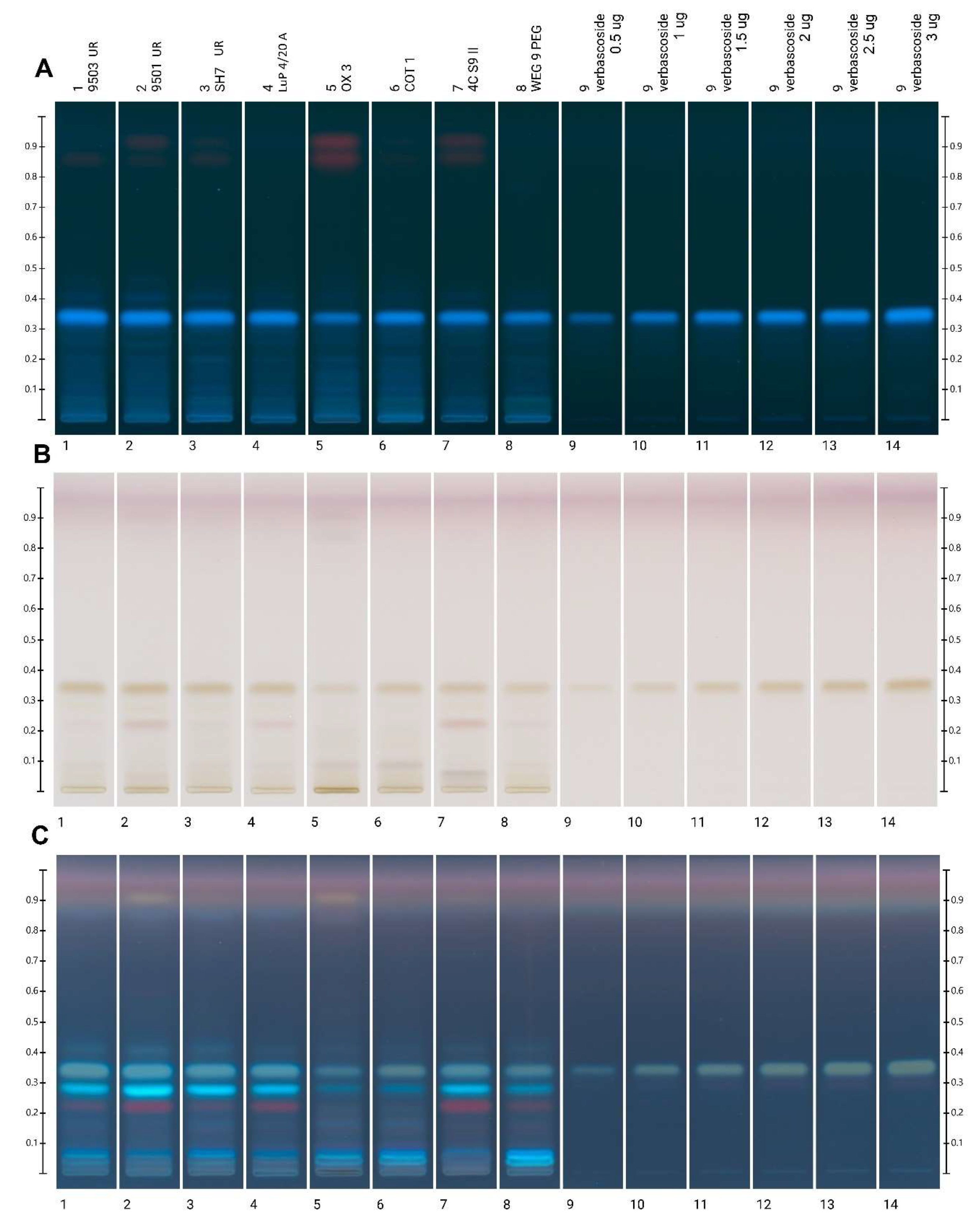
2.8. Polyphenolic Profiles by HPTLC and Verbascoside Quantification

Analysis of dry extracts reconstituted in 50% ethanol in concentration of 1 mg/mL were performed on HPTLC Silica Gel 60 F254 plates (20 cm × 10 cm) purchased from Merck (Darmstadt, Germany). Ten microliters of each extract were applied to the plate as 11 mm bands from the lower edge of the plate at rate of 50 nL/s using a semi-automated HPTLC application device (Linomat 5, CAMAG, Muttenz, Switzerland). The chromatographic separation was performed in a chromatographic tank saturated for 20 min with the mobile phase composed of ethyl acetate:water:formic acid:acetic acid [15:2:1:1 v/v/v/v], and developed to a distance 70 mm. The obtained results were documented using an HPTLC imaging device (TLC Visualizer, CAMAG) under white light, 254 and 366 nm. In addition, each plate was derivatized with p-anisaldehyde sulfuric acid reagent using automated derivatizer of TLC plates (CAMAG Derivatizer). After derivatization, the plates were heated at 110 °C for 10 min and imaged under white light and 366 nm. The obtained chromatographic images were analyzed using the HPTLC software (Vision CATS, CAMAG).
The quantitative analysis was performed by applying a verbascoside solution in the range of 0.5 to 3 µg on the HPTLC plate. The image of the plate after derivatization in visible light was used for the quantitative analysis. A standard curve (y = 2.639·10−9x + 1.373·10−3, R2 = 0.9890) was constructed taking into account the peak area of the chromatograms generated by the software.

2.9. Antibacterial Activity

Five different bacterial strains were used for antibacterial determination of plant extracts: Gram positive: Staphylococcus aureus ATCC 25923, Bacillus cereus PCM 482 and Gram negative ones: Escherichia coli PCM 2561, Yersinia enterocolitica PCM 2080 and Salmonella enterica PCM 840. The antimicrobial properties of paulownia extracts (after ethanol evaporation and replacement by water) were determined by agar well diffusion method according to National Committee of Clinical Laboratory Standards [39] and described by Borycka et al. [40]. Briefly, each bacterial culture was incubated for 24 h at 37 °C on TSA (Tryptic Soy Agar Medium, Biomaxima, Poland). Then, agar plates with MHA medium (Mueller–Hinton agar, BTL, Poland) were inoculated with each of bacterial suspension containing 106 CFU/mL, spread on the agar plates. Subsequently, wells of 8 mm diameter were punched into the agar medium with a sterile cork borer and filled with 100 μL of plant extract and allowed to diffuse at room temperature for 2 h. Bacteria were incubated at 37 °C for 24 h. After incubation, the diameters of the growth inhibition zones were measured in mm. Gentamycin (30 µg) was used as a positive control.

2.10. Statistical Analyses

All calculations were done in three replications and presented as means with standard deviations. The correlations among obtained parameters were analyzed by Pearson coefficient (r). The significant differences were calculated by a one-way analysis of variance followed by Tukey’s honest significant difference test (p < 0.05). Principal components analysis (PCA) was applied to find the relation between tested parameters for paulownia leaves. All calculations were made using the Statistica 13.1 software (StatSoft, Tulsa, OK, USA).

3. Results

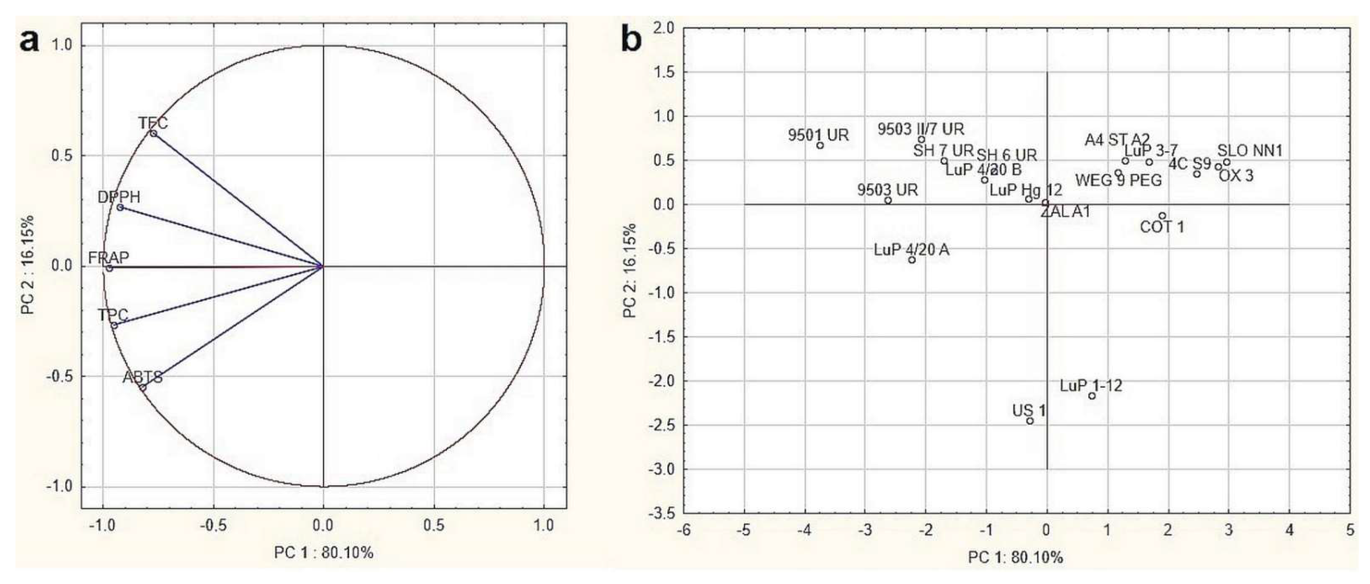
3.1. Screening for the Clone of the Best Antioxidant Potential

The antioxidant potential of paulownia leaf extracts was found to be very diverse regardless of the analytical method used (Table 2). The highest values (more than twice) of tested parameters were found for sample no. 3 (9501 UR), and the lowest for sample no. 1 (OX 3). The observed differences were significant (p < 0.05) in the most cases for blades (Tables S1–S5) and petioles (Tables S6–S10). Regardless the sample, great differences between leaf blades and petioles extracts activity were found, depending the method by 4–9 times lower in petioles. The samples with higher values of antioxidant measures were abundant in polyphenols and the share of flavonoids in the polyphenol fraction ranged from 60–86%, excluding US 1 and LuP 1–12 clones (by 24% only).
The results for the antioxidant activity of leaf extracts showed strong positive correlations with the DPPH, FRAP, ABTS, TPC, and TFC parameters both for the blades (Table S11) and petioles (Table S12) measured by Pearson correlation coefficient (r = 0.838–0.929). The relationships between the determined parameters for leaf blades were only verified by principal component analysis (PCA) (Figure 1). Variables displayed in Figure 1a confirmed the strongest positive correlation between the DPPH, FRAP, ABTS, TPC, and TFC methods, demonstrated by close proximity. Moreover, it can be noticed that all examined parameters had a significant impact on overall leaf extract quality due to their location close to the projection circle. The analysis showed that the tested samples were characterized by high heterogeneity (Figure 1b); however, based on the tested parameters it was possible to classify the samples in terms of paulownia species. The leaf extracts samples were divided into four separate groups (Figure 1b). Samples with the high values of tested parameters were located on the upper-left part of the graph, these were mainly P. tomentosa × P. fortunei clones and some P. tomentosa exhibited higher activity. A particularly significant difference was observed for samples with lower activity, localized in the upper-right, including P. elongata × P. fortunei hybrids OX 3 and COT 1. Samples with very low content of flavonoids (US 1 and LuP 1–12) were located separately in different sides of the plot.

3.2. Detailed Characteristic of Biological Activity of Selected Paulownia Clones Leaves

For the next step of study the leaf blades of 8 paulownia clones: 4 with the highest (‘9503 UR’, ‘9501 UR’, ‘SH7UR’, ‘LuP 4/20A’) and 4 with the lower (‘OX3’, ‘COT1’, ‘4CS 9’, ‘WEG 9 PEG’) antioxidant activity were selected. The extracts were subjected to the following analyses: antioxidant activity of freeze-dried extracts, FT-IR spectra analysis, polyphenolic HPTLC profile, verbascoside content and antibacterial activity.

3.2.1. Antioxidant Activity

The highest antioxidant activity, polyphenols and flavonoid content for the samples obtained for P. tomentosa × P. fortunei hybrids were observed whereas the lowest one for P. elongata × P. fortunei clones (Table 3). Due to concentration effect the results are relatively higher; however, are consistent with data presented for crude extracts (Table 2).

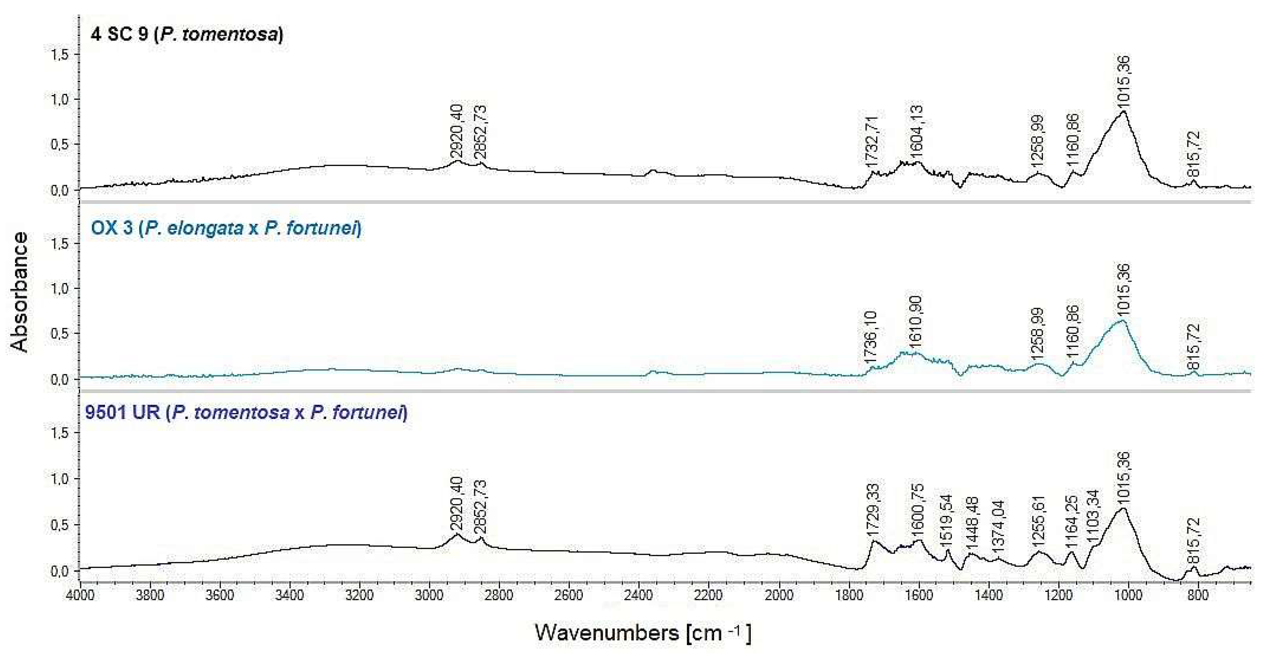
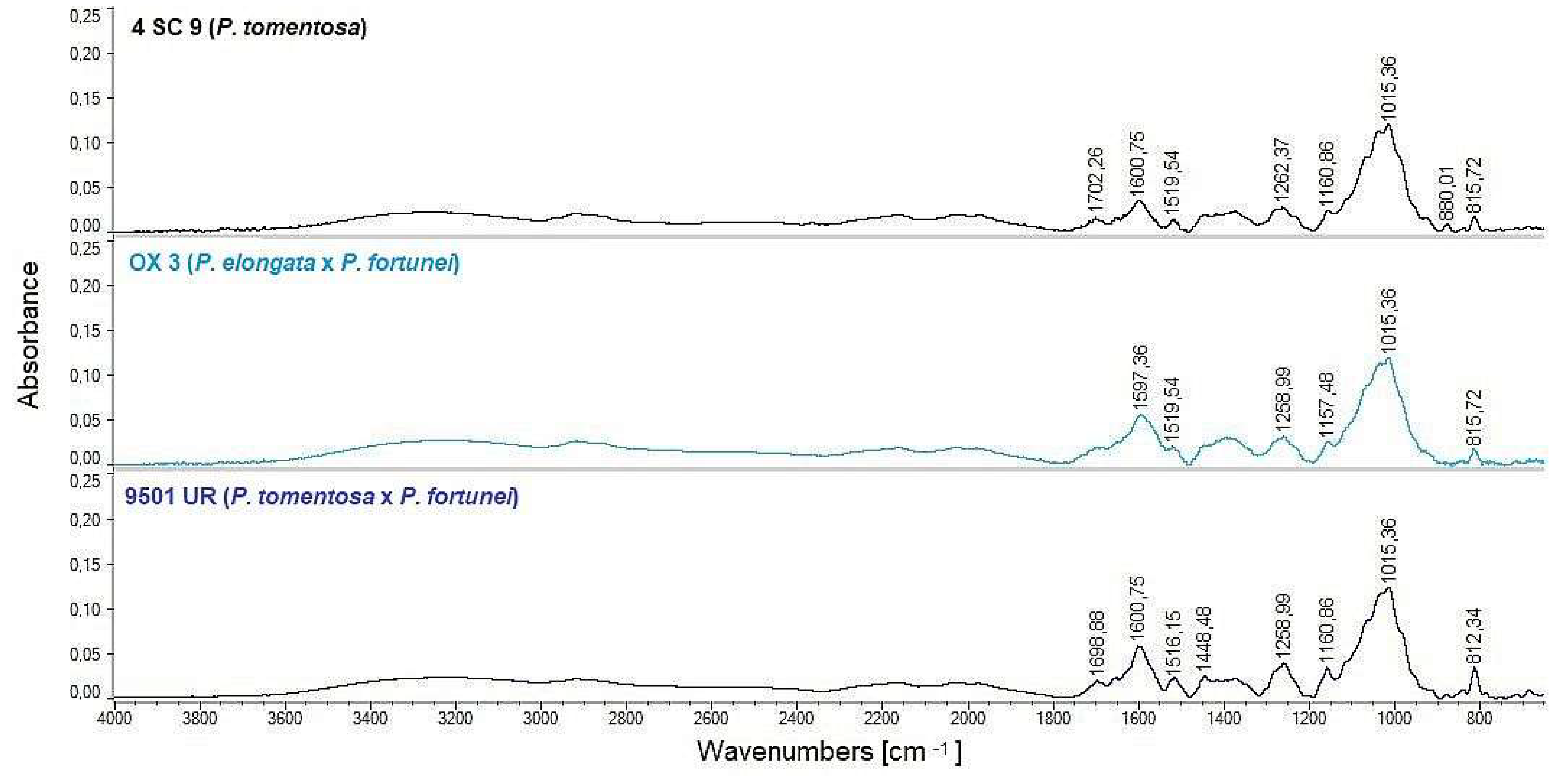
3.2.2. FT-IR Spectra Analysis

FT-IR spectra were made for selected clones including powdered leaf blades (Figure 2) and dry extracts (Figure 3) samples. Chosen spectra for representatives of individual clones are only shown.
On the FT-IR spectra (Figure 2), for powdered leaf blades, the bands and regions with the greatest differences among the studied clones were indicated. These are the bands at 2840–2870, 1733, 1516, 1157 and 810 cm−1. The indicated bands are most visible in the spectrum for the hybrid clone P. tomentosa × P. fortunei, in the remaining cases they are less intense or almost indistinguishable.
On the spectra obtained for dry extracts (Figure 3), several bands distinguishing spectra obtained from various clones compared to raw plant material spectra were found. The bands at 1601, 1148 and 812 cm−1 were significantly enhanced, while the bands in the range 2840–2870 and at 1733 cm−1 are significantly weakened.

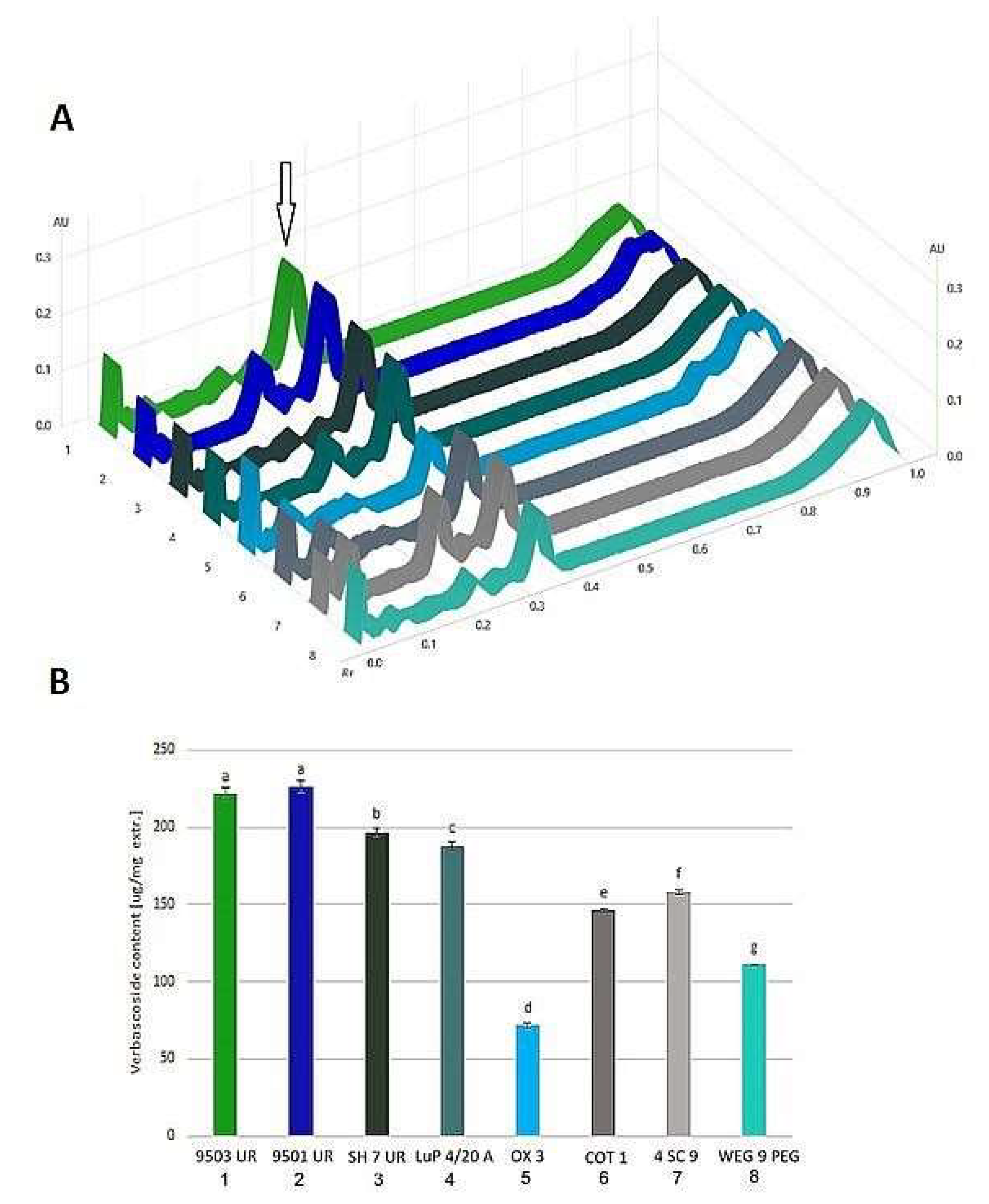
3.2.3. HPTLC Analysis

Leaf extracts of selected paulownia clones were analyzed by HPTLC for the polyphenol profile. Detection was performed by visualizing the chromatogram under UV light 366 nm and also by derivatization with reagent p-anisaldehyde sulfuric acid reagent (Figure 4). Under UV light (Figure 4A) numerous blue bands are visible, the most intense at Rf = 0.33. Some samples also have orange bands with high Rf values (0.8–0.9). Derivatization caused the strands to be dyed mostly green-brown (Figure 4B). In UV light, after derivatization (Figure 4C) the bands assumed different colors: pale yellow in the range of Rf 0.8–0.9, gray at Rf = 0.41, 0.33 and 0.19, intense blue at Rf = 0.25 and 0.07, and red at Rf = 0.23.
On the basis of the standard curve of verbascoside applied to the plate, its content in individual samples was estimated (Figure 5A). Determined verbascoside content was highest in the four clones selected as “stronger”: from 188 for ‘LuP 4/20 A’ to 228.8 µg/mg of extract for ‘9501 UR’ clone. The four clones selected as “weaker” contained significantly less of this compound (p < 0.05), the least in the ‘OX 3’ clone (71.5 µg/mg of extract) (Figure 5B). Verbascoside content was also positively correlated with antioxidant capacity results with Pearson’s coefficient values 0.79, 0.78, 0.73, and 0.76 for FRAP, DPPH, ABTS, and TPC, respectively.

3.2.4. Antibacterial Activity

Leaf extracts of paulownia clones showed different antibacterial activity against five tested pathogenic bacteria (Table 4). The antibacterial potential of all tested extracts was observed mainly against Gram positive bacteria—S. aureus and B. cereus. However, there were two exceptions—clones ‘OX3’ and ‘COT1’ did not inhibit B. cereus growth and had the lowest antibacterial potential against S. aureus. The best inhibitory activity against S. aureus was observed for clones ‘9501 UR’, ‘LuP 4/20A’, and ‘4CS9’ with inhibition zones from 19.33 to 21.75 mm. Moderate antibacterial potential against S. aureus was observed for ‘9503 UR’, ‘SH7 UR’ and ‘WEG 9PEG’ clones. The most resistant bacteria were Gram negative S. enterica and E. coli as no inhibition growth was observed for all tested leaves extracts. Clones ‘4CS9’, ‘WEG 9PEG’ and ‘9501 UR’ inhibited growth of Y. enterocolitica the most, while clones ‘OX 3’ and ‘COT 1’ did not inhibit them.

4. Discussion

4.1. Clone-Specific Antioxidant Activity of Leaves

Paulownia, considered as a rich source of biologically active secondary metabolites, is traditionally used in Chinese herbal medicine. More than 130 physiologically active constituents have been isolated from different parts of the paulownia plant [32]. Their biological activity has been tested using both the isolated compounds and different types of extracts. As wide variety bioactive compounds occurring in plant extracts, mutually interacted to neutralize free radical groups the evaluation of antioxidant capacity of extract is usually a first step of sample bioactivity assessment. Thus, in the first step of the study the comparison of antioxidant activity of rested paulownias leaves was performed. Regarding the species origin of the leaves the antioxidant activity increased in order:
P. elongata × P. fortunei < P. tomentosa < P. tomentosa × P. fortunei
The most active were P. tomentosa × P. fortunei hybrid clones selected in University of Rzeszow from Chinese seed strains (‘9501’, ‘9503’ and ‘SH’) The antioxidant activity was highly correlated with total polyphenolics and flavonoids content for blades (Table S11) and petioles (Table S12) extracts. The greatest variability was observed in the case of P. tomentosa, where the best results for ‘LuP 4/20 A’ and the weakest for ‘LuP 1–12’ clones were found. It seems that increasing the level of ploidy from diploid to tetraploid slightly change the biochemical properties of Paulownia clones (‘WEG 9’ in comparison with ‘4C S9). The antioxidant activity of petiole extracts was much lower than leaf blade extracts (Table 2), by 2–11 times regardless of the used method. Similarly, the total polyphenols content was significantly lower in petioles compared to leaves whereas a much lower (about 10 times) content of flavonoids was noted (Table 2). As the petioles were notably poorer in antioxidant components the only leaf blades were chosen for the further study.

4.2. FT-IR Comparison of Chemical Composition

The comparison of FT-IR spectra of powdered leaf blades between paulownia clones support some information about differences in chemical composition. The identification of the major chemical groups of the examined compounds is based on the “fingerprint” region (950–1200 cm−1) of the spectra. The position (frequency) and intensity of the bands are characteristic of each compound. Based on earlier findings [41,42], specific main ranges of wave numbers can be superimposed to main components: 2840–2870 cm−1 to protein (peptide bonds), 3270–3290 cm−1 to carbohydrates, and 2840–2870 cm−1 to lipids. When comparing the spectra for tested paulownia species leaves regarding mentioned bands, it can be concluded that there are some differences in the chemical composition of the leaves among species and clones. Two bands in the 2840–2870 cm−1 range, especially visible in the P. tomentosa and P. tomentosa × P. fortunei clones, are attributed to the lipid vibration. The band at 1733 cm−1, clearly outlined in the spectrum of P. tomentosa × P. fortunei leaves, comes from the vibration of C=O bonds in ester groups (phospholipids, sterols, hemicellulose and pectin esters) [41]. This may indicate a greater abundance of such compounds in these clones. The range from 800 to 1400 cm−1, with the most intense band at 1014 cm−1 corresponds mainly to the vibrations of glycosidic and ester bonds in polysaccharides building cell walls [41,42]. In this range, for the analyzed clones, differences in the intensity of the bands derived from cellulose (1145 cm−1) and in the range of 800–840 cm−1 are visible. The latter range may be due to the vibrations of the aromatic rings [42], which may be related to the higher phenolic content in the P. tomentosa × P. fortunei clones. This observation is consistent with spectrophotometric analysis results.
Analyzing the FT-IR spectra obtained for dry extracts of leaves, a few changes of chemical composition were observed: some compounds were eliminated and others were concentrated.
The bands assigned to lipids were almost as invisible (2840–2870 cm−1) as the applied extraction system eliminates lipids, extracting only more polar compounds. The bands at 1254 cm−1 (pectin) and at 812 cm−1 (C–H bending vibrations of aromatic compounds) found in extracts of P. tomentosa × P. fortunei were more intense. The higher intensity of these bands may be related to the higher content of phenolic compounds in the analyzed hybrid. Similar FT-IR spectra for P. tomentosa leaf extracts were previously obtained by Pontaza-Licona et al. [43]. The authors indicate that the form of the FT-IR spectrum depends on the solvent used for the extraction. They also pointed to the polyphenol bands in aqueous ethanol extract at 1328 and 1274 cm−1 (bending vibrations of O-H and C-O bonds, respectively). In our study, the band close to 1274 cm−1 was clearly outlined in the spectrum of P. tomentosa × P. fortunei hybrid extract (average peak intensity 0.146 mAU), compared to 0.097 and 0.120 for P. tomentosa and P. elongata × P. fortunei, respectively. The intensity of this band was significantly correlated with the content of phenolic compounds (r = 0.908). The use of this rapid analytical technique allows an easy comparison of the overall chemical composition of both the raw material and the obtained extracts.

4.3. HPTLC Polyphenolic Profile and Verbascoside Quantification

The HPTLC technique was previously used to characterize the phenol profiles of individual paulownia species, and the antioxidant activity of individual metabolites was also analyzed by derivatization with DPPH solution [44]. In chromatograms obtained by us, a main compound was verbascoside visible at Rf = 0.33 as an intensely blue band (in UV 366 nm, Figure 4A) or after derivatization with p-anisaldehyde sulfuric acid reagent as a green-brown (in white light) or grey-blue band (in UV 366 nm, Figure 4B,C). Verbascoside (acteoside) is the dominant secondary metabolite of paulownia, belonging to the group of phenylethanoid glycosides [27,45,46]. This metabolite is known for its beneficial effects as a strong antioxidant, anti-inflammatory, anti-neoplastic, neuroprotective and wound healing agent [47]. Adach et al. [46] studied verbascoside content in P. elongata × P. fortunei Clone 112 in vitro. In methanolic extract of leaves they found a content ranging from 74.6 to 424.7 mg/g of dry extract. Šmejkal et al. [27], found the verbascoside content in the ethyl acetate and n-butanol extracts of P. tomentosa fruits in the range of 0.07 and 0.09 mol/100 g of dry extract, respectively, which corresponds to 437 and 562 µg/mg of extract. It is obvious that the content of verbascoside as well as other secondary metabolites is strictly dependent on the part of the plant used as raw material, plant development phase and the extraction method used. Despite the lower content of verbascoside in the leaves, determined by us, it should be stated that it is a better source of it, due to its easier availability and greater mass. Moreover, paulownia flowers are not available every season and their harvest is much troublesome.
Apart from verbascoside, there are some bands from other compounds, possibly its isomers (gray-blue bands at Rf = 0.41 after p-anisaldehyde derivatization). The intense blue bands visible in UV after derivatization (especially at Rf = 0.29 and 0.05, Figure 4C) probably came from flavonoid compounds. The leaves of paulownia contain numerous flavonoids, including luteolin, quercetin, apigenin and their derivatives and wide group of C-geranylated flavonoids [32,48]. Among unidentified compounds, particularly interesting was the band with Rf = 0.22, which glowed red in UV light after p-anisaldehyde derivatization. It was practically absent in P. elongata × P. fortunei hybrid sample profiles. Thus, it can be assumed as a specific compound for P. tomentosa. Such a metabolite could potentially be a good chemical marker for species discrimination. In UV light (before derivatization), signals in the form of orange bands from chlorophylls were also visible, with Rf = 0.85 and 0.91), especially intense in the case of OX 3 and 4C S9 II clones. An interesting combination of HPTLC techniques with the analysis of antibacterial properties (bioautography) was applied to the studies of methanolic leaf extracts of P. tomentosa by Moricz et al. [49]. They showed the presence of compounds such as p-coumaric acid and apigenin and their strong activity against Bacillus subtilis and Aliivibrio fischerii bacteria.

4.4. Clone-Specific Antibacterial Activity

Phenolic compounds are secondary metabolites in plants and are considered as important natural molecules due to their bioactive properties [50]. These compounds present in different concentrations in paulownia leaves extracts can be responsible for their antibacterial potential, which was confirmed by positive correlation between antioxidant properties, phenolic content and antibacterial activity. The weakest antibacterial properties of two clones (‘OX 3’ and ‘COT1’) was correlated with their lowest antioxidant potential and total phenolic content. On the other hand the highest antibacterial activity was observed for clones with the highest phenolic content and antioxidant activity. Moreover, one of the most dominated secondary metabolite in paulownia is verbascoside. This compound has several biological properties such as anti-inflammatory, antioxidant, and antitumor, can be a promising therapeutic agent against different microbial infections and can synergistically increase the antimicrobial effectivity of antibiotics, like gentamycin [51,52,53]. We can observe that clones with the lowest verbascoside content (‘OX3’ and’ COT1’) have the weakest antibacterial properties, while those with the highest verbascoside content show the strongest antibacterial properties. The possible mechanism of antibacterial action of verbascoside is correlated with its capability to modulate membrane-dependent cellular processes [49].
Antibacterial properties of P. elongata water leaves extracts were also observed by Popova and Baykov [22] against such microorganisms as: Salmonella enterica, Pseudomonas aeruginosa, Staphylococcus aureus, Streptococcus pyogenes, Paenibacillus alvei, and Candida albicans. We did not observed antibacterial potential against S. enterica in paulownia ethanolic leaves extracts. It can be explained by using different solvent for extraction or different species of paulownia.
We proved that paulownia leaves could be a good source for biologically active substances with high antioxidant and antibacterial potential. It is a common observation that juvenile (and pseudojuvenile) paulownia plants form huge leaves even more than 50 cm in width [4,54]. Moreover, we found also that fresh mass of leaves surpassed the fresh mass of several month old stems (Litwińczuk, personal observation, data not presented). As paulownia plantations in Poland could yield more than 10 t/ha stems they could produce similar mass of leaves [54]. A paulownia tree that is 8–10 years old is reported to have 100 kg fresh leaves and produces 350–400 kg of branches [2]. Thus, paulownia leaves could be considered as easily accessible and abundant source of verbascoside or can serve as effective antimicrobial feed additive.

5. Conclusions

Leaves, separately blades and petioles, collected from 18 paulownia clones, including eleven P. tomentosa, five hybrids P. tomentosa × P. fortunei, and two P. elongata × P. fortunei, were examined. Among tested clones, self-selected in vitro P. tomentosa × P. fortunei hybrids (marked UR*) were characterized by the best biological activity. Analysis of antioxidant properties showed higher potential for leaf blades than petioles. The newly generated hybrid clones P. tomentosa × P. fortunei stood out for the highest antioxidant capacity as well as total polyphenols and flavonoids content. Such species-dependent differences were also observed on FT-IR spectra of raw material and dry extracts. Within P. tomentosa clones, a great variability of tested parameters was observed. The weakest properties for hybrids P. elongata × P. fortunei were found. Verbascoside, a dominant metabolite identified by HPTLC, was chosen as a bioactivity marker and quantified in selected samples. Its content was significantly correlated with antioxidant activity and total polyphenols. The high inhibitory effect against some tested bacteria, including S. aureus, B. cereus and Y. enterocolitica, was found in the case of P. tomentosa × P. fortunei clones, whereas P. elongata × P. fortunei was far less effective. The obtained results indicate a potential application of selected paulownia clones in pharmaceutical and fed industry.

Supplementary Materials

The following are available online at https://www.mdpi.com/article/10.3390/agronomy11102001/s1, Table S1. Statistical differences for results obtained with FRAP method for paulownia leaf blades. Table S2. Statistical differences for results obtained with DPPH method for paulownia leaf blades. Table S3. Statistical differences for results obtained with ABTS method for paulownia leaf blades. Table S4. Statistical differences for total phenolic content (TPC) for paulownia leaf blades. Table S5. Statistical differences for total flavonoid content (TFC) for paulownia leaf blades. Table S6. Statistical differences for results obtained with FRAP method for paulownia leaf petioles. Table S7. Statistical differences for results obtained with DPPH method for paulownia leaf petioles. Table S8. Statistical differences for results obtained with ABTS method for paulownia leaf petioles. Table S9. Statistical differences for total phenolic content (TPC) for paulownia leaf petioles. Table S10. Statistical differences for total flavonoid content (TFC) for paulownia leaf petioles. Table S11. Correlation matrix for paulownia leaf blades. Table S12. Correlation matrix for paulownia leaf petioles.

Author Contributions

Conceptualization, W.L. and M.D.; methodology, M.D.; software, M.M.; validation, M.M. and D.G.-L.; formal analysis, W.L.; investigation, M.M., D.G.-L., J.H. and B.J.; resources, W.L.; data curation, M.M. and D.G.-L.; writing—original draft preparation, M.M., D.G.-L. and W.L.; writing—review and editing, M.D.; visualization, M.M.; supervision, M.D. and W.L.; project administration, M.D.; funding acquisition, W.L. All authors have read and agreed to the published version of the manuscript.

Funding

This work was supported by the project financed under the program of the Minister of Science and Higher Education of Poland entitled “Regional Initiative of Excellence” in 2019–2022 (project no. 026/RID/2018/19).

Institutional Review Board Statement

Not applicable.

Informed Consent Statement

Not applicable.

Data Availability Statement

The data presented in this study are available in the article and supplementary material.

Conflicts of Interest

The authors declare no conflict of interest.

References

  1. Icka, P.; Damo, R.; Icka, E. Paulownia Tomentosa, a Fast Growing Timber. Ann. Valahia Univ. Targoviste Agric. 2016, 10, 14–19. [Google Scholar] [CrossRef] [Green Version]
  2. Woods, V.B. Paulownia as a Novel Bio-Mass Crop for Northern Ireland? A Review of Current Knowledge; Occasional Publ. Agri-Food and Biosciences Institute: Hillsborough, FL, USA, 2008. [Google Scholar]
  3. Akyildiz, M.H.; Kol, H.S. Some technological properties and uses of paulownia (Paulownia tomentosa Steud.) wood. J. Environ. Biol. 2010, 31, 351–355. [Google Scholar] [PubMed]
  4. Zhu, Z.H.; Chao, C.J.; Lu, X.Y.; Xiong, Y.G. Paulownia in China: Cultivation and Utilization; Asian Network for Biological Sciences: Singapore, 1986; pp. 1–65. [Google Scholar]
  5. Barton, I.L.; Nicholas, I.D.; Ecroyd, C.E. Paulownia. For. Res. Bull. 2007, 231, 5–68. [Google Scholar]
  6. Essl, F. From ornamental to detrimental? The incipient invasion of Central Europe by Paulownia tomentosa. Preslia 2007, 79, 377–389. [Google Scholar]
  7. Yadav, N.K.; Vaidya, B.N.; Henderson, K.; Lee, J.F.; Stewart, W.M.; Dhekney, S.A.; Joshee, N. A Review of PaulowniaBiotechnology: A Short Rotation, Fast Growing Multipurpose Bioenergy Tree. Am. J. Plant Sci. 2013, 4, 2070–2082. [Google Scholar] [CrossRef] [Green Version]
  8. Snow, W.A. Ornamental, crop, or invasive? The history of the Empress tree (Paulownia) in the USA. For. Trees Livelihoods 2015, 24, 85–96. [Google Scholar] [CrossRef]
  9. TISI 2019. The Texas Invasive Species Institute. Available online: http://www.tsusinvasives.org/home/database/paulownia-tomentosa (accessed on 29 April 2021).
  10. DAISIE. Delivering Alien Invasive Species Inventories for Europe. 2018. Available online: http://www.europe-aliens.org/speciesFactsheet.do?speciesId=18222 (accessed on 29 April 2021).
  11. GISD. Global Invasive Species Database. 2018. Available online: http://www.iucngisd.org/gisd/search.php (accessed on 29 April 2021).
  12. Berg, E.C.; Zarnoch, S.J.; McNab, W.H. Survivorship, attained diameter, height and volume of three Paulownia species after 9 years in the southern Appalachians, USA. J. For. Res. 2020, 31, 2181–2191. [Google Scholar] [CrossRef] [Green Version]
  13. Doumett, S.; Lamperi, L.; Checchini, L.; Azzarello, E.; Mugnai, S.; Mancuso, S.; Petruzzelli, G.; Del Bubba, M. Heavy metal distribution between contaminated soil and Paulownia tomentosa, in a pilot-scale assisted phytoremediation study: Influence of different complexing agents. Chemosphere 2008, 72, 1481–1490. [Google Scholar] [CrossRef]
  14. Stankovic, D.; Nikolic, M.S.; Krstic, B.; Vilotic, D. Heavy metals in the leaves of tree species paulownia elongata s.y.hu in the region of the city of Belgrade. Biotechnol. Biotechnol. Equip. 2009, 23, 1330–1336. [Google Scholar] [CrossRef]
  15. Wang, J.; Zhang, C.B.; Jin, Z.X. The distribution and phytoavailability of heavy metal fractions in rhizosphere soils of Paulowniu fortunei (seem) Hems near a Pb/Zn smelter in Guangdong, PR China. Geoderma 2009, 148, 299–306. [Google Scholar] [CrossRef]
  16. Azzarello, E.; Pandolfi, C.; Giordano, C.; Rossi, M.; Mugnai, S.; Mancuso, S. Ultramorphological and physiological modifications induced by high zinc levels in Paulownia tomentosa. Environ. Exp. Bot. 2012, 81, 11–17. [Google Scholar] [CrossRef]
  17. Ayan, S.; Sivacioglu, A.; Bilir, N. Growth variation of Paulownia Sieb, and Zucc. species and origins at the nursery stage in Kastamonu-Turkey. J. Environ. Biol. 2006, 27, 499–504. [Google Scholar]
  18. López, F.; Pérez, A.; Zamudio, M.A.M.; De Alva, H.E.; García, J.C. Paulownia as raw material for solid biofuel and cellulose pulp. Biomass Bioenergy 2012, 45, 77–86. [Google Scholar] [CrossRef]
  19. Durán Zuazo, V.H.; Jiménez Bocanegra, J.A.; Perea Torres, F.; Rodríguez Pleguezuelo, C.R.; Francia Martínez, J.R. Biomass Yield Potential of Paulownia Trees in a Semi-Arid Mediterranean Environment (S Spain). Int. J. Renew. Energy Res. 2013, 3, 789–793. [Google Scholar]
  20. Ayan, S.; Sağlam, İ.; Sıvacıoğlu, A. A new exotic genus for multi-purpose uses in Kastamonu-Turkey. Decision Support for Multiple Purpose. Forestry 2003, 4, 23–25. [Google Scholar]
  21. Stewart, W.M.; Vaidya, B.N.; Mahapatra, A.K.; Terrill, T.H.; Joshee, N. Potential Use of Multipurpose Paulownia elongata Tree as an Animal Feed Resource. Am. J. Plant Sci. 2018, 9, 1212–1227. [Google Scholar] [CrossRef] [Green Version]
  22. Popova, T.P.; Baykov, B.D. Antimicrobial activity of aqueous extracts of leaves and silage from Paulownia elongata. Am. J. Biol. Chem. Pharm. Sci. 2013, 1, 8–15. [Google Scholar]
  23. He, T.; Vaidya, B.; Perry, Z.; Parajuli, P.; Joshee, N. Paulownia as a Medicinal Tree: Traditional Uses and Current Advances. Eur. J. Med. Plants 2016, 14, 1–15. [Google Scholar] [CrossRef]
  24. Woźniak, M.; Gałazka, A.; Grzadziel, J.; Frac, M. Microbial diversity of Paulownia spp. leaves-A new source of green manure. BioResources 2019, 13, 4807–4819. [Google Scholar] [CrossRef]
  25. Yamauchi, T.; Date, H.; Saihara, Y.; Osada, K. Deodorants Containing Hydrogen Peroxide and Plant Extracts. Jpn. Kokai Tokkyo Koho 1987, 62–357. [Google Scholar]
  26. Si, C.L.; Liu, S.C.; Hu, H.Y.; Jiang, J.Z.; Yu, G.J.; Ren, X.D.; Xu, G.H. Activity-guided screening of the antioxidants from Paulownia tomentosa var. Tomentosa bark. BioResources 2013, 8, 628–637. [Google Scholar] [CrossRef]
  27. Smejkal, K.; Holubova, P.; Zima, A.; Muselik, J.; Dvorska, M. Antiradical activity of Paulownia tomentosa (Scrophulariaceae) extracts. Molecules 2007, 12, 1210–1219. [Google Scholar] [CrossRef] [PubMed]
  28. Kim, M.-S.; Lee, D.-C.; Hong, J.-E.; Chang, I.-S.; Cho, H.-Y.; Kwon, Y.-K.; Kim, H.-Y. Antimicrobial Effects of Ethanol Extracts from Korean and Indonesian Plants. Korean J. Food Sci. Technol. 2000, 32, 949–958. [Google Scholar]
  29. Jo, N.-Y.; Kim, K.-T. Antioxidant Activity and Anti-inflammatory Effect of Extracts from Paulownia tomentosa in LPS-stimulated RAW264.7 macrophage cells. J. Korean Med. 2019, 40, 72–83. [Google Scholar] [CrossRef] [Green Version]
  30. Jiang, T.F.; Du, X.; Shi, Y.P. Determination of Flavonoids from Paulownia tomentosa (Thunb) Steud by Micellar Electrokinetic Capillary Electrophoresis. Chromatographia 2004, 59, 255–258. [Google Scholar] [CrossRef]
  31. Oprea, E.; Radulescu, V.; Chiliment, S. The Analysis of the Volatile and Semi-volatile Compounds of the Paulownia tomentosa Flowers by Gas Chromatography Coupled with Mass Spectrometry. Rev. Chim. 2004, 55, 410–412. [Google Scholar]
  32. Schneiderová, K.; Šmejkal, K. Phytochemical profile of Paulownia tomentosa (Thunb). Steud. Phytochem. Rev. 2015, 14, 799–833. [Google Scholar] [CrossRef]
  33. Litwińczuk, W.; Bochnia, E. Development of royal Paulownia (Paulownia tomentosa Steud.) in vitro shoot cultures Under the influence of different saccharides. Acta Sci. Pol. Hortorum Cultus 2012, 11, 3–13. [Google Scholar]
  34. Bertoncelj, J.; Doberšek, U.; Jamnik, M.; Golob, T. Evaluation of the phenolic content, antioxidant activity and colour of Slovenian honey. Food Chem. 2007, 105, 822–828. [Google Scholar] [CrossRef]
  35. Blois, M.S. Antioxidant Determinations by the Use of a Stable Free Radical. Nature 1958, 181, 1199–1200. [Google Scholar] [CrossRef]
  36. Re, R.; Pellegrini, N.; Proteggente, A.; Pannala, A.; Yang, M.; Rice-Evans, C. Antioxidant activity applying an improved ABTS radical cation decolorization assay. Free Rad. Biol. Med. 1999, 26, 1231–1237. [Google Scholar] [CrossRef]
  37. Singleton, V.L.; Rossi, J.A. Colorimetry of Total Phenolics with Phosphomolybdic-Phosphotungstic Acid Reagents. Am. J. Enol. Vitic. 1965, 16, 144–158. [Google Scholar]
  38. Biju, J.; Reddy, V.R.K.; Suleiman, C.T. Total Phenolics and Flavonoids in Selected Justicia Species. J. Pharmacol. Pharmacogn. 2015, 2, 51–52. [Google Scholar]
  39. NCCLS. Performance Standards for Antimicrobial Disc Suspectibility Tests; Approved Standard NCCLS Publication M2-A5: Villanova, PA, USA, 1993. [Google Scholar]
  40. Borycka, K.; Grabek-Lejko, D.; Kasprzyk, I. Antioxidant and antibacterial properties of commercial bee pollen products. J. Apicult. Res. 2015, 54, 491–502. [Google Scholar] [CrossRef]
  41. Wang, Y. Application of fourier transform infrared microspectroscopy (FTIR) and thermogravimetric analysis (TGA) for quick identification of Chinese herb Solanum lyratum. Plant Omics 2012, 5, 508–513. [Google Scholar]
  42. Durak, T.; Depciuch, J. Effect of plant sample preparation and measuring methods on ATR-FTIR spectra results. Environ. Exp. Bot. 2020, 169, 103915. [Google Scholar] [CrossRef]
  43. Pontaza-Licona, Y.S.; Ramos-Jacques, A.; Cervantes-Chavez, J.; López-Miranda, J.L.; de Jesús Ruíz-Baltazar, Á.; Maya-Cornejo, J.; Rodríguez-Morales, A.L.; Esparza, R.; Estevez, M.; Pérez, R.; et al. Alcoholic extracts from Paulownia tomentosa leaves for silver nanoparticles synthesis. Results Phys. 2019, 12, 1670–1679. [Google Scholar] [CrossRef]
  44. Hawrył, A.; Hawrył, M.; Litwińczuk, W.; Bogucka-Kocka, A. Thin-layer chromatographic fingerprint of selected Paulownia species with chemometrics and antioxidant activity. J. Liq. Chromatogr. Relat. Technol. 2020, 43, 367–374. [Google Scholar] [CrossRef]
  45. Schilling, G.; Hügel, M.; Mayer, W. Verbascosid und Isoverbascosid aus Paulownia tomentosa Steud. Zeitschrift fur Naturforsch. -Sect. B J. Chem. Sci. 1982, 37, 1633–1635. [Google Scholar] [CrossRef] [Green Version]
  46. Adach, W.; Zuchowski, J.; Moniuszko-Szajwaj, B.; Szumacher-Strabel, M.; Stochmal, A.; Olas, B.; Cieslak, A. Comparative phytochemical, antioxidant, and hemostatic studies of extract and four fractions from paulownia clone in vitro 112 leaves in human plasma. Molecules 2020, 25, 4371. [Google Scholar] [CrossRef]
  47. Alipieva, K.; Korkina, L.; Orhan, I.E.; Georgiev, M.I. Verbascoside-A review of its occurrence, (bio)synthesis and pharmacological significance. Biotechnol. Adv. 2014, 32, 1065–1076. [Google Scholar] [CrossRef] [PubMed]
  48. Cheng, C.-L.; Jia, X.-H.; Xiao, C.-M.; Tang, W.-Z. Paulownia C-geranylated flavonoids: Their structural variety, biological activity and application prospects. Phytochem. Rev. 2019, 18, 549–570. [Google Scholar] [CrossRef]
  49. Móricz, Á.M.; Ott, P.G.; Knaś, M.; Długosz, E.; Krüzselyi, D.; Kowalska, T.; Sajewicz, M. Antibacterial potential of the phenolics extracted from the Paulownia tomentosa L. leaves as studied with use of high-performance thin-layer chromatography combined with direct bioautography. J. Liq. Chromatogr. Relat. Technol. 2019, 42, 282–289. [Google Scholar] [CrossRef]
  50. Takó, M.; Beáta Kerekes, E.; Zambrano, C.; Kotogán, A.; Papp, T.; Krisch, J.; Vágvölgyi, C. Plant phenolics and phenolic-enriched extracts as antimicrobial agents against. Antioxidants 2020, 9, 165. [Google Scholar] [CrossRef] [PubMed] [Green Version]
  51. Georgiev, M.; Pastore, S.; Lulli, D.; Alipieva, K.; Kostyuk, V.; Potapovich, A.; Panetta, M.; Korkina, L. Verbascum xanthophoeniceum-derived phenylethanoid glycosides are potent inhibitors of inflammatory chemokines in dormant and interferon-gamma-stimulated human keratinocytes. J. Ethnopharmacol. 2012, 144, 754–760. [Google Scholar] [CrossRef]
  52. Ghaemi, E.O.; Khorshidi, D.; Moradi, A.; Seifi, A.; Mazendrani, M.; Bazouri, M.; Mansourian, A.R. The efficacy of ethanolic extract of Lemon verbena on the skin infection due to Staphylococcus aureus in an animal model. Pak. J. Biol. Sci. 2007, 10, 4132–4135. [Google Scholar] [CrossRef]
  53. Fazly Bazzaz, B.S.; Khameneh, B.; Zahedian Ostad, M.R.; Hosseinzadeh, H. In vitro evaluation of antibacterial activity of verbascoside, lemon verbena extract and caffeine in combination with gentamicin against drug-resistant Staphylococcus aureus and Escherichia coli clinical isolates. Avicenna J. Phytomed. 2018, 8, 246–253. [Google Scholar]
  54. Jacek, B.; Litwińczuk, W. The selected biomass properties of Paulownia tomentosa strains cultivated for energy purposes in the first two years of vegetation. Ann. Wars. Univ. Life Sci. SGGW. Agric. (Agric. For. Eng.) 2016, 68, 61–66. [Google Scholar]
Figure 1. Statistical analysis results: (a) projection of chosen variables as a function of the PC1 vs. PC2, (b) PCA score plot of leaf blades extract. PC—principal component, TFC—total flavonoid content, TPC—total phenolic content, DPPH—(2,2-diphenyl-1-picrylhydrazyl scavenging assay, ABTS—2,2′-azino-bis(3-ethylbenzothiazoline-6-sulfonic acid antioxidant assay, FRAP—Ferric Reducing Antioxidant Power assay. For sample identification legend, see Table 1.
Agronomy 11 02001 g001
Figure 2. FT-IR spectra of powdered leaves for selected representatives of individual clones.
Agronomy 11 02001 g002
Figure 3. FT-IR spectra of dry extracts for selected representatives of individual clones.
Agronomy 11 02001 g003
Figure 4. HPTLC chromatograms of selected clones dry extracts and verbascoside calibration curve. (A)—without derivatization in UV 366 nm, (B)—after p-anisaldehyde sulfuric acid reagent derivatization in white light, (C)—after p-anisaldehyde sulfuric acid reagent derivatization in UV 366 nm.
Agronomy 11 02001 g004
Figure 5. Verbascoside quantification in leaf blades dry extracts of chosen clones. (A)—HPTLC polyphenolic profiles with marked verbascoside peak (Rf = 0.33; arrow), (B)—calculated verbascoside content [μg/mg of dry extract]. Means marked with the same letters do not differ significantly (p < 0.05). Rf – retention factor.
Agronomy 11 02001 g005
Table 1. Short description of studied paulownia clones.
NoCodeSpeciesOrigin
1OX 3P. elongata × P. fortunei‘Oxytree’ (in vitro clone 112)
2COT 1P. elongata × P. fortunei‘Cotevisa 2’ (‘Cotte Visa 2’)
39501 URP. tomentosa × P. fortuneiUR * clone obtained from fast growing seedling of Chinese seed strain ‘9501’ in vitro
49503 URP. tomentosa × P. fortuneiUR clone obtained from fast growing seedling of Chinese seed strain ‘9503’ in vitro
59503 II/7 URP. tomentosa × P. fortuneiUR clone obtained from fast growing seedling of Chinese seed strain ‘9503’ in vitro; deep dark-leaf variant of 9503
6SH 6 URP. tomentosa × P. fortuneiUR clone obtained from fast growing seedling of Chinese seed strain ‘Shan Tong’ in vitro
7SH 7 URP. tomentosa × P. fortuneiUR clone obtained from fast growing seedling of Chinese seed strain ‘Shan Tong’ in vitro
8US 1P. tomentosaUR clone obtained from fast growing seedling of US seed strain
9SLO NN1P. tomentosaUR clone obtained from fast growing seedling of Slovakian seed strain
10WEG 9 PEGP. tomentosaUR clone obtained from fast growing PEG-tolerant seedling of Hungarian seed strain
114C S9P. tomentosaTetraploid mutant obtained from ‘WEG 9 PEG’
12ZAL A1P. tomentosaUR clone obtained in vitro from Polish seed strain ‘LuD’ through undirect morphogenesis
13A4 ST A2P. tomentosaUR clone obtained in vitro from Polish seed strain ‘LuD’ through undirect morphogenesis
14LuP 3–7P. tomentosaUR clone obtained from specimen of fastest growing Polish seed strain ‘LuP’ in the field
15LuP 1–12P. tomentosaUR clone obtained from specimen ‘1–12’ of fastest growing Polish seed strain ‘LuP’ in the field
16LuP Hg 12P. tomentosaUR clone obtained in vitro from Hg-tolerant seedling of Polish seed strain ‘LuP’
17LuP 4/20 AP. tomentosaUR clone obtained in vitro from low pH and Al-tolerant seedling A of Polish seed strain ‘LuP’
18LuP 4/20 BP. tomentosaUR clone obtained in vitro from low pH and Al-tolerant seedling B of Polish seed strain ‘LuP’
* UR—breeding clone obtained in Department of Plant Physiology and Biotechnology, University of Rzeszow, Poland.
Table 2. Antioxidant activity, total phenolics and flavonoids contents of Paulownia spp. leaf (separately blades and petioles) grouped by species: P. elongata × P. fortunei (no. 1–2) P. tomentosa × P. fortunei (no. 3–7), P. tomentosa (no. 8–18).
No.CloneAntioxidant ActivityTPC [mg GAE/g s.m.]TFC [mg QE/g s.m.]
FRAP [μmol TE/g s.m.]DPPH [μmol TE/g s.m.]ABTS [μmol TE/g s.m.]
BladesPetiolesBladesPetiolesBladesPetiolesBladesPetiolesBladesPetioles
1OX 3194.80 ± 2.5126.69 ± 0.35162.99 ± 5.4018.13 ± 0.80278.61 ± 14.07140.96 ± 13.2939.40 ± 2.535.89 ± 0.0627.82 ± 0.162.13 ± 0.00
2COT 1230.72 ± 3.0727.83 ± 4.19180.97 ± 4.1323.33 ± 0.32401.34 ± 95.38132.12 ± 13.2945.87 ± 0.166.38 ± 0.1328.92 ± 0.972.44 ± 0.21
39501 UR504.08 ± 24.00101.50 ± 6.91335.19 ± 1.5982.66 ± 0.00641.81 ± 16.42490.34 ± 33.6287.67 ± 2.7622.01 ± 0.5175.64 ± 1.756.82 ± 0.16
49503 UR479.21 ± 4.4778.90 ± 3.14334.07 ± 1.9163.84 ± 1.84583.21 ± 10.16370.38 ± 6.2580.47 ± 2.8415.22 ± 0.1951.63 ± 1.314.72 ± 0.19
59503 II/7 UR425.13 ± 7.2665.43 ± 14.51296.75 ± 37.5263.84 ± 1.20512.45 ± 8.60311.23 ± 10.1678.18 ± 1.5014.00 ± 0.9862.86 ± 1.264.06 ± 0.21
6SH 6 UR329.21 ± 17.8651.76 ± 3.28271.35 ± 14.3141.70 ± 2.00535.12 ± 18.76194.04 ± 10.1665.74 ± 1.5810.31 ± 0.0654.14 ± 4.083.35 ± 0.11
7SH 7 UR362.37 ± 10.6170.95 ± 4.88299.00 ± 8.9081.87 ± 1.60542.86 ± 0.00331.13 ± 7.0376.12 ± 0.7914.60 ± 0.0659.50 ± 1.174.65 ± 0.16
8US 1360.69 ± 11.8658.12 ± 0.70180.23 ± 26.8831.38 ± 5.99680.44 ± 27.71118.01 ± 6.3676.67 ± 2.0513.53 ± 0.3518.49 ± 0.491.66 ± 0.11
9SLO NN1205.07 ± 3.6378.45 ± 0.14129.27 ± 0.3261.19 ± 2.40285.80 ± 35.18328.40 ± 18.7636.33 ± 1.5015.02 ± 0.5431.98 ± 1.124.42 ± 0.19
10WEG 9 PEG280.26 ± 0.5673.37 ± 1.19190.42 ± 0.9558.93 ± 0.32407.97 ± 59.42328.37 ± 4.6948.49 ± 1.0313.95 ± 0.0941.32 ± 0.837.31 ± 1.23
114C S9206.64 ± 4.75138.85 ± 2.09162.76 ± 16.5383.95 ± 0.24316.21 ± 7.82587.08 ± 20.3242.52 ± 1.2624.84 ± 0.0931.36 ± 0.538.05 ± 0.11
12ZAL A1342.83 ± 6.9888.57 ± 7.61221.89 ± 7.9574.69 ± 1.36481.50 ± 89.91393.60 ± 21.8960.94 ± 1.5817.39 ± 0.0943.56 ± 1.075.81 ± 0.50
13A4 ST A2273.55 ± 12.8466.22 ± 2.79220.32 ± 22.8956.22 ± 0.16356.56 ± 2.35276.96 ± 5.4748.77 ± 0.9513.26 ± 0.5135.73 ± 0.684.96 ± 0.08
14LuP 3–7277.11 ± 7.8270.26 ± 1.40212.45 ± 13.6757.23 ± 1.28343.30 ± 3.91333.34 ± 11.7347.21 ± 7.5811.70 ± 2.3432.63 ± 0.873.69 ± 0.10
15LuP 1–12278.29 ± 45.3680.67 ± 13.19167.72 ± 24.8446.25 ± 1.43620.61 ± 24.45221.16 ± 6.9667.86 ± 0.3221.25 ± 0.4416.17 ± 0.282.48 ± 0.08
16Lu Hg 12344.01 ± 14.7930.59 ± 6.70261.23 ± 6.9924.18 ± 2.00468.78 ± 9.38153.13 ± 0.7864.96 ± 1.586.72 ± 0.1640.47 ± 2.232.90 ± 0.11
17LuP 4/20 A401.64 ± 58.89113.78 ± 3.07319.46 ± 4.1381.64 ± 0.00669.45 ± 13.29436.17 ± 75.8379.80 ± 1.5820.58 ± 0.5746.44 ± 1.754.80 ± 0.15
18LuP 4/20 B374.80 ± 8.6560.35 ± 4.95292.71 ± 15.2651.24 ± 1.44504.16 ± 12.51267.01 ± 0.7867.13 ± 1.5010.65 ± 1.2947.03 ± 1.513.89 ± 0.06
Table 3. The antioxidant activity as well as polyphenolic and flavonoids content of freeze-dried extracts of paulownia leaf blades.
CloneOriginFRAP [mmol TE/g]DPPH [mmol TE/g]ABTS [mmol TE/g]TPC [mg GAE/g]TFC [mg QE/g]
High activity9503 URP. tomentosa × P. fortunei1.66 ± 0.02 a0.73 ± 0.02 a2.84 ± 0.13 a349.70 ± 6.31 a164.12 ± 0.00 a
9501 URP. tomentosa × P. fortunei1.78 ± 0.03 b0.76 ± 0.02 a3.12 ± 0.06 b375.74 ± 3.16 b233.59 ± 10.80 e
SH 7 URP. tomentosa × P. fortunei1.56 ± 0.01 c0.70 ± 0.00 a2.68 ± 0.05 a331.10 ± 1.05 c166.03 ± 7.01 a
LuP 4/20AP. tomentosa1.31 ± 0.01 d0.57 ± 0.00 b2.23 ± 0.05 c290.17 ± 0.00 d118.32 ± 0.00 b c
Low activityOX 3P. elongata × P. fortunei1.09 ± 0.01 e0.53 ± 0.06 b c2.21 ± 0.08 c248.51 ± 0.00 e147.71 ± 4.86 f
COT 1P. elongata × P. fortunei0.99 ± 0.01 f0.45 ± 0.01 c2.22 ± 0.05 c215.77 ± 4.21 f123.66 ± 4.32 b
4C S9P. tomentosa1.09 ± 0.02 e0.54 ± 0.06 b c1.86 ± 0.05 d226.93 ± 1.05 g110.69 ± 2.16 c
WEG 9 PEGP. tomentosa1.26 ± 0.01 g0.53 ± 0.02 b c1.94 ± 0.11 d267.11 ± 3.15 h162.60 ± 6.48 a
a,b,c,d,e,f,g,h—means sharing the same superscript letter do not differ statistically (Tukey’s test, p < 0.05).
Table 4. Antibacterial properties of paulownia extracts determined by agar well diffusion method.
CloneS. aureusB. cereusY. enterocoliticaS. entericaE. coli
Diameter of growth inhibition zone with standard deviation (mm)
9503 UR17.00 ± 1.00 ab9.50 ± 0.71 a---
9501 UR21.20 ± 1.92 bd11.00 ± 1.22 ab14.50 ± 1.29 ab--
SH 7 UR17.67 ± 0.58 b11.00 ± 1.00 ab11.67 ± 1.52 a--
LuP 4/20A21.75 ± 1.50 bd12.33 ± 2.08 ab12.67 ± 0.58 ab--
OX 311.50 ± 3.54 ac----
COT 110.67 ± 1.53 c----
4CS919.33 ± 2.52 b11.00 ± 1.73 ab16.67 ± 1.53 b--
WEG 9 PEG16.80 ± 1.48 ab14.33 ± 2.08 b14.5 ± 2.3 ab--
Gentamycin 30 μg23.00 ± 1.41 d23.50 ± 0.71 c22.50 ± 0.71 c24.50 ± 0.7122.00 ± 0.00
“-”—no growth inhibition, a,b,c,d—means sharing the same superscript letter do not differ statistically (Tukey’s test, p < 0.05).
Publisher’s Note: MDPI stays neutral with regard to jurisdictional claims in published maps and institutional affiliations.

