Article

Current Classification and Diversity of Fusarium Species Complex, the Causal Pathogen of Fusarium Wilt Disease of Banana in Malaysia

by
Anysia Hedy Ujat
1,2,
Ganesan Vadamalai
1,
Yukako Hattori
2,
Chiharu Nakashima
2,
Clement Kiing Fook Wong
3 and
Dzarifah Zulperi
1,4,*
1
Department of Plant Protection, Faculty of Agriculture, Universiti Putra Malaysia, Serdang 43400, Selangor, Malaysia
2
Graduate School of Bioresources, Mie University, Tsu, Mie 514-0001, Japan
3
Department of Agricultural and Food Science, Faculty of Science, Universiti Tunku Abdul Rahman, Kampar 31900, Perak, Malaysia
4
Laboratory of Sustainable Resources Management, Institute of Tropical Forestry and Forest Products, Universiti Putra Malaysia, Serdang 43400, Selangor, Malaysia
*
Author to whom correspondence should be addressed.
Agronomy 2021, 11(10), 1955; https://doi.org/10.3390/agronomy11101955
Submission received: 26 August 2021 / Revised: 11 September 2021 / Accepted: 14 September 2021 / Published: 28 September 2021

Abstract

:
The re-emergence of the Fusarium wilt caused by Fusarium odoratissimum (F. odoratissimum) causes global banana production loss. Thirty-eight isolates of Fusarium species (Fusarium spp.) were examined for morphological characteristics on different media, showing the typical Fusarium spp. The phylogenetic trees of Fusarium isolates were generated using the sequences of histone gene (H3) and translation elongation factor gene (TEF-1α). Specific primers were used to confirm the presence of F. odoratissimum. The phylogenetic trees showed the rich diversity of the genus Fusarium related to Fusarium wilt, which consists of F. odoratissimum, Fusarium grosmichelii, Fusarium sacchari, and an unknown species of the Fusarium oxysporum species complex. By using Foc-TR4 specific primers, 27 isolates were confirmed as F. odoratissimum. A pathogenicity test was conducted for 30 days on five different local cultivars including, Musa acuminata (AAA, AA) and Musa paradisiaca (AAB, ABB). Although foliar symptoms showed different severity of those disease progression, vascular symptoms of the inoculated plantlet showed that infection was uniformly severe. Therefore, it can be concluded that the Fusarium oxysporum species complex related to Fusarium wilt of banana in Malaysia is rich in diversity, and F. odoratissimum has pathogenicity to local banana cultivars in Malaysia regardless of the genotype of the banana plants.

1. Introduction

Edible banana, genus Musa, belongs to the family Musaceae of the Zingiberales, is noted for its nutritional content, is rich in potassium, and is one of Asia’s staple foods [1,2]. In 2019, the export of bananas in Asia was 4.5 million tonnes and increased 18% compared to 2018 [3]. Banana plants are known as one of the earliest crops to be domesticated [4]. The early banana export trade was established based on Panama’s cultivar ‘Gros-Michel (AAA)’. However, the fungal wilt disease, caused by Fusarium oxysporum f. sp. cubense (Foc), was then infested the banana plantations. A while later, the disease spread throughout the cropping areas globally, and the banana plantation suffered catastrophic damage [1]. The cultivar ‘Cavendish’ resistant to the Fusarium wilt was introduced to the farms and replaced ‘Gros-Michel (AAA)’ in the world. In the 1990s, a different race of the same fungus, Tropical Race 4 (TR4), arose in Southeast Asia. This race can infect and disease the cultivar ‘Cavendish’, shows similar wilting symptoms, and eventually dies out [5]. About 80% of cultivars of world banana production are susceptible varieties to Fusarium wilt, such as ‘Cavendish’, ‘Highland & ABB bananas’, and ‘Gros-Michel’. However, the susceptibility of various local banana varieties in Malaysia against Fusarium wilt caused by Fusarium oxysporum f. sp. cubense TR4 has not been evaluated yet.
Maryani et al. [6] recently reclassified the taxonomical position of Fusarium oxysporum f. sp. cubense based on its phylogenetic position and phenotypic characters, such as morphology and pathogenicity. As a result, it has been named Fusarium odoratissimum, which is an independent species of the Fusarium oxysporum species complex. To date, there is no effective control method against this disease due to the nature of the causal agent itself [7]. Therefore, existing disease management policies, including quarantine procedures and standard operating procedures on agronomic practices [8], need to understand the fungal biodiversity to develop control strategies.
In Malaysia, several studies of the genetic diversity of Foc have been conducted based on the molecular phylogeny and VCG analyses, and those results suggest the existence of highly divergent variations of strains of Foc-TR4 (F. odoratissimum) [9,10]. Morphological characterization is a fundamental initial classification that has been used to identify the fungal at the species level [11]. The histone (H3) gene was used to classify filamentous ascomycetes and deuteromycetes as it could amplify the conserved gene’s introns [12] to observe the polymorphism. Since the development of this primer set, it has been widely used to detect Fusarium spp., including Fusarium oxysporum and Fusarium subglutinans [13,14,15]. In contrast, the translation elongation factor 1–alpha (TEF-1α) has been one of the most reliable species-level markers of choice as it is highly informative for Fusarium spp. It was first developed to investigate the Fusarium oxysporum species complex lineage [16]. A recent study by Zeng et al. [14] shows that both the H3 gene and TEF-1α are comparable and produced powerful resolution.
This study aims to grasp the current diversity of the Fusarium species complex infecting the Malaysian local banana related to the Fusarium wilt and the susceptibility of the other local banana cultivar against F. odoratissimum (TR4).

2. Materials and Methods

2.1. Sample Collection

Symptomatic banana plants of various genome types were collected from 17 locations throughout Malaysia, including Sarawak’s state. These samples were stored at the Biological Control Laboratory, Universiti Putra Malaysia, Selangor, Malaysia. Thirty-eight fungal isolates were obtained from symptomatic stems, corms, and roots (Table 1).

2.2. Morphological Identification of Fusarium Species

All isolates were grown on potato dextrose agar (PDA) medium for seven days in the incubator at 25 °C to observe the colony colour and growth rate.
Carnation Leaves Agar (CLA) was prepared with modification [17]. Carnation leaves were obtained from a local florist at Kea Farm Market, Cameron Highlands, Pahang. Carnation leaves were washed under running tap water for 1 h, cut into 1cm × 1cm, and dried at 45 °C for 2 h, where leaves should remain green but brittle. The dried leaves were sterilised by autoclaving at 121 °C for 15 min. Two pieces of sterilised carnation leaves were placed on 2% water agar before the agar solidified. A 5 mm mycelial plug was placed beside the leaves and incubated for 30 days at 12 h light and 12 h dark cycle at 25 °C to induce sporulation.
Spezieller Nährstoffarmer agar (SNA) were prepared according to Nirenberg [18]. The medium was autoclaved at 121 °C for 15 min and poured into the Petri dishes. A five mm mycelial plug was placed in the centre of the agar and incubated under continuous light at 25 °C for 10 days to promote branching of the conidiophore. Aerial conidiophores will be observed for microconidia and sporodochia production.
After 7, 10, and 30 days of incubation in CLA, SNA, and PDA, microscopic observation was conducted by preparing the fungal isolates with lactophenol cotton blue (LCB) dye and Shear Mounting Fluid [19]. Microscopic images were taken at 400× and 1000× magnification using Canon EOS D1000, and images were analysed using ImageJ 1.52q [20]. Size measurement of 30 conidia per representative group was recorded.

2.3. Molecular Identification of Fusarium Species

To identify the Fusarium species related to Fusarium wilt, two protein-coding regions known as species-level barcodes, histone H3 coding gene (H3) and translation elongation factor 1-alpha coding gene (TEF-1α), were analysed.
All the isolates of Fusarium spp. were grown on PDA for seven days. Genomic DNA was extracted from isolates using the cetyl trimethyl ammonium bromide (CTAB) method described by Umesha et al. [21].
The H3 gene was amplified using the H3-1a and H3-1b primers [12], and the TEF-1a gene was amplified using primer EF1 and EF2 [16], respectively. To confirm the presence of F. odoratissimum, all fungal DNA extract was subjected to Foc-TR4 specific PCR amplification analysis using Foc-TR4 specific primers, FocTR4-F and FocTR4-R [22].
The PCR condition was as follows: for H3 gene [12]: PCR was carried out in a 12.5 μL reaction volume containing 8.2 μL ddH2O, 0.38 μL MgCl2 (Nippon Gene), 1.25 μL buffer solution (Nippon Gene), 1 μL dNTPs (Nippon Gene), 0.31 μL of forward primer H3-1a, and 0.31 μL of reverse primer H3-1b and 1 μL DNA template. PCR amplification was performed as following protocols; initial denaturation 94 °C for 2 min; 30 cycles of denaturation at 94 °C for 1 min, annealing at 56 °C for 1 min and elongation at 72 °C for 1 min; final extension of 72 °C for 5 min; for TEF-1α [16]: PCR was carried out in a 12.5 μL reaction volume containing 6.25 μL master mix, 8.2 μL ddH2O, 0.38 μL MgCl2 (Nippon Gene), 1.25 μL buffer solution (Nippon Gene), 1 μL dNTPs (Nippon Gene), 0.31 μL of forward primer EF1, and 0.31 μL of reverse primer EF2 and 1 μL DNA template. PCR amplification was performed as following protocols; initial denaturation 95 °C for 2 min; 35 cycles of denaturation at 95 °C for 30 s, annealing at 57 °C for 30 s and elongation at 72 °C for 1 min 30 s; final extension of 72 °C for 10 min; and for TR4 specific primer [22]: PCR was carried out in a 12.5 μL reaction volume containing 6.25 μL master mix, 4.65 μL ddH2O, 0.3 μL of forward primer FocTR4-F, and 0.3 μL of reverse primer FocTR4-R and 1 μL DNA template. The PCR amplification was performed as the following protocols; initial denaturation at 95 °C for 2 min; 30 cycles of denaturation at 95 °C for 1 min, annealing at 60 °C for 1 min and elongation at 72 °C for 3 min; final extension of 72 °C for 10 min.
The sequences of all purified PCR products of H3 and TEF-1α genes were analysed on an Applied Biosystems 3730xl DNA Analyzer (Life Technologies, Carlsbad, CA, USA) installed at the Advanced Science Research Promotion Center, Mie University, Mie, Japan. Sequences were assembled and manually edited using MEGA 7 version software [23]. All sequences were later re-assembled and aligned with similar sequences retrieved from the GenBank database (Supplementary Material).

2.4. Phylogenetic Analysis

Phylogenetic analysis in this study was based on Bayesian inference (BI), maximum-likelihood (ML) and maximum parsimony (MP). The MP analysis was conducted by PAUP v.4.0 b8 [24], where heuristic search options with 1000 random taxon addition and TBR were used as branch swapping algorithms were applied. ML analysis was conducted with raxmlHPC-PTHREADS [25], and branch strength was tested by bootstrap analysis by 1000 replication. Bayesian inference (BI) analysis was performed by using BEAST v.2.5.1 [26]. The Markov Chain Monte Carlo (MCMC) algorithm was used to calculate the posterior probability (PP) whereby the settings were adjusted to run for 10,000,000 generations and sampled at every 1000 generations. The initial 25% of phylogenies were discarded as the “burn-in” phase, and posterior probability was determined from the consensus phylogenies.

2.5. Pathogenicity Test

Pathogenicity test was conducted over 30 days in triplicate by using an isolate that was confirmed to be of high virulence [10] on five different cultivars of local banana, consisting of banana plantlet of genome type AAA (Dwarf Cavendish), AA (Lakatan), AAB (Raja and Laknau), and ABB (Saba). The inoculum was the conidial suspension of F. odoratissimum (MUCC2841), which was prepared by mung bean medium (MBM: 5 g mung bean with 1 L of ddH2O) [27]. A seven day old mycelial plug was inoculated to MBM. At seven days of inoculation on a 100 rpm rotary shaker at 25 °C, the medium was filtered using two layers of sterile cheesecloth to remove the hyphal fragment. The conidia concentration was counted using a hemocytometer, with 106 conidia per mL. Finally, 200 mL of the inoculum was directly added to a close potting system of the banana plantlet.
The severity of wilting was rated following the disease scale, where 1 indicates no symptom/healthy, 2 denotes initial yellowing mainly on the lower leaves, 3 means yellowing of all the lower leaves, including some discolouration on the younger leaves, 4 indicates intense yellowing on all leaves, and 5 means “plant dead” or complete wilting [10]. Additionally, pseudostem splitting was observed, and the plantlet was uprooted, cleaned, and cut at rhizome, observing for corm rot, and rated as the following: 1 indicates no discolouration observed, 2 means discolouration on isolated points, 3 denotes about 30% discolouration, 4 indicates up to 50% discolouration, 5 means discolouration up to 90%, and 6 denotes plant decay [27].
Disease severity percentage was calculated by using the formula,
D S ,   % =   Σ ( a × b )   N · c × 100 % ,  
where Σ (a × b): sum of symptomatic plant and their score scale, N: total number of sampled plants, and c: highest score scale.

2.6. Statistical Analysis

One way analysis of variance (ANOVA) was performed to measure the conidia size and the disease severity of inoculated plantlets for the pathogenicity test. In addition, Fisher’s least significant difference (LSD) was performed to determine the significant differences between groups, where the result was considered significantly different at a 95% confidence level. The statistical analysis was conducted by using Statistical Analysis System (SAS) University Edition software [28].

3. Results

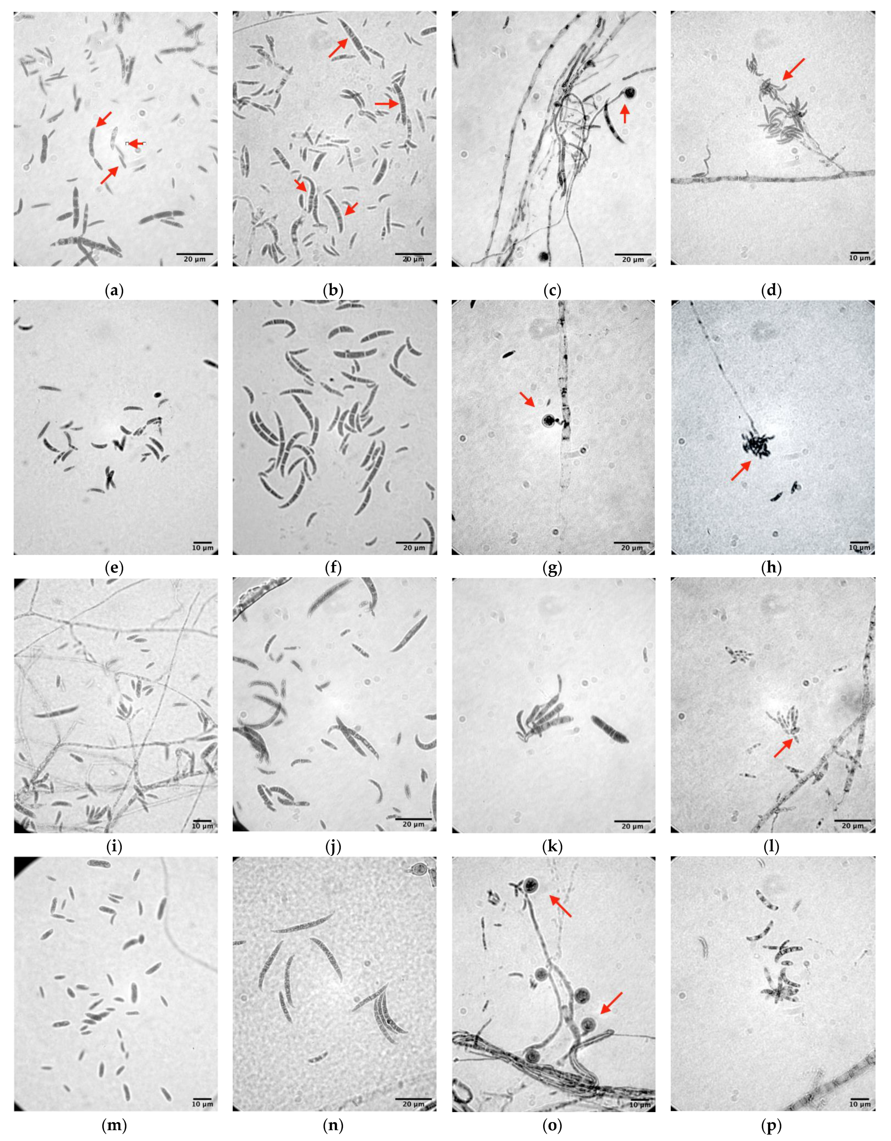
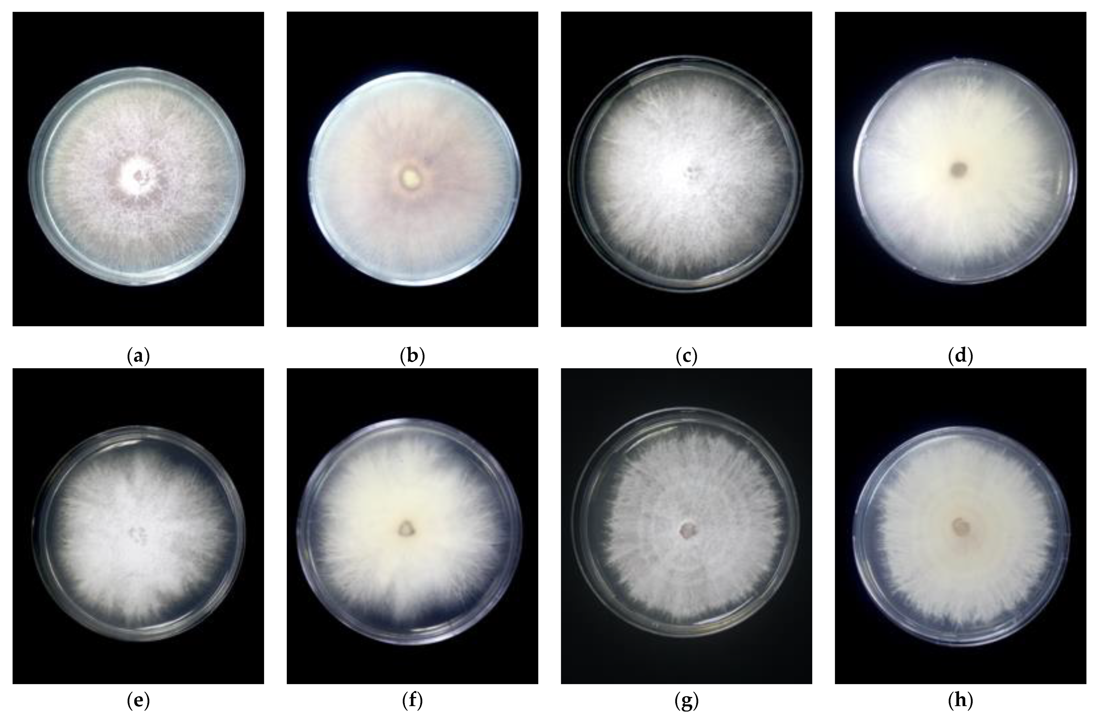
3.1. Morphological Identification

After seven days of incubation on the PDA, the examined 38 isolates could be clustered into four different groups based on the cultural characters. Colony characteristics and growth rate of the isolates are listed as follows (Table 2). For each group, a representative isolate was chosen randomly for further analysis, which were MUCC2839 (Group 1), MUCC2841 (Group 2), MUCC2867 (Group 3), and MUCC2858 (Group 4).
Representative strains showed typical colonies of Fusarium spp. (Figure 1; Table 2). The morphological characteristics of the fungal pathogen on the CLA medium showed no significant differences between the macroconidia and microconidia sizes and shape of the isolates (Figure 2; Table 3).

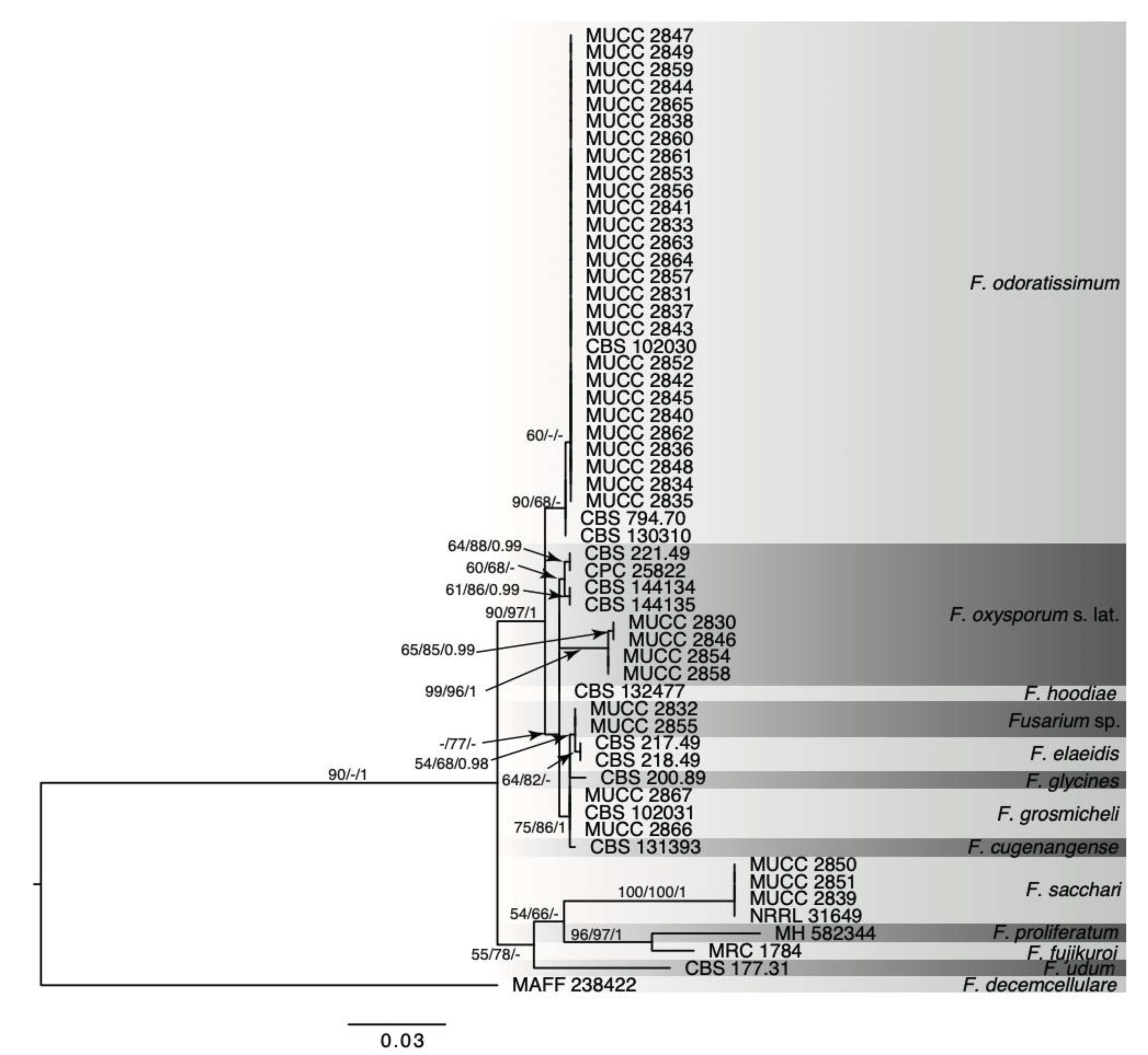
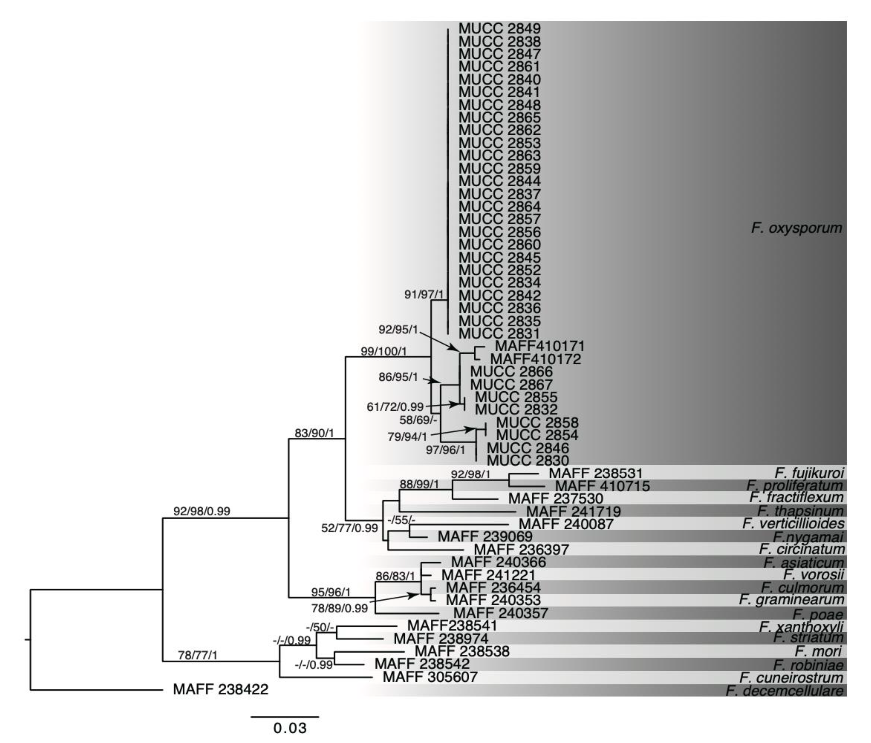
3.2. Molecular Analysis

PCR amplification using H3 and TEF-1α genes was successful, and all sample sequences were deposited into the GenBank repository under the accession number LC545766 to 545836 (Table 1). All amplification by the H3 was successful. Nonetheless, a total of six samples were not successfully sequenced after several attempts. Reference strains were taken from two different databases for the H3 and TEF-1α genes. For the H3 gene, all reference sequences were taken from the Approved Strain Database of Fusarium Species from the National Agriculture and Food Research Organization, Japan. In contrast, for the TEF-1α gene, all reference sequences were retrieved from the GenBank database. On generated trees using the H3 gene sequences matrix (Figure 3), 33 examined isolates formed a well-supported clade of Fusarium oxysporum species complex with two isolates of F. oxysporum (MAFF 410171, MAFF 410172) (MP-BS/ML-BS/Bayesian PP = 99/100/1).
In the analysis by TEF-1α coding gene sequences, known as a species barcode region for Fusarium fujikuroi species complex, the generated tree (Figure 4) revealed the phylogenetic position of examined isolates. Three isolates from the phylogenetic tree were recognised as Fusarium sacchari (MP-BS/MP-BS/Bayesian PP = 100/100/1). Meanwhile, 27 isolates were clustered with F. odoratissimum (MP-BS/MP-BS/Bayesian PP = 90/68/-). In addition, although the low statistical support, two isolates were recognised as Fusarium grosmichelii, four isolates were identified as Fusarium oxysporum s. lat., and two isolates as Fusarium spp. The summary of the findings was presented in Table 4.
The result of amplification using the Foc-TR4 specific primers shows that 27 isolates, grouped with F. odoratissimum reference strains on the TEF-1α phylogeny, were positively identified as F. odoratissimum.

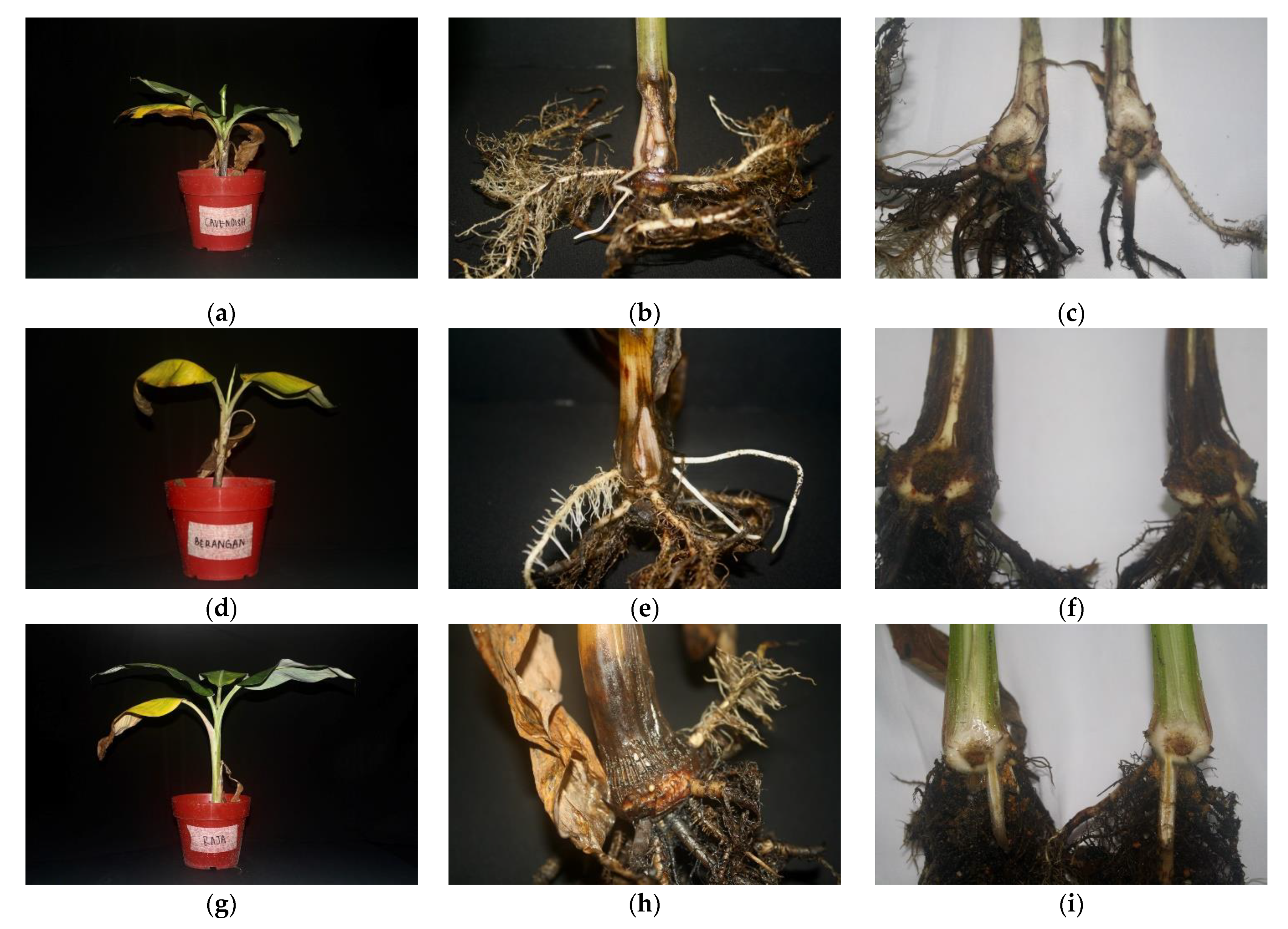
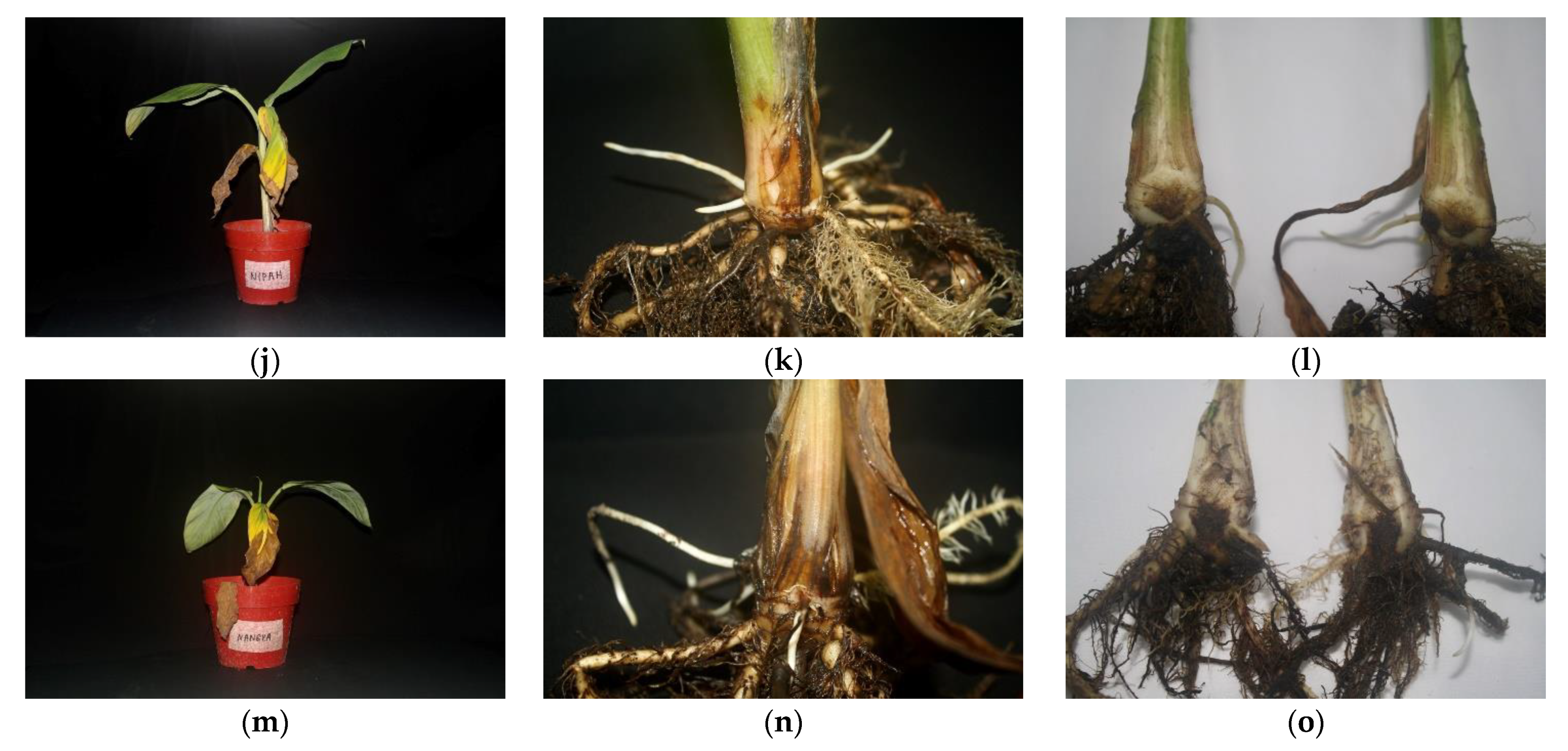
3.3. Pathogenicity Test

All inoculated plantlets showed typical symptoms of Fusarium wilt, such as yellowing of leaves, chlorosis, and death of older leaves at 30 days post-inoculation (Table 5). In a more severe condition, plant wilting was observed (Figure 5b). In this study, all inoculated plantlets showed foliar symptoms of infection with a different degree of severity. Apart from the foliar symptom, the banana plant was also observed lodging of plantlet, splitting at pseudostem, and corm rotting, the typical symptoms of Fusarium wilt. All five examined cultivars of inoculated bananas showed the splitting symptom at the pseudostem (Figure 5b,e,h,k,n). Although the degree of severity of the foliar symptom varied at the different cultivars, the internal symptoms of all cultivars showed severe corm rotting and discolouration (Figure 5c,f,i,l,o).
Statistical analysis shows a significant difference between control plantlets and inoculated plantlets for both foliar and corm symptoms as presented in Table 6. To verify the symptoms were caused by the inoculum, the pathogen was re-isolated from the corm of inoculated plantlets. It was identified as F. odoratissimum based on the morphology and PCR using the TR4 specific primer, which fulfils Koch’s postulates.

4. Discussion

From the phenotypic characters, phylogenetic relationships, and Foc-TR4 specific PCR amplification analysis, these examined isolates were identified as F. grosmichelii, F. odoratissimum, F. oxysporum s. lat., and F. sacchari. There was an observable difference in colony morphology on the phenotypic characters of the isolates, which is the colony’s colour as described by Leslie and Summerell [11]. Still, no significant difference was observed in conidia size. However, the mycelia of the genus Fusarium are floccose, sparse, or abundant with white to pale violet, and it mutates readily to flat and wet with yellow to orange. The size of macroconidia and microconidia was parallel with previous findings on the characteristics of Fusarium spp. The septation of both macro and microconidia was described by the Fusarium manual [11] and updated research on the morphology of Fusarium spp. [6].
For identification of Fusarium spp., the key features are cultural and morphological characteristics on the artificial media, SNA [18], CLA [17], and PDA. However, these characteristics could not distinctly differentiate at the forma specialis or the race of F. oxysporum species complex. Therefore, a more detailed examination, such as molecular characterization, is required to identify the species to grasp the fungal biodiversity and true pathogen related to the wilt of bananas, especially in the developing countries in which the plantations are developed.
In this study, we used the H3 and TEF-1α coding genes for phylogenetic analyses of species and the species complex of the Fusarium genus [6,12,16]. Zeng et al. [14] insisted that the H3 and TEF-1α coding genes have similarly good resolution on the internal groups within Foc isolates, related to the variety of pathogenicity. Our results showed that isolates of F. odoratissimum, identified by the specific primer analysis, formed a monophyletic clade on the H3 and TEF-1α phylogenetic trees despite the differentiation of the pathogenicity. The identification using both the H3 and TEF-1α coding gene sequences could only be conducted up to the genus level for the genus Fusarium. In this study, the results revealed that the TEF-1α gene was better in the detection of F. odoratissimum (Foc-TR4), known as the primary pathogen of Fusarium wilt for various genomes types of bananas. The result produced a more specific finding from the TEF-1α analysis as this could be due to the high volume of research conducted on the identification of Fusarium spp. Thus, this gene yielded more datasets for comparison [6,9,10,16,27,29]. One possible reason for the better result obtained using TEF-1α is that this protein is highly conservative and was identified as an essential genetic marker in the Fusarium study [12,30]. The presence of SNP in the TEF region was exploited to develop a specific primer for the detection of F. odoratissimum [22].
The diversity in the Fusarium spp. could be founded, despite the lack of sexual reproduction due to horizontal gene transfer [6,31], resulting in the formation of new lineages. However, as for F. odoratissimum, they usually were of the clonal population instead of multiple origins [10]. These results suggest that F. odoratissimum in this study was of the same lineage. On the other hand, Fusarium spp. related to the wilted banana has been reported in the 1990s in Malaysia, there are several hypotheses to the origin of these Fusarium species in the Indo-Malaya region [4] and that fungal pathogen co-evolved with the host plant itself. From the previous studies of diversity of strains based on the TEF-1α coding gene [9,10,32], numerous strains of Fusarium oxysporum f. sp. cubense, of which the race is unknown according to Leong et al. [9] or Tropical Race 4 Lineage V and Lineage I-II-V in Wong et al. [10], are widely disseminated across Peninsular Malaysia and were found from the different local banana cultivars. However, in this study, only F. odoratissimum (Foc-TR4), which is monophyletic on TEF- and H3 phylogenetic trees, was detected from the entire of Malaysia. These results may indicate the simplification of the diversity of F. odoratissimum by applying selection pressure of agricultural chemicals or environmental events such as simplifying the variety of cultivating bananas.
Fusarium sacchari isolated from symptomatic banana plants is a member of Fusarium fujikuroi species complex. According to a recent taxonomical study by Maryani et al. [33], it is non-pathogenic to Cavendish and spends an endophytic life cycle. However, there is a probability that the banana plant serves as an intermediary host for Pokkah boeng disease of sugarcane [34]. Although the total crop yields are not significant in Malaysia, F. sacchari as a sugarcane pathogen should be observed in the field.
In our study, two isolates of Fusarium grosmichelii, which is F. oxysporum f. sp. cubense race 1, which is known to affect cultivar ‘Gros-Michel’, ‘Silk’, and ‘Pome’ in Malaysia, were obtained. These isolates were obtained from Pisang Nangka (AAB). A previous study showed that the banana of genome AAB is susceptible to F. grosmichelii (R1) [1,2,35]. Thus, this race is the causal pathogen of the first wave of Fusarium wilt of banana that wiped out the world’s cultivar ‘Gros Michael’. Although the diversification of cropping varieties with local varieties is a generally efficacious approach for controlling the diseases, this result shows that local varieties are still facing the re-emergence of the forgotten disease.
For the pathogenicity test, the high virulence isolate of F. odoratissimum (MUCC2841) was inoculated to the local banana varieties in the previous study [10]. The foliar symptoms of all inoculated plants indicated susceptibility to F. odoratissimum. The foliar symptoms, including wilting and chlorosis of leaves, vary for each banana cultivar, even though it was infected with the same fungal isolate (Table 5). Cultivar ‘Lakatan’ (AAA) suffered the most severe infection as seen on the foliar symptom, whereby all of the leaves that were yellowing started to wilt. For pseudostem and internal symptoms, all inoculated plantlets showed pseudostem splitting, corm rot, and internal discolouration.
Although foliar symptoms suggest the establishment of infection by a pathogen, it is often an unreliable criterion for disease severity assessment [36]. García-Bastidas et al. [27] stated that morphological changes observed on foliar symptoms might be due to the trimming of the root for the root dip method. It causes atypical chlorosis resulting in similar symptoms of Fusarium wilt [31]. Furthermore, Chen et al. indicated that the wilting symptom without corm discolouration was uninfected at the xylem and was considered uninfected by the fungal pathogen [34,36]. The internal symptoms on pseudostem and corm are a more reliable parameter for disease assessment for Fusarium wilt from the previous study, which applied a different inoculation method yield a consistent result in the discolouration [36].
In the detection of Fusarium wilt, the molecular method was developed for the rapid detection of samples collected in the field [23]. Due to the lack of accessibility to access the international collection of VCG samples and the longer time needed to conduct VCG testing, the TR4 specific method is a highly specific detection method with high accuracy [27]. A previous study conducted on Malaysian isolates by Leong et al. [9] concluded that Malaysian Fusarium oxysporum is polyphyletic. However, there is no mention of its pathogenicity or race. A study conducted by Wong et al. [10] shows that all F. odoratissimum (TR4) are monophyletic. This study agrees with both findings, whereby variations could be founded in the F. oxysporum species complex, but F. odoratissimum (TR4) itself is monophyletic. A previous study conducted by Dita et al. [27] also mentions that the VCG of the Fusarium itself does not correlate with pathogenicity.
Our results support the previous study that M. acuminata (AAA) variants, cultivar ‘Berangan’ (AAA), are highly susceptible to F. odoratissimum [10]. The banana cultivars used in this study are in extremely high demand and widely planted in Malaysia. Once this fungus is transmitted on the banana plantation, it may lead to severe economic damage. Although diversification of different crop varieties is generally a productive approach to control disease, the F. odoratissimum was found to infect all banana cultivars in this study regardless of genotype. Therefore, resistance cultivar is one of the best choices for managing Fusarium wilt [7]. However, it is not obtained yet for cropping in a standard field except the transgenic Cavendish bananas in the test fields [37]. Therefore, early detection with rapid diagnoses and digging up the diseased plant and soil disinfection are crucial for controlling this disease as it stands now. Further studies with local varieties of bananas are required to understand the diversity of tolerance against the various strains of Fusarium wilt pathogens.

5. Conclusions

The findings of this study demonstrate that there is a diverse community of Fusarium spp. incorporated to Fusarium wilt disease of banana in Malaysia. Susceptibility of local commercial banana cultivar against Fusarium wilt was also assessed at a preliminary level, regardless of the genotype of the banana plants. Thus, this study provides new insight into the diversity of Fusarium spp. presence in the Malaysian banana plantation. As there is no practical method to control this disease, it would be challenging for the local banana breeders to search for resistance cultivars for cropping purposes.

Supplementary Materials

The following are available online at https://www.mdpi.com/article/10.3390/agronomy11101955/s1, Figure S1: His H3 Bayesian tree with posterior probability (PP) with 1,000,000 generation. MCMC chain were sampled at every 1000 generation. Branch length unit is substitution per site. Figure S2: His H3 Maximum parsimony (MP) tree with bootstrap value. Tree length (TL) = 343, Consistency Index (CI) = 0.633, Retention Index (RI) = 0.833, Rescaled Consistency (RC) = 0.527. Figure S3: TEF-1a H3 Bayesian tree with posterior probability (PP) with 1,000,000 generation. MCMC chain were sampled at every 1,000 generation. Branch length unit is substitution per site. Figure S4: TEF-1a Maximum parsimony (MP) tree with bootstrap value. Tree length (TL) = 235, Consistency Index (CI) = 0.868, Retention Index (RI) = 0.929, Rescaled Consistency (RC) = 0.806. Table S1: GenBank accessions numbers of Fusarium reference strains for phylogenetic analyses.

Author Contributions

Conceptualisation, D.Z., G.V. and C.N.; methodology, A.H.U. and C.N.; software, Y.H.; validation, D.Z. and C.K.F.W.; formal analysis, A.H.U. and Y.H.; investigation, A.H.U.; resources, D.Z. and C.N.; data curation, A.H.U. and Y.H.; writing—original draft preparation, A.H.U.; writing—review and editing, D.Z., G.V., C.N. and C.K.F.W.; visualization, A.H.U. and Y.H.; supervision, D.Z.; project administration, D.Z.; funding acquisition, D.Z. and C.N. All authors have read and agreed to the published version of the manuscript.

Funding

This research was funded by the Ministry of Higher Education of Malaysia (MOHE) through Fundamental Research Grant Scheme (FRGS/1/2018/WAB01/UPM/02/9), GP-GERAN PUTRA/ 9551700 and GP-IPS/9546600. This project was partially supported by the Institute for Fermentation, Osaka (IFO), Japan, and JSPS KAKENHI (17K07837 and 20K06146 to CN).

Institutional Review Board Statement

Not applicable.

Informed Consent Statement

Not applicable.

Acknowledgments

The first author is grateful for the Tun Taib scholarship provided by the Sarawak Foundation, Malaysia. All of the authors would like to thank all involving party and colleague in involvement of this study.

Conflicts of Interest

The authors declare no conflict of interest.

References

  1. Ploetz, R.C. Fusarium wilt of banana. Phytopathology 2015, 105, 1512–1521. [Google Scholar] [CrossRef] [PubMed] [Green Version]
  2. Mostert, D.; Molina, A.B.; Daniells, J.; Fourie, G.; Hermanto, C.; Chao, C.P.; Fabregar, E.; Sinohin, V.G.; Masdek, N.; Thangavelu, R.; et al. The distribution and host range of the banana Fusarium wilt fungus, Fusarium oxysporum f. sp. cubense in Asia. PLoS ONE 2017, 12, e0181630. [Google Scholar] [CrossRef] [PubMed] [Green Version]
  3. FAO Banana Market Review. 2018. Available online: http://www.fao.org/fileadmin/templates/est/COMM_MARKETS_MONITORING/Bananas/Documents/Banana_Market_Review_Prelim_Results_2018.pdf (accessed on 4 March 2019).
  4. Ploetz, R.; Pegg, K. Fusarium wilt of banana and Wallace’s line: Was the disease originally restricted to his Indo-Malayan region? Australas. Plant Pathol. 1997, 26, 239–249. [Google Scholar] [CrossRef]
  5. Su, H.J.; Hwang, S.C.; Ko, W.H. Fusarium wilt of Cavendish bananas in Taiwan. Plant Dis. 1986, 70, 814–818. [Google Scholar]
  6. Maryani, N.; Lombard, L.; Poerba, Y.S.; Subandiyah, S.; Crous, P.W.; Kema, G.H.J. Phylogeny and genetic diversity of the banana Fusarium wilt pathogen Fusarium oxysporum f. sp. cubense in the Indonesian centre of origin. Stud. Mycol. 2019, 92, 155–194. [Google Scholar] [CrossRef]
  7. Ploetz, R.C. Management of Fusarium wilt of banana: A review with special reference to tropical race 4. Crop. Prot. 2015, 73, 7–15. [Google Scholar] [CrossRef]
  8. Malik, T.M.T.A.; Roff, M.N.M.; Rozeita, L.; Maimun, T. Socio-economic Impact, Research and Development, and Policy-making: Regulatory of Fusarium Wilt on Banana in Malaysia. Retrived Dec. 2013, 26, 2019. [Google Scholar]
  9. Leong, S.K.; Latiffah, Z.; Baharuddin, S. Genetic diversity of Fusarium oxysporum f. sp. cubense isolates from Malaysia. Afr. J. Microbiol. Res. 2010, 4, 1026–1037. [Google Scholar]
  10. Wong, C.K.F.; Zulperi, D.; Vadamalai, G.; Saidi, N.B.; Teh, C.Y. Phylogenetic Analysis of Fusarium oxysporum f. sp. cubense Associated with Fusarium Wilt of Bananas from Peninsular Malaysia. Sains Malays. 2019, 48, 1593–1600. [Google Scholar] [CrossRef]
  11. Leslie, J.F.; Summerell, B.A. The Fusarium Laboratory Manual, 1st ed.; Blackwell Publishing Ltd.: Milton, Australia, 2008. [Google Scholar]
  12. Glass, N.L.; Donaldson, G.C. Development of primer sets designed for use with the PCR to amplify conserved genes from filamentous ascomycetes. Appl. Environ. Microb. 1995, 61, 1323–1330. [Google Scholar] [CrossRef] [PubMed] [Green Version]
  13. Jiménez-Gasco, M.M.; Milgroom, M.G.; Jiménez-Díaz, R.M. Gene genealogies support Fusarium oxysporum f. sp. ciceris as a monophyletic group. Plant Pathol. 2002, 51, 72–77. [Google Scholar] [CrossRef] [Green Version]
  14. Zeng, L.; Liu, W.; Zhao, Z.; Wang, F.; Zhou, J.; Li, H.; Du, C.-X. The identification on race of Fusarium oxysporum f. sp. cubense based on multi-gene sequence analysis. Mycosystema 2014, 33, 867–882. [Google Scholar] [CrossRef]
  15. Geiser, D.M.; Jiménez-Gasco, M.D.M.; Kang, S.; Makalowska, I.; Veeraraghavan, N.; Ward, T.J.; Zhang, N.; Gretchen, A.K.; O’Donnell, K. FUSARIUM-ID v. 1.0: A DNA sequence database for identifying Fusarium. Eur. J. Plant Pathol. 2004, 110, 473–479. [Google Scholar] [CrossRef]
  16. O’Donnell, K.; Kistler, H.C.; Cigelnik, E.; Ploetz, R.C. Multiple evolutionary origins of the fungus causing Panama disease of banana: Concordant evidence from nuclear and mitochondrial gene genealogies. Proc. Natl. Acad. Sci. USA 1998, 95, 2044–2049. [Google Scholar] [CrossRef] [Green Version]
  17. Fisher, N.L.; Burgess, L.W.; Toussoun, T.A.; Nelson, P.E. Carnation Leaves as a Substrate and for Preserving Cultures of Fusarium species. Phytopathology 1982, 72, 151–153. [Google Scholar] [CrossRef]
  18. Nirenberg, H.I. A simplified method for identifying Fusarium spp. occurring on wheat. Can. J. Bot. 1981, 59, 1599–1609. [Google Scholar] [CrossRef]
  19. Chupp, C. Further Notes on Double Cover-Glass Mounts. Mycologia 1940, 32, 269–270. [Google Scholar] [CrossRef]
  20. Schneider, C.A.; Rasband, W.S.; Eliceiri, K.W. NIH Image to ImageJ: 25 years of image analysis. Nat. Methods 2012, 9, 671–675. [Google Scholar] [CrossRef]
  21. Umesha, S.; Manukumar, H.M.; Raghava, S. A rapid method for isolation of genomic DNA from food-borne fungal pathogens. 3 Biotech 2016, 6, 123. [Google Scholar] [CrossRef] [Green Version]
  22. Dita, M.A.; Waalwijk, C.; Buddenhagen, I.W.; Souza, J.T.; Kema, G.H.J. A molecular diagnostic for tropical race 4 of the banana Fusarium wilt pathogen. Plant Pathol. 2010, 59, 348–357. [Google Scholar] [CrossRef]
  23. Kumar, S.; Stecher, G.; Tamura, K. MEGA7: Molecular Evolutionary Genetics Analysis Version 7.0 for Bigger Datasets. J. Mol. Evol. 2016, 33, 1870–1874. [Google Scholar] [CrossRef] [Green Version]
  24. Swofford, D.L. PAUP^*: Phylogenetic Analysis Using Parsimony (and Other Methods) 4.0 b8; Sinauer: Sunderland, MA, USA, 2001. [Google Scholar]
  25. Stamatakis, A. RAxML-VI-HPC: Maximum likelihood-based phylogenetic analyses with thousands of taxa and mixed models. Bioinformatics 2006, 22, 2688–2690. [Google Scholar] [CrossRef]
  26. Bouckaert, R.; Vaughan, T.G.; Barido-Sottani, J.; Duchêne, S.; Fourment, M.; Gavryushkina, A.; Heled, J.; Jones, G.; Kühnert, D.; De Maio, N.; et al. BEAST 2.5: An advanced software platform for Bayesian evolutionary analysis. PLoS Comput. Biol. 2019, 15, e1006650. [Google Scholar] [CrossRef] [PubMed] [Green Version]
  27. García-Bastidas, F.A.; Van der Veen, A.J.T.; Nakasato-Tagami, G.; Meijer, H.J.G.; Arango-Isaza, R.E.; Kema, G.H.J. An Improved Phenotyping Protocol for Panama Disease in Banana. Front. Plant Sci. 2019, 10, 1006. [Google Scholar] [CrossRef] [PubMed] [Green Version]
  28. Cody, R. Biostatistic by Example Using SAS ® Studio, 2nd ed.; SAS Institutle Inc: Cary, NC, USA, 2016; pp. 113–128. [Google Scholar]
  29. O’Donnell, K.; Ward, T.J.; Robert, V.A.; Crous, P.W.; Geiser, D.M.; Kang, S. DNA sequence-based identification of Fusarium: Current status and future directions. Phytoparasitica 2015, 43, 583–595. [Google Scholar] [CrossRef] [Green Version]
  30. Roger, A.J.; Sandblom, O.; Doolittle, W.F.; Philippe, H. An evaluation of elongation factor 1 alpha as a phylogenetic marker for eukaryotes. Mol. Biol. Evol. 1999, 16, 218–233. [Google Scholar] [CrossRef] [Green Version]
  31. Liu, S.; Wu, B.; Lv, S.; Shen, Z.; Li, R.; Yi, G.; Li, C.; Guo, X. Genetic Diversity in FUB Genes of Fusarium oxysporum f. sp. cubense Suggests Horizontal Gene Transfer. Front. Plant Sci. 2019, 10, 1069. [Google Scholar] [CrossRef] [PubMed]
  32. Bentley, S.; Pegg, K.G.; Moore, N.Y.; Davis, R.D.; Buddenhagen, I.W. Genetic variation among vegetative compatibility groups of Fusarium oxysporum f. sp. cubense analysed by DNA fingerprinting. Phytopathology 1998, 88, 1283–1293. [Google Scholar] [CrossRef] [PubMed] [Green Version]
  33. Maryani, N.; Sandoval-Denis, M.; Lombard, L.; Crous, P.W.; Kema, G.H.J. New endemic Fusarium species hitch-hiking with pathogenic Fusarium strains causing Panama disease in small-holder banana plots in Indonesia. Persoonia 2019, 43, 48. [Google Scholar] [CrossRef] [Green Version]
  34. Matsumoto, K.; Barbosa, M.L.; Souza, L.A.C.; Teixeira, J.B. Race 1 Fusarium wilt tolerance on banana plants selected by fusaric acid. Euphytica 1995, 84, 67–71. [Google Scholar] [CrossRef]
  35. Ploetz, R.C. Fusarium wilt of banana is caused by several pathogens referred to as Fusarium oxysporum f. sp. cubense. Phytopathology 2006, 96, 653–656. [Google Scholar] [CrossRef] [Green Version]
  36. Chen, A.; Sun, J.; Matthews, A.; Armas-Egas, L.; Chen, N.; Hamill, S.; Mintoff, S.; Tran-Nguyen, L.T.; Batley, J.; Aitken, E.A. Assessing Variations in Host Resistance to Fusarium oxysporum f. sp. cubense Race 4 in Musa Species, with a Focus on the Subtropical Race 4. Front. Microbiol. 2019, 10, 1062. [Google Scholar] [CrossRef] [PubMed] [Green Version]
  37. Dale, J.; James, A.; Paul, J.Y.; Khanna, H.; Smith, M.; Peraza-Echeverria, S.; Garcia-Bastidas, F.; Kema, G.; Waterhouse, P.; Mengersen, K.; et al. Transgenic Cavendish bananas with resistance to Fusarium wilt tropical race 4. Nat. Commun. 2017, 8, 1496. [Google Scholar] [CrossRef] [PubMed] [Green Version]
Figure 1. Isolates growth on PDA after seven days of incubation; (a,b) aerial and backplate view of the isolate MUCC2839; (c,d) aerial and backplate view of the isolate MUCC2841; (e,f) aerial and backplate view of the isolates MUCC2867; (g,h) aerial and backplate view of the isolates MUCC2858.
Agronomy 11 01955 g001
Figure 2. Micromorphology of MUCC2893: (a) microconidia; (b) macroconidia; (c) chlamydospores; (d) false head. Micromorphology of MUCC2841: (e) microconidia; (f) macroconidia; (g) chlamydospores; (h) False HEAD. Micromorphology of MUCC2867: (i) microconidia; (j) macroconidia; (k,l) false head. Micromorphology of MUCC2858: (m) microconidia; (n) macroconidia; (o) chlamydospores; (p) false head.
Agronomy 11 01955 g002
Figure 3. Phylogenetic tree constructed using a maximum-likelihood method based on the H3 gene sequence. The MP and ML bootstrap values and Bayesian posterior probability (PP) value are denoted near the branch (MP/ML/PP) where MP/MP/PP (>50/50/0.96) indication of support.
Agronomy 11 01955 g003
Figure 4. Phylogenetic tree constructed using a Maximum-likelihood method based on the TEF-1α gene sequence. The MP and ML bootstrap values and Bayesian posterior probability (PP) value are denoted near the branch (MP/ML/PP) where MP/MP/PP (>50/50/0.96) indication of support.
Agronomy 11 01955 g004
Figure 5. Pathogenicity test on local banana cultivars exhibiting wilting symptom (a) Dwarf Cavendish; (d) Lakatan; (g) Raja; (j) Laknau; (m) Saba. Plantlet shows pseudostem splitting on (b) Dwarf Cavendish; (e) Lakatan; (h) Raja; (k) Laknau; (n) Saba. Corm rotting (c) Dwarf Cavendish; (f) Lakatan; (i) Raja; (l) Laknau; (o) Saba.
Agronomy 11 01955 g005aAgronomy 11 01955 g005b
Table 1. Sample collection locations, parts, isolate numbers, and GenBank accessions.
Sample LocationGPS CoordinateParts SampledSample CodeGenBank Accessions
TEF-1aHIS H3
Universiti Putra Malaysia, SelangorN 2 59.510 E 101 42.963StemMUCC2830LC545806LC545766
Kluang, JohorN 2 02.996 E 103 17.847StemMUCC2831LC545832LC545774
Batu Pahat, JohorN 1 50.863 E 103 05.339RootMUCC2832LC545800LC545770
Batu Pahat, JohorN 1 52.001 E 102 55.593StemMUCC2833LC545831-
Ayer Hitam, JohorN 1 56.237 E 103 11.050StemMUCC2834LC545830LC545775
Ayer Hitam, JohorN 1 56.237 E 103 11.050StemMUCC2835LC545829LC545776
Ayer Hitam, JohorN 1 56.237 E 103 11.050StemMUCC2836LC545833LC545777
Jasin, MelakaN 2 18.912 E 102 25.800SoilMUCC2837LC545828LC545778
Kuala Pilah Tengah, Negeri SembilanN 2 41.494 E 102 11.839CormMUCC2838LC545827LC545779
Jabatan Pertanian Lekir, PerakN 4 08.673 E 100 43.742CormMUCC2839LC545836-
Kuala Pilah Tengah, Negeri SembilanN 2 41.494 E 102 11.839CormMUCC2840LC545826LC545780
Kuala Pilah Tengah, Negeri SembilanN 2 41.494 E 102 11.839StemMUCC2841LC545825LC545781
Kuala Pilah Tengah, Negeri SembilanN 2 41.494 E 102 11.839StemMUCC2842LC545824LC545782
Universiti Putra Malaysia, SelangorN 2 59.510 E 101 42.963StemMUCC2843LC545823-
Universiti Putra Malaysia, SelangorN 2 59.510 E 101 42.963StemMUCC2844LC545822LC545783
Universiti Putra Malaysia, SelangorN 2 59.510 E 101 42.963StemMUCC2845LC545821LC545784
Universiti Putra Malaysia, SelangorN 2 59.510 E 101 42.963StemMUCC2846LC545805-
Kampung Poh, Bidor, PerakN 4 05.185 E 101 20.019StemMUCC2847LC545820LC545785
Kampung Banir, Bidor, PerakN 4 12.849 E 101 10.580StemMUCC2848LC545819LC545786
Kampung Poh, Bidor PerakN 4 05.185 E 101 20.019RootMUCC2849LC545818LC545787
Chetok, KelantanN 6 02.727 E 100 12.341StemMUCC2850LC545835-
Chetok, KelantanN 6 02.727 E 100 12.341SoilMUCC2851LC545834-
Kampung Perlis, Pulau PinangN 5 18.796 E 100 12.341RootMUCC2852LC545817LC545788
Kampung Perlis, Pulau PinangN 5 18.796 E 100 12.341RootMUCC2853LC545816LC545789
Kampung Karu, SarawakN 1 17.119 E 110 16.936StemMUCC2854LC545804LC545768
Kampung Pulau Manis, TerengganuN 5 14.763 E 103 01.730CormMUCC2855LC545799LC545771
Kampung Sungai Maong, SarawakN 1 32.537 E 110 18.313CormMUCC2856LC545815LC545790
Kampung Sungai Maong, SarawakN 1 32.537 E 110 18.313CormMUCC2857LC545814LC545791
Kampung Karu, SarawakN 1 17.119 E 110 16.936CormMUCC2858LC545803LC545796
Chetok, KelantanN 6 02.727 E 102 08.661CormMUCC2859LC545813LC545792
Tumpat, KelantanN 6 07.184 E 102 13.275CormMUCC2860LC545812LC545793
Tumpat, KelantanN 6 07.184 E 102 13.275CormMUCC2861LC545811LC545794
Sungai Atong, PahangN 3 52.953 E 103 09.777StemMUCC2862LC545810LC545798
Kampung Ria Semantan, PahangN 3 56.334 E 101 50.423CormMUCC2863LC545809LC545795
Kampung Tanjung Besar, PahangN 3 28.015 E 102 28.121RootMUCC2864LC545808LC545796
Kampung Ria Semantan, PahangN 3 56.334 E 101 50.342RootMUCC2865LC545807LC545797
Kampung Ria Semantan, PahangN 3 56.334 E 101 50.342CormMUCC2866LC545802LC545772
Kampung Ria Semantan, PahangN 3 56.334 E 101 50.342CormMUCC2867LC545801LC545773
Table 2. Characteristic culture morphology and growth rate of isolates grouping.
GroupIsolatesCulture CharacteristicGrowth Rate (PDA)
cm/day
PDASNACLA
1MUCC2839Magenta pigmentation on both sides of the plateSparse mycelia“Wet” mycelia with pale yellow to orange sporodochia0.57 ± 0.20
2MUCC2841White colony with cottony and dense myceliaFloccose myceliaSparse mycelia, pale yellow to white sporodochia0.60 ± 0.10
3MUCC2867White colony with cottony and floccose myceliaAbundance myceliaDense mycelia with yellow sporodochia0.48 ± 0.13
4MUCC2858White colony with purple pigmentation, floccose and sparse myceliaAbundance floccose myceliaSparse mycelia with yellow to orange sporodochia0.52 ± 0.11
PDA: Potato Dextrose Agar; CLA: Carnation Leaves Agar; SNA: Spezieller Nährstoffarmer Agar.
Table 3. Macro- and micro-conidia size, shape, and septation of representative isolates on CLA.
IsolatesMacroconidiaMicroconidia
LengthWidthSeptateShapeLengthWidthSeptateShape
MUCC283915.07–30.99 (24.94 ± 4.09)1.39–3.94 (2.59 ± 0.53)3–5Falcate3.58–12.54 (7.02 ± 2.15)0.86–2.43 (1.45 ± 0.43)0–1Oval to ellipsoid
MUCC284116.89–33.84 (25.06 ± 4.94)1.76–3.65 (2.32 ± 0.54)0–6Falcate3.29–12.34 (6.97 ± 2.75)0.77–2.42 (1.48 ± 0.47)0–3Oval to ellipsoid
MUCC286719.93–31.85 (25.42 ± 3.65)1.03–3.94 (2.38 ± 0.65)0–5Falcate4.78–11.48 (6.13 ± 1.46)0.86–2.93 (1.64 ± 0.48)0–2Oval to ellipsoid
MUCC285816.97–38.07 (26.48 ± 5.41)1.25–3.27 (2.24 ± 0.61)0–4Falcate3.79–12.43 (7.14 ± 1.96)1.18–2.18 (1.64 ± 0.28)0–1Oval to ellipsoid
Table 4. Summary of the findings for all isolates obtained in this study using the H3 and TEF-1α genes amplification.
Sample CodeH3TEF-1α
MUCC2830F. oxysporumF. oxysporum s. lat.
MUCC2831F. oxysporumF. odoratissimum
MUCC2832F. oxysporumF. oxysporum
MUCC2833Not availableF. odoratissimum
MUCC2834F. oxysporumF. odoratissimum
MUCC2835F. oxysporumF. odoratissimum
MUCC2836F. oxysporumF. odoratissimum
MUCC2837F. oxysporumF. odoratissimum
MUCC2838F. oxysporumF. odoratissimum
MUCC2839Not availableF. odoratissimum
MUCC2840F. oxysporumF. odoratissimum
MUCC2841F. oxysporumF. odoratissimum
MUCC2842F. oxysporumF. odoratissimum
MUCC2843Not availableF. odoratissimum
MUCC2844F. oxysporumF. odoratissimum
MUCC2845F. oxysporumF. odoratissimum
MUCC2846Not availableF. oxysporum s. lat.
MUCC2847F. oxysporumF. odoratissimum
MUCC2848F. oxysporumF. odoratissimum
MUCC2849F. oxysporumF. odoratissimum
MUCC2850Not availableF. sacchari
MUCC2851Not availableF. sacchari
MUCC2852F. oxysporumF. odoratissimum
MUCC2853F. oxysporumF. odoratissimum
MUCC2854F. oxysporumF. oxysporum s. lat.
MUCC2855F. oxysporumF. oxysporum
MUCC2856F. oxysporumF. odoratissimum
MUCC2857F. oxysporumF. odoratissimum
MUCC2858F. oxysporumF. oxysporum s. lat.
MUCC2859F. oxysporumF. odoratissimum
MUCC2860F. oxysporumF. odoratissimum
MUCC2861F. oxysporumF. odoratissimum
MUCC2862F. oxysporumF. odoratissimum
MUCC2863F. oxysporumF. odoratissimum
MUCC2864F. oxysporumF. odoratissimum
MUCC2865F. oxysporumF. odoratissimum
MUCC2866F. oxysporumFusarium grosmichelii
MUCC2867F. oxysporumFusarium grosmichelii
Table 5. Evaluation of susceptibility of local varieties of banana against Fusarium odoratissimum.
Banana Cultivar
(Genome Type)
Pathogenicity RatingNotes
Leaf aCorm b
Musa acuminata cv. ‘Dwarf Cavendish’ (AAA)35Discolouration of younger leaves; pseudostem splitting; corm rot and discolouration.
Musa acuminata cv. ‘Lakatan’ (AA)46All leaves yellowing; pseudostem splitting; corm rotted and discoloured.
Musa × paradisiaca cv. ‘Raja’ (AAB)15Initial yellowing on older leaves; pseudostem splitting; corm rot and discolouration.
Musa × paradisiaca cv. ‘Laknau’ (AAB)25Older leaves yellowing; pseudostem splitting; corm rot and discolouration.
Musa × paradisiaca cv. Saba (ABB)25Older leaves yellowing; pseudostem splitting; corm rot and discolouration.
a Severity of wilting was rated following the disease scale [10]; 1: no symptom/healthy, 2: initial yellowing mainly on the lower leaves, 3: yellowing of all the lower leaves including some discolouration on the younger leaves, 4: intense yellowing on all leaves, 5: plant dead/complete wilting. b Corm rot severity was rated following the disease scale [27]: 1 indicates no discolouration observed, 2 means discolouration on isolated points, 3 denotes about 30% of discolouration, 4 indicates up to 50% discolouration, 5 means discolouration up to 90%, and 6 denotes corm decay.
Table 6. Leaf and corm discolouration percentage of control and inoculated plantlet.
Banana Cultivar
(Genome Type)
LeafCorm
ControlInoculatedControlInoculated
Musa acuminata cv. ‘Dwarf Cavendish’ (AAA)26.38 a97.91 d0 a94.44 c
Musa acuminata cv. ‘Lakatan’ (AA)25.00 a70.07 c0 a88.89 b
Musa × paradisiaca cv. ‘Raja’ (AAB)25.00 a69.23 c0 a83.33 b
Musa × paradisiaca cv. ‘Laknau’ (AAB)25.00 a67.29 bc0 a83.33 b
Musa × paradisiaca cv. Saba (ABB)25.00 a59.23 b0 a83.33 b
a no significant difference with control group. b, c, d significantly different from control group.
Publisher’s Note: MDPI stays neutral with regard to jurisdictional claims in published maps and institutional affiliations.

