Article

Field Performance and Genetic Stability of Micropropagated Gooseberry Plants (Ribes grossularia L.)

Department of Applied Biology, Research Institute of Horticulture, Konstytucji 3 Maja 1/3 Str., 96-100 Skierniewice, Poland
*
Author to whom correspondence should be addressed.
Agronomy 2021, 11(1), 45; https://doi.org/10.3390/agronomy11010045
Submission received: 6 November 2020 / Revised: 18 December 2020 / Accepted: 22 December 2020 / Published: 28 December 2020

Abstract

:
Gooseberry (Ribes grossularia L.) is a small fruit crop producing valuable fruits, which is constantly gaining importance. In vitro propagation of this species can significantly support the production of virus-free planting material and accelerate the introduction of new cultivars to the market. The aim of presented study was to assess field performance and genetic stability of micropropagated plants (MPs) of four gooseberry cultivars, “Captivator”, “Hinnonmaki Rot”, “Invicta”, and “Resika”. The growth vigor and yield of MPs and plants propagated by standard methods from softwood cuttings (ST) were evaluated in a field experiment. Microscopic observations of the number and length of the stomata of MP and ST plants were carried out. Two DNA-based techniques, amplified fragment length polymorphism (AFLP) and inter simple sequence repeat (ISSR), were used to assess genetic stability of MP plants. For analysis of genetic stability of ST plants, the ISSR technique was applied. For three cultivars, Captivator, Hinnonmaki Rot, and Invicta, the plants’ growth vigor and fruit yield were greater in MP plants than in ST plants. In the case of Resika, most of these parameters were higher in ST plants. Microscopic observations of the stomata indicated a lack of differences in the length between MP and ST plants, while the stomata frequency on leaves of MP plants was higher than that of ST plants. The genetic variability of MP plants, assessed by AFLP, ranged from 0.35% for Hinnonmaki Rot to 2.12% for Resika. The results of ISSR analysis of MP plants showed variability from 0% in the case of Hinnonmaki Rot and Resika to 4% and 8.69% for Captivator and Invicta, respectively. No polymorphism was detected among ST plants of all analyzed gooseberry cultivars.

1. Introduction

Gooseberry (Ribes grossularia L.) is a small fruit crop producing valuable fruits, which is constantly gaining importance. Gooseberry fruits are a rich source of bioactive compounds such as vitamins, especially vitamin C, mineral salts, organic acids, dietary fiber, and antioxidant polyphenols, which are considered beneficial to human health [1,2,3]. Gooseberries are grown on large commercial plantations in some European countries with favorable agroclimatic conditions, including Poland, which is the second largest producer of these fruits in Europe [3].
Gooseberries are propagated vegetatively by cuttings or layering, but the rooting efficiency is below 50% [4]. There are several scientific reports on gooseberry micropropagation [5,6,7,8,9,10,11]. In vitro propagation of this species is difficult due to the appearance of shoot necrosis during initiation and multiplication stages, lack of shoot elongation, leaf yellowing, and hyperhydration. For the rapid multiplication of selected gooseberry clones, an effective method of micropropagation using meta-topolin (m-T) instead of benzylaminopurine (BAP) was developed [12]. This method can significantly support the production of virus-free planting material and accelerate the introduction of new cultivars to the market.
The successful application of in vitro techniques in plant multiplication depends on, among other things, maintaining genetic stability of the genotype. In plant tissue cultures, especially those maintained for a longer time, somaclonal variation (SV) is a common problem [13,14]. SV, either of genetic or epigenetic nature, appearing among clones propagated in in vitro, results either from pre-existing genetic variation within explants or is induced during micropropagation [15]. The character of SV includes, among other things, changes in the number and structure of chromosomes, changes in nucleotide sequences and the number of gene copies, activation of transposons, and alterations in DNA methylation pattern [13,14,16].
SV can be identified by analyzing morphological, biochemical, physiological, and genetic characteristics of plants. Methods based on molecular markers more precisely detect the variability than other techniques, and they are easy in application and guarantee high repeatability of results. Molecular markers such as random amplified polymorphic DNA (RAPD), amplified fragment length polymorphism (AFLP), inter simple sequence repeat (ISSR), or other microsatellite markers are successfully used as tools to study not only somaclonal variability, but also to identify genotypes, study germplasm genetic diversity, and identify putative DNA markers of valuable traits that might be used in marker-assisted selection in many fruit plant species [4,13,17,18,19], including the genus Ribes [20,21,22,23,24,25,26,27].
There are few reports available concerning gooseberry DNA markers. Lanham and Brennan [21] used RAPD, ISSR, and AFLP markers to estimate the genetic diversity within this species. Unique profiles for each of the 12 tested gooseberry genotypes were generated only by AFLP. The other techniques did not allow the distinguishing of genotypes. Molecular markers based on RAPD and ISSR techniques were applied to characterize gooseberry genotypes collected in the germplasm collection of the Research Institute of Horticulture in Skierniewice, Poland, used in breeding programs [17]. The results confirmed the usefulness of both techniques in estimating the genetic distance between the 12 gooseberry genotypes tested; however, the ISSR analysis was assessed as being more precise and repetitive.
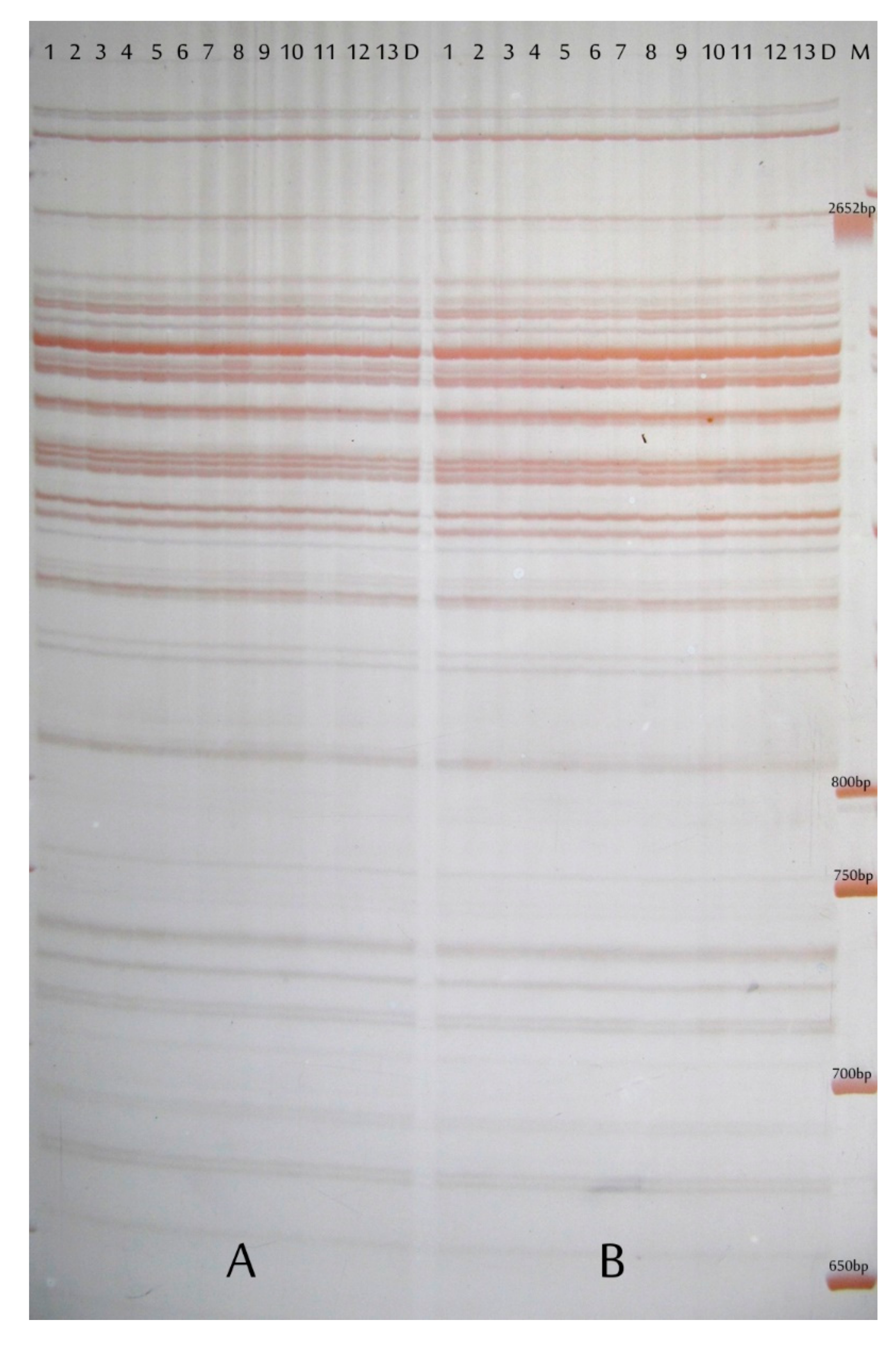
To date, no scientific reports on SV generated by in vitro gooseberry cultures have been published. In our research, we decided to use the AFLP technique, as it was considered to be the most effective in detecting DNA polymorphism in gooseberry [21], and is widely used in other species [28,29,30,31]. In order to increase the reliability of the analysis, a combination of two or more markers was used [32,33,34]. In our research, besides AFLP, ISSR markers were applied. ISSR markers were also used to compare the level of genetic diversity within in vitro-derived and vegetative seedlings.

2. Materials and Methods

2.1. Plant Material

The research was carried out on four gooseberry cultivars: “Captivator”, “Hinnonmaki Rot”, “Invicta”, and “Resika”. Plant material was taken from the gooseberry germplasm collection of the Department of Horticultural Crop Breeding, the Research Institute of Horticulture in Skierniewice, Poland. For experimental purposes, micropropagated plants (MP plants) and those propagated by the standard method from softwood cuttings (ST plants) of each genotype were obtained and planted in an experimental field.

2.1.1. Culture Medium and Growth Conditions

In vitro cultures of gooseberry shoots were initiated and multiplied based on a method developed previously [11,12]. The shoots were multiplied on media containing salts and vitamins set by Murashige and Skoog (MS) [35]: 100 mg L−1 inositol, 85.45 mg L−1 MgSO4, 0.1 mg L−1 gibberellic acid (GA3), 0.1 mg L−1 indole 3-acetic acid (IAA), 0.5 mg L−1 meta-topolin (mT), sucrose 30 g L−1, and Bacto agar 7.0 g L−1 with a pH of 5.8. All nutrient components were purchased from Duchefa (Haarlem, the Netherlands), except the Bacto agar, which was produced by Benton Dikinson (Franklin Lakes, NJ, USA). Shoots were maintained in tissue cultures for two years, the shoots were subcultured every 6 weeks. Rooting was carried out in in vitro conditions on the medium composed of ½ macro and micro elements of MS and 1.0 mg L−1 indole-3-butyric acid (IBA) for five days in the dark, and then transferred to the medium without growth regulators, containing 4 g L−1 of activated carbon. MP plants were acclimatized to ex vitro conditions in a greenhouse for four weeks, then they were transplanted into pots and grown in greenhouse conditions.

2.1.2. Plants Propagation from Softwood Cuttings

In June, softwood cuttings of about 10 cm long were collected from mother plants. Cuttings were rooted in a 1:1 mixture of peat and perlite, using the rooting agent Rhizopon AA (Rhizopon, Rijndijk, the Netherlands) (powder 1%) and high humidity and shading was provided. Rooted ST plants were transplanted into pots and grown in greenhouse conditions.

2.1.3. Conditions of the Field Experiment

The following year, ten-month-old seedlings of MP and ST plants were planted in an experimental field, in a random block system, in three replications, five plants per plot. Standard agrotechnical treatments for the species were used to maintain the experimental plots. Plants were supplied annually with mineral fertilizer containing nitrogen (12% N w/w), phosphorus (12% P2O5 w/w), potassium (36% K2O w/w), and the essential micronutrients (Mila; Yara, Oslo, Norway). The fertilizer was broadcast at soil surface along bush rows at the swollen bud stage at rate of 300 kg ha−1.

2.2. Morphological Measurements and Observations

In the second and third growing seasons, the growth vigor and yield of MP and ST plants were assessed. In the spring and autumn of the second growing season, and in the autumn of the third growing season, the following measurements were carried out: the height and width of bushes and the number of one-year-old shoots. On the basis of these measurements, the increases of the height and width of MP and ST plants in the second and third growing seasons were calculated. In the third year of the field experiment, the fruit yield per plot was also assessed.
Microscopic observations of the number and length of the stomata of MP and ST plants were carried out. The leaves were collected in August in the third year of the experiment. Samples of the abaxial epidermis isolated from the third leaf, 10 cm from the tip, were mounted on microscopic slides and stained with toluidine blue according to the procedure of Dyki and Habdas [36]. For each genotype, the frequency of stomata per 1 mm2 and the length of the stomata (100 stomata/per genotype) were determined. The observations were performed using a Nikon Eclipse 80i microscope (Eclipse 80i, Nikon, Tokyo, Japan) with the NIS-Elements BR program, version 2.30 (Nikon Instruments Inc., Tokyo, Japan), at 100- and 400-times magnification.
Results for the growth parameters, yield, and microscopic observations were statistically analyzed by one-way analysis of variance (Statistica 13.1, StatSoft Inc., Tulsa, OK, USA), separately for each growing season and genotype using Tukey’s test at p = 0.05.

2.3. Genetic Stability Assessment

Genetic stability of 15 Captivator, 13 Hinnonmaki Rot, 12 Invicta, and 14 Resika MP plants were assessed with the use of AFLP markers. ISSR analysis was used to evaluate 10 MP and 10 ST plants of each cultivar. Along with the MP and ST plants, the donor plants were analyzed by both methods.

2.3.1. DNA Extraction

The DNA from gooseberry leaves was isolated using three various commercial kits. For AFLP, the extraction was performed with DNeasy Plant Mini Kit® (Qiagen, Hilden, Germany; first extraction) and NucleoSpin® Plant II (Macherey-Nagel, Düren, Germany; second extraction). For ISSR, the first extraction was done with DNeasy Plant Mini Kit® (Qiagen) and the second with Plant/Fungi DNA Isolation Kit (Norgen Biotek Corp., Thorold, Canada). The extractions were performed according to the instructions provided by the manufacturers. The DNA preparations were analyzed in duplicate, using an Epoch spectrophotometer (BioTek, Winooski, VT, USA). Based on spectrophotometric measurements, the concentration of DNA was calculated and their purity was estimated (260/280 nm factor). The mean concentration of DNA as well as mean value of 260/280 nm factor in the preparations obtained with the use of individual isolation kits was calculated.

2.3.2. AFLP Analysis

AFLP analysis was performed according to Zabeau and Vos [37] and Money et al. [38]. Fifty nanograms of genomic DNA were digested with PstI and MseI restriction endonucleases (Thermo Fisher Scientific™, Waltham, MA, USA) and the restriction fragments were ligated using T4 DNA ligase (Thermo Scientific™, Waltham, MA, USA) to double-stranded 20-mer adapters, specific for either the PstI or the MseI restriction sites. Amplification of DNA fragments was performed in two steps: pre-selective amplification with primers complementary to adapter sequences and selective amplification using primers with an additional two or three nucleotides at the 3′ end as described by Money et al. [38]. Five primer pairs were used in selective amplification: Pst-AT/Mse-CG, Pst-TA/Mse-GA, Pst-AGC/Mse-CT, Pst-CAG/Mse-TG, and Pst-GTC/Mse-AC. Products of amplification were analysed by separation in 6% denaturing polyacrylamide gels run in 0.5 × TBE buffer at 80 W for 2 h in the Dual Dedicated Height Nucleic Acid Sequencer (C.B.S. Scientific, Waltham, MA, USA) and silver stained [39]. Distinct monomorphic and polymorphic bands were scored. The analyses were done in two replications.

2.3.3. ISSR Analysis

Eighteen arbitrary primers of the 800 series (University of British Columbia, Vancouver, BC, Canada) were screened in ISSR-PCR with the DNA of the four tested gooseberry genotypes. Ten of the tested primers which produced rich profiles containing reproducible and polymorphic bands, were selected for analysis (Table 1). For each gooseberry cultivar, amplification reactions were carried out with the use of five ISSR primers that produced the best banding profiles for the genotype. ISSR analyses were performed twice in a T100 Thermal Cycler (Bio-Rad, Hercules, CA, USA). The reaction mixture of 40 µL in volume contained 2 µL DNA at a concentration of 10 ng µL−1, 0.4 µL U Taq polymerase (DreamTaq DNA™ Green Polymerase, Thermo Fisher Scientific) (5 U µL−1), 4 µL 10 × PCR buffer, 1.5 µL dNTPs (10 mM), and 3 µL primer (10 µM). The PCR parameters were as follows: 3 min of initial DNA denaturation at 95 °C, 40 cycles of amplification (30 sec of denaturation at 95 °C, 40 sec of annealing from 50 to 55 °C, 90 sec of elongation at 72 °C), and 5 min of final elongation at 72 °C. The PCR products were separated by electrophoresis on 2% agarose gel (EURx, Gdańsk, Poland) supplemented with Simple Safe fluorescent dye (5 µL 100 m L−1 gel, EURx), in 0.5 × TBE buffer. The ISSR products were observed under UV light and documented with a set consisting of a transilluminator and a camera connected to a computer (Syngen Biotech, Wrocław, Poland). The presence, number, and size of monomorphic and polymorphic ISSR products was evaluated.

3. Results

3.1. Morphological Measurements and Observations

In general, the growth vigor of MP Captivator, Hinnonmaki Rot, and Invicta bushes was stronger compared to ST plants in both the second and third growing seasons: the height and width increases of bushes were greater and they produced more one-year-old shoots (except for height of Captivator in the second growing season) (Table 2). For these three cultivars, the fruit yield from MP plants was higher than that from ST plants (Table 2). For the Resika cultivar, the height and width increase of bushes as well as the fruit yield were higher in ST plants than in MP plants, only the number of one-year-old shoots was higher for the MP bushes (Table 2).
Microscopic observations of the stomata indicated a lack of differences in their length, except for the Invicta cultivar, while the stomata frequency on leaves of MP plants was higher than those of ST plants (Table 3).

3.2. Genetic Stability Assessment

3.2.1. DNA Extraction

The mean concentrations of DNA and mean values of 260/280 nm factor in preparations obtained from gooseberry leaves with the use of different isolation kits were: 26.5 ng μL−1 ± 1.38 and 1.81. ± 0.006 (DNeasy Plant Mini Kit®, Qiagen), 34.1 ng μL−1 ± 2.5 and 1.82 ± 0.02 (NucleoSpin® Plant II, Macherey-Nagel), and 19.3 ng μL−1 ± 0.53 and 1.81 ± 0.007 (Plant/Fungi DNA Isolation Kit, Norgen Biotek Corp.). The purity of DNA obtained with all three isolation kits was satisfactory. DNA concentrations in the preparations were low but sufficient for ISSR and AFLP analyses. The type of kit used did not influence further analyses.

3.2.2. AFLP Analysis

The number of products generated by the AFLP primer pairs ranged from 29 to 81 (Table 4), with an average of 54.9. The highest total number of amplification products was obtained in the analysis of Captivator (291), and the lowest in the reactions with the DNA of Invicta (247) (Table 5). Genetic variability analyzed by the AFLP method in MP gooseberry plants varied for individual cultivars and ranged from 0.35% for Hinnonmaki Rot to 2.12% for Resika (Table 5). An example photo of the electrophoresis of AFLP products is shown in Figure 1.

3.2.3. ISSR Analysis

As a result of polymorphism analysis of four gooseberry cultivars with five ISSR primers, two to eight amplification products per profile were obtained (Table 6). The sizes of the obtained amplification products ranged from 250 to 2900 bp, depending on the primer and cultivar used (Table 6). The ISSR-PCR analysis of MP plants showed a different degree of polymorphism among the studied genotypes, from 0% in the case of Hinnonmaki Rot and Resika to 4% and 8.69% for Captivator and Invicta, respectively (Table 7, Figure 2 and Figure 3). No polymorphism was detected among ST plants of all analyzed gooseberry cultivars (Table 7).

4. Discussion

In vitro plant propagation is one of the most important methods of vegetative reproduction used in horticultural production. The main advantage of micropropagation is the possibility of obtaining, in a short time, a large number of true-to-type, high-quality, and pathogen-free plants. Micropropagation of fruit plants is successfully used for the production of elite nursery material, in breeding work and in the mass production of seedlings of species difficult to reproduce in a traditional way [40,41,42,43].
It has been demonstrated, in many scientific reports, that plants produced in in vitro tissue cultures show greater juvenility and productivity. Berry plants obtained from in vitro cultures are characterized by increased vegetative growth and more intense rhizome and fruit formation than plants obtained by standard multiplication methods. This phenomenon has been observed in strawberry [44,45], raspberry [46], blueberry [47,48,49], cranberry [50], and lingonberry [51].
There are no scientific reports on the growth of in vitro-derived gooseberry plants under field conditions except for one publication by Wainwright and Flegmann [7]. The authors found that in vitro-derived gooseberry plants cv. Invicta developed more shoots at the base of the bushes and had more thorns compared to plants propagated by the standard method. In our field experiment, for three studied cultivars, the plants’ growth vigor and fruit yield were greater in in vitro-derived plants than in plants propagated from softwood cuttings. In the case of one cultivar, Resika, most of these parameters were higher in plants propagated by the standard method. This phenomenon can be explained by the influence of plant growth regulators, mainly cytokinins, used during in vitro propagation; their residues may increase plant vigor and yield in ex vitro conditions [45,52].
Many scientific reports indicate changes in the structure, shape, and density of the stomata in the leaves of plants grown in in vitro cultures compared to those grown in a greenhouse or in the field. Noe and Bonini [53] reported that leaves of Vaccinium corymbosum collected from in vitro shoots had a higher number of smaller stomata (361 mm−2) compared to leaves taken from plants grown in the field (241 mm−2). Brainerd and Fuchigami [54] made similar observations on apple trees and Blanke and Belcher [55] and Capellades et al. [56] on rose. Joshi et al. [57] observed that the leaves of in vitro seedlings showed twice the frequency of stomata compared to traditionally propagated seedlings of Wrightia tomentosa. The opposite results were found for the plum cv. Pixi in a study by Brainerd and co-workers [58]. In our field experiment, a higher frequency of stomata on leaves of MP plants was demonstrated for all studied gooseberry cultivars. Stomatal morphology and structure are highly affected by environmental conditions, such as relative humidity, and light level. In in vitro conditions, these two parameters (high humidity and low light levels) differ significantly from those ex vitro [55,59]. The high humidity in the culture vessel causes the transpiration in in vitro conditions to be low [54]. The water imbalance exhibited by micropropagated plants can result in deformation and malfunctioning of the stomata [55,59,60].
Somaclonal variability appearing in plant tissue cultures depends on many factors, such as the type of explant, the culture conditions, including the composition of the propagation medium, especially the type and concentration of growth regulators and culture age [13,14,15,16]. Of the many factors, genotype is considered to be the most important determinant of variability [14,16]. We did not observe phenotypic variability among the obtained MP plants of all the gooseberry cultivars tested. In order to assess the influence of the applied in vitro propagation method on the appearance of SV among progeny plants, we examined variability at the DNA level. In our research, the level of genetic variability in micropropagated gooseberry plants varied depending on the cultivar. Using both marker systems, the lowest genetic variability was detected in Hinnonmaki Rot and the highest in Invicta. Mohamed and co-workers [61] assessed the genetic stability of in vitro-derived plants of several strawberry cultivars using RAPD markers. The study also revealed that the frequency of genetic variation depends on the cultivar studied; plants of Tudla, depending on the culture conditions, showed polymorphism within 2.86–11.77%, while the variability in the cultivar Festival ranged from 10.35 to 30.77%.
The age of the culture is another factor considered to have a strong impact on inducing variability in in vitro cultures [13,14]. The MP plants used for the research were obtained from stabilized two-year-old shoot cultures, subcultured every 6 weeks. In these plants, the level of genetic variation was low and ranged from 0.35% to 2.12% (AFLP markers) and from 0% to 8.69% (ISSR markers). Nookaraju and Agrawal [62] demonstrated clonal homogeneity of six-month-old tissue culture raised plants of grapevine cv. Crimson Seedless compared to donor plants using ISSR and simple sequence repeat (SSR) markers. In a study by Khan [63], the variability of in vitro shoots of blackcurrant cv. Ben Sark increased significantly when plants were kept in cultures for a long time. As assessed with the use of RAPD markers, micropropagated shoots showed no genetic variation up to the fourteenth subculture, the first polymorphic products were detected in the fifteenth subculture, and the final genetic diversity detected in the study was 13.4% (after 16 subcultures). The same analysis was performed for micropropagated raspberry cv. Autumn Bliss. Genetic variability was already detected in the fifth subculture and the maximum variation observed was 26% [63]. In some plant species, genetic variability can reach a very high level, especially when the conditions of the culture are favorable for this phenomenon. In plants derived from four-year pineapple in vitro cultures, the analysis, with the use of RAPD markers, showed as much as 95.9% variability [64]. On the other hand, Borsai and co-workers [65], using sequence-related amplified polymorphism (SRAP) and RAPD markers, did not detect any polymorphism in plants of blackberry cv. Loch Ness and Chester Thornless propagated for both 3 and 11 subcultures. Similarly, no genetic variability was observed by Negi and Saxena [66] using ISSR markers in Bambusa balcooa plants multiplied up to 33 passages or by Singh and co-workers [67] in Dendrocalamus asper in vitro raised shoots up to the thirtieth passage, checked with the use of RAPD, ISSR, SSR, and AFLP markers.
In research using molecular markers, the quality of the DNA preparations used in the analyses is of great importance. The isolation of genomic DNA from plants of the genus Ribes is most often carried out by the method according to Doyle and Doyle [68], based on an extraction buffer containing CTAB (hexadecyltrimethylammonium bromide) [17,26] or the method developed by Milligan [20,21,22,24,
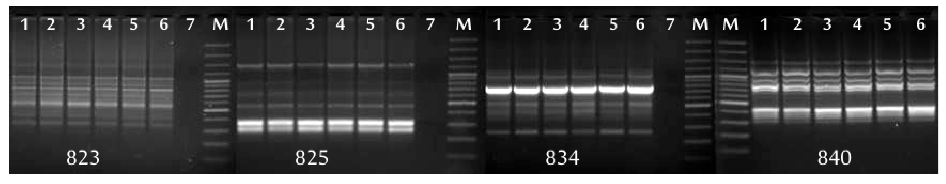
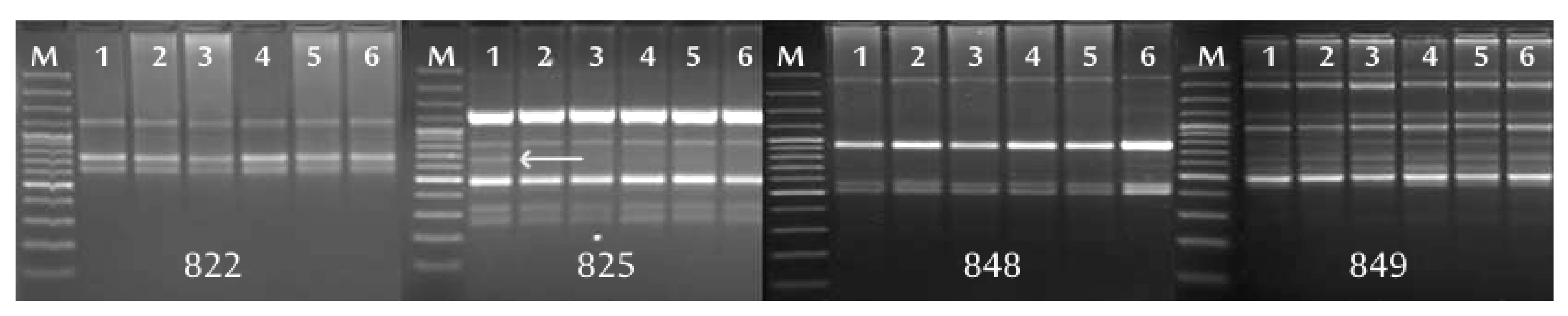
69]. Compared to the technique using commercial DNA isolation kits, these methods are longer and more labor-intensive. All commercial kits used in our study enabled to isolate DNA from small amounts of plant material and to obtain preparations of genomic DNA with sufficient DNA content and purity for AFLP and ISSR analyses. In our study, AFLP markers detected genetic variability in MP plants of all gooseberry genotypes, while ISSR markers only did so in Captivator and Invicta. In order to assess the genetic variability of berry plants, the RAPD and ISSR markers are most frequently used. In several research papers, the use of AFLP, SSR, and SRAP markers was also reported [17,19,21,25,61,62,63,65,70,71,72]. AFLP analysis proved to be more reliable and sensitive than other methods not only in our research, but also in the studies of Lanham and Brennan [21] and Costa and co-workers [31]. The AFLP marker system is believed to be a very sensitive and reproducible method [13,30,31]; no prior knowledge of DNA sequences is required and it can check large fragments of the genome, but it also has disadvantages—it is time-consuming, technically demanding and expensive. In terms of the facility and speed of performing the analysis, microsatellite markers are more advantageous than AFLP. In our research, ISSR analysis proved to be less reliable than the AFLP. Presumably, the reason is that only five primers were used to analyze each gooseberry cultivar; therefore, a small number of products for analysis was obtained.
The results obtained showed that the method of in vitro propagation of four gooseberry cultivars used in our research is suitable for obtaining plants with high genetic fidelity. It supports the opinion that variability is very rare when the technique of shoot regeneration from lateral buds is used in micropropagation and is consistent with the results obtained by Saker et al. [73] and Bhatia et al. [74] for date palm and gerbera.

Author Contributions

Conceptualization, D.W. and D.K.; funding acquisition, D.K.; investigation, D.W., A.T., and D.K.; delivery of plant material, D.K.; methodology, D.W., A.T., and D.K.; data curation, visualization, D.W., A.T., and D.K.; statistical analysis, A.T.; project administration, D.W.; writing—Original draft, D.W., A.T., and D.K.; writing—Review and editing, D.W. and A.T. All authors have read and agreed to the published version of the manuscript.

Funding

This research was supported by the Ministry of Agriculture and Rural Development as a grant for Biological Progress in Crop Production (No 79).

Institutional Review Board Statement

Not applicable.

Informed Consent Statement

Not applicable.

Data Availability Statement

Not applicable.

Acknowledgments

The authors would like to thank Grażyna Korpas, Katarzyna Mynett, Lucyna Ogórek, and Angelika Niewiadomska-Wnuk for great technical support.

Conflicts of Interest

The authors declare no conflict of interest.

References

  1. Pantelidis, G.E.; Vasilakakis, M.; Manganaris, G.A.; Diamantidis, G.R. Antioxidant capacity, phenol, anthocyanin and ascorbic acid contents in raspberries, blackberries, red currants, gooseberries and Cornelian cherries. Food Chem. 2007, 102, 777–783. [Google Scholar] [CrossRef]
  2. Pinto, M.D.S.; Kwon, Y.-I.; Apostolidis, E.; Lajolo, F.M.; Genovese, M.I.; Shetty, K. Evaluation of Red Currants (Ribes Rubruml.), Black Currants (Ribes Nigruml.), Red and Green Gooseberries (Ribes Uva-Crispa) for Potential Management of Type 2 Diabetes and Hypertension Usingin Vitromodels. J. Food Biochem. 2010, 34, 639–660. [Google Scholar]
  3. Pluta, S. Gooseberry—Ribes uva-crispa, sin. R. grossularia L. In Exotic Fruits Reference Guide; Rodrigues, S., De Oliveira Silva, E., Sousa de Brito, E., Eds.; Elsevier, Academic Press: London, UK, 2018; pp. 211–218. [Google Scholar]
  4. Debnath, S.C. Micropropagation of small fruits. In Micropropagation of Woody Trees and Fruits; Jain, S.M., Ishii, K., Eds.; Springer: Dordrecht, The Netherlands, 2003; Volume 2, pp. 465–506. [Google Scholar]
  5. Welander, M. Micropropagation of gooseberry, Ribes grossularia. Sci. Hortic. 1985, 26, 267–272. [Google Scholar] [CrossRef]
  6. Wainwright, H.; Flegmann, A. The micropropagation of gooseberry (Ribes uva-crispa L.): I. Establishmentin vitro. J. Hortic. Sci. 1985, 60, 215–221. [Google Scholar] [CrossRef]
  7. Wainwright, H.; Flegmann, A.W. The micropropagation of gooseberry (Ribes uva-crispaL.): II.In vitroproliferation andin vivoestablishment. J. Hortic. Sci. 1985, 60, 485–491. [Google Scholar] [CrossRef]
  8. Mokrá, A. Meristem culture and micropropagation of gooseberry. Zahradnictvi 1994, 21, 83–92, (In Czech, English Abstract). [Google Scholar]
  9. Kolbanova, E.V.; Kukharchik, N.V. Influence of triiodobenzoic acid on gooseberry in vitro development. Fruit-Grow. Sci. Pap. 2012, 24, 129–136, (In Russian, English Summary). [Google Scholar]
  10. Overchenko, O.V.; Kliuvadenko, A.A.; Likhanov, A.F.; Kostenko, S.M.; Melnychuck, M.D. The reproduction of gooseberries varieties (Grossularia reclinata L.) in vitro culture. Nauk. Vìsn. NLTU Ukr. 2014, 248, 87–93, (In Ukrainian, English Abstract). [Google Scholar]
  11. Kucharska, D.; Niewiadomska-Wnuk, A.; Kiszczak, W. Factors affecting the initiation, stabilization and in vitro propagation of gooseberry (Ribes grossularia L.). Sci. J. Inst. Hortic. 2017, 25, 5–17, (In Polish, English Abstract). [Google Scholar]
  12. Kucharska, D.; Orlikowska, T.; Maciorowski, R.; Kunka, M.; Wójcik, D.; Pluta, S. Application of meta-Topolin for improving micropropagation of gooseberry (Ribes grossularia). Sci. Hortic. 2020, 272, 109529. [Google Scholar] [CrossRef]
  13. Bairu, M.W.; Aremu, A.O.; Van Staden, J. Somaclonal variation in plants: Causes and detection methods. Plant Growth Regul. 2011, 63, 147–173. [Google Scholar] [CrossRef]
  14. Krishna, H.; Alizadeh, M.; Singh, D.; Singh, U.; Chauhan, N.; Eftekhari, M.; Sadh, R.K. Somaclonal variations and their applications in horticultural crops improvement. 3 Biotech 2016, 6, 1–18. [Google Scholar] [CrossRef] [PubMed] [Green Version]
  15. Deepthi, V.P. Somaclonal variation in micro propagated bananas. Adv. Plants Agric. Res. 2018, 8, 624–627. [Google Scholar]
  16. Rani, V.; Raina, S.N. Genetic fidelity of organized meristem-derived micropropagated plants: A critical reappraisal. Vitr. Cell Dev. Biol. Plant 2000, 36, 319–330. [Google Scholar] [CrossRef]
  17. Korbin, M.; Kuras, A.; Zurawicz, E. Fruit plant germplasm characterisation using molecular markers generated in RAPD and ISSR-PCR. Cell. Mol. Biol. Lett. 2002, 7, 785–794. [Google Scholar]
  18. Keller-Przybylkowicz, S.; Lewandowski, M.; Korbin, M. Molecular screening of apple (Malus domestica) cultivars and breeding clones for their resistance to fire blight. J. Fruit Ornam. Plant Res. 2009, 17, 31–43. [Google Scholar]
  19. Lācis, G. Characterisation of Latvia Fruit Crop Genetic Resources by Application of Molecular Genetics Methods. Proc. Latv. Acad. Sci. Sect. B Nat. Exact Appl. Sci. 2013, 67, 84–93. [Google Scholar] [CrossRef]
  20. Lanham, P.G.; Brennan, R.M. Characterization of the genetic resources of redcurrant (Ribes rubrum: Subg. Ribesia) using anchored microsatellite markers. Theor. Appl. Genet. 1998, 96, 917–921. [Google Scholar] [CrossRef]
  21. Lanham, P.G.; Brennan, R.M. Genetic characterization of gooseberry (Ribes grossularia subgenus Grossularia) germplasm using RAPD, ISSR and AFLP markers. J. Hortic. Sci. Biotechnol. 1999, 74, 361–366. [Google Scholar] [CrossRef]
  22. Lanham, P.G.; Korycinska, A.; Brennan, M. Genetic diversity within a secondary gene pool for Ribes nigrum L. revealed by RAPD and ISSR markers. J. Hortic. Sci. Biotechnol. 2000, 75, 371–375. [Google Scholar] [CrossRef]
  23. Keller-Przybyłkowicz, S.; Korbin, M.; Gwozdecki, J. RAPD and ISSR markers of black and green colour of blackcurrant (Ribes nigrum) fruits. J. Fruit Ornam. Plant Res. 2006, 14, 45–52. [Google Scholar]
  24. Brennan, R.; Jorgensen, L.; Hackett, C.; Woodhead, M.; Gordon, S.; Russell, J. The development of a genetic linkage map of blackcurrant (Ribes nigrum L.) and the identification of regions associated with key fruit quality and agronomic traits. Euphytica 2007, 161, 19–34. [Google Scholar] [CrossRef]
  25. Cavanna, M.; Marinoni, D.T.; Beccaro, G.L.; Bounous, G. Microsatellite-based evaluation of Ribes spp. germplasm. Genome 2009, 52, 839–848. [Google Scholar] [CrossRef] [PubMed]
  26. Mazeikiene, I.; Bendokas, V.; Stanys, V.; Siksnianas, T. Molecular markers linked to resistance to the gall mite in blackcurrant. Plant Breed. 2012, 131, 762–766. [Google Scholar] [CrossRef]
  27. Mazeikiene, I.; Juskyte, A.D.; Stanys, V. Application of marker-assisted selection for resistance to gall mite and Blackcurrant reversion virus in Ribes genus. Zemdirbyste 2019, 106, 359–366. [Google Scholar] [CrossRef]
  28. Bahulikar, R.; Stanculescu, D.; Preston, A.C.; Baldwin, I.T. ISSR and AFLP analysis of the temporal and spatial population structure of the post-fire annual, Nicotiana attenuata, in SW Utah. BMC Ecol. 2004, 4, 12. [Google Scholar] [CrossRef] [Green Version]
  29. Meudt, H.M.; Clarke, A.C. Almost Forgotten or Latest Practice? AFLP applications, analyses and advances. Trends Plant Sci. 2007, 12, 106–117. [Google Scholar] [CrossRef]
  30. Kumar, A.; Misra, P.; Batra, S. Amplified fragment length polymorphism: An adept technique for genome mapping, genetic differentiation, and intraspecific variation in protozoan parasites. Parasitol. Res. 2012, 112, 457–466. [Google Scholar] [CrossRef]
  31. Costa, R.; Pereira, G.; Garrido, I.; Tavares-De-Sousa, M.M.; Espinosa, F. Comparison of RAPD, ISSR, and AFLP Molecular Markers to Reveal and Classify Orchardgrass (Dactylis glomerata L.) Germplasm Variations. PLoS ONE 2016, 11, e0152972. [Google Scholar] [CrossRef] [Green Version]
  32. Podwyszyńska, M.; Niedoba, K.; Korbin, M.; Marasek-Ciolakowska, A. Somaclonal variation in micropropagated tulips determined by phenotype and DNA markers. Acta Hortic. 2006, 714, 211–220. [Google Scholar] [CrossRef]
  33. Bhattacharyya, P.; Kumaria, S.; Job, N.; Tandon, P. En-masse production of elite clones of Dendrobium crepidatum: A threatened, medicinal orchid used in Traditional Chinese Medicine (TCM). J. Appl. Res. Med. Aromat. Plants 2016, 3, 168–176. [Google Scholar] [CrossRef]
  34. Espinosa-Leal, C.A.; Puente-Garza, C.A.; García-Lara, S. In vitro plant tissue culture: Means for production of biological active compounds. Planta 2018, 248, 1–18. [Google Scholar] [CrossRef]
  35. Murashige, T.; Skoog, F. A Revised Medium for Rapid Growth and Bio Assays with Tobacco Tissue Cultures. Physiol. Plant. 1962, 15, 473–497. [Google Scholar] [CrossRef]
  36. Dyki, B.; Habdas, H. The method of isolation of epidermis of tomato and cucumber leaves for microscopic investigation of pathogenic fungus development. Acta Agrobot. 1996, 49, 123–129, (In Polish, English Abstract). [Google Scholar] [CrossRef] [Green Version]
  37. Zabeau, M.; Vos, P. Selective restriction fragment amplification: A general method for DNA fingerprinting. European Patent EP0534858A1, 31 March 1993. [Google Scholar]
  38. Money, T. AFLP-based mRNA fingerprinting. Nucleic Acids Res. 1996, 24, 2616–2617. [Google Scholar] [CrossRef] [PubMed] [Green Version]
  39. Chalhoub, B.A.; Thibault, S.; Laucou, V.; Rameau, C.; Höfte, H.; Cousin, R. Silver staining and recovery of AFLP amplification products on large denaturing polyacrylamide gels. BioTechniques 1997, 22, 216–220. [Google Scholar] [CrossRef] [PubMed] [Green Version]
  40. Dale, A.; Hughes, B.R.; Donnelly, D. The Role of Micropropagation in Producing Specific Pathogen-tested Plants. HortScience 2008, 43, 74–77. [Google Scholar] [CrossRef] [Green Version]
  41. Isac, V.; Coman, M. Validated protocols for micropropagation of some fruit species to produce pre-basic plant material. Fruit Grow. Res. 2012, 28, 20. [Google Scholar]
  42. Sedlák, J.; Paprštein, F. In vitro establishment and proliferation of red currant cultivars. Hortic. Sci. 2012, 39, 21–25. [Google Scholar] [CrossRef]
  43. Carrasco, B.; Meisel, L.; Gebauer, M.; García-Gonzales, R.; Silva, H. Breeding in peach, cherry and plum: From a tissue culture, genetic, transcriptomic and genomic perspective. Biol. Res. 2013, 46, 219–230. [Google Scholar] [CrossRef]
  44. Boxus, P.; Damiano, C.; Brasseur, E. Strawberry. In Handbook of Plant Cell Culture; Amirato, D.A., Evans, P.V., Sharp, W.R., Yamada, Y., Eds.; MacMillan: New York, NY, USA, 1984; Volume 3, pp. 453–486. [Google Scholar]
  45. Debnath, S.C. Characteristics of strawberry plants propagated byin vitrobioreactor culture andex vitropropagation method. Eng. Life Sci. 2009, 9, 239–246. [Google Scholar] [CrossRef]
  46. O’Dell, C.R. Berries: Marketing raspberries. Am. Fruit Grower 1989, 109, 34–35. [Google Scholar]
  47. Morrison, S.; Smagula, J.M.; Litten, W. Morphology, Growth, and Rhizome Development of Vaccinium angustifolium Ait. Seedlings, Rooted Softwood Cuttings, and Micropropagated Plantlets. HortScience 2000, 35, 738–741. [Google Scholar] [CrossRef] [Green Version]
  48. Jamieson, A.R.; Nickerson, N.L. Field performance of the lowbush blueberry propagated by seed, stem cuttings and micropropagation. Acta Hortic. 2003, 626, 423–428. [Google Scholar] [CrossRef]
  49. Debnath, S.C. Influence of indole-3-butyric acid and propagation method on growth and development of in vitro- and ex vitro-derived lowbush blueberry plants. Plant Growth Regul. 2007, 51, 245–253. [Google Scholar] [CrossRef]
  50. Debnath, S.C. Zeatin-induced one-step in vitro cloning affects the vegetative growth of cranberry (Vaccinium macrocarpon Ait.) micropropagules over stem cuttings. Plant Cell Tiss. Org. Cult. 2008, 93, 231–240. [Google Scholar] [CrossRef]
  51. Gustavsson, B.A.; Stanys, V. Field Performance of ‘Sanna’ Lingonberry Derived by Micropropagation vs. Stem Cuttings. HortScience 2000, 35, 742–744. [Google Scholar] [CrossRef] [Green Version]
  52. Leposavić, A.; Ružić, D.; Karaklajić-Stajić, Ž.; Cerović, R.; Vujović, T.; Żurawicz, E.; Mitrović, O. Field performance of micropropagated Rubus species. Acta Sci. Pol. Hortorum Cultus 2016, 15, 3–14. [Google Scholar]
  53. Noé, N.; Bonini, L. Leaf anatomy of highbush blueberry grownin vitro and during acclimatization to ex vitro conditions. Biol. Plant. 1996, 38, 19–25. [Google Scholar] [CrossRef]
  54. Brainerd, K.E.; Fuchigami, L.H. Acclimatization of aseptically cultured apple plants to low relative humidity. J. Am. Soc. Hortic. Sci 1981, 106, 515–518. [Google Scholar]
  55. Blanke, M.B.; Belcher, A.R. Stomata of apple leaves cultured in vitro. Plant Cell Tiss. Org. Cult. 1989, 19, 85–89. [Google Scholar] [CrossRef]
  56. Capellades, M.; Fontarnau, R.; Candia, C.; Debergh, P. Environment influences anatomy of stomata and epidermal cells in tissue-cultured Rosa multiflora. J. Am. Soc. Hortic. Sci. 1990, 115, 141–145. [Google Scholar] [CrossRef] [Green Version]
  57. Joshi, P.; Joshi, N.; Purohit, S.D. Stomatal characteristics during micropropagation of Wrightia tomentosa. Biol. Plant. 2006, 50, 275–278. [Google Scholar] [CrossRef]
  58. Brainerd, K.E.; Fuchigami, L.H.; Kwiatkowski, S.; Clark, C.S. Leaf anatomy and water stress of aseptically cultured ’Pixy’ plum grown under different environments. Hortic. Sci. 1981, 16, 173–175. [Google Scholar]
  59. Preece, J.E.; Sutter, E.G. Acclimatization of micropropagated plants to the greenhouse and field. In Micropropagation. Technology and Application; Debergh, P.C., Zimmerman, R.H., Eds.; Springer: Dordrecht, Germany, 1991; pp. 71–93. [Google Scholar] [CrossRef]
  60. Isah, T. Adjustment to in vitro culture conditions and associated anomalies in plants. Acta Biol. Cracov. Bot. 2015, 57, 9–28. [Google Scholar] [CrossRef]
  61. Mohamed, F.H.; Abd El-Hamed, K.E.; Omar, F.A.; El-Shahat, A.A. Ex vitro performance and genetic stability of strawberry plants derived from different in vitro propagation methods. HJSC 2017, 6, 39–50. [Google Scholar] [CrossRef] [Green Version]
  62. Nookaraju, A.; Agrawal, D.C. Genetic homogeneity of in vitro raised plants of grapevine cv. Crimson Seedless revealed by ISSR and microsatellite markers. S. Afr. J. Bot. 2012, 78, 302–306. [Google Scholar] [CrossRef] [Green Version]
  63. Khan, S. Variation arising through tissue culture in soft fruits. Ph.D. Thesis, The University of Edinburgh, Edinburgh, UK, 1997; p. 181. Available online: http://hdl.handle.net/1842/28357 (accessed on 28 December 2020).
  64. Roostika, I.; Khumaida, N.; Ardie, S.W. RAPD Analysis to Detect Somaclonal Variation of Pineapple In Vitro Cultures during Micropropagation. Biotropia 2015, 22, 109–119. [Google Scholar]
  65. Borsai, O.; Hârța, M.; Szabo, K.; Kelemen, C.-D.; Andrecan, F.A.; Codrea, M.-M.; Clapa, D. Evaluation of genetic fidelity of in vitro-propagated blackberry plants using RAPD and SRAP molecular markers. Hortic. Sci. 2020, 47, 21–27. [Google Scholar] [CrossRef]
  66. Negi, D.; Saxena, S. Ascertaining clonal fidelity of tissue culture raised plants of Bambusa balcooa Roxb. using inter simple sequence repeat markers. New For. 2009, 40, 1–8. [Google Scholar] [CrossRef]
  67. Singh, S.R.; Dalal, S.; Singh, R.; Dhawan, A.K.; Kalia, R.K. Evaluation of genetic fidelity of in vitro raised plants of Dendrocalamus asper (Schult. & Schult. F.) Backer ex K. Heyne using DNA-based markers. Acta Physiol. Plant. 2013, 35, 419–430. [Google Scholar] [CrossRef]
  68. Doyle, J.J.; Doyle, J.L. Isolation of plant DNA from fresh tissue. Focus 1990, 12, 13–15. [Google Scholar]
  69. Milligan, B.G. Plant DNA isolation. In Molecular Genetics of Populations: A Practical Approach; Hoelzed, A.R., Ed.; IRL Press: Oxford, UK, 1992; pp. 59–88. [Google Scholar]
  70. Brennan, R.M.; Jorgensen, L.; Woodhead, M.; Russell, J. Development and characterization of SSR markers in Ribes species. Mol. Ecol. Notes 2002, 2, 327–330. [Google Scholar] [CrossRef]
  71. Debnath, S.C.; Vyas, P.; Goyali, J.C.; Igamberdiev, A.U. Morphological and molecular analysis in micropropagated berry plants acclimatized under ex vitro condition. Can. J. Plant Sci. 2012, 92, 1065–1073. [Google Scholar] [CrossRef]
  72. Wojtania, A.; Markiewicz, M.; Góraj-Koniarska, J. Ex vitro rooting and acclimatization of Lonicera caerulea var. kamtschatica shoots, and determination of genetic stability of plantlets using AFLP and ISSR markers. J. Hortic. Res. 2020, 28. Available online: https://content.sciendo.com/view/journals/johr/ahead-of-print/issue.xml (accessed on 28 December 2020). [CrossRef]
  73. Saker, M.M.; Adawy, S.S.; Mohamed, A.A.; El-Itriby, H.A. Monitoring of cultivar identity in tissue culture-derived date palms using RAPD and AFLP analysis. Biol. Plant. 2006, 50, 198–204. [Google Scholar] [CrossRef]
  74. Bhatia, R.; Singh, K.; Jhang, T.; Sharma, T. Assessment of clonal fidelity of micropropagated gerbera plants by ISSR markers. Sci. Hortic. 2009, 119, 208–211. [Google Scholar] [CrossRef]
Figure 1. The fragment of banding pattern of AFLP-PCR products obtained with primers Pst-AT/Mse-CG and DNA of gooseberry cv. Hinnonmaki Rot extracted with the use of DNeasy Plant Mini Kit® (Qiagen) (A) and NucleoSpin® Plant II (Macherey-Nagel) (B), lane 1–13 micropropagated plants (MP), D—donor plant, M—50 bp DNA Ladder (Thermo Fisher Scientific).
Agronomy 11 00045 g001
Figure 2. Banding pattern of ISSR-PCR products using primers 823, 825, 834, 840 for cv. Hinnonmaki Rot, lane 1–3 micropropagated plants (MP), 4–6 plants propagated from softwood cuttings (ST), 7—negative control, M—GeneRuler™ 100 bp DNA Ladder Plus (Thermo Fisher Scientific).
Agronomy 11 00045 g002
Figure 3. Banding pattern of ISSR-PCR products using primers 822, 825, 848, 849 for cv. Captivator, lane 1–3 micropropagated plants (MP), 4–6 plants propagated from softwood cuttings (ST), 7—negative control, M—GeneRuler™ 100 bp DNA Ladder Plus (Thermo Fisher Scientific). The arrow indicates a polymorphic band.
Agronomy 11 00045 g003
Table 1. Nucleotide sequences of primers used in inter simple sequence repeat (ISSR) analysis.
Code of PrimerNucleotide Sequence 5′ to 3′
821(GT)8T
822(TC)8A
823(TC)8C
825(AC)8T
830(TG)8G
834(AG)8YT
840(GA)8YT
848(CA)8RG
849(GT)8YA
853(TC)8RT
Table 2. Vigor and yield of micropropagated plants (MP) and plants propagated with a standard method (ST) of four gooseberry cultivars in the second and third growing seasons (n = 15, 3 replications, 5 plant per replication).
Growing SeasonHeight Increase (cm) ± SEWidth Increase (cm) ± SENo. of One-Year-Old Shoots/Bush ± SEFruit Yield (g plot−1) ± SE
MP STMPSTMPSTMPST
Captivator
2nd7.3 b* ± 1.5011.1 a ± 1.1911.8 a ± 1.9510.0 a ± 1.624.6 a ± 1.083.2 a ± 0.76n.d. **n.d.
3rd14.9 a ± 2.579.8 b ± 2.1916.0 a ± 2.6815.5 a ± 2.3620.0 a ± 1.7211.1 b ± 1.061485.4 a ± 97.841215.4 b ± 87.76
Hinnonmaki Rot
2nd13.1 a ± 2.1510.0 b ± 1.7017.1 a ± 2.149.2 b ± 1.007.8 a ± 1.155.6 b ± 1.08n.d.n.d.
3rd10.9 a ± 0.77.6 b ± 0.816.9 a ± 3.3813.1 b ± 2.0827.1 a ± 2.6723.0 b ± 3.003941.0 a ± 150.611853.4 b ± 115.15
Invicta
2nd10.8 a ± 2.155.5 b ± 1.1513.2 a ± 2.194.9 b ± 1.005.8 a ± 1.303.3 b ± 0.70n.d.n.d.
3rd12.3 a ± 1.4210.0 b ± 1.4017.5 a ± 3.268.9 b ± 1.3013.7 a ± 2.108.8 b ± 2.151502.0 a ± 90.95607.1 b ± 13.75
Resika
2nd17.3 a ± 2.2618.1 a ± 2.199.5 b ± 1.1515.2 a ± 2.804.4 a ± 1.022.3 b ± 0.6n.d.n.d.
3rd14.3 b ± 2.1417.6 a ± 2.7024.6 a ± 2.9925.2 a ± 2.0017.4 a ± 2.1614.5 b ± 1.72502.1 b ± 10.00636.1 a ± 10.15
* means within the row for each parameter followed by the same letter were not significantly different using Tukey’s multiple range test at p = 0.05, SE—standard error; ** n.d.—not determined.
Table 3. The frequency and length of stomata of micropropagated plants (MP) and plants propagated with a standard method (ST) of four gooseberry cultivars.
Stomata Frequency (No. mm−2) ± SELength of Stomata (µm) ± SE
MPSTMPST
Captivator
103.0 a ± 1.1599.0 b* ± 0.5730.09 a ± 0.4129.06 a ± 0.38
Hinnonmaki Rot
92.3 a ± 5.9382.0 b ± 2.6433.90 a ± 0.4532.92 a ± 0.36
Invicta
141.3 a ± 10.2886.0 b ± 11.3727.24 b ± 0.3632.51 a ± 0.42
Resika
176.3 a ± 16.4099.3 b ± 11.8531.04 a ± 0.5633.44 a ± 1.21
* means within the row for each parameter followed by the same letter were not significantly different using Tukey’s multiple range test at p = 0.05, ± SE—standard error.
Table 4. Primer combinations, the size and number of scorable bands and percentage of polymorphic bands obtained in amplified fragment length polymorphism (AFLP) analysis of micropropagated plants (MP) of four gooseberry cultivars in relation to donor plants.
CultivarAFLP Primer Combinations The Size of the Products (bp)Number of Amplification Products per Profile% of Polymorphic Bands
TotalMonomorphicPolymorphic
CaptivatorPst-AT/Mse-CG320–2300707000.00
Pst-TA/Mse-GA280–2500767600.00
Pst-AGC/Mse-CT310–3200414012.43
Pst-CAG/Mse-TG280–2200605911.66
Pst-GTC/Mse-AC270–2300444400.00
Hinnonmaki RotPst-AT/Mse-CG330–3200626200.00
Pst-TA/Mse-GA290–2300818100.00
Pst-AGC/Mse-CT330–2700292813.44
Pst-CAG/Mse-TG270–2600606000.00
Pst-GTC/Mse-AC300–2500464600.00
InvictaPst-AT/Mse-CG290–2800656500.00
Pst-TA/Mse-GA290–2700666600.00
Pst-AGC/Mse-CT310–2300313013.22
Pst-CAG/Mse-TG250–2600494900.00
Pst-GTC/Mse-AC240–26003632411.11
ResikaPst-AT/Mse-CG290–2600636123.17
Pst-TA/Mse-GA350–2700787800.00
Pst-AGC/Mse-CT340–2400333039.09
Pst-CAG/Mse-TG260–2800727200.00
Pst-GTC/Mse-AC260–2500363512.77
Table 5. Summary results of AFLP analysis with 5 primers pairs of 15 Captivator, 13 Hinnonmaki Rot, 12 Invicta, and 14 Resika micropropagated plants (MP) in relation to donor plant.
CultivarTotal No. of Amplified BandsAverage No. of Band/Primer pairsTotal No. of Polymorphic Bands% of Polymorphism
Captivator29158.220.68
Hinnonmaki Rot27855.610.35
Invicta24749.452.02
Resika28256.462.12
Table 6. Nucleotide sequences of primers, the size and number of scorable bands, and the percentage of polymorphic bands obtained in ISSR analysis of micropropagated plants (MP) and plants propagated from softwood cuttings (ST) of four gooseberry cultivars in relation to donor plants.
CultivarISSR PrimerNucleotide Sequence (5′-3′)The Size of the Products (bp)Total No. of Amplified BandsMPST
Polymorphic% of Polymorphic BandsPolymorphic% of Polymorphic Bands
Captivator822(TC)8A650–1300300.0000.00
825(AC)8T280–12006116.6600.00
830(TG)8G500–1000500.0000.00
848(CA)8RG500–2800400.0000.00
849(GT)8YA500–2900700.0000.00
Hinnonmaki Rot823(TC)8C450–1100400.0000.00
825(AC)8T280–1300600.0000.00
834(AG)8YT280–1050500.0000.00
840(GA)8YT450–1500500.0000.00
853(TC)8RT700–1400300.0000.00
Invicta821(GT)8T550–20004125.0000.00
825(AC)8T250–12005120.0000.00
834(AG)8YT260–750200.0000.00
849(GT)8YA400–2000800.0000.00
853(TC)8RT600–1500400.0000.00
Resika821(GT)8T350–2000500.0000.00
822(TC)8A650–1250300.0000.00
825(AC)8T250–1300400.0000.00
834(AG)8YT500–1200600.0000.00
849(GT)8YA500–1200400.0000.00
Table 7. Summary results of ISSR analysis of 10 micropropagated plants (MP) and 10 plants propagated from softwood cuttings (ST) of four gooseberry cultivars in relation to donor plant.
CultivarTotal No. of Amplified BandsAverage No. of Bands per PrimerMPST
Total No. of Polymorphic Bands% of PolymorphismTotal No. of Polymorphic Bands% of Polymorphism
Captivator255.014.0000.00
Hinnonmaki Rot234.600.0000.00
Invicta234.628.6900.00
Resika224.400.0000.00
Publisher’s Note: MDPI stays neutral with regard to jurisdictional claims in published maps and institutional affiliations.

