Article

Temporal Responses to Direct and Induced Iron Deficiency in Parietaria judaica

1
Department of Agricultural and Environmental Sciences: Production, Landscape, Agroenergy, University of Milan, 20133 Milan, Italy
2
Department of Life Sciences and Systems Biology, Innovation Centre, University of Turin, 10125 Turin, Italy
3
Faculty of Science and Technology, Free University of Bolzano, 39100 Bolzano, Italy
*
Author to whom correspondence should be addressed.
Agronomy 2020, 10(7), 1037; https://doi.org/10.3390/agronomy10071037
Submission received: 15 June 2020 / Revised: 15 July 2020 / Accepted: 15 July 2020 / Published: 18 July 2020
(This article belongs to the Special Issue Role of Iron in Plant Nutrition, Growth and Metabolism)

Abstract

:
Parietaria judaica grows in highly calcareous environments, overcoming the low bioavailability of Fe caused by elevated pH. The aim of this work was to investigate the temporal dynamics of root exudation of P. judaica under Fe deficiency conditions. As high concentrations of bicarbonate and Ca2+ in calcareous soils interfere with the general plant mineral nutrition, two different alkaline growing conditions were applied to distinguish the effects due to the high pH from the responses induced by the presence of high calcium carbonate concentrations. Growth parameters and physiological responses were analyzed during a 7 day time course—shoot and root biomass, chlorophyll and flavonoid contents in leaves, root accumulation, and exudation of organic acids and phenolics were determined. Different responses were found in plants grown in the presence of bicarbonate and in the presence of an organic pH buffer, revealing a time- and condition-dependent response of P. judaica and suggesting a stronger stress in the buffer treatment. The high tolerance to alkaline conditions may be related to an earlier and greater exudation rate of phenolics, as well as to the synergistic effect of phenolics and carboxylic acids in root exudates in the late response. The identification of the main functional traits involved in tolerance to low Fe availability in a wild species could offer crucial inputs for breeding programs for application to crop species.

1. Introduction

In plants, iron (Fe) is required for a wide range of biological functions, such as respiration, photosynthesis, and chlorophyll biosynthesis. Although Fe is the fourth most abundant element in the Earth’s crust, it is not readily available for plant uptake due to its low solubility. Severe lack of Fe affects chlorophyll synthesis, leading to chlorosis, and could cause acute decrease in biomass due to photosynthesis impairment. Consequently, plants endeavor to ensure the availability of iron in order to survive and compete successfully.
Dicot and monocot plants (except Poaceae) have developed metabolic adaptations to cope with low Fe availability, known as strategy I responses, which are characterized by a sustained root activity to uptake Fe from the soil. Overall, strategy I involves a ferric chelate reductase (FCR) located at the root cell plasma membrane that converts Fe3+-chelates to Fe2+, which can be taken up by the cell through the activity of the Fe-regulated transporter (IRT1) [1,2]. Moreover, in several species an H+ pumping activity carried out by the plasma membrane H+-ATPase is increased under Fe deficiency. The extrusion of H+ from the cell acidifies the apoplast and the rhizosphere, increasing Fe solubility and generating an electrochemical gradient useful to drive cation uptake [3,4,5,6]. Furthermore, Fe-deficiency-induced activity involves the accumulation of other compounds in tissues, as well as in the rhizosphere, such as organic acids (i.e., citric and malic acid) and phenolics [7,8,9]. Accumulation of organic acids might trigger different roles in Fe-deficient roots, including: (i) Fe translocation through the formation of different species of Fe(III)-citrate [10]; (ii) control of cytoplasmic pH by the pH-stat mechanism; (iii) control of apoplastic pH [11,12,13]; (iv) by acting as an Fe-chelating agent in the apoplast and the rhizosphere; (v) by acting as a Ca-chelating agent to solubilize P in the soil [14,15]; and (vi) to capture excessive Ca2+ inside the cell [16]. Phenolic compounds have also been reported to be the main components in root exudates in Fe-deficient conditions, and their implication in Fe uptake has been suggested, either by increasing Fe3+ solubility or to support the reducing capacity of Fe3+ on the root surface [17,18,19]. In particular, coumarins are involved in Fe mobilization, as well as in recruiting microbes within the rhizosphere [20].
It is well known that the presence of carbonates in soils causes a decrease in the solubility of Fe and other micronutrients [21,22]. In fact, at a high soil pH there are several essential elements (notably P, Fe, and Mn) that are barely present in soluble or easily available forms. Fe-deficiency-induced chlorosis, otherwise known as lime chlorosis, is a major nutritional disorder in crops growing in calcareous soils. Calcareous soils, representing 30% of the Earth’s land surface, are characterized by high pH values and may contain high concentrations of HCO3 ions in the soil solution. On the one hand, alkaline pH dramatically reduces Fe solubility in soil, but on the other it has been suggested that the presence of HCO3 interferes with the physiological processes of Fe uptake. Plant roots grown under calcareous conditions exhibited a higher Fe content in comparison to those grown on neutral and non-calcareous conditions, indicating that low Fe availability in soils is not necessarily a problem, but rather a matter of Fe uptake from the apoplast [23]. In the soil solution, HCO3 acts by buffering the rhizosphere and the root apoplast, thus neutralizing the H+ extruded by the H+-ATPase. Furthermore, some studies have shown that alkaline pH, and especially high HCO3 concentrations, strongly inhibit the activity of the FCR, which is the necessary step in Fe acquisition by strategy I plants [24]. A study conducted on graminaceous species (sorghum, barley, and maize) grown in different concentrations of HCO3 suggested that the impaired Fe acquisition observed at high concentrations of HCO3 is attributable to root growth inhibition [25]. Studies conducted on wild plants grown in calcareous soils have proposed that chlorosis is caused by the immobilization of Fe in non-active forms rather than by a decrease of its total concentration in leaves or roots [26,27]. Lucena et al. [28], working with Arabidopsis and dicotyledonous crop species (pea, tomato, and cucumber), suggested that high contents of HCO3 in the medium could cause Fe deficiency symptoms by inhibiting the expression of several genes related to Fe acquisition. The exact mechanism by which HCO3 acts has not been completely elucidated so far. However, many plant species, despite the low concentration and availability of Fe in calcareous soils, can develop successfully without showing any evident sign of chlorosis.
Parietaria judaica is a wild plant that grows successfully in extremely calcareous environments, such as wall cracks, showing an elevated Fe efficiency. Therefore, it is considered a calcicole plant. P. judaica exhibits the typical strategy I responses to Fe deficiency [29,30,31], including increased FCR activity and a high capacity to acidify the rhizosphere through H+ extrusion. Donnini et al. [29] found an enhanced activity of enzymes directly involved in the biosynthesis of phenolic compounds, e.g., shikimate dehydrogenase (SDH) and phenylalanine ammonia lyase (PAL), and a significant increase in malic and citric acids in both root tissues and in exudates when plants were grown in the presence of bicarbonate.
Understanding the adaptation mechanism of wild plants such as P. judaica to calcareous conditions represents a promising strategy to identify new insights involved in Fe-deficiency-induced mechanisms. To address this issue, it is crucial to establish experimental growth conditions able to mimic calcareous conditions, as well as to distinguish the effects of low Fe availability from those caused by the presence of carbonate and alkaline conditions. Therefore, the aim of this work was to investigate the effects of direct or indirect induced Fe deficiency conditions on root exudation of P. judaica in a time course analysis. Direct induced Fe deficiency was applied by limiting the Fe availability in the nutrient solution, while indirect Fe deficiency was established by applying two different alkaline growing conditions with and without the addition of bicarbonate in the nutrient solution. Our results highlight a greater exudation of phenolics in the early responses to low Fe availability compared with the calcareous or alkaline condition, while malic and citric acid exudation occurred later.

2. Materials and Methods

2.1. Plant Material

Cuttings of P. judaica were taken from a mother plant and transplanted into an aerated half-strength nutrient solution for 10 days. Rooted cuttings were then transferred to 10 L plastic tanks (40 plants per tank) for hydroponic culture under four different nutritional conditions: C (control, full nutrient solution adjusted to pH 6.2); –Fe (direct deficiency: full nutrient solution in the total absence of Fe, brought to pH 6.2 with NaOH,); Bic (induced deficiency: full nutrient solution with addition of 0.5 g L1 CaCO3 and 15 mM NaHCO3, which brought the pH to 8.3); and Tric (full nutrient solution, buffered with Tricine and adjusted to pH 8.3). When required, pH was adjusted with NaOH. The composition of the full nutrient solution was as follows: 2 mM Ca(NO)3, 0.75 mM K2SO4, 0.65 mM MgSO4, 0.5 mM KH2PO4, 10 μM H3BO3, 1 μM MnSO4, 0.5 μM CuSO4, 0.5 μM ZnSO4, 0.05 μM (NH4)Mo7O24, and 0.1 mM Fe-EDTA (when added). Treatments were carried out for 7 days in a growth chamber under a 16/18 h light/dark regime with cool-white light at 200 µmol m1 s1, 27/21 °C temperature range, at 65–75% relative humidity. For the time course analysis, plant samples were collected for the determination of the various parameters at the first (T1), third (T2), fifth (T3), and seventh (T4) days of the overall treatment.

2.2. Biomass Parameters

Fresh weights (FW) of shoots and roots were determined as the means of 9 independent measurements per given time and treatment (n = 9). Dry weight (DW) was determined by drying shoot and root samples in an oven at 60 °C until a constant value was recorded. Biomass partitioning patterns were calculated by comparing the root-to-shoot ratio (on a FW basis) over the time course analysis.

2.3. Optical Measurements of Leaf Chlorophyll and Flavonoids

Measurements of chlorophyll and flavonoids were taken from the youngest fully developed leaves using Dualex (FORCE-A, Paris, France) over the time course analysis. Measurements were made on both the adaxial and abaxial sides of 5 randomly chosen leaves at each given time and for each treatment. For chlorophyll calculations, Dualex units were converted into mg g−1 units, determining the chlorophyll content of leaf disks at the end of the treatment according to Lichtenthaler [32] and constructing calibration curves for each treatment. Flavonoid content was considered as the sum of the adaxial and abaxial Dualex readings and was expressed in Dualex units. Means and standard errors for each sample (n = 5) at a given time were calculated.

2.4. Determination of Apoplastic Fe

Apoplastic Fe was determined during the time course. Roots from 5 plants per treatment were transferred to a beaker with 0.5 mM CaSO4 under vigorous aeration. After 10–15 min, roots were transferred to 40 mL tubes with 21 mL of 10 mM 2-(N-morpholino)ethanesulfonic acid (MES), 0.5 mM Ca(NO3)2, 1.5 mM 2,2′ bipyridyl (pH 5.5) at 25 °C. Tubes were covered with a cotton plug and N2 was bubbled through the solution. After 5 min, 1 mL of 250 mM Na2S2O4 was added. The A520 of the solution (A520 of 1mM Fe[bipyridyl]3 = 8.650) was determined on 2 mL aliquots, as reported by Bienfait et al. [33]. The aliquots employed for the determinations were returned to the tube and left for 1 h in the dark. The determination was carried out every 1 h 30 min until a constant value was obtained. Five independent replicates were performed for each treatment at each given time (n = 5). Roots were then collected and oven-dried at 60 °C until a constant weight (dry weight, DW) was reached.

2.5. Collection of Root Exudates

In total, 15 seven-day-old plants for each treatment were transferred to 250 mL distilled water with 10% (v/v) Micropur (Katadyn, Minneapolis, MN, USA) to prevent microbial activity. Root exudates were collected for a period of 24 h under continuous aeration. The exudate solution collected for the determination of total phenolic compounds was acidified to pH 3.5–4.0 with HCl (1 N) and then filtered through 0.45 µm Millex HN filters (Merck Millipore, Milano, Italy) before being freeze-dried. For all the other measurements, the exudate solutions were filtered and freeze-dried without adjusting the pH, and thereafter resuspended in 3 mL distilled water.

2.6. Determination of Organic Acids

Root samples were collected, carefully rinsed in distilled water, and homogenized in 10% (v/v) perchloric acid following a 1:1 ratio (w/v) and centrifuged at 10,000× g for 15 min. The pH was brought to 7.5 with 0.5 M K2CO3 to neutralize the acidity and to precipitate the perchlorate. Extracts were then centrifuged at 15,000× g for 15 min and the supernatant was recovered. Malic and citric acid concentrations in both root extracts and exudates were determined, as reported previously [34 and references therein]—five independent assays were performed for each treatment at each given time (n = 5).

2.7. Determination of Total Phenolic Content

Phenolic content in fresh root tissues was determined in two different extraction media—100% distilled water and 100% methanol. Root samples from the different treatments were homogenized with mortar and pestle in the extraction medium with a 1:1 volume ratio (w/v). Homogenates were centrifuged at 10,000× g for 15 min and the supernatants were collected. The concentrations of phenolics in root extracts and exudates were determined spectrophotometrically at 750 nm with the Folin–Ciocalteau reagent (Sigma-Aldrich, Milano, Italy) using gallic acid as a standard [29]. Five independent experiments were performed for each treatment at each given collection time (n = 5).

2.8. Enzyme Activity Assays

Roots sampled from different treatments were cut, rinsed in distilled water and homogenized at 24 °C in a buffer (1:1, w/v) in the presence of 20% (w/w) Poly(vinylpolypyrrolidone) (PVPP). The homogenization buffer contained: 50 mM Tris-HCl pH 7.5, 10 mM MgCl2, 10% glycerol, 1 mM EDTA, 14 mM β mercaptoethanol, 1mM PMSF, and 10 mg mL−1 leupeptin. The homogenates were filtered through 4 layers of gauze and centrifuged at 13,000× g at 4 °C for 15 min. Supernatants were centrifuged at 100,000× g at 4 °C for 30 min. The final supernatants were collected and dialyzed for 3 h at 4 °C against 2 L of (leupeptine-free) homogenization solution. The dialyzed extracts were used for enzymatic assays of phosphoenolpyruvate carboxylase (PEPCase). PEPCase (EC.4.1.1.31) activity was determined according to De Nisi and Zocchi [34]. Citrate synthase (CS) activity was assessed on mitochondrial enriched fractions from roots according to Vigani et al. [35]. PAL activity was performed according to Donnini et al. [29].

2.9. Statistical Analysis

The statistical analyses were carried out with SPSS v.15.0.1 (SPSS Inc., IBM Corporation, License provided by University of Turin). Levene’s test was carried out to assess the equality of variances and a general linear model was used to test any statistical differences among the data. Differences at p < 0.05 were considered to be significant. Statistical differences among values are expressed by different letters.

3. Results

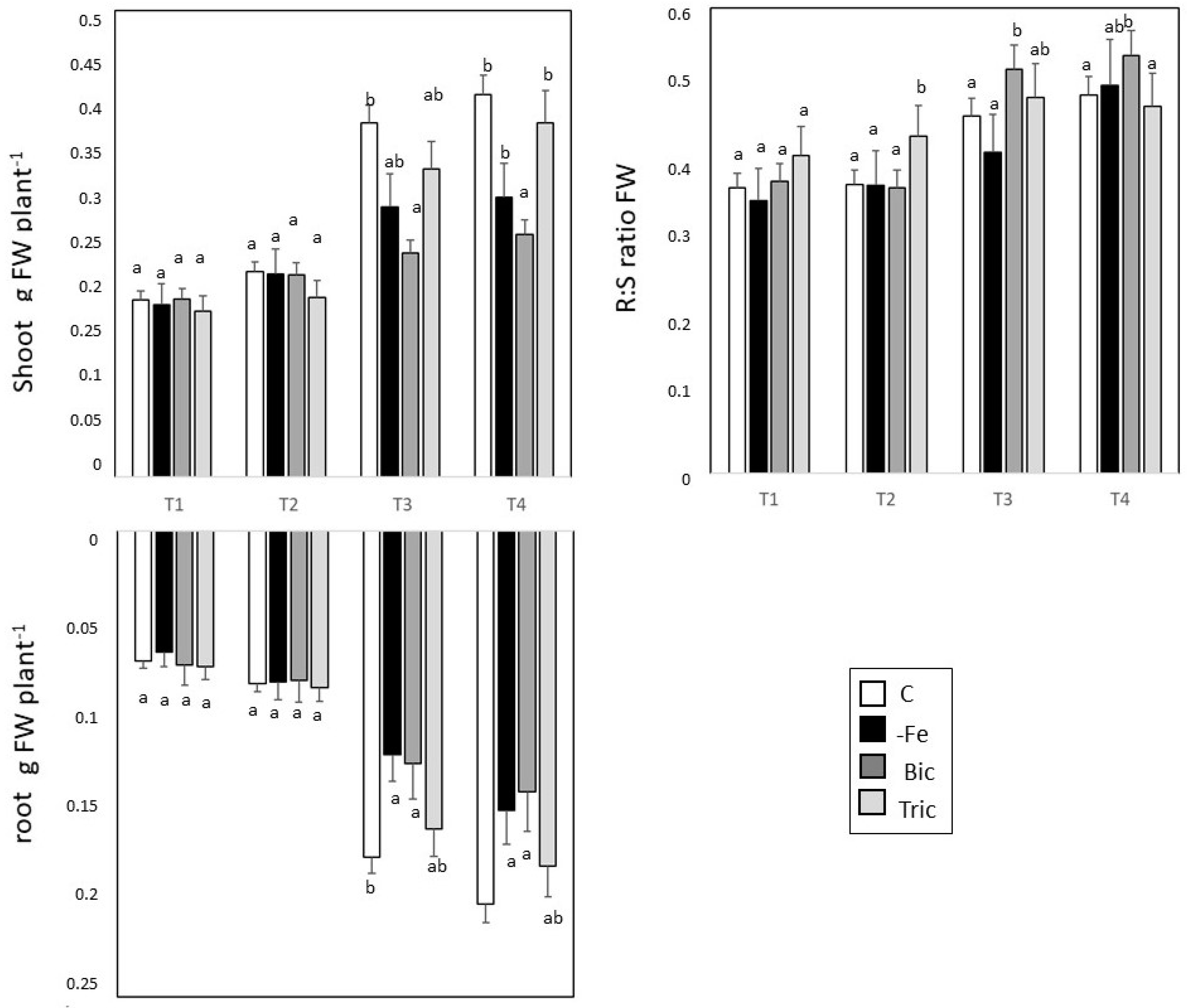
3.1. Direct and Induced Fe Deficiency Affect Plant Growth during Time

Direct and induced Fe deficiency treatments differentially affected plant growth. Shoot and root FW and DW showed that Fe and Bic strongly affected plant growth in comparison to C and Tric treatments (Figure 1, Figure S1). Figure 1 shows that shoot and root FW were significantly impaired mainly under –Fe and Bic treatments at the T3 and T4 in comparison with C plants, while plants grown in alkaline conditions (Tric) did not display any differences in shoot and root biomass. The root-to-shoot (R:S) ratio showed higher values only under Bic treatment 5 (T3) and 7 (T4) days after the imposition of stress conditions (Figure 1).
According to the observations at the biomass level, treatments differentially affected chlorophyll and total flavonoid contents in leaves (Figure 2). The chlorophyll contents in leaves grown under C and Tric conditions exhibited increases of approximately 1-fold along the time course analysis, reaching similar values at the final sampling time (Figure 2, left panel). In Bic-treated leaves, the chlorophyll content increased by about 35% at T3 compared to T1, reaching chlorophyll contents comparable to those found under C and Tric treatments. However, at the end of the experimental time, Bic-treated leaves showed a chlorophyll content that was lower by nearly 30% than the C and Tric treatments. Under direct Fe deficiency (–Fe), the leaf chlorophyll content slightly increased up to T2 and then decreased thereafter, reaching the lowest value at the end of the treatment (2.5-fold decrease with respect to the control). The flavonoid contents under C and Tric conditions showed a trend similar to that of chlorophyll (Figure 2, right panel). Under Bic conditions, the flavonoid content showed an increased by about 45% at the end of the treatment, while with Fe leaf conditions the flavonoid content remained quite constant over the course of the experiment.

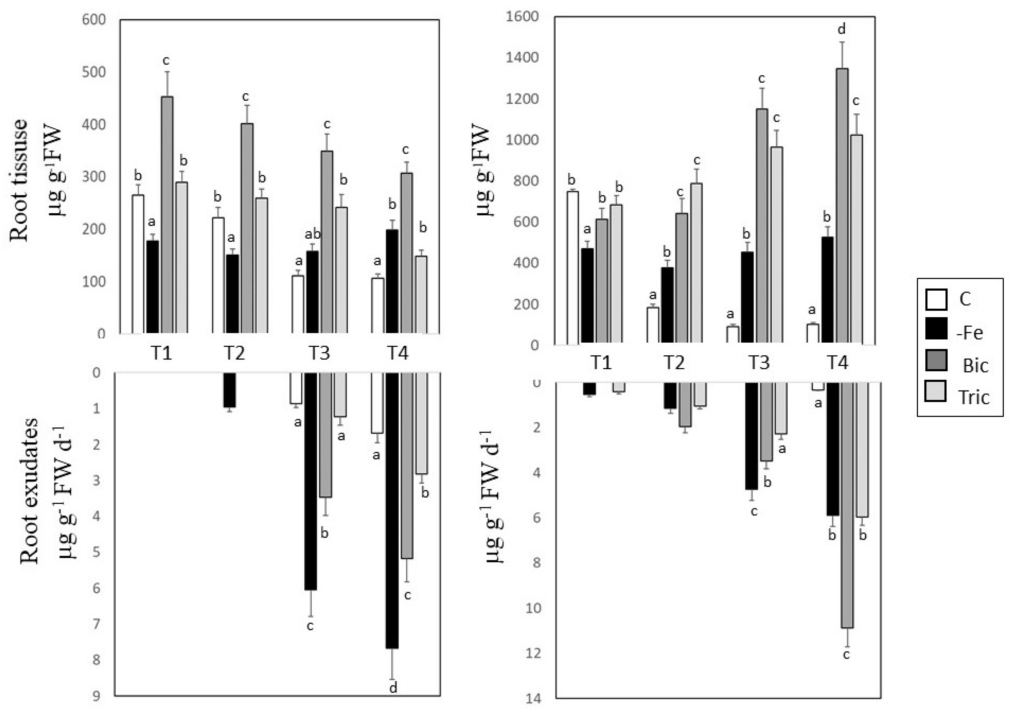
3.2. Malic and Citric Acid Contents in Root Tissues and Exudates

Malic acid contents gradually diminished during the treatment in C, Bic, and Tric conditions (Figure 3). The malic acid accumulated early in Bic-treated plants (T1) compared with the C plants. Despite an initial decrease, the malic acid content increased in –Fe plants at T4. Tric-treated plants displayed an increase in malic acid content at T4. In the root exudate fraction, the malic acid increased early in –Fe plants (T2), while it increased in Bic at 5 (T3) and 7 (T4) days after treatment. However, root exudates from –Fe plants showed the highest malic acid content at T2, T3, and T4 compared with other treatments. According to the observations in root tissues, the malic acid content in the root exudates from Tric-treated plants did not change compared with C plants (Figure 3).
The citric acid content showed different trends (Figure 3). Bic and Tric conditions exhibited the highest accumulation of citric acid (2- and 1.5-fold, respectively) at the end of the treatment compared with their contents at T1. Despite an initial decrease, citric acid also accumulated in the roots of –Fe plants. In the root exudates, citric acid was detected mainly in –Fe, Bic, and Tric plants at T2, T3, and T4 time points. However, citric acid content strongly increased in root exudates from Bic plants at the end (T4) of the experiment.

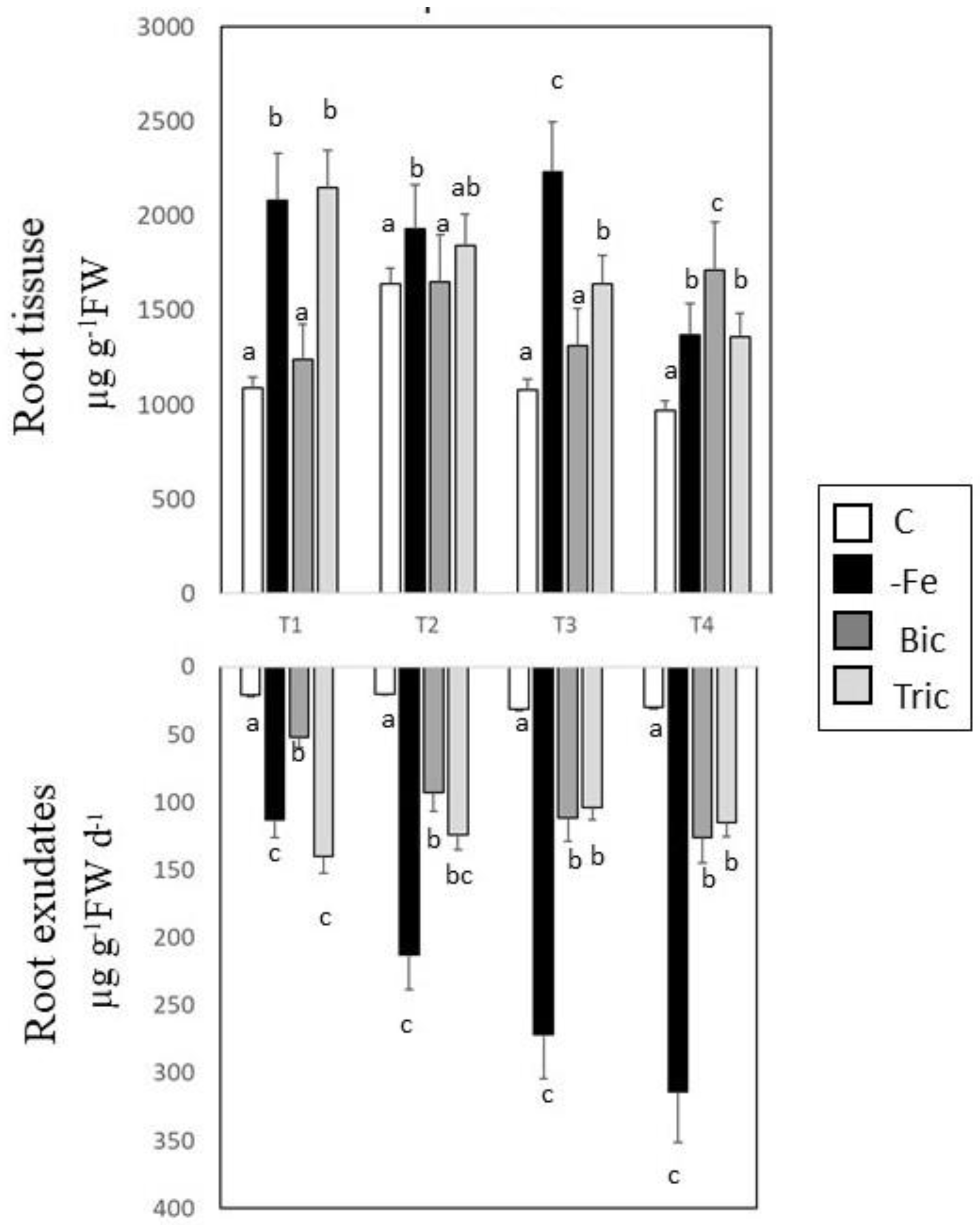
3.3. Phenolic Compounds in Root Extracts and Exudates

The water-soluble content of phenolics in C plants exhibited equivalent concentrations at T4 as at the beginning of the time course, while in –Fe and Tric conditions it decreased with respect to their initial concentrations by 34% and 38%, respectively (Figure 4). The three low-iron treatments showed higher accumulation of water-soluble phenolics in comparison with C at the end of the time course (T4). Bicarbonate (Bic)-treated plants displayed an increased accumulation of phenolics by about 76%, while –Fe- and Tric-treated plants displayed phenolic content increases of about 40% compared with the C treatment. The methanol-soluble phenolics content showed a variable trend between treatments (Table S1). The Tric condition demonstrated no significant differences between T1 and T4. Bic-treated plants showed a 1-fold increase with respect increase their initial values. However, phenolic compounds determined in methanol extracts showed consistent increases in all stressed conditions at the final sampling (T4) date when compared to the C conditions. Bicarbonate treatment resulting in an almost 3-fold increase in accumulation of phenolics compared with C, while –Fe and Tric increased by about 80% and 82%, respectively. Similarly, to what was registered for water extracts, –Fe- and Tric-treated roots exhibited similar phenolic contents at the end of the time course.
The phenolic contents in root exudates collected from C, –Fe, Bic, and Tric plants are shown in Figure 4 (lower panel). The results obtained were one order of magnitude higher in comparison to a previous work [35]. In fact, the method used to collect exudates was modified in the present study. Phenolics are more stable at low pH and precipitate at high pH. Thus, to avoid loss of material and to maintain the structure of the compounds, the collected solutions were acidified to pH 3.5 before being freeze-dried.
Phenolic compounds were detected in exudates at the first sampling date (T1) in all treatments. At T1, higher concentrations of phenolics were found in all treatments in comparison to the control; in particular, –Fe- and Tric-treated plants showed approximately 4-fold and 6-fold increases, respectively, compared with the control. Exudates from –Fe and Bic showed a gradual increase in phenolic content over the time course, reaching maximum levels at the end of the treatment, which were 1.8- and 1.4-fold higher, respectively, than their initial values. In C conditions, the concentrations remained constant throughout the treatment. Plants grown under –Fe, Bic, and Tric conditions exhibited high contents of phenolics in exudates with respect to C. The highest content of phenolics was found in –Fe exudates that exhibited an 11-fold increase with respect to the control at the end of the treatment, while Bic and Tric treatments resulted in 4.3-fold and 4-fold increases, respectively.

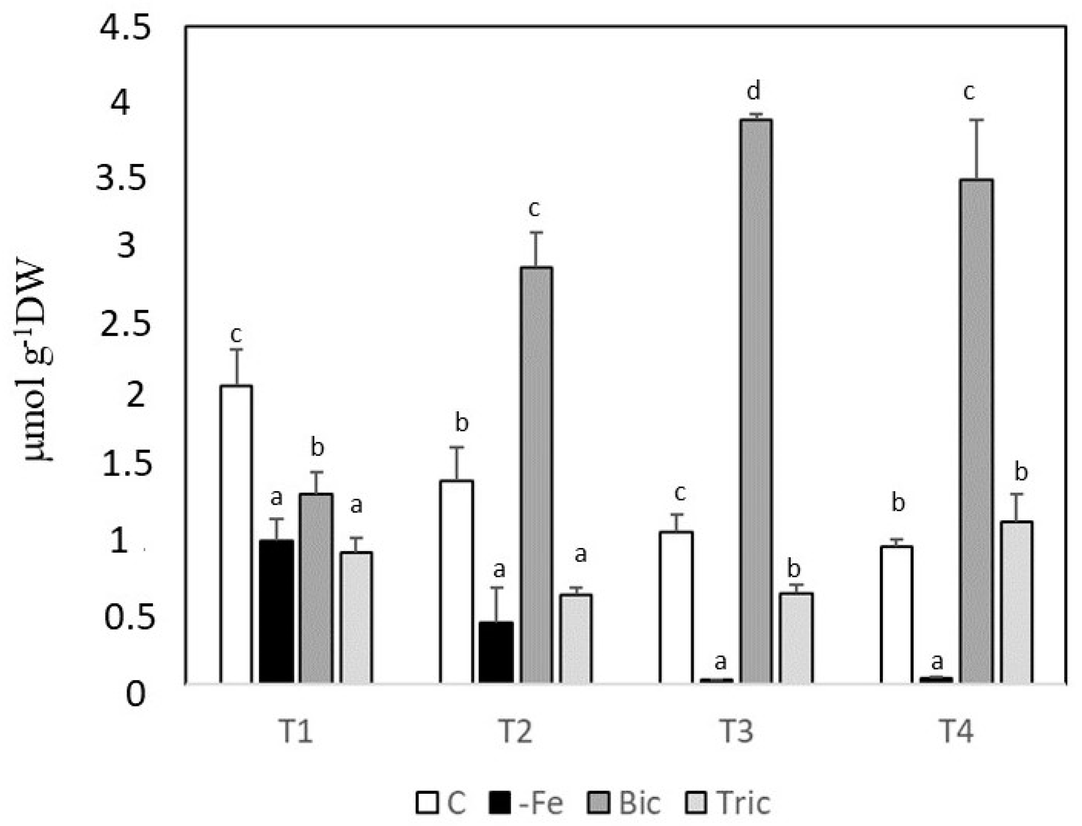
3.4. Root Apoplastic Fe

The results of the apoplastic root Fe contents obtained along the time course for C, –Fe, Bic, and Tric treatments are shown in Figure 5. The initial concentrations of apoplastic Fe in plants were determined (T0) before imposing the treatments (1.41–0.17 µmol g−1 DW). In –Fe conditions, the content of apoplastic Fe decreased, reaching a very low value at T3. Under Bic conditions, apoplastic Fe exhibited a strong accumulation of Fe, reaching a 2.7-fold increase at T3 with respect to T0 and remaining almost constant until the end of the treatment. Furthermore, a 3.5-fold increase in accumulation of apoplastic Fe was observed with respect to C results at the end of the treatment. In Tric conditions, no statistical variation was recorded throughout the experiment or at T4 in comparison with C.

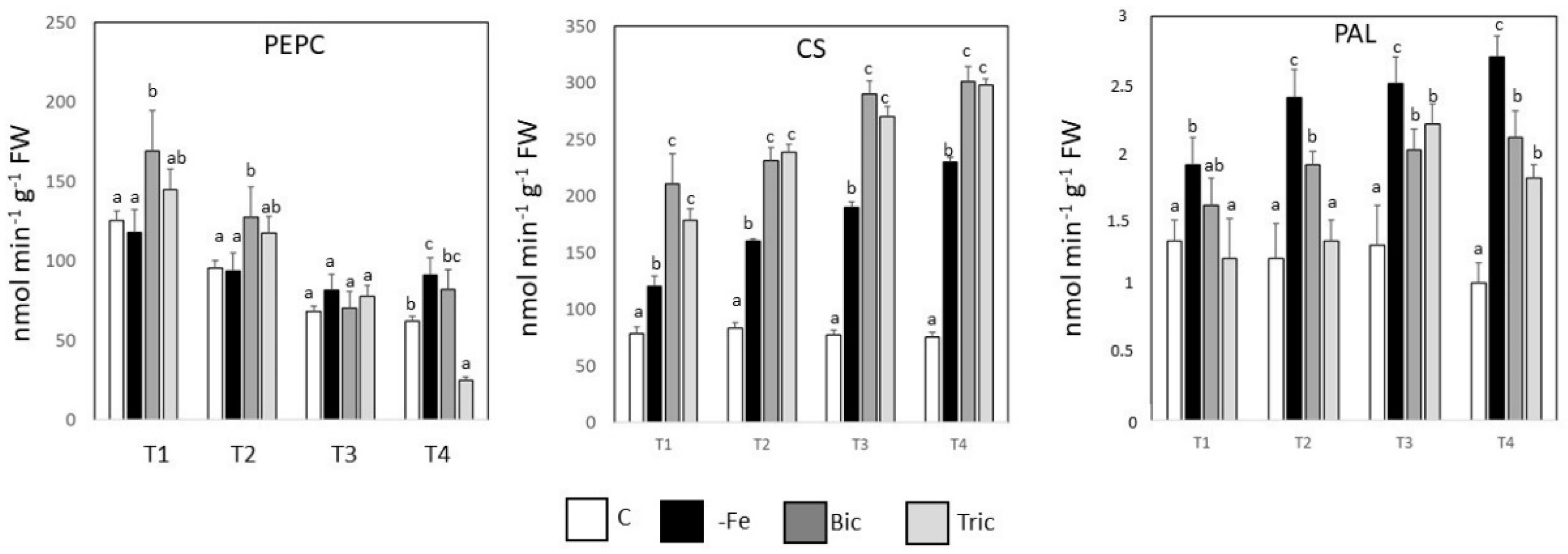
3.5. PEPCase, CS, and PAL Activities

In order to investigate the metabolic modulation occurring in P.judaica under direct and induced Fe deficiency, the activity of three different enzymes was determined: phosphoenol pyruvate carboxylase (PEPCase), citrate synthase (CS), and phenylalanine ammonia lyase (PAL). PEPCase is considered a key enzyme under Fe deficiency conditions, both in terms of pH stat regulation and as a driving force for glycolysis [34]. PEPCase activity showed a general and gradual decrease in all treatments during the time course—the decrease was sharper in Tric conditions (83%), leading to lower enzymatic activity at the end of the treatment. In –Fe conditions, the PEPCase activity decreased at the beginning of the treatment, then reached a constant value. However, comparing PEPCase activities at T4 with respect to C, increases were registered for –Fe and Bic (45% and 34%, respectively), while Tric showed a strong decrease of 60%. The CS activity had already increased in –Fe, Bic, and Tric conditions after 24 h of treatment compared with the control. However, CS activity was higher in both Bic and Tric plants compared with –Fe and C plants during the experimental time considered. By contrast, PAL activity was activated early in –Fe plants, displaying higher values than the other treatments. However, PAL activity increased later in Bic and Tric plants (Figure 6).

4. Discussion

Iron deficiency in plants is particularly evident in calcareous soils, where the presence of carbonate along with the alkaline pH reduces the solubility of Fe and consequently reduces its availability for plants. Furthermore, in calcareous media the availability of P is also affected, as it reacts with Ca+2 forming Ca3(PO4)2 precipitates, thus limiting its bioavailability. Therefore, under such conditions plants face shortages of both Fe and P. In order to discriminate between the different contributors to low Fe availability, we assessed the adaptation strategies of P. judaica in different growing conditions, namely under direct (–Fe) and induced Fe deficiency (Bic and Tric). Indeed, treatments differentially affected plant growth, as revealed by shoot and root biomass results (Figure 1). While alkaline (Tric) conditions did not affect shoot or root biomass, –Fe- and Bic-treated plants displayed growth reductions. This might be attributable to the low availability of Fe. The presence of high concentrations of bicarbonate in the growing medium leads to an immobilization of Fe within the plant [36,37]. Despite the lower chlorophyll concentration (about 30% lower than the control, (Figure 2), Bic plants did not present any symptoms of chlorosis, indicating that the consistent decrease in the growth rate observed in P. judaica under Bic conditions probably contributed to the maintenance of a physiologically compatible chlorophyll concentration in tissues. Moreover, Zohlen and Tyler [27] found that many wild calcicole grass species are able to maintain a much higher metabolically active Fe fraction in leaves, avoiding Fe chlorosis. In Tric conditions, where Fe deficiency is due only to a high pH, the FW and DW results for shoots and roots, as well as for biomass partition ratios, reached similar values to the control (Figure 1). Taken together, these results provide evidence that P. judaica displayed a high degree of tolerance to alkaline conditions, as well as to the presence of HCO3.
Countless works have pointed out the important roles that organic acids play under Fe deficiency conditions [13], both inside the plant and as root exudates. It has been well documented that roots exude a variety of organic compounds that play multiple functions in plant nutrient acquisition. Indeed, organic acid exudation might be considered a critical checkpoint in plant nutrition, since such molecules can (i) protect the roots from the entry of toxic ions (i.e., Al3+) and (ii) enhance nutrient uptake by modifying root development, and in turn soil exploration [38]. An increase in organic acid root exudation has been reported in several dicotyledonous species under Fe deficiency conditions, and the involvement of exudates in the mobilization of Fe has been documented as well [39]. Some studies have pointed out the enhanced rate of exudation of citric and malic acids in calcicole species compared with calcifuges as a strategy characterizing their efficient acquisition of P and Fe [40].
P. judaica greatly increased the malic and citric acid exudation when subjected to direct and induced Fe deficiency (Figure 3). Both malic and citric acids may undergo complexation with target metals such as Fe(III) once exuded, improving their solubility, particularly in calcareous soils [12,41]. However, citric acid demonstrates a better metal extraction capability than malic acid in alkaline conditions [42]. Citric acid release was higher than that of malic acid, particularly in induced Fe-deficient conditions (Bic and Tric treatments). On the other hand, under direct Fe deficiency (–Fe), malic acid release was higher than citric acid (Figure 3). Interestingly, both organic acids displayed a time- and condition-dependent exudation pattern. Indeed, the release of organic compounds is closely linked to the physiological status of the plants and the physical, chemical, and biological characteristics of the growing medium, thus leading to great temporal variability [43]. Indeed, in the present study, citric acid was already detected in root exudates of P. judaica after 24 h (T1), while malic acid started to be released only at T2 and only if plants were subjected to direct Fe deficiency (Figure 3). Furthermore, the release of citric acid over time also differs depending on the imposed conditions, with the highest release in direct Fe-deficient conditions occurring at T3 and the highest release in induced Fe-deficient conditions occurring at T4. This differential behavior suggests that (i) citric acid plays a predominant role as the primary response to Fe deficiency and (ii) that citric acid exudation is both time- and substrate-dependent, as previously observed in other plant species, such as white lupin and rapeseed [43]. Citric acid can also chelate Ca2+, enhancing Ca-P mineral dissolution [40,41,42], thus increasing P availability. Hence, the high content of citric acid in Bic exudates at the end of the treatment could represent a dual response to both low Fe and P availability in a treatment at high Ca2+ concentrations.
Calcareous conditions also affected the content of both malic and citric acids in root tissues (Figure 3). The highest root accumulation was observed in the induced Fe-deficient conditions (Bic and Tric). Such observations are in line with the variation of the Fe content in the root apoplast—Bic-treated plants displayed a strong accumulation of Fe in the root apoplast with respect to the other treatments (Figure 5). Recent evidence suggested that malic acid accumulation in the apoplast is involved in the chelation and accumulation of Fe in the apoplast, driving root developmental changes under P starvation [38]. However, such an effect was strictly related to the presence of carbonate in the growing solution, while the alkaline condition alone did not affect malic acid or apoplast Fe contents in the roots. In –Fe conditions, the predominance of malic acid exudation could be attributable to a specific metabolic state. Plants grown in Fe deficiency conditions activate the strategy I mechanisms, increasing ATP and reducing power requirements that cannot be fulfilled through the respiratory chain, which is impaired by the lack of Fe. In fact, the impaired mitochondrial electron chain leads to a low turnover of NADH/NAD+; accordingly Fe-starved plants have to face the problem of refilling NAD+. The high activity of PEPCase in –Fe roots converts PEP to oxaloacetate (OAA), which can be reduced to malate, since a high NADH/NAD+ ratio would favor the reaction and could be a way to regenerate NAD+. Furthermore, the malic acid might be synthesized from the tricarboxylic acid (TCA) cycle [35]. Here, we observed that under both direct (–Fe) and Bic-induced Fe deficiency conditions, the PEPCase activity was significantly higher than under normal Fe supply conditions. Furthermore, CS activity was induced under both direct and induced Fe deficiency conditions, with higher induction under calcareous or alkaline conditions. Such findings are in agreement with findings for citric acid accumulation in roots as well as in root exudates, indicating that the induction of citric acid is driven mainly by higher pH conditions. Moreover, the accumulation of citric acid in roots constitutes an advantage, as it performs multiple functions in cells (pH-stat, signaling, xylem transport of Fe, carbon skeleton precursor functions) [44,45].
The high Fe accumulation in the apoplast (Figure 5) could also be related to enhanced production and release of pectates, i.e., the main components of the root apoplast. A triggered release of these high molecular weight organic compounds has been observed in aluminum-tolerant plants, enhancing the extracellular absorption of the element and preventing its plant uptake [46]. An increase in negative binding sites might, thus, lead to the higher Fe accumulation and act as an Fe reservoir for the plants. This phenomenon was particularly triggered in the induced Fe-deficient conditions (Bic, Figure 5), suggesting that a pronounced release of pectates or the modification of their chemical properties might represent an additional adaptation strategy of calcicole plants.
Other than organic acids, environmental stresses induce the accumulation in roots and exudation out of the roots of phenolic compounds in plants. Phenolics have been reported to have multiple biological effects, including antioxidant and chelating activities [47]. The antioxidant activity of phenolics is mainly due to their redox properties, which allow them to act as reducing agents, hydrogen donors, and singlet oxygen and free radical scavengers. Furthermore, their metal chelation activity [47] facilitates Fe acquisition by plants [18,19,20,48]. P. judaica growing under –Fe mainly showed a high accumulation of phenolic compounds in root tissues at the early stages, while plants grown in the presence of Bic accumulated water-soluble phenols later. This is likely because highly calcareous conditions lead plants to face cumulative stress, due to the low availability of Fe and P. Decreased Fe and P in plants induces high levels of reactive oxygen species (ROS), promoting oxidative stress [49,50]. The lower concentration of phenolics in Tric-treated roots with respect to Bic could be due to an overall lower stress condition, as explained above. The root apoplastic Fe has been suggested to be an important Fe source for plants, as 75% of total Fe in the roots is located in the apoplast [19]. The decrease of the Fe content in the apoplast of –Fe plants is in agreement with Zhang et al. [51]. In fact, under –Fe conditions, the Fe immobilized in the apoplast is the sole available source of Fe for the plant. In Bic conditions, the concentration of apoplastic Fe increased due to the alkaline pH, which dramatically decreases Fe solubility. In addition, it has been pointed out that high apoplastic pH depresses FCR activity, consequently decreasing the reduction and uptake of Fe [25]. Jin et al. [18] found that removal of phenolics from the growing medium in red clover decreased the Fe content in the shoots, resulted in leaf chlorosis, and increased FCR activity and proton extrusion, suggesting the direct involvement of phenolics in apoplastic Fe reutilization.
In the root exudate fraction, –Fe treated plants displayed higher accumulation of phenolics compared with the other treatments, indicating that exudation of such compounds is mainly dependent on the Fe availability in the external medium. Phenolic concentrations in root exudates of P. judaica subjected to direct (–Fe) and induced Fe deficiency increased in almost all treatments along the time course and reached higher concentrations than the control at the end of the time course (Figure 4). In Bic conditions, the initial concentration of exuded phenolics was lower with respect to Tric, but it strongly increased along the time course; conversely, in Tric conditions a slight decrease in phenolic exudates was registered. The highest concentration of phenolics was found in –Fe conditions at T4. The immediate and massive release of phenolics might be linked to the triggered Fe accumulation in the apoplast, constituting a kind of reservoir of the essential nutrient for the plant. The fast depletion of apoplastic Fe in –Fe roots could be then supported by the high levels of phenolics found in the root exudates. In Tric conditions, the exudation of phenolics most probably resulted in the acquisition of Fe, as it was present in the solution and also in the apoplast, meaning the plant could retrieve Fe, making further secretion of exudates unnecessary.
Notably, the concentration of released phenolic compounds (Figure 4) was one order of magnitude higher than for citric and malic acids (Figure 3). This is in agreement with several works reporting that phenolic compounds are the main components in root exudates in response to Fe deficiency [18,52]. In addition, phenolic compounds were detected earlier than organic acids in the exudates. These data clearly highlight different time-dependent release patterns for the two classes of compounds, with differences between the two organic acids. Indeed, phenolics and organic acids have different functions in the rhizosphere. Citric acid is involved in ligand exchange reactions favoring the mobilization of P, also acting as a complexing agent for Fe and Ca. Comparing the stability constants of the two organic acids released by P. judaica, citric acid exhibits a higher affinity towards Fe and Ca ions than does malic acid [43]. Thus, the release of this multifunctional citric acid in the early stage (T1) could represent a further advantage of the adaptation strategy.

5. Conclusions

In conclusion, the results presented in this work show the great flexibility in P. judaica responses to Fe deficiency. Overall, temporal analysis of plant responses revealed that the root exudate pattern is time-dependent, and that direct and induced –Fe deficiency conditions differentially affect root exudation of malic acid, citric acid, and phenolics. Indeed, direct Fe deficiency induced a higher exudation of malic acid and phenolics, while calcareous conditions led to a strong accumulation of citric acid. Alkaline conditions resulted in intermediate behaviors, suggesting that conditions inducing a decreased Fe availability (i.e., absence in the media and immobilization by carbonate) might differentially affect temporal responses of root exudation. On one hand, this work revealed that plant responses to induced Fe deficiency are more complex than those to direct Fe deficiency, while on the other hand revealing that the high degree of tolerance to alkaline conditions of P. judaica may be linked to earlier and greater phenolics exudation rates, as well as to the combination of phenolics and carboxylic acids in root exudates in the late responses. Investigating the adaptation mechanisms of wild plant species such as P. judaica is challenging, and at this stage we cannot rule out the presence of other specific strategies explaining the calcicole behavior of this plant. Further efforts should be addressed in this direction in order to identify new mechanisms underlying the tolerance traits to Fe deficiency in plants.

Supplementary Materials

The following are available online at https://www.mdpi.com/2073-4395/10/7/1037/s1.

Author Contributions

Conceptualization, G.V. and G.Z.; methodology, L.T. and T.M.; validation, L.T.; formal analysis, L.T. and M.I.; resources, G.Z. and G.V.; data curation, G.V.; writing—original draft preparation, L.T. and G.V.; writing—review and editing, T.M, G.Z., and G.V. All authors have read and agreed to the published version of the manuscript.

Funding

This research received no external funding.

Conflicts of Interest

The authors declare no conflict of interest.

References

  1. Marschner, H. Mineral Nutrition of Higher Plants; Academic Press: London, UK, 1995. [Google Scholar]
  2. Kim, S.A.; Guerinot, M.L. Mining iron: Iron uptake and transport in plants. FEBS Lett. 2007, 581, 2273–2280. [Google Scholar] [CrossRef] [PubMed] [Green Version]
  3. Rabotti, G.; Zocchi, G. Plasma membrane-bound H+-ATPase and reductase activities in Fe-deficient cucumber roots. Physiol. Plant. 1994, 90, 779–785. [Google Scholar] [CrossRef]
  4. Dell’Orto, M.; Santi, S.; De Nisi, P.; Cesco, S.; Varanini, Z.; Zocchi, G. Development of Fe deficiency responses in cucumber (Cucumis sativus L.) roots: Involvement of plasma membrane H+-ATPase activity. J. Exp. Bot. 2001, 51, 695–701. [Google Scholar]
  5. Santi, S.; Cesco, S.; Varanini, Z.; Pinton, R. Two plasma membrane H+-ATPase genes are differentially expressed in iron-deficient cucumber plants. Plant Physiol. Biochim. 2005, 43, 287–292. [Google Scholar] [CrossRef] [PubMed]
  6. Santi, S.; Schmidt, W. Dissecting iron deficiency-induced proton extrusion in Arabidopsis roots. New Phytol. 2009, 183, 1072–1084. [Google Scholar] [CrossRef]
  7. Zocchi, G. Metabolic changes in iron-stressed dicotyledonous plants. In Iron Nutrition in Plants and Rhizospheric Microorganisms; Barton, L.L., Abadía, J., Eds.; Springer: Dordrecht, The Netherlands, 2006; pp. 359–370. [Google Scholar]
  8. Abadía, J.; López-Millán, A.F.; Rombolà, A.; Abadía, A. Organic acids and Fe deficiency: A review. Plant Soil 2002, 241, 75–86. [Google Scholar] [CrossRef]
  9. Jelali, N.; Wissala, M.; Dell’Orto, M.; Abdelly, C.; Gharsalli, M.; Zocchi, G. Changes of metabolic responses to direct and induced Fe deficiency of two Pisum sativum cultivars. Environ. Exp. Bot. 2010, 68, 238–246. [Google Scholar] [CrossRef]
  10. López-Millán, A.F.; Morales, F.; Abadía, A.; Abadía, J. Effects of iron deficiency on the composition of the leaf apoplastic fluid and xylem sap in sugar beet. Implications for iron and carbon transport. Plant Physiol. 2000, 124, 873–884. [Google Scholar] [CrossRef] [Green Version]
  11. Rellán-Álvarez, R.; Andaluz, S.; Rodríguez-Celma, J.; Wohlgemuth, G.; Zocchi, G.; Álvarez-Fernández, A.; Fiehn, O.; López-Millán, A.F.; Abadía, J. Changes in the proteomic and metabolic profiles of Beta vulgaris root tips in response to iron deficiency and resupply. BMC Plant Biol. 2010, 10, 120. [Google Scholar] [CrossRef] [Green Version]
  12. López-Bucio, J.; Nieto-Jacobo, M.F.; Ramírez-Rodríguez, V.; Herrera-Estrella, L. Organic acid metabolism in plants: From adaptive physiology to transgenic varieties for cultivation in extreme soils. Plant Sci. 2000, 160, 1–13. [Google Scholar] [CrossRef]
  13. Reid, R.; Smith, F.A. The cytoplasmic pH Stat. In Handbook of Plant Growth: pH as the Master Variable; Rengel, Z., Ed.; Marcel Dekker: New York, NY, USA, 2002; pp. 47–67. [Google Scholar]
  14. Canarini, A.; Kaiser, C.; Merchant, A.; Richter, A.; Wanek, W. Root exudation of primary metabolites: Mechanism and their roles in plant responses to environmental stimuli. Front Plant Sci. 2019, 10, 157. [Google Scholar] [CrossRef] [PubMed] [Green Version]
  15. Tyler, G.; Strom, L. Differing organic acid exudation patterns explain calcifuge and acidifuge behaviour of plants. Ann. Bot. 1995, 75, 75–78. [Google Scholar] [CrossRef] [Green Version]
  16. Bush, D.S. Calcium regulation in plant cells and its role in signaling. Annu. Rev. Plant Phys. 1995, 46, 95–122. [Google Scholar] [CrossRef]
  17. Dakora, F.D.; Phillips, D.A. Root exudates as mediators of mineral acquisition in low nutrient environments. Plant Soil 2002, 245, 35–47. [Google Scholar] [CrossRef]
  18. Cesco, S.; Neumann, G.; Tommasi, N.; Pinton, R.; Weisskopf, L. Release of plant-borne flavonoids into the rhizosphere and their role in plant nutrition. Plant Soil 2010, 329, 1–25. [Google Scholar] [CrossRef]
  19. Jin, C.W.; You, G.Y.; He, Y.F.; Tang, C.; Wu, P.; Zheng, S.J. Iron deficiency-induced secretion of phenolics facilitates the reutilization of root apoplastic iron in red clover. Plant Physiol. 2007, 144, 278–285. [Google Scholar] [CrossRef] [PubMed] [Green Version]
  20. Voges, M.J.E.E.E.; Bai, Y.; Schulze-Lefert, P.; Sattely, E.S. Plant-derived coumarins shape the composition of an Arabidopsis synthetic root microbiome. Proc. Natl. Acad. Sci. USA 2019, 116, 12558–12565. [Google Scholar] [CrossRef] [Green Version]
  21. Lindsay, W.L.; Schwab, A.P. The chemistry of iron in soils and its availability to plants. J. Plant Nutr. 1982, 5, 821–840. [Google Scholar] [CrossRef]
  22. Schenkeveld, W.D.C.; Kraemer, S.M. Constrints to Synergistic Fe mobilization from calcareous soil by a phytosiderophore and a reductant. Soil Syst. 2018, 2, 67. [Google Scholar] [CrossRef] [Green Version]
  23. Mengel, K. Iron availability in plant tissues—iron chlorosis on calcareous soils. Plant Soil 1994, 165, 275–283. [Google Scholar] [CrossRef]
  24. Kosegarten, H.; Hoffmann, D.; Rroco, E.; Grolig, F.; Glüsenkamp, F.K.; Mengel, K. Apoplastic pH and FeIII reduction in young sunflower (Helianthus annuus) roots. Physiol. Plant 2004, 122, 95–106. [Google Scholar] [CrossRef]
  25. Alhendawi, R.A.; Römheld, V.; Kirkby, E.A.; Marschner, H. Influence of increasing bicarbonate concentrations on plant growth, organic acid accumulation in roots and iron uptake by barley, sorghum, and maize. J. Plant Nutr. 1997, 20, 1731–1753. [Google Scholar] [CrossRef]
  26. Zohlen, A.; Tyler, G. Immobilization of tissue iron on calcareous soil: Differences between calcicole and calcifuge plants. Oikos 2000, 89, 95–106. [Google Scholar] [CrossRef]
  27. Zohlen, A. Chlorosis in wild plants: Is it a sign of iron deficiency? J. Plant Nutr. 2002, 25, 2205–2228. [Google Scholar] [CrossRef]
  28. Lucena, C.; Romera, F.J.; Rojas, C.L.; García, M.J.; Alcántara, E.; Pérez-Vicente, R. Bicarbonate blocks the expression of several genes involved in the physiological responses to Fe deficiency of Strategy I plants. J. Funct. Plant Biol. 2007, 34, 1002–1009. [Google Scholar] [CrossRef]
  29. Donnini, S.; De Nisi, P.; Gabotti, G.; Tato, L.; Zocchi, G. Adaptive strategies of Parietaria diffusa (M.K.) to calcareous habitat with limited iron availability. Plant Cell Environ. 2012, 35, 1171–1184. [Google Scholar] [CrossRef]
  30. Dell’Orto, M.; De Nisi, P.; Pontiggia, A.; Zocchi, G. Fe deficiency responses in Parietaria diffusa: A calcicole plant. J. Plant Nutr. 2003, 26, 10–11. [Google Scholar] [CrossRef]
  31. Tato, L.; De Nisi, P.; Donnini, S.; Zocchi, G. Low iron availability and phenolic metabolism in a wild plant species (Parietaria judaica L.). Plant Physiol. Biochem. 2013, 72, 145–153. [Google Scholar] [CrossRef]
  32. Lichtenthaler, H.K. Chlorophyll and carotenoids: Pigments of photosynthetic biomembranes. Methods Enzymol. 1987, 148, 350–382. [Google Scholar]
  33. Bienfait, H.F.; van den Briel, W.; Mesland-Mul, N.T. Free space iron pools in roots. Generation and mobilization. Plant Physiol. 1985, 78, 596–600. [Google Scholar] [CrossRef] [Green Version]
  34. De Nisi, P.; Zocchi, G. Phosphoenolpyruvate carboxylase in cucumber (Cucumis sativus L.) roots under iron deficiency: Activity and kinetic characterization. J. Exp. Bot. 2000, 352, 1903–1909. [Google Scholar] [CrossRef] [PubMed] [Green Version]
  35. Vigani, G.; Bashir, K.; Ishimaru, Y.; Lehmann, M.; Casiraghi, F.M.; Nakanishi, H.; Seki, M.; Geigenberger, P.; Zocchi, G.; Nishizawa, N.K. Knocking down Mitochondrial Iron Transporter (MIT) reprograms primary and secondary metabolism in rice plants. J. Exp. Bot. 2016, 67, 1357–1368. [Google Scholar] [CrossRef] [PubMed] [Green Version]
  36. Nikolic, M.; Kastori, R. Effect of bicarbonate and Fe supply on Fe nutrition of grapevine. J. Plant Nutr. 2000, 23, 1619–1627. [Google Scholar] [CrossRef]
  37. Tagliavini, M.; Rombolà, A.D. Iron deficiency and chlorosis in orchard and vineyard ecosystems. Eur. J. Agron. 2001, 15, 71–92. [Google Scholar] [CrossRef]
  38. Mora-Macias, J.; Ojeda-Rivera, J.O.; Gutierrez-Alanis, D.; Yong-Villalobos, L.; Oropeza-Aburto, A.; Raya-Gonzalez, J.; Jiménez-Domínguez, G.; Chávez-Calvillo, G.; Rellán-Álvarez, R.; Herrera-Estrella, L. Malate-dependent Fe accumulation is a critical checkpoint in the root developmental response to low phosphate. Proc. Natl. Acad. Sci. USA 2017, 114, E3563–E3572. [Google Scholar] [CrossRef] [Green Version]
  39. Farrar, J.F.; Jones, D.L. The control of carbon acquisition by roots. New Phytol. 2000, 147, 43–53. [Google Scholar] [CrossRef]
  40. Ström, L.; Owen, A.G.; Godbold, D.L.; Jones, D.L. Organic acid behavior in calcareous soil implication for rhizosphere nutrient cycling. Soil Biol. Biochem. 2005, 37, 2046–2054. [Google Scholar] [CrossRef]
  41. Abadía, J.; Vázquez, S.; Rellán-Álvarez, R.; El-Jendoubi, E.; Abadía, A.; Álvarez-Fernández, A.; López-Millán, A.F. Towards a knowledge-based correction of iron chlorosis. Plant Physiol. Biochem. 2011, 49, 471–482. [Google Scholar] [CrossRef]
  42. Terzano, R.; Cuccovillo, G.; Gattullo, C.E.; Medici, L.; Tomasi, N.; Pinton, R.; Mimmo, T.; Cesco, R. Combined effect of organic acids and flavonoids on the mobilization of major and trace elements from soils. Biol. Fert. Soil 2015, 51, 685–695. [Google Scholar] [CrossRef]
  43. Mimmo, T.; Hann, S.; Jaitz, L.; Cesco, S.; Gessa, C.E.; Puschenreiter, M. Time and substrate dependent exudation of carboxylates by Lupinus albus and Brassica napus L. Plant Physiol. Biochem. 2011, 49, 1272–1278. [Google Scholar] [CrossRef]
  44. Vigani, G.; Zocchi, G.; Bashir, K.; Philippar, K.; Briat, J.F. Signals from chloroplasts and mitochondria for iron homeostasis regulation. Trends Plant Sci. 2013, 18, 305–311. [Google Scholar] [CrossRef]
  45. Vigani, G.; Pii, Y.; Celletti, S.; Maver, M.; Mimmo, T.; Cesco, S.; Astolfi, S. Mitochondrial dysfunction under Fe and S deficiency: Is citric acid involved in the regulation of nutrient-responsive genes? Plant Physiol Biochem. 2018, 2018 126, 86–96. [Google Scholar] [CrossRef]
  46. Horst, W.J.; Wang, Y.; Eticha, D. The role of the root apoplast in aluminium induced inhibition of root elongation and in aluminium resistance of plants: A review. Ann. Bot. 2010. 106, 185–197. [CrossRef]
  47. Rice-Evans, C.A.; Miller, N.J.; Paganga, G. Antioxidant properties of phenolic compounds. Trends Plant Sci. 1997, 2, 152–159. [Google Scholar] [CrossRef]
  48. Andjelkovic, M.; Van Camp, J.; De Meuleraer, B.; Depaemelaere, G.; Socaciu, C.; Verloo, M.; Verhe, R. Iron chelation properties of phenolic acids bearing catechol and galloyl groups. Food Chem. 2006, 98, 23–31. [Google Scholar] [CrossRef]
  49. Tewari, R.K.; Kumar, P.; Sharma, P.N. Oxidative stress and antioxidant responses in young leaves of mulberry plants grown under nitrogen, phosphorus or potassium deficiency. J. Integr. Plant Biol. 2007, 49, 313–322. [Google Scholar] [CrossRef]
  50. Hajiboland, R. Effect of micronutrient deficiencies on plant stress responses. In Abiotic Stress Responses in Plants; Hyderabad, A.P., Parvaiz, A., Eds.; Springer: New York, NY, USA, 2007; pp. 330–389. [Google Scholar]
  51. Zhang, F.S.; Romheld, V.; Marschner, H. Role of the root apoplasm for iron acquisition by wheat plants. Plant Physiol. 1991, 97, 1302–1305. [Google Scholar] [CrossRef] [PubMed] [Green Version]
  52. Schmid, N.B.; Giehl, R.F.; Döll, S.; Mock, H.P.; Strehmel, N.; Scheel, D.; Kong, X.; Hider, R.C.; von Wirén, N. Feruloyl-CoA 6′-Hydroxylase1-dependent coumarins mediate iron acquisition from alkaline substrates in Arabidopsis. Plant Physiol. 2014, 164, 160–172. [Google Scholar] [CrossRef] [Green Version]
Figure 1. Determination of fresh weight (FW) of shoots and roots and root-to-shoot (R/S) ratio of plants grown in control condition (C), direct Fe deficiency conditions (–Fe), and under induced Fe deficiency in the presence of bicarbonate (Bic) or alkaline organic buffer (Tric). T1, T2, T3, and T4 correspond to sampling at 1, 3, 5, and 7 days. The R/S ratio was calculated on a FW basis. Bars indicate the means ± SE (standard error) (n = 9). Different letters indicate statistical differences (p < 0.05) within each treatment.
Agronomy 10 01037 g001
Figure 2. Leaf chlorophyll contents (mg g−1 FW) (left panel) and total flavonoid contents (expressed in Dualex units) (right panel) in leaves of plants grown in control conditions (C), direct iron deficiency (–Fe), and under induced Fe deficiency in the presence of bicarbonate (Bic) or alkaline organic buffer (Tric). T1, T2, T3, and T4 correspond to sampling at 1, 3, 5, and 7 days. Error bars represent the means ± SE (standard error) (n = 5). Different letters indicate statistical differences (p < 0.05) within each treatment.
Agronomy 10 01037 g002
Figure 3. Time course of the concentrations of malic and citric acids in root tissues and exudates of plants grown in control conditions (C), direct iron deficiency conditions (–Fe), and under induced Fe deficiency in presence of bicarbonate (Bic) or alkaline organic buffer (Tric). Malic acid contents are reported in the left panels; citric acid contents are reported in the right panels. T1, T2, T3, and T4 correspond to sampling at 1, 3, 5, and 7 days. Data are the means ± SE (standard error) (n = 5). Different letters indicate statistical differences (p < 0.05) within each treatment.
Agronomy 10 01037 g003
Figure 4. Contents of phenolic compounds in root tissues and exudates of plants grown in control conditions (C), direct iron deficiency conditions (–Fe), and under induced Fe deficiency in the presence of bicarbonate (Bic) or alkaline organic buffer (Tric). T1, T2, T3, and T4 correspond to sampling at 1, 3, 5, and 7 days. Values are expressed as the means ± SE (standard error) (n = 5). Different letters indicate statistical differences (p < 0.05) within each treatment.
Agronomy 10 01037 g004
Figure 5. Apoplastic Fe concentration (µmol g−1 DW) in roots of plants grown in control conditions (C), direct iron deficiency conditions (–Fe), and under induced Fe deficiency in the presence of bicarbonate (Bic) or alkaline organic buffer (Tric). T1, T2, T3, and T4 correspond to sampling at 1, 3, 5, and 7 days. Data are the means ± SE (standard error) (n = 5). Different letters indicate statistical differences (p < 0.05) within each treatment.
Agronomy 10 01037 g005
Figure 6. Enzymatic activity (phosphoenol pyruvate carboxylase - PEPCase, citrate synthase -CS, and phenylalanine ammonia lyase -PAL).) in the roots of plants grown in control conditions (C), direct iron deficiency conditions (–Fe), and under induced Fe deficiency in the presence of bicarbonate (Bic) or alkaline organic buffer (Tric). T1, T2, T3, and T4 correspond to sampling at 1, 3, 5, and 7 days. Data are expressed as the means ± SE (n = 5). Enzyme activity is expressed as nmol min−1mg−1prot. Significant differences are labelled with different letters (p < 0.05) within each treatment.
Agronomy 10 01037 g006

