Article

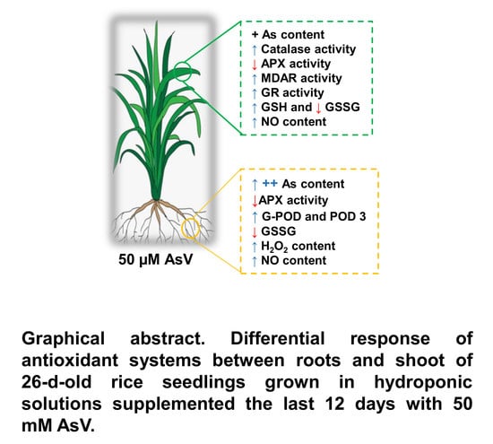
Reactive Oxygen Species (ROS) Metabolism and Nitric Oxide (NO) Content in Roots and Shoots of Rice (Oryza sativa L.) Plants under Arsenic-Induced Stress

by
Ernestina Solórzano
1,
Francisco J. Corpas
2,
Salvador González-Gordo
2 and
José M. Palma
2,*
1
Departmento de Medio Ambiente, Instituto de Ciencias y Tecnologías Aplicadas (INSTEC), Quinta de los Molinos, Ave. Salvador Allende, 1110, Plaza de la Revolución, La Habana, Cuba
2
Group of Antioxidants, Free Radicals and Nitric Oxide in Biotechnology, Food and Agriculture, Department of Biochemistry, Cell and Molecular Biology of Plants, Estación Experimental del Zaidín, CSIC, Apartado 419, E-18080 Granada, Spain
*
Author to whom correspondence should be addressed.
Agronomy 2020, 10(7), 1014; https://doi.org/10.3390/agronomy10071014
Submission received: 25 May 2020 / Revised: 29 June 2020 / Accepted: 12 July 2020 / Published: 14 July 2020
(This article belongs to the Special Issue Functional Metabolism in Crops/Agronomy-Series Ⅱ)

Abstract

:
Arsenic (As) is a highly toxic metalloid for all forms of life including plants. Rice is the main food source for different countries worldwide, although it can take up high amounts of As in comparison with other crops, showing toxic profiles such as decreases in plant growth and yield. The induction of oxidative stress is the main process underlying arsenic toxicity in plants, including rice, due to an alteration of the reactive oxygen species (ROS) metabolism. The aim of this work was to gain better knowledge on how the ROS metabolism and its interaction with nitric oxide (NO) operate under As stress conditions in rice plants. Thus, physiological and ROS-related biochemical parameters in roots and shoots from rice (Oryza sativa L.) were studied under 50 μM arsenate (AsV) stress, and the involvement of the main antioxidative systems and NO in the response of plants to those conditions was investigated. A decrease of 51% in root length and 27% in plant biomass was observed with 50 μM AsV treatment, as compared to control plants. The results of the activity of superoxide dismutase (SOD) isozymes, catalase, peroxidase (POD: total and isoenzymatic), and the enzymes of the ascorbate–glutathione cycle, besides the ascorbate and glutathione contents, showed that As accumulation provoked an overall significant increase of most of them, but with different profiles depending on the plant organ, either root or shoot. Among the seven identified POD isozymes, the induction of the POD-3 in shoots under As stress could help to maintain the hydrogen peroxide (H2O2) redox homeostasis and compensate the loss of the ascorbate peroxidase (APX) activity in both roots and shoots. Lipid peroxidation was slightly increased in roots and shoots from As-treated plants. The H2O2 and NO contents were enhanced in roots and shoots against arsenic stress. In spite of the increase of most antioxidative systems, a mild oxidative stress situation appears to be consolidated overall, since the growth parameters and those from the oxidative damage could not be totally counteracted. In these conditions, the higher levels of H2O2 and NO suggest that signaling events are simultaneously occurring in the whole plant.

Graphical Abstract

1. Introduction

Arsenic (As) is one of the major environmental pollutants distributed worldwide, although it concentrates especially in certain areas, since this metalloid is a byproduct of industrial and agricultural (through the use of fertilizers) activities. Arsenic is highly toxic for all forms of life including plants, and can affect all links in the trophic chain [1,2,3,4,5,6,7,8]. Consequently, As-contaminated groundwater and food are serious health risks, because As may enter organisms by different ways [1]. In animals, As interrupts the mitochondrial ATP production at different levels, but it also disrupts the cellular electrolytic function interfering on the voltage-gated potassium channels (VGKCs). It results in neurological imbalances, high blood pressure, cardiovascular episodes, anemia and even death [9,10,11,12]. In plants, As provokes developmental negative effects [13], although plant sensitivity can significantly vary among species and among cultivars within the same species.
Plants undergo contamination by arsenic due to the anionic forms of the metalloid, both arsenate (AsV) and arsenite (AsIII). This latter form reacts with protein sulfhydryl (-SH) groups triggering cellular dysfunction which can lead to cell death. AsV is an analog to phosphate, one of the most important macronutrients in plants, thus competing through phosphate transporters and nodulin 26-like intrinsic channels in the root uptake mechanism [14]. Once in the cytoplasm it can disrupt the cell metabolism by replacing phosphate in ATP, thus giving rise to the unstable form ADP/As (adenosine-5′-diphosphate/arsenate). In root cells, AsV is reduced to AsIII by the arsenate reductase, which is a more toxic species. This reaction is accompanied by the concomitant conversion of reduced glutathione (GSH) to the oxidized form (GSSG) of this tripeptide. AsIII can be detoxified through soluble thiols such as the plant’s own GSH, but also via phytochelatins (PCs), small polypeptides rich in GSH [4]. Arsenite can also be mobilized for its vacuolar sequestration by virtue of the activity of ABC (ATP-binding cassette) transporters [15,16].
In plants, all tissues are prone to be adversely affected by arsenic and diverse approaches have been developed to accomplish this research. Although much of the knowledge on the biochemical and molecular mechanisms underlying As toxicity in plant cells has been obtained from the use of plant models such as Arabidopsis thaliana [17,18,19,20,21,22,23,24,25], the transport, metabolism, accumulation, toxicity and detoxification of As have been thoroughly studied in many species under different perspectives including the existence of hyper-accumulation mechanisms, because it occurs in some plant species, as well as the synthesis of phytochelatins, and the metabolism of reactive oxygen species (ROS), among others [1,4,17,20,21,22,23,26,27]. In fact, the induction of oxidative stress is the main process underlying arsenic toxicity in plants. Accordingly, plants basically respond to oxidative stress by increasing the production of antioxidant enzymes [4,27,28,29,30,31]. Thus, the activity of catalase (CAT), superoxide dismutase (SOD), ascorbate peroxidase (APX), and glutathione reductase (GR) from rapeseed (Brassica napus) has been reported to follow a positive correlation with increasing As concentrations [30] and, similarly, a correlation between arsenic accumulation in the different organs and the antioxidative response of garlic (Allium sativum) was found [4,27,31]. On the contrary, it was also recently reported that As inhibits CAT activity in Arabidopsis thaliana.
Commonly, agronomic species with a low tolerance against arsenic show toxic profiles such as a decrease in plant growth and crop yield. Among the main agronomic species consumed worldwide, rice (Oryza sativa L.) is one of the most studied under As stress conditions [28,32,33,34,35,36,37,38,39,40]. Rice is the main food source for many countries and it is well known that this species is susceptible to accumulating moderate/high As concentrations in whole plants and grains [33,34,38]. Thus, rice usually takes up arsenic in the form of methylated arsenic (o-As) and inorganic arsenic species (i-As) [34,36]. This As uptake notably influences the translocation of nutrients into rice plants and, consequently, the nutritional mineral composition in countries where this cereal is the basis for the human diet [32]. Diverse effects exerted by the metalloid in rice plants have been reported. A common response consisting of the synthesis of PCs to complex arsenic has been described, as well as a thiolic metabolite synthesis coordinated and proportional to the tolerance to such metalloid [29,32,35,36]. On the other hand, early studies with arsenic treatment in this species showed that an accumulation of As occurred, accompanied by an up-regulation of SOD, APX, GR, and peroxidase along with an increased malondialdehyde (MDA) content [28]. This redox pattern, which matches with oxidative stress conditions, was later reported but was also associated with imbalances of the profiles of certain amino acids such as proline, cysteine, glycine, glutamic acid and methionine, the content of which was higher in rice varieties, showing a higher tolerance to As [29,37]. All this evidence associates As to oxidative stress, although knowledge gaps still remain on some aspects, such as the whole redox metabolic picture displayed in both roots and aerials parts, and what the interconnection is regarding the ROS/antioxidant metabolism with some modulating molecules such as nitric oxide (NO). In fact, NO is a gas radical which, together with its derived reactive nitrogen species (RNS), display powerful regulatory functions in many physiological processes and under stress conditions in plants [41,42].
The aim of this work was to gain better knowledge on how the ROS metabolism and its interaction with NO operate under As stress conditions in rice plants. Thus, physiological and ROS-related biochemical parameters in roots and shoots from rice (Oryza sativa L.) were studied under 50 μM arsenate (AsV) stress, and the involvement of the main antioxidative systems and NO in the response of plants to those conditions was investigated. The pattern of two important signaling molecules such as H2O2 and NO, along with that of MDA, suggests that As at 50 μM provokes a situation of mild oxidative stress where the interaction between H2O2 and NO might be responsible for the responses issued by plants.

2. Materials and Methods

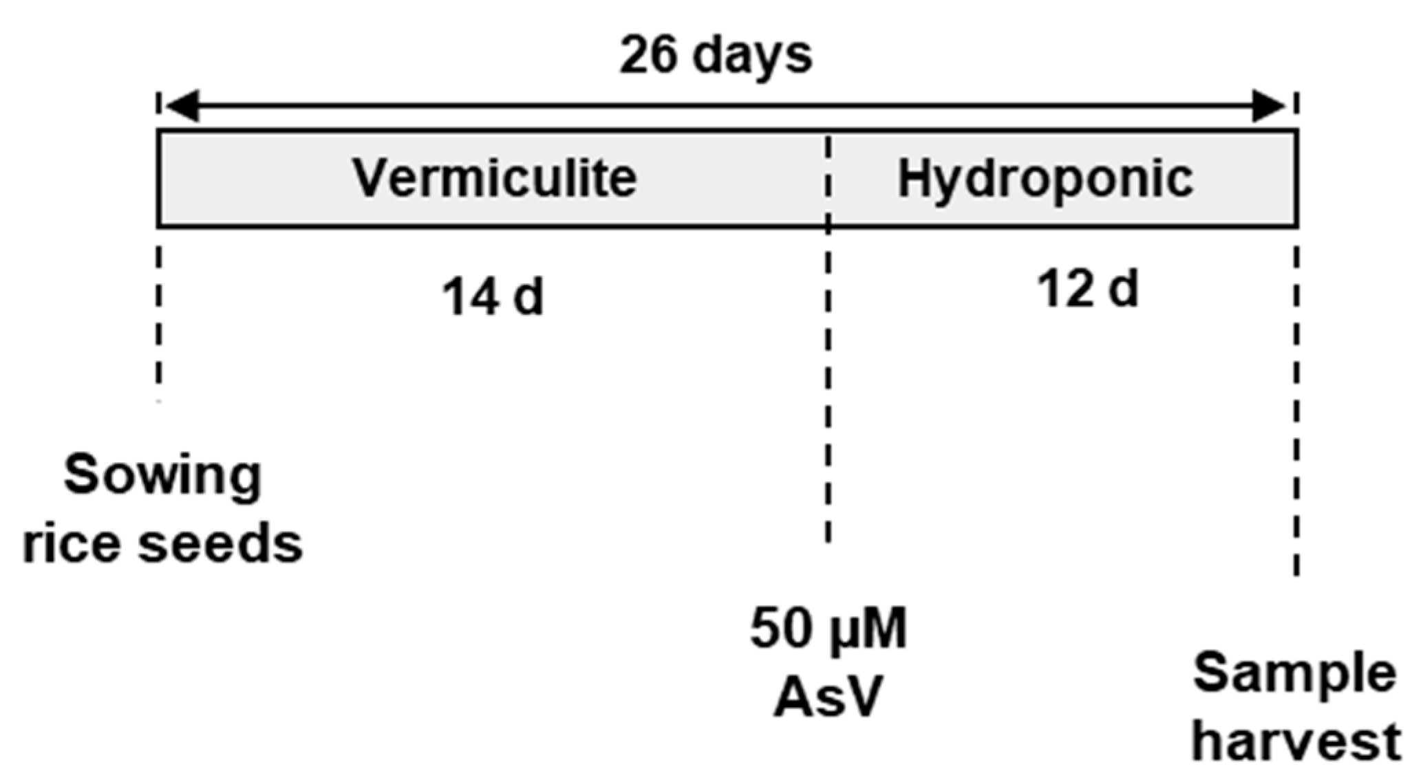
2.1. Plant Material and Growth Conditions

Rice (Oryza sativa L) seeds were planted and grown in vermiculite for 14 d under controlled conditions. Healthy and vigorous seedlings (n = 36) were selected and transplanted to hydroponic culture in 4 L plastic pots in aerated full nutrient media under optimal conditions in a growth chamber. After 14 d, nutrient solution was added with 0, 10, 25, 50, 100, and 200 μM potassium dihydrogen arsenate (KH2AsO4) which corresponds to the AsV form. The AsV form was used in our study since AsIII is predominant under anaerobic flooded conditions, whereas AsV is the most appropriate to improve crop yield under P fertilization [32]. Our experimental conditions, with aerated full nutrient media, including P, fit better with the use of AsV. Then, plants were grown for 12 d at 22/18 °C, day/night temperatures, a photoperiod of 16 h, and 100–120 μmol m−2s−1 irradiance (Figure 1). The composition of the nutrient solution was: 1 mM Ca(NO3)2,1 mM KH2PO4, 1 mM KNO3, 1 mM MgSO4, 50 μM Na–Fe–EDTA, 46 μM H3BO3, 10 μM MnSO4, 0.77 μM ZnSO4, 0.32 μM CuSO4, 0.58 μM Na2MoO4, 0.01 μM CoCl2 and pH adjusted to 6.0 with KOH. After the growth period, roots and leaves from the control and AsV-treated plants were harvested and used for analytical and biochemical assays. Length and fresh weight were measured in roots, shoots and whole plants from all treatments.

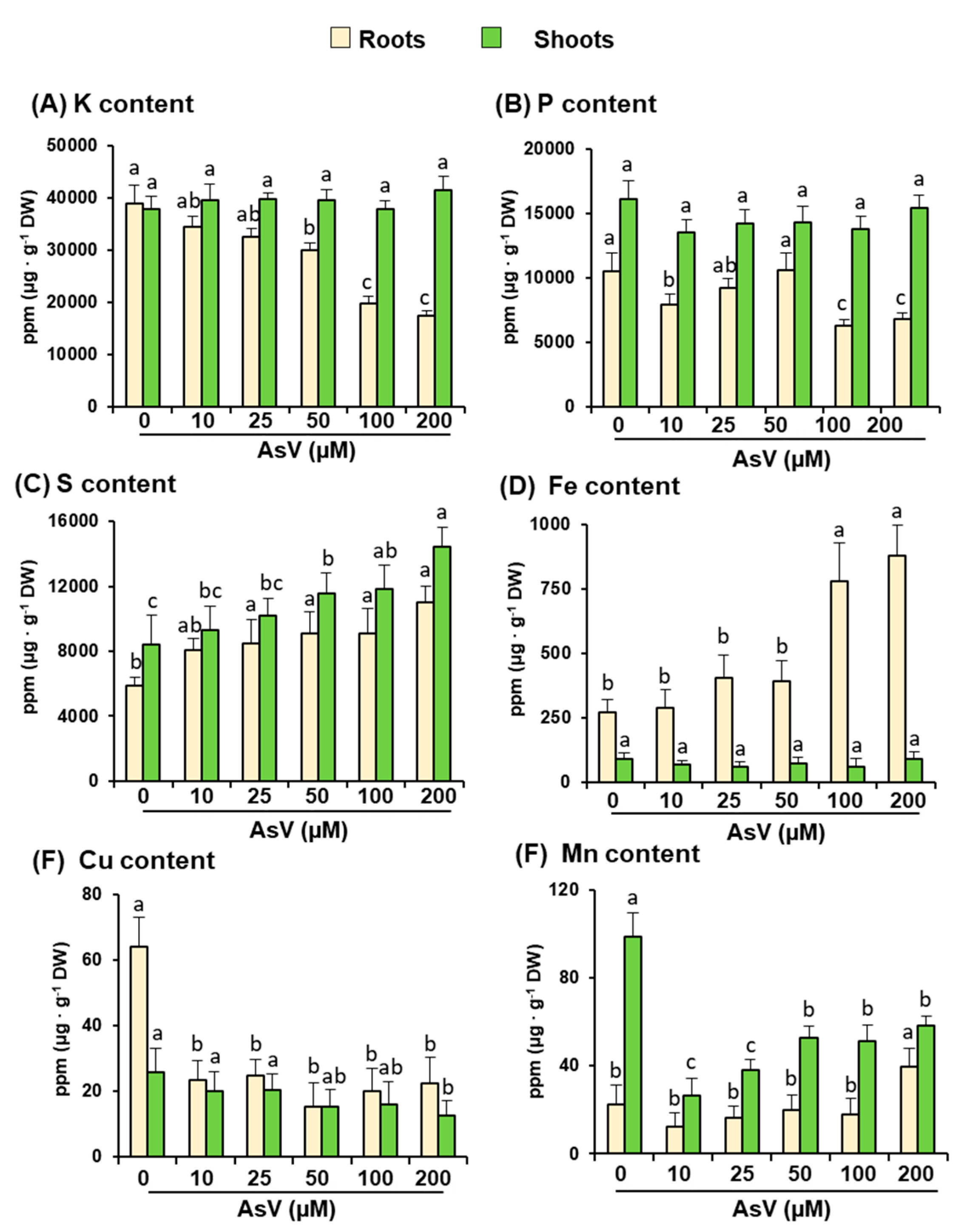
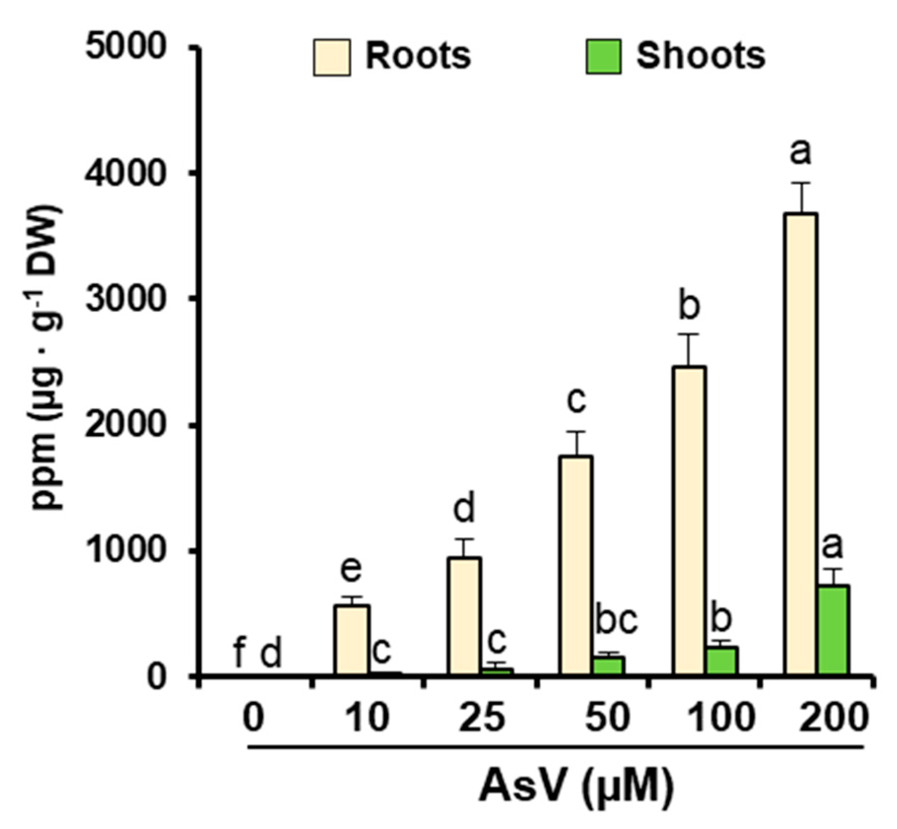
2.2. Arsenic and Main Nutrient Contents

Arsenic-treated and untreated full plants were washed with Milli-Q water containing 0.1 mM ethylene tetraacetic acid (EDTA). Then, roots and shoots were separated, dried for 8 h at 80 °C, pulverized, and digested for 90 min (total time) in a HNO3:HClO4 (2:1; v/v) solution. Mineral arsenic, and some macro- (K, P, S), and micro-nutrients (Fe, Cu and Mn) were analyzed by inductively coupled plasma spectrometry using a Varian720-ES ICP-OES spectrometer (Varian ICP 720-ES, Santa Clara, CA, USA). The root to shoot translocation factor (TFroot to shoot) was considered as the ability of plants to translocate As from roots to the aerial parts and was estimated through the formula TFroot to shoot = [As shoot]/[As root] [4].

2.3. Preparation of Crude Extracts for Enzyme and Lipid Peroxidation Assays

Plant material was frozen under liquid N2 and pulverized in a mortar with a pestle. The powder was suspended in a homogenizing medium composed of 50 mM Tris-HCl, pH 7.8, 0.1 mM EDTA, 10% (v/v) glycerol, 0.2% (v/v) Triton X-100, and 5 mM dithiothreitol (DTT). For the determination of APX activity, 2 mM ascorbate was added to the homogenizing medium to preserve the enzyme activity [43]. Homogenates were centrifuged 25 min at 15,000 g, 4 °C, and supernatants were used immediately for assays.

2.4. Enzyme Activity Assays

Catalase (EC 1.11.1.6) activity was determined at 240 nm by following the breakdown of H2O2 [44]. Ascorbate peroxidase (APX; EC 1.11.1.11) was plotted by monitoring the initial ascorbate oxidation by H2O2 at 290 nm [45]. Monodehydroascorbate reductase (MDAR; EC 1.6.5.4) was measured by assaying the monodehydroascorbate-dependent NADH oxidation. In these assays, the monodehydroascorbate was generated through the ascorbate/ascorbate oxidase system, as reported earlier [46]. The rate of monodehydroascorbate-independent NADH oxidation (without ascorbate oxidase and ascorbate) was subtracted from the monodehydroascorbate-dependent reaction. GR (EC 1.6.4.2) was analyzed by recording the NADPH oxidation coupled to the reduction of GSSG to the GSH form [47]. The GR reaction rate in the assays was corrected for the very small, nonenzymatic oxidation of NADPH by GSSG. Guaiacol peroxidase (G-POD; EC 1.11.1.7) activity was determined by monitoring the guaiacol oxidation at 470 nm [48].
For the detection of SOD (EC 1.15.1.1) and peroxidase (POD) isozymes, nondenaturing polyacrylamide gel electrophoresis (PAGE) and specific staining methods were followed. Thus, SOD isozymes were separated on 10% acrylamide gels and visualized in-gel by the photochemical nitroblue tetrazolium (NBT) reduction method [49]. Acromatic bands appeared over a blue background. To identify the different SOD isozymes, before staining preincubation of gels in the presence of specific inhibitors, either 5 mM KCN or 5 mM H2O2, was carried out. Cyanide inhibits copper zinc-containing SODs (CuZn-SODs), H2O2 inactivates CuZn-SODs and iron-containing SODs (Fe-SODs), and manganese-containing SODs (Mn-SODs) are resistant to both chemicals [50,51]. For POD isozymes, 8% acrylamide gels were prepared and the in-gel activity was developed by immersing gels for 20 min in a solution (pH 5.5) containing 0.1 M sodium acetate, 1 mM 3,3-diaminobenzidine (DAB) and 0.03% (v/v) H2O2 [52]. Band intensity of SOD and POD isozymes was quantified using ImageJ 1.45 software (National Institutes of Health, Bethesda, MD, USA).
Protein concentration was determined by the method of Bradford [53], with the Bio-Rad (Hercules, CA, USA) protein assay solution and bovine serum albumin as standard.

2.5. Detection and Quantification of Ascorbate, GSH and GSSG by Liquid Chromatography-Electrospray Mass Spectrometry (LC-ES/MS)

Rice roots and shoots were ground under liquid N2 with the use of a mortar and a pestle. Then, 0.4 g from each powdered organ were suspended into 1 mL of 0.1 M HCl and centrifuged at 15,000 g for 20 min at 4 °C. Supernatants were filtered through 0.22 μm polyvinylidene fluoride filters and immediately analyzed, with all procedures performed at 4 °C and protected from light to avoid potential degradation of the analytes. All samples were analyzed by liquid chromatography–electrospray/mass spectrometry (LC-ES/MS) using an Allience 2695 HPLC system connected to a Micromass Quattro micro API triple quadrupole mass spectrometer, both obtained from the Waters Corporation (Milford, Massachusetts, USA). HPLC was carried out using an Atlantis® T3 3 μm 2.1 × 100 mm Column obtained from the Waters Corporation. For the instrument control, data collection, analysis and management, the MassLynx 4.1 software (Waters Corporation (Milford, Massachusetts, USA) package was used. This method allowed for simultaneous detection and quantification of ascorbate, GSH, and GSSG [54,55]. The concentration of analytes was calculated using external standards and expressed as nmol g−1 of fresh weight (FW).

2.6. Other Assays

Lipid peroxidation was estimated by measuring the concentration of MDA through the thiobarbituric acid reacting substances (TBARS) method [56]. Nitric oxide (NO) was determined by a spectrofluorometric assay using 10 µM 4,5-diaminofluorescein (4,5-diaminofluorescein, DAF-2), (Calbiochem®, EMD Chemicals, San Diego, CA, USA) as fluorescence probe and excitation and emission wavelengths of 485 and 515 nm, respectively [57]. H2O2 was detected and quantified according to a peroxidase-coupled assay using 4-aminoantipyrine and phenol as donor substrates [58].

2.7. Statistical Analysis
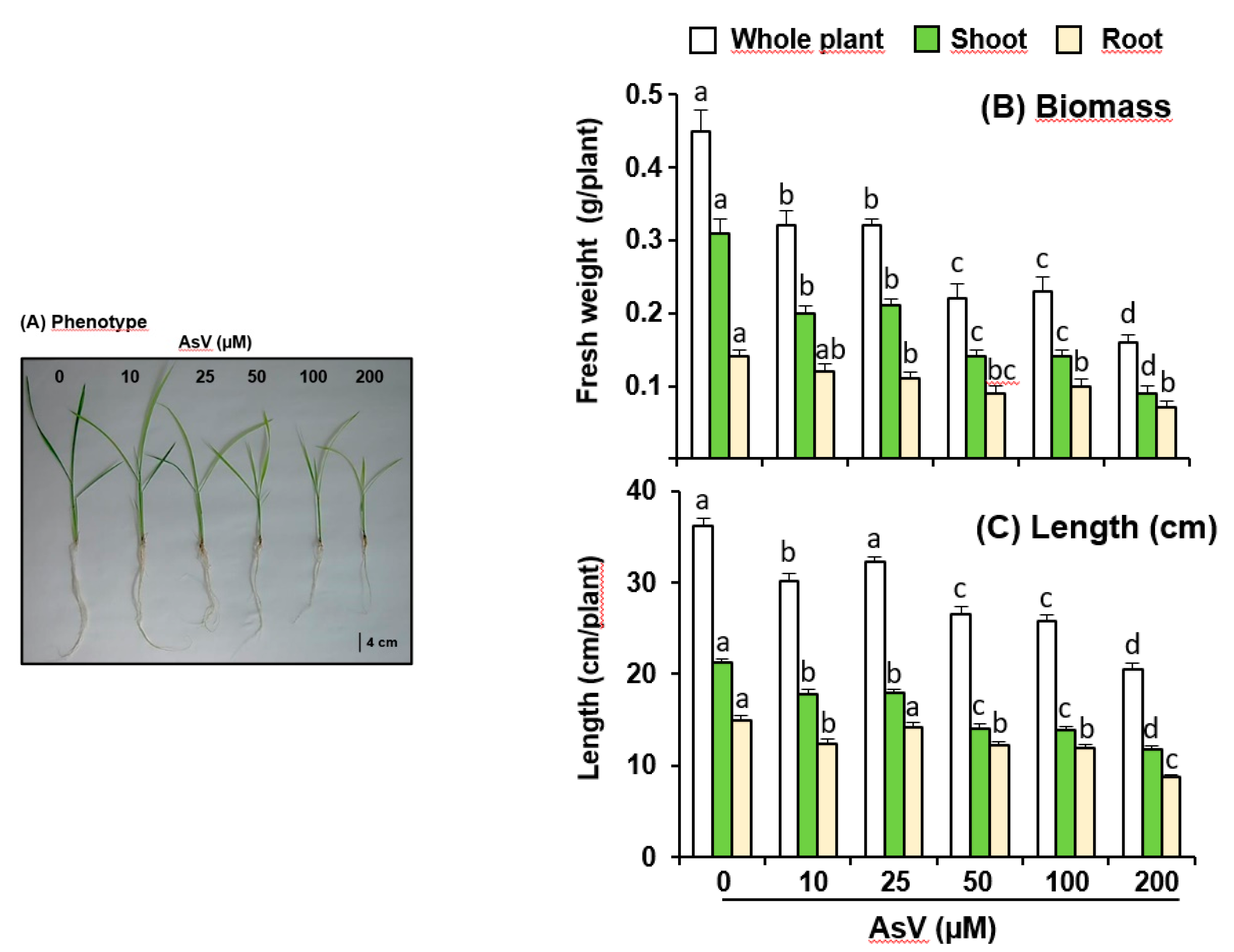
All data are means from three to five sets of independent experiments with three repetitions each. One-way Anova was used, as indicated in the respective figure legends, for comparisons between means using the program Statgraphics Centurion (Statgraphics.Net, Madrid, Spain). t-Student was also used to compare data from As-treated plants with control ones, as given in the respective figure legends.

3. Results

As an approach to investigate the effect of AsV on the ROS metabolism in rice plants, a preliminary analysis of the growth parameters under different As (form V) concentrations was carried out. Figure 2A shows a representative image of the appearance of 26-d-old rice plants in the six treatments followed in this work (0–200 μM). According to the visual analysis, a slight chlorosis seems to be promoted by arsenic. As observed in Figure 2, As provoked a diminution of growth parameters, from the lowest concentration used in this study (10 μM). This effect was clearer from data on fresh weight (Figure 2B) than those on length (Figure 2C). Thus, the observed changes in growth were basically due to the drastic effect exerted on shoot biomass and length, whereas the roots were less affected.
The content of some macro- and micro-nutrients was also analyzed. Thus, Figure 3 (panels A to F) shows the K, P, S, Fe, Cu and Mn contents in roots and leaves of 26-d-old rice seedlings grown under hydroponic conditions and exposed to 0, 10, 25, 50, 100 and 200 μM AsV during the last 12 days of the experiment. In roots, K content followed a progressive decrease as the AsV concentration was enhanced, reaching the lowest K content (55%) at an AsV concentration of 200 μM. This micronutrient was unaffected in shoots (Figure 3A). Figure 3B shows the P content in rice roots and shoots exposed to AsV, with values in roots lowering around 40% at AsV concentrations of 100–200 μM. However, in the shoots the P content was also unaffected. Regarding S content both organs showed a progressive significant enhancement from AsV 25 μM, being 1.8-fold higher in roots and 1.7-fold in shoots at 200 μM As, compared to untreated plants (Figure 3C). Figure 3D illustrates the Fe content which significantly increased from AsV 25 μM in roots, being 3.2-fold higher in AsV of 200 μM. However, in shoots the Fe content was unaffected by AsV. Cu content decreased in both organs but most drastically in roots (2.9-fold lower at 200 μM AsV), rather than in shoots (2.0-fold at maximum AsV concentration) (Figure 3E). Figure 3F shows the Mn content, which had an irregular and opposite behavior in both organs, since it only increased in roots at 200 μM AsV (1.8-fold) but lowered in shoots from 10 μM AsV up to 3.7 times.
A global analysis of the above parameters on growth and mineral contents led us to set 50 μM as the threshold concentration. The analysis of the As content in both roots and shoots at all assayed As concentrations showed that the metalloid was mostly concentrated in roots, although at the highest concentration (200 μM), shoots displayed a fifth of the values obtained in roots (Figure 4). As shown in Table 1, the TF indicated that As was transported from roots to shoots in parallel with the As increasing concentrations in the nutrient solutions. Again, considering the data from both parts of plants, 50 μM AsV was the level from which this parameter displayed the clearest changes. Accordingly, this concentration of 50 μM was set to achieve further study of the effect of As on the ROS metabolism of rice. For that purpose, enzymatic and nonenzymatic antioxidants were investigated.
A battery of antioxidative enzymes, including catalase, those from the ascorbate–glutathione cycle (AGC) APX, MDAR and GR, peroxidases and SOD was evaluated. As depicted in Figure 5A, catalase increased in both roots and shoots as a consequence of the treatment with 50 μM AsV. Interestingly, the other important H2O2-scavenging enzyme in plant cells, the APX, behaved in an opposite way, lowering its activity after the As treatment (Figure 5B). The other two enzymes of the AGC, MDAR and GR, which use NAD(P)H and NADPH, respectively, to carry out their catalytic reaction were only enhanced significantly in the aerial parts (Figure 5C,D). Guaiacol peroxidase (G-POD), another enzyme that consumes H2O2, increased significantly in roots, and no changes were detected in shoots (Figure 5E)
The isoenzyme pattern of SODs and peroxidases were achieved by nondenaturing PAGE and the respective abundance of each isoenzyme was quantified by using the image analyzer software ImageJ (Figure 6). Thus, it was found that the SOD system consisted of four isoenzymes, which according to their sensibility to the specific inhibitors cyanide and H2O2 and the increasing electrophoretic mobilities, were designated as Mn-SOD, Fe-SOD, CuZn-SOD I, CuZn-SOD II and CuZn-SOD III (Figure 6A). In control plants, the most abundant isozymes in the roots were Mn-SOD and CuZn-SOD I, whereas in shoots both CuZn-SOD I and CuZn-SOD II showed the highest activities and the Fe-SOD could not be nearly detected. CuZn-SOD III could only be found in shoots. No significant changes on the SOD isoenzyme profiles were detected as the effect of As, excepting slight decreases of Mn-SOD and Fe-SOD in roots and of CuZn-SOD I in shoots, along with a little enhancement of CuZn-SOD III in shoots (Figure 6A). With respect to peroxidases, up to 7 isoenzymes could be detected after specific DAB-staining of gels which were designated POD 1—POD 7 depending of their increasing electrophoretic mobilities (Figure 6B). POD 1, POD 2 and POD 4 were present in roots, whereas the shoots contained all isozymes excepting POD 2. Neither qualitative nor quantitative changes promoted by As treatment were observed in the isozyme pattern of roots. On the contrary, in shoots POD 3 was induced by As while POD 1, 4, 6 and 7 were down-regulated by effect of the metalloid (Figure 6B).
On the other hand, as indexes of the antioxidative capacity of both organs under As treatment, two of the most powerful and abundant cell antioxidants were determined, ascorbate, and glutathione (reduced and oxidized form). Globally, all these compounds were more abundant in shoots than in roots (Figure 7). The ascorbate content was enhanced by As in both organs (Figure 7A). With respect to GSH, the treatment with As provoked an increase in the aerial part, whilst no variation was observed in roots (Figure 7B). The behavior of GSSG was opposite in both roots and shoots as a consequence of the metalloid effect. Thus, while As promoted a significant rise in roots, this parameter lowered in shoots from As-treated plants (Figure 7C). The redox reduced/oxidized (GSH/GSSG) state of glutathione was determined. In roots, this ratio lowered from 51.0 under control conditions to 12.1 in As-treated plants. Regarding shoots, this ratio rose from 12.6 (control) to 30.8 (As).
The content of two important signaling molecules (H2O2 and NO) in plant physiology were also determined in these experiments. In all cases both species increased significantly due to the metalloid (Figure 8A,B, respectively). Finally, lipid peroxidation, a known index of oxidative stress, was analyzed. It was shown that the formation of MDA species increased moderately, but significantly, in both roots and shoots of plants subjected to As treatment (Figure 8C).

4. Discussion

According to the analysis of the mineral content carried out in this work, it is remarkable that the root was the organ that underwent the most significant differences under AsV-stress, with reductions in the content of K, P and Cu whereas S, Fe and Mn increased. However, the content of these minerals in shoots was less affected, with the rise of S content and the reduced content of Cu and Mn being the most remarkable.
It has been thoroughly reported that arsenic (As), either as AsIII or AsV, provokes negative effects in plant development with significant alterations at cellular, biochemical and molecular levels. Thus, the most consensual mechanism by which As affects plant metabolism implies oxidative stress events [4,5,13,14,21,22,23,25,59,60]. Nevertheless, the sensitivity to and the response against As can significantly differ among plant species, among cultivars within the same species, between roots and aerial parts in the same plant, but they also may depend on the concentration and the active form of the metalloid (AsIII or AsV) and the geographical locations [14,27,34,36,39,61,62,63]. Due to the relevance at agronomical and economical levels and its wide distribution in some geographical areas, rice (Oryza sativa) has been the major crop species where these studies have been achieved, with the coincidence that rice grains, which are the nutritional basis for many countries worldwide, are able to accumulate this metalloid, thus favoring the risk to transmit As to the human population and the rest of the food chain [64]. Up until today, a diversity of effects of As on the physiology and metabolism of rice plants has been reported, but some gaps in the intimate mechanism of As toxicity in this crop are still unknown due to the diversity of interactions with metabolic and signaling processes.
To perform our study, we initially used a wide range of arsenate concentrations (10–200 μM AsV) to set the potential threshold at which the metalloid starts provoking significant effects. Arsenic promoted a decrease in growth parameters just from the lowest concentration used, although it was more significantly evident in fresh weight. This pattern agrees with previously reported data in this plant species [28,31,37,62,65]. Additionally, the mineral contents of K, P, S, Fe, Cu and Mn was analyzed in rice seedlings under AsV stress. It is remarkable that the root was the organ that underwent the most prominent differences, with significant reductions in the content of K, P and Cu whereas S, Fe and Mn increased due to As treatment. However, the content of these minerals in shoots was less affected, with the increase of S content and the reduced content of Cu and Mn being the most remarkable.
Taking into account the growth parameters, 50 μM AsV was considered as the threshold concentration to achieve our further research. It was also found that from this As concentration and above, the metalloid was translocated to the aerial parts more efficiently, so it indicates that this was the best concentration to follow the effects in the oxidative metabolism in shoots. The TFroot to shoot values obtained in this work are mostly in the range reported before for rice (0.05-1.43) [36], but above most TFroot to shoot displayed by other species such as lettuce (Lactuca sativa), romaine lettuce (Lactuca sativa var. longifolia), garlic (Allium sativum), water spinach (Ipomea aquatica), celery (Apium graveolens) or pea (Pisum sativum), which showed values of 0.02–0.17 [4,66]. Although the obtained values in rice indicate that most of the metalloid remains in the roots, the comparison with other species with lower TFroot to shoot values might explain why As is accumulated in reproductive tissues (grains) which are part of the aerial organs in this crop.
As in other plant species investigated so far, the main symptom in rice plants as a consequence of high As concentrations is the induction of oxidative stress characterized by an alteration of the antioxidative systems and increases of lipid peroxidation levels. This assertion is based on results obtained from a diversity of experimental designs where As has been used as AsIII or AsV at different concentrations and for various experimental lengths [5,28,29,31,33,37,60,62,65]. Our work establishes a whole picture on how the main antioxidant systems (both enzymatic and nonenzymatic) respond to a threshold (sub-toxic) As concentration at both root and aerial parts after 12 d treatment.
Globally, roots and shoots displayed parallel activity patterns in all antioxidative enzymes. The H2O2-scavenging enzymes catalase (CAT) and guaiacol peroxidase (G-POD) increased in both organs, but with different profiles. Thus, whereas CAT showed greater augmentation in shoots, G-POD did in roots. Catalase is the main enzyme responsible for removing H2O2 in plants, and is mainly relevant in photosynthetic tissues where, due to its high KM, in the mM range, it is able to decompose important amounts of the ROS [67,68,69,70,71]. Regarding peroxidase, the pattern observed in both organs by determining the activity spectrophotometrically differed from the isoenzyme profile observed after nondenaturing PAGE. Thus, no changes were found in roots after As treatment, whereas the metalloid greatly induced isozyme POD 3 in shoots. This points towards an important role of this isozyme, and more research is necessary to address the full identification and nature of POD 3. Recently, it has been reported that OsPRX38, a class HI peroxidase is upregulated under As stress in rice. This isozyme has been overexpressed in Arabidopsis thaliana enhancing the tolerance of the mutants to As and this was accompanied by increased SOD, POD and glutathione-S-transferase activities and lower H2O2 and malondialdehyde contents [72]. The POD isoenzyme pattern reported here differs from that reported earlier where only four less defined isozymes were detected. Moreover, distinct AsV concentrations (100 and 500 μM) were used in such work [28].
Interestingly, APX, the enzyme which finely tunes the H2O2 level in cooperation with catalase and peroxidases in plant cells, displayed an opposite behavior to these last enzymes in both organs, with down regulated activity promoted by arsenic. It has been reported that rice contains a total of eight APX isozymes present in the different subcellular compartments [73]. In this context, it has been shown in cytosolic APX1 and APX2 double knockdown rice plants that abiotic stress promotes an induction of POD activity to compensate the loss of APX [74]. This mechanism would allow keeping the appropriate H2O2 levels for both signaling and oxidative protection. This trend is in good agreement with the observed increase of POD 3 activity in shoots under As stress. The profile of APX activity depicted in this work is also contrary to the upregulating effect of the metalloid on the ascorbate content. This antioxidant was greatly increased by As and this was more visible in shoots. This opposite evolution of ascorbate and APX indicates that the AGC is possibly uncoupled and not properly functioning due to the As. This eventuality is supported by the fact that the two other AGC determined in this work (MDAR and GR) are upregulated by As in shoots. It could be explained in two ways. On the one hand, both MDAR and GR might be implicated in the regeneration of ascorbate once it has been oxidized by direct action over ROS, without the participation of the APX; on the other hand, the GR activity may work independently to provide reduced GSH for other purposes such as the synthesis of phytochelatins (PCs). The profile of the glutathione levels supports this last assumption since the reduced form (GSH) shares the same tendency displayed by the GR activity. The induction of the synthesis of PCs is a common event which is triggered by arsenic as it has been demonstrated in several plant species [4,5,75,76,77] and also in rice [29,33,35,36]. Very recently, it has been also reported that O. sativa L. cv. Dasan plants cultivated for 14 d in the presence of 15 μM AsIII and sprayed with GSH reduced the oxidative stress promoted by arsenic, improved the antioxidant defense systems through maintaining the redox homeostasis and increased the ascorbate and GSH levels [65]. Nevertheless, the same authors also reported that, using the cultivar Dasan and the As treatment reported above, the metalloid provoked a decrease in the ascorbate and GSH contents. Therefore, this issue needs further research to better understand the dynamics of GSH in combination with ascorbate and the AGC.
Regarding the SOD isozymes detected in nondenaturing PAGE, no qualitative differences were promoted by AsV, and only slight changes in the activity of the isozymes were detected in both organs. The isoenzymatic pattern obtained in this work is somehow different to the one reported earlier by Shri and colleagues [28]. The cultivar, the growth and the As treatment conditions, besides the preparation of samples and the electrophoresis handling, could be responsible of such differences. The electrophoretic and staining resolution reported in the present work confirms that five SODs are present in rice plants: one Mn-SOD, one Fe-SOD and three CuZn-SODs (I, II and III). Both roots and shoots share the isoenzymes Mn-SOD, Fe-SOD (very low abundance in shoots), CuZn-SOD I and CuZn-SOD II, whereas CuZn-SOD III could only be detected in the aerial parts. These differences in the abundance and distribution of the SOD isozymes in both rice organs is not unusual, although it has been observed in other species under diverse stress situations [50,78]. Thus, in 14-d-old Arabidopsis seedlings grown under in vitro conditions in the presence of 500 µM arsenate, one Mn-SOD, one Fe-SOD and three CuZn-SODs were identified, but any of them showed significant differences [22]. A similar behavior was observed in roots and leaves from 27-day-old Arabidopsis plants grown in hydroponic conditions under 500 µM AsV where the SOD isozyme pattern showed no significant changes [24]. Likewise, in garlic (Allium sativum L.) plants grown in hydroponic conditions in the presence of AsV 200 µM, the SOD isoenzyme pattern in roots and shoots was also unaffected [4]. Nevertheless, in pea plants grown under AsV 50 µM the zymogram was quite different. Thus, in roots from untreated plants, one Mn-SOD and two CuZn-SODs (I and II) were identified, while in leaves, an additional Fe-SOD isozyme was detected. Under AsV stress, roots showed a significant decrease of the different isozymes (70% of the Mn-SOD, 36% of the CuZn-SOD I and 68% of the CuZn-SOD II), while all leaf SOD isozymes were unaffected by AsV [19]. All these data indicate that the antioxidant defense against As stress seems to be quite different depending on the analyzed organs and the plant species, but it may also be affected by length of application, the plant age, etc.
Two markers of oxidative stress were determined in this work, the H2O2 content and the lipid peroxidation level. The increase of H2O2 in both organs as a consequence of the arsenic treatment would explain the profile of the catalase and peroxidase activities referred to above. This higher content of H2O2 along with the slight increase of lipid peroxidation indicates that a mild oxidative stress seems to occur under the AsV concentration used in our study, thus suggesting that the metabolic network from rice plants, under our experimental conditions, has the capacity to palliate the oxidative stress which is usually associated with arsenic stress.
The profile followed by the H2O2 should be also viewed under the perspective of the nitric oxide (NO) content observed after treatment with arsenic in both organs. NO is a gas radical with important signaling and regulatory functions in plants. Thus, it has been demonstrated that NO is involved in germination, development, senescence, and fruit ripening [79,80,81,82] and in the response against biotic and abiotic stresses [83,84,85]. It has been found that the application of exogenous NO provides resistance to metal stress including cadmium [86] and aluminum [87], but also arsenic [88]. Thus, it has been also reported that the treatment of Vicia faba with sodium nitroprusside (SNP), a NO donor, improves the tolerance to arsenic (As) toxicity by increasing the antioxidant attributes of plants [89]. Likewise, the exogenous supplementation of SNP to Brassica juncea seedlings palliated the inhibitory role of As-III through the alteration of nutrients, amino acids and auxin redistribution [90]. In O. sativa seedling the treatment with NO in combination with AsIII diminished the accumulation of As, improved the changes underwent by the root architecture, and reduced the ROS generation among, other advantages [91]. At the cell/tissue levels, NO can interact with H2O2 [92], and this may allow setting up a series of complex biochemical networks to help the plant responds against the arsenic impact.

5. Conclusions and Future Prospects

Rice grains accumulate arsenic at a certain level, which is a threat for a huge population, especially for citizens living in Western Asia where this cereal is a major food and part of their daily intake. Arsenic reduces plant growth and provokes oxidative stress in most crops of agronomic interest. Besides issuing and improving the policies against those practices that favor environmental pollution (including direct soil contamination by As and through irrigation water), a good knowledge on how the metabolism of the rice plant responds against the harm induced by As is elementary to developing biotechnological strategies to alleviate the metalloid effects. Setting the threshold concentration which triggers most of the metabolic responses provoked by As may help to devise priming programs. Our results indicate that a mild oxidative stress occurs at 50 μM AsV and this could be considered as the threshold concentration. Thus, under our experimental conditions, As-induced stress is accompanied by a differential response of the ROS metabolism between roots and shoots. The enzymes involved in the H2O2 redox homeostasis are the most significantly affected (catalase, APX and POD). A remarkable and significant induction of POD 3 in roots could compensate for the loss of the APX activity in this organ.
Our data also provide evidence for a relevant role of NO in coordination with other signaling molecules such as H2O2, as was also proposed elsewhere [93,94]. Therefore, due to its signaling and regulatory potentialities, the development of biotechnological tools based on the NO biochemistry could help to obtain plants with improved capacity to cope against As. This perspective can be also combined with alternative proposals to alleviate As damage such as those which involve treatment with the nontoxic selenium (Se) [8,39,40,95], salicylic acid, 24-epi-brassinolide and silicon [60], or even leaf extracts from certain plants such as neem (Azadirachta indica) and Tulsi (Ocimum sanctum) [31].

Author Contributions

Performed the experimental works, E.S.; participated in the data analyses, S.G.-G.; designed the research, discussed the data and participated in writing the manuscript, J.M.P. and F.J.C. All authors have read and agreed to the published version of the manuscript.

Funding

This work was supported by ERDF-cofinanced grants from the Ministry of Science and Innovation (Recupera 2020-20134R056 and PID2019-103924GB-I00), Junta de Andalucía (group BIO192), and CSIC (COOPB20171), Spain.

Acknowledgments

The helpful assistance of the technicians Carmelo Ruiz Torres and María Jesús Campos is acknowledged. LC-ES/MS and mineral analyses were carried out at the Instrumental Technical Services of the Estación Experimental del Zaidín (CSIC).

Conflicts of Interest

Authors declare no conflict of interest.

References

  1. Zhao, F.J.; McGrath, S.P.; Meharg, A.A. Arsenic as a food chain contaminant: Mechanisms of plant uptake and metabolism and mitigation strategies. Annu. Rev. Plant Biol. 2010, 61, 535–559. [Google Scholar] [CrossRef] [Green Version]
  2. Sarkar, A.; Paul, B. The global menace of arsenic and its conventional remediation—A critical review. Chemosphere 2016, 158, 37–49. [Google Scholar] [CrossRef] [PubMed]
  3. Clemens, S.; Ma, J.F. Toxic heavy metal and metalloid accumulation in crop plants and foods. Annu. Rev. Plant Biol. 2016, 67, 489–512. [Google Scholar] [CrossRef] [Green Version]
  4. Ruiz-Torres, C.; Feriche-Linares, R.; Rodríguez-Ruíz, M.; Palma, J.M.; Corpas, F.J. Arsenic-induced stress activates sulfur metabolism in different organs of garlic (Allium sativum L.) plants accompanied by a general decline of the NADPH-generating systems in roots. J. Plant Physiol. 2017, 211, 27–35. [Google Scholar] [CrossRef] [PubMed]
  5. Abbas, G.; Murtaza, B.; Bibi, I.; Shahid, M.; Niazi, N.K.; Khan, M.I.; Amjad, M.; Hussain, M.; Tahir, N. Arsenic uptake, toxicity, detoxification, and speciation in plants: Physiological, biochemical, and molecular aspects. Int. J. Environ. Res. Public Health 2018, 15, 59. [Google Scholar] [CrossRef] [Green Version]
  6. Rodríguez-Ruiz, M.; Aparicio-Chacón, M.V.; Palma, J.M.; Corpas, F.J. Arsenate disrupts ion balance, sulfur and nitric oxide metabolisms in roots and leaves of pea (Pisum sativum L.) plants. Environ. Exp. Bot. 2019, 161, 143–156. [Google Scholar] [CrossRef]
  7. Alam, M.Z.; Hoque, M.A.; Ahammed, G.J.; McGee, R.; Carpenter-Boggs, L. Arsenic accumulation in lentil (Lens culinaris) genotypes and risk associated with the consumption of grains. Sci. Rep. 2019, 9, 9431. [Google Scholar] [CrossRef] [Green Version]
  8. Ali, W.; Zhang, H.; Junaid, M.; Mao, K.; Xu, N.; Chang, C.Y.; Rasool, A.; Aslam, M.; Ali, J.; Yang, Z.G. Insights into the mechanisms of arsenic-selenium interactions and the associated toxicity in plants, animals, and humans: A critical review. Crit. Rev. Environ. Sci. Technol. 2020. [Google Scholar] [CrossRef]
  9. Ventura-Lima, J.; Bogo, M.R.; Monserrat, J.M. Arsenic toxicity in mammals and aquatic animals: A comparative biochemical approach. Ecotoxicol. Environ. Saf. 2011, 74, 211–218. [Google Scholar] [CrossRef] [Green Version]
  10. Abdul, K.S.; Jayasinghe, S.S.; Chandana, E.P.; Jayasumana, C.; De Silva, P.M. Arsenic and human health effects: A review. Environ. Toxicol. Pharmacol. 2015, 40, 828–846. [Google Scholar] [CrossRef]
  11. Escudero-Lourdes, C. Toxicity mechanisms of arsenic that are shared with neurodegenerative diseases and cognitive impairment: Role of oxidative stress and inflammatory responses. Neurotoxicology 2016, 53, 223–235. [Google Scholar] [CrossRef] [PubMed]
  12. Palma-Lara, I.; Martínez-Castillo, M.; Quintana-Pérez, J.C.; Arellano-Mendoza, M.G.; Tamay-Cach, F.; Valenzuela-Limón, O.L.; García-Montalvo, E.A.; Hernández-Zavala, A. Arsenic exposure: A public health problem leading to several cancers. Regul. Toxicol. Pharmacol. 2020, 110. [Google Scholar] [CrossRef]
  13. Islam, E.; Khan, M.T.; Irem, S. Biochemical mechanisms of signaling: Perspectives in plants under arsenic stress. Ecotoxicol. Environ. Saf. 2015, 114, 126–133. [Google Scholar] [CrossRef]
  14. Abedi, T.; Mojiri, A. Arsenic uptake and accumulation mechanisms in rice species. Plant 2020, 9, 129. [Google Scholar] [CrossRef] [Green Version]
  15. Song, W.Y.; Mendoza-Cózatl, D.G.; Lee, Y.; Schroeder, J.I.; Ahn, S.N.; Lee, H.S.; Wicker, T.; Martinoia, E. Phytochelatin-metal(loid) transport into vacuoles shows different substrate preferences in barley and Arabidopsis. Plant Cell Environ. 2014, 37, 1192–1201. [Google Scholar] [CrossRef] [PubMed] [Green Version]
  16. Tang, Z.; Chen, Y.; Miller, A.J.; Zhao, F.J. The C-type ATP-binding cassette transporter OsABCC7 is involved in the root-to-shoot translocation of arsenic in rice. Plant Cell Physiol. 2019, 60, 1525–1535. [Google Scholar] [CrossRef]
  17. Schmöger, M.E.; Oven, M.; Grill, E. Detoxification of arsenic by phytochelatins in plants. Plant Physiol. 2000, 122, 793–801. [Google Scholar] [CrossRef] [PubMed] [Green Version]
  18. Lee, D.A.; Chen, A.; Schroeder, J.I. ars1, an Arabidopsis mutant exhibiting increased tolerance to arsenate and increased phosphate uptake. Plant J. 2003, 35, 637–646. [Google Scholar] [CrossRef]
  19. Quaghebeur, M.; Rengel, Z. Arsenic uptake, translocation and speciation in pho1 and pho2 mutants of Arabidopsis thaliana. Physiol. Plant. 2004, 120, 280–286. [Google Scholar] [CrossRef]
  20. Li, Y.; Dhankher, O.P.; Carreira, L.; Lee, D.; Chen, A.; Schroeder, J.I.; Balish, R.S.; Meagher, R.B. Overexpression of phytochelatin synthase in Arabidopsis leads to enhanced arsenic tolerance and cadmium hypersensitivity. Plant Cell Physiol. 2004, 45, 1787–1797. [Google Scholar] [CrossRef] [PubMed] [Green Version]
  21. Bienert, G.P.; Jahn, T.P. Major intrinsic proteins and arsenic transport in plants: New players and their potential role. Adv. Exp. Med. Biol. 2010, 679, 111–125. [Google Scholar]
  22. Leterrier, M.; Airaki, M.; Palma, J.M.; Chaki, M.; Barroso, J.B.; Corpas, F.J. Arsenic triggers the nitric oxide (NO) and S-nitrosoglutathione (GSNO) metabolism in Arabidopsis. Environ. Pollut. 2012, 166, 136–143. [Google Scholar] [CrossRef] [PubMed]
  23. Leterrier, M.; Barroso, J.B.; Palma, J.M.; Corpas, F.J. Cytosolic NADP-isocitrate dehydrogenase in Arabidopsis leaves and roots. Biol. Plant. 2012, 56, 705–710. [Google Scholar] [CrossRef]
  24. Corpas, F.J.; Aguayo-Trinidad, S.; Ogawa, T.; Yoshimura, K.; Shigeoka, S. Activation of NADPH-recycling systems in leaves and roots of Arabidopsis thaliana under arsenic-induced stress conditions is accelerated by knock-out of Nudix hydrolase 19 (AtNUDX19) gene. J. Plant Physiol. 2016, 192, 81–89. [Google Scholar] [CrossRef] [PubMed]
  25. Park, J.H.; Han, Y.S.; Seong, H.J.; Ahn, J.S.; Nam, I.H. Arsenic uptake and speciation in Arabidopsis thaliana under hydroponic conditions. Chemosphere 2016, 154, 283–288. [Google Scholar] [CrossRef]
  26. Finnegan, P.M.; Chen, W. Arsenic toxicity: The effects on plant metabolism. Front. Physiol. 2012, 3, 182. [Google Scholar] [CrossRef] [PubMed] [Green Version]
  27. Kofroňová, M.; Hrdinová, A.; Mašková, P.; Tremlová, J.; Soudek, P.; Petrová, S.; Pinkas, D.; Lipavská, H. Multi-Component antioxidative system and robust carbohydrate status, the essence of plant arsenic tolerance. Antioxidants 2020, 9, 283. [Google Scholar] [CrossRef] [PubMed] [Green Version]
  28. Shri, M.; Kumar, S.; Chakrabarty, D.; Trivedi, P.K.; Mallick, S.; Misra, P.; Shukla, D.; Mishra, S.; Srivastava, S.; Tripathi, R.D.; et al. Effect of arsenic on growth, oxidative stress, and antioxidant system in rice seedlings. Ecotoxicol. Environ. Saf. 2009, 72, 1102–1110. [Google Scholar] [CrossRef] [PubMed]
  29. Dave, R.; Tripathi, R.D.; Dwivedi, S.; Tripathi, P.; Dixit, G.; Sharma, Y.K.; Trivedi, P.K.; Corpas, F.J.; Barroso, J.B.; Chakrabarty, D. Arsenate and arsenite exposure modulate antioxidants and amino acids in contrasting arsenic accumulating rice (Oryza sativa L.) genotypes. J. Hazard. Mater. 2013, 262, 1123–1131. [Google Scholar] [CrossRef]
  30. Farooq, M.A.; Li, L.; Ali, B.; Gill, R.A.; Wang, J.; Ali, S.; Gill, M.B.; Zhou, W. Oxidative injury and antioxidant enzymes regulation in arsenic-exposed seedlings of four Brassica napus L. cultivars. Environ. Sci. Pollut. Res. Int. 2015, 22, 10699–10712. [Google Scholar] [CrossRef]
  31. Gautam, A.; Pandey, A.K.; Dubey, R.S. Azadirachta indica and Ocimum sanctum leaf extracts alleviate arsenic toxicity by reducing arsenic uptake and improving antioxidant system in rice seedlings. Physiol. Mol. Biol. Plants 2020, 26, 63–81. [Google Scholar] [CrossRef] [PubMed]
  32. Duan, G.; Liu, W.; Chen, X.; Hu, Y.; Zhu, Y. Association of arsenic with nutrient elements in rice plants. Metallomics 2013, 5, 784–792. [Google Scholar] [CrossRef]
  33. Dave, R.; Singh, P.K.; Tripathi, P.; Shri, M.; Dixit, G.; Dwivedi, S.; Chakrabarty, D.; Trivedi, P.K.; Sharma, Y.K.; Dhankher, O.P.; et al. Arsenite tolerance is related to proportional thiolic metabolite synthesis in rice (Oryza sativa L.). Arch. Environ. Contam. Toxicol. 2013, 64, 235–242. [Google Scholar] [CrossRef] [PubMed]
  34. Lei, M.; Tie, B.; Zeng, M.; Qing, P.; Song, Z.; Williams, P.N.; Huang, Y. An arsenic-contaminated field trial to assess the uptake and translocation of arsenic by genotypes of rice. Environ. Geochem. Health 2013, 35, 379–390. [Google Scholar] [CrossRef] [PubMed]
  35. Tripathi, P.; Tripathi, R.D.; Singh, R.P.; Dwivedi, S.; Chakrabarty, D.; Trivedi, P.K.; Adhikari, B. Arsenite tolerance in rice (Oryza sativa L.) involves coordinated role of metabolic pathways of thiols and amino acids. Environ. Sci. Pollut. Res. Int. 2013, 20, 884–896. [Google Scholar] [CrossRef]
  36. Batista, B.L.; Nigar, M.; Mestrot, A.; Rocha, B.A.; Barbosa Junior, F.; Price, A.H.; Raab, A.; Feldmann, J. Identification and quantification of phytochelatins in roots of rice to long-term exposure: Evidence of individual role on arsenic accumulation and translocation. J. Exp. Bot. 2014, 65, 1467–1479. [Google Scholar] [CrossRef] [PubMed] [Green Version]
  37. Begum, M.C.; Islam, M.S.; Islam, M.; Amin, R.; Parvez, M.S.; Kabir, A.H. Biochemical and molecular responses underlying differential arsenic tolerance in rice (Oryza sativa L.). Plant Physiol. Biochem. 2016, 104, 266–277. [Google Scholar] [CrossRef] [PubMed]
  38. Chen, Y.; Han, Y.H.; Cao, Y.; Zhu, Y.G.; Rathinasabapathi, B.; Ma, L.Q. Arsenic transport in rice and biological solutions to reduce arsenic risk from rice. Front Plant Sci. 2017, 8, 268. [Google Scholar] [CrossRef] [PubMed] [Green Version]
  39. Kalita, J.; Pradhan, A.K.; Shandilya, Z.M.; Tanti, B. Arsenic stress responses and tolerance in rice: Physiological, cellular and molecular approaches. Rice Sci. 2018, 25, 235–249. [Google Scholar] [CrossRef]
  40. Singh, R.; Upadhyay, A.K.; Singh, D.P. Regulation of oxidative stress and mineral nutrient status by selenium in arsenic treated crop plant Oryza sativa. Ecotoxicol. Environ. Saf. 2018, 148, 105–113. [Google Scholar] [CrossRef]
  41. Corpas, F.J.; González-Gordo, S.; Cañas, A.; Palma, J.M. Nitric oxide and hydrogen sulfide in plants: Which comes first? J. Exp. Bot. 2019, 70, 4391–4404. [Google Scholar] [CrossRef] [PubMed]
  42. Palma, J.M.; Freschi, L.; Rodríguez-Ruiz, M.; González-Gordo, S.; Corpas, F.J. Nitric oxide in the physiology and quality of fleshy fruits. J. Exp. Bot. 2019, 70, 4405–4417. [Google Scholar] [CrossRef] [PubMed]
  43. Miyake, C.; Asada, K. Inactivation mechanism of ascorbate peroxidase at low concentrations of ascorbate: Hydrogen peroxide decomposes compound I of ascorbate peroxidase. Plant Cell Physiol. 1996, 36, 661–668. [Google Scholar] [CrossRef] [Green Version]
  44. Aebi, H. Catalase in vitro. Methods Enzymol. 1984, 105, 121–126. [Google Scholar] [PubMed]
  45. Hossain, M.A.; Asada, K. Inactivation of ascorbate peroxidase in spinach chloroplasts on dark addition of hydrogen peroxide: Its protection by ascorbate. Plant Cell Physiol. 1984, 25, 1285–1295. [Google Scholar]
  46. Hossain, M.A.; Nakano, Y.; Asada, K. Monodehydroascorbate reductase in spinach chloroplast and its participation in regeneration of ascorbate for scavenging of hydrogen peroxide. Plant Cell Physiol. 1984, 25, 385–395. [Google Scholar]
  47. Edwards, E.A.; Rawsthone, S.; Mullineaux, P.M. Subcellular distribution ofmultiple forms of glutathione reductase in leaves of pea (Pisum sativum L.). Planta 1990, 180, 278–284. [Google Scholar] [CrossRef]
  48. Quessada, M.P.; Macheix, J.J. Caractérisation d’une peroxydase impliquée spécifiquement dans la lignification, en relation avec l’incompatibilité au greffage chez l’Abricotier. Physiol. Végét. 1984, 22, 533–540. [Google Scholar]
  49. Beauchamp, C.; Fridovich, I. Superoxide dismutase: Improved assays and an assay applicable to acrylamide gels. Anal. Biochem. 1971, 44, 276–287. [Google Scholar] [CrossRef]
  50. Houmani, H.; Rodríguez-Ruiz, M.; Palma, J.M.; Abdelly, C.; Corpas, F.J. Modulation of superoxide dismutase (SOD) isozymes by organ development and high long-term salinity in the halophyte Cakile maritima. Protoplasma 2016, 253, 885–894. [Google Scholar] [CrossRef]
  51. Pinilla, M.; Iglesias-Moya, J.; Campos, M.J.; Corpas, F.J.; Palma, J.M. Pomegranate (Punica granatum L.) Fruits: Characterization of the main enzymatic antioxidants (peroxisomal catalase and SOD isozymes) and the NADPH-regenerating system. Agronomy 2019, 9, 338. [Google Scholar] [CrossRef] [Green Version]
  52. Ádám, A.L.; Bestwick, C.S.; Barna, B.; Mansfield, J.W. Enzymes regulating the accumulation of active oxygen species during the hypersensitive reaction of bean to Pseudomonas syringae pv. phaseolicola. Planta 1995, 197, 240–249. [Google Scholar]
  53. Bradford, M.M. A rapid and sensitive method for the quantitation of microgram quantities of protein utilizing the principle of protein-dye binding. Anal. Biochem. 1976, 72, 248–254. [Google Scholar] [CrossRef]
  54. Airaki, M.; Sánchez-Moreno, L.; Leterrier, M.; Barroso, J.B.; Palma, J.M.; Corpas, F.J. Detection and quantification of S-nitrosoglutathione (GSNO) in pepper (Capsicum annuum L.) plant organs by LC-ES/MS. Plant Cell Physiol. 2011, 52, 2006–2015. [Google Scholar] [CrossRef] [PubMed] [Green Version]
  55. Rodríguez-Ruiz, M.; Mateos, R.M.; Codesido, V.; Corpas, F.J.; Palma, J.M. Characterization of the galactono-1,4-lactone dehydrogenase from pepper fruits and its modulation in the ascorbate biosynthesis. Role of nitric oxide. Redox Biol. 2017, 12, 171–181. [Google Scholar] [CrossRef] [PubMed]
  56. Buege, J.A.; Aust, S.D. Microsomal lipid peroxidation. Methods Enzymol. 1978, 52, 302–310. [Google Scholar] [PubMed]
  57. Chaki, M.; Álvarez de Morales, P.; Ruiz, C.; Begara-Morales, J.C.; Barroso, J.B.; Corpas, F.J.; Palma, J.M. Ripening of pepper (Capsicum annuum) fruit is characterized by an enhancement of protein tyrosine nitration. Ann. Bot. 2015, 116, 637–647. [Google Scholar] [CrossRef] [PubMed] [Green Version]
  58. Palma, J.M.; Garrido, M.; Rodríguez-García, M.I.; del Río, L.A. Peroxisome proliferation and oxidative stress mediated by activated oxygen species in plant peroxisomes. Arch. Biochem. Biophys. 1991, 287, 68–74. [Google Scholar] [CrossRef]
  59. Chandrakar, V.; Naithani, S.C.; Keshavkant, S. Arsenic-induced metabolic disturbances and their mitigation mechanisms in crop plants: A review. Biologia 2016, 71, 367–377. [Google Scholar] [CrossRef]
  60. Maghsoudi, K.; Arvin, M.J.; Ashraf, M. Mitigation of arsenic toxicity in wheat by the exogenously applied salicylic acid, 24-epi-brassinolide and silicon. J. Soil Sci. Plant Nutr. 2020, 2, 577–588. [Google Scholar] [CrossRef]
  61. Kumarathilaka, P.; Seneweera, S.; Meharg, A.; Bundschuh, J. Arsenic speciation dynamics in paddy rice soil-water environment: Sources, physico-chemical, and biological factors—A review. Water Res. 2018, 140, 403–414. [Google Scholar] [CrossRef] [PubMed]
  62. Majumder, B.; Das, S.; Mukhopadhyay, S.; Biswas, A.K. Identification of arsenic-tolerant and arsenic-sensitive rice (Oryza sativa L.) cultivars on the basis of arsenic accumulation assisted stress perception, morpho-biochemical responses, and alteration in genomic template stability. Protoplasma 2019, 256, 193–211. [Google Scholar] [CrossRef] [PubMed]
  63. Otero, X.L.; Atiaga, O.; Estrella, R.; Tierra, W.; Ruales, J.; Zayas, L.; Souza, V., Jr.; Ferreira, T.O.; Nóbrega, G.N.; Oliveira, D.P.; et al. Geographical variations in arsenic contents in rice plants from Latin America and the Iberian Peninsula in relation to soil conditions. Environ. Geochem. Health 2020. [Google Scholar] [CrossRef] [PubMed]
  64. Chowdhury, N.R.; Das, A.; Joardar, M.; De, A.; Mridha, D.; Das, R.; Rahman, M.M.; Roychowdhury, T. Flow of arsenic between rice grain and water: Its interaction, accumulation and distribution in different fractions of cooked rice. Sci. Total Environ. 2020, 731, 138937. [Google Scholar] [CrossRef] [PubMed]
  65. Jung, H.I.; Kong, M.S.; Lee, B.R.; Kim, T.H.; Chae, M.J.; Lee, E.J.; Jung, G.B.; Lee, C.H.; Sung, J.K.; Kim, Y.H. Exogenous glutathione increases arsenic translocation into shoots and alleviates arsenic-induced oxidative stress by sustaining ascorbate-glutathione homeostasis in rice seedlings. Front. Plant Sci. 2019, 10, 1089. [Google Scholar] [CrossRef] [PubMed]
  66. Kumwimba, M.N.; Zeng, X.; Bai, L.; Wang, J. A preliminary study on genetic variation of arsenic concentration in 32 different genotypes of leafy vegetable. Environ. Pollut. 2014, 3, 72–87. [Google Scholar] [CrossRef] [Green Version]
  67. Mhamdi, A.; Queval, G.; Chaouch, S.; Vanderauwera, S.; Van Breusegem, F. Catalase function in plants: A focus on Arabidopsis mutants as stress-mimic models. J. Exp. Bot. 2010, 61, 4198–4220. [Google Scholar] [CrossRef] [Green Version]
  68. Mhamdi, A.; Noctor, G.; Baker, A. Plant catalases: Peroxisomal redox guardians. Arch. Biochem. Biophys. 2012, 525, 1–194. [Google Scholar] [CrossRef]
  69. Anjum, N.A.; Sharma, P.; Gill, S.S.; Hasanuzzaman, M.; Khan, E.A.; Kachhap, K.; Mohamed, A.A.; Thangavel, P.; Devi, G.D.; Vasudhevan, P.; et al. Catalase and ascorbate peroxidase-representative H2O2-detoxifying heme enzymes in plants. Environ. Sci. Pollut. Res. 2016, 23, 19002–19029. [Google Scholar] [CrossRef]
  70. Rodríguez-Ruiz, M.; González-Gordo, S.; Cañas, A.; Campos, M.J.; Paradela, A.; Corpas, F.J.; Palma, J.M. Sweet pepper (Capsicum annuum L.) fruits contain an atypical peroxisomal catalase that is modulated by reactive oxygen and nitrogen species. Antioxidants 2019, 8, 374. [Google Scholar] [CrossRef] [Green Version]
  71. Palma, J.M.; Mateos, R.M.; López-Jaramillo, J.; Rodríguez-Ruiz, M.; González-Gordo, S.; Lechuga-Sancho, A.M.; Corpas, F.J. Plant catalases as NO and H2S targets. Redox Biol. 2020, 34, 101525. [Google Scholar] [CrossRef]
  72. Kidwai, M.; Dhar, Y.V.; Gautam, N.; Tiwari, M.; Ahmad, I.Z.; Asif, M.H.; Chakrabarty, D. Oryza sativa class HI peroxidase (OsPRX38) overexpression in Arabidopsis thaliana reduces arsenic accumulation due to apoplastic lignification. J. Hazard. Mater. 2019, 362, 383–393. [Google Scholar] [CrossRef] [PubMed]
  73. Teixeira, F.K.; Menezes-Benavente, L.; Margis, R.; Margis-Pinheiro, M. Analysis of the molecular evolutionary history of the ascorbate peroxidase gene family: Inferences from the rice genome. J. Mol. Evol. 2004, 59, 761–770. [Google Scholar] [CrossRef] [PubMed]
  74. Bonifacio, A.; Martins, M.O.; Ribeiro, C.W.; Fontenele, A.V.; Carvalho, F.E.; Margis-Pinheiro, M.; Silveira, J.A. Role of peroxidases in the compensation of cytosolic ascorbate peroxidase knockdown in rice plants under abiotic stress. Plant Cell Environ. 2011, 34, 1705–1722. [Google Scholar] [CrossRef]
  75. Gupta, D.K.; Tripathi, R.D.; Mishra, S.; Srivastava, S.; Dwivedi, S.; Rai, U.N.; Yang, X.E.; Huanji, H.; Inouhe, M. Arsenic accumulation in root and shoot vis-a-visits effects on growth and level of phytochelatins in seedlings of Cicer arietinum L. J. Environ. Biol. 2008, 29, 281–286. [Google Scholar]
  76. Kim, D.Y.; Park, H.; Lee, S.H.; Koo, N.; Kim, J.G. Arsenate tolerance mechanism of Oenothera odorata from a mine population involves the induction of phytochelatins in roots. Chemosphere 2009, 75, 505–512. [Google Scholar] [CrossRef] [PubMed]
  77. Zhang, H.; Xu, W.; Guo, J.; He, Z.; Ma, M. Coordinated responses of phytochelatins and metallothioneins to heavy metals in garlic seedlings. Plant Sci. 2005, 169, 1059–1065. [Google Scholar] [CrossRef]
  78. del Río, L.A.; Corpas, F.J.; López-Huertas, E.; Palma, J.M. Plant superoxide dismutases: Function under abiotic stress conditions. In Antioxidants and Antioxidant Enzymes in Higher Plants; Gupta, D.K., Palma, J.M., Corpas, F.J., Eds.; Springer International Publishing: Cham, Switzerland, 2018; pp. 1–26. [Google Scholar]
  79. Yu, M.; Lamattina, L.; Spoel, S.H.; Loake, G.J. Nitric oxide function in plant biology: A redox cue in deconvolution. New Phytol. 2014, 202, 1142–1156. [Google Scholar] [CrossRef]
  80. Corpas, F.J.; Palma, J.M. Nitric oxide on/off in fruit ripening. Plant Biol. (Stuttg.) 2018, 20, 805–807. [Google Scholar] [CrossRef]
  81. Takahashi, M.; Morikawa, H. Nitrate, but not nitrite, derived from nitrogen dioxide accumulates in Arabidopsis leaves following exposure to 15N-labeled nitrogen dioxide. Plant Signal. Behav. 2019, 14, 1559579. [Google Scholar] [CrossRef] [Green Version]
  82. Kolbert, Z.; Barroso, J.B.; Brouquisse, R.; Corpas, F.J.; Gupta, K.J.; Lindermayr, C.; Loake, G.J.; Palma, J.M.; Petřivalský, M.; Wendehenne, D.; et al. A forty year journey: The generation and roles of NO in plants. Nitric Oxide 2019, 93, 53–70. [Google Scholar] [CrossRef] [PubMed] [Green Version]
  83. Chaki, M.; Fernández-Ocaña, A.M.; Valderrama, R.; Carreras, A.; Esteban, F.J.; Luque, F.; Gómez-Rodríguez, M.V.; Begara-Morales, J.C.; Corpas, F.J.; Barroso, J.B. Involvement of reactive nitrogen and oxygen species (RNS and ROS) in sunflower-mildew interaction. Plant Cell Physiol. 2009, 50, 265–279. [Google Scholar] [CrossRef] [Green Version]
  84. León, J.; Castillo, M.C.; Coego, A.; Lozano-Juste, J.; Mir, R. Diverse functional interactions between nitric oxide and abscisic acid in plant development and responses to stress. J. Exp. Bot. 2014, 65, 907–921. [Google Scholar] [CrossRef]
  85. Houmani, H.; Rodríguez-Ruiz, M.; Palma, J.M.; Corpas, F.J. Mechanical wounding promotes local and long distance response in the halophyte Cakile maritima through the involvement of the ROS and RNS metabolism. Nitric Oxide 2018, 74, 93–101. [Google Scholar] [CrossRef] [PubMed]
  86. Laspina, N.V.; Groppa, M.D.; Tomaro, M.L.; Benavides, M.P. Nitric oxide protects sunflower leaves against Cd-induced oxidative stress. Plant Sci. 2005, 169, 323–330. [Google Scholar] [CrossRef]
  87. Wang, Y.S.; Yang, Z.M. Nitric oxide reduces aluminum toxicity by preventing oxidative stress in the roots of Cassia tora L. Plant Cell Physiol. 2005, 46, 1915–1923. [Google Scholar] [CrossRef] [Green Version]
  88. Singh, A.P.; Dixit, G.; Kumar, A.; Mishra, S.; Singh, P.K.; Dwivedi, S.; Trivedi, P.K.; Chakrabarty, D.; Mallick, S.; Pandey, V.; et al. Nitric oxide alleviated arsenic toxicity by modulation of antioxidants and thiol metabolism in rice (Oryza sativa L.). Front. Plant Sci. 2016, 6, 1272. [Google Scholar] [CrossRef] [Green Version]
  89. Ahmad, P.; Alam, P.; Balawi, T.H.; Altalayan, F.H.; Ahanger, M.A.; Ashraf, A. Sodium nitroprusside (SNP) improves tolerance to arsenic (As) toxicity in Vicia faba through the modifications of biochemical attributes, antioxidants, ascorbate-glutathione cycle and glyoxalase cycle. Chemosphere 2020, 244, 125480. [Google Scholar] [CrossRef] [PubMed]
  90. Praveen, A.; Pandey, A.; Gupta, M. Nitric oxide alters nitrogen metabolism and PIN gene expressions by playing protective role in arsenic challenged Brassica juncea L. Ecotoxicol. Environ. Saf. 2019, 176, 95–107. [Google Scholar] [CrossRef]
  91. Praveen, A.; Gupta, M. Nitric oxide confronts arsenic stimulated oxidative stress and root architecture through distinct gene expression of auxin transporters, nutrient related genes and modulates biochemical responses in Oryza sativa L. Environ. Pollut. 2018, 240, 950–962. [Google Scholar] [CrossRef]
  92. Gupta, D.K.; Palma, J.M.; Corpas, F.J. (Eds.) Nitric Oxide and Hydrogen Peroxide in Higher Plants; Springer: Cham, Switzerland, 2019. [Google Scholar]
  93. Terrón-Camero, L.C.; Peláez-Vico, M.A.; Del-Val, C.; Sandalio, L.M.; Romero-Puertas, M.C. Role of nitric oxide in plant responses to heavy metal stress: Exogenous application versus endogenous production. J. Exp. Bot. 2019, 70, 4477–4488. [Google Scholar] [CrossRef] [PubMed]
  94. Piacentini, D.; Corpas, F.J.; D’Angeli, S.; Altamura, M.M.; Falasca, G. Cadmium and arsenic-induced-stress differentially modulates Arabidopsis root architecture, peroxisome distribution, enzymatic activities and their nitric oxide content. Plant Physiol. Biochem. 2020, 148, 312–323. [Google Scholar] [CrossRef] [PubMed]
  95. Camara, A.Y.; Wan, Y.N.; Yu, Y.; Wang, Q.; Wang, K.; Li, H.F. Effect of endogenous selenium on arsenic uptake and antioxidative enzymes in as-exposed rice seedlings. Int. J. Environ. Res. Public Health 2019, 16, 3350. [Google Scholar] [CrossRef] [PubMed] [Green Version]
Figure 1. Experimental design of the arsenate effect on 26-d-old rice seedlings grown in hydroponic solutions supplemented the last 12 days with 50 µM arsenate (AsV).
Agronomy 10 01014 g001
Figure 2. Effect of arsenate (AsV) on 26-d-old rice seedlings grown in hydroponic solutions supplemented the last 12 days with different AsV concentrations. (A) Representative image of rice seedlings at all As concentrations used in this work. (B) Plant biomass (g/plant). (C) Plant length (cm/plant). Data are means ± SEM of at least three independent experiments. One-way Anova was used for comparisons between means using the program Statgraphics Centurion. Different letters over the columns for each organ (either whole plant, shoot or root) indicate that differences with respect to control (0 μM AsV) values were statistically significant at p < 0.05.
Agronomy 10 01014 g002
Figure 3. Content of K, P, S, Fe, Cu and Mn in roots and shoots of 26-d-old rice seedlings grown in hydroponic solutions supplemented the last 12 days with different AsV concentrations. (A) Potassium. (B) Phosphorus. (C) Sulfur (D) Iron. (F) Copper. (E) Manganese. Data are means ± SEM of at least three independent experiments. One-way Anova was used for comparisons between means using the program Statgraphics Centurion. Different letters over the columns for each organ (either shoot or root) indicate that differences with respect to control (0 μM AsV) values were statistically significant at p < 0.05.
Agronomy 10 01014 g003
Figure 4. Arsenic content in roots and shoots of 26-d-old rice seedlings grown in hydroponic solutions supplemented the last 12 days with different AsV concentrations. Data are means ± SEM of at least three independent experiments. One-way Anova was used for comparisons between means using the program Statgraphics Centurion. Different letters over the columns for each organ (either shoot or root) indicate that differences with respect to control (0 μM AsV) values were statistically significant at p < 0.05. DW, dry weight.
Agronomy 10 01014 g004
Figure 5. Activity of diverse antioxidant enzymes in roots and shoots of 26-d-old rice seedlings grown in hydroponic solutions supplemented the last 12 days without (control) and with 50 μM AsV. (A) Catalase. (B) APX, ascorbate peroxidase. (C) MDAR, monodehydroascorbate reductase. (D) GR, glutathione reductase. (E) G-POD, guaiacol peroxidase. Data are means ± SEM of at least three independent experiments. t-Student was used to compare data from As-treated plants with control ones. Asterisks indicate that differences with respect to control (0 μM AsV) values were statistically significant at p < 0.05 (*), p < 0.01 (**) and p < 0.001 (***).
Agronomy 10 01014 g005
Figure 6. Superoxide (SOD) and peroxidase (POD) isoenzyme activity in roots and shoots of 26-d-old rice seedlings grown in hydroponic solutions supplemented the last 12 days without (control) and with 50 μM AsV. (A) SOD. (B) POD. Isoenzymes were separated by nondenaturing PAGE in 10% (SOD) and 8% (POD) acrylamide gels. Then, the respective in-gel activity was detected by the NBT reduction method and the DAB oxidation method, respectively. Isozymes from both enzymatic systems are depicted on the left of each panel. Similar protein amount (80 μg) per lane was loaded in each gel. For each isoenzyme profile, a representative picture, from at least three experiments, is shown. Relative quantification of band intensities was performed using the image analyzer software ImageJ.
Agronomy 10 01014 g006
Figure 7. Content of ascorbate and glutathione (reduced and oxidized) in roots and shoots of 26-d-old rice seedlings grown in hydroponic solutions supplemented the last 12 days without (control) and with 50 μM AsV. (A) Ascorbate. (B) Reduced glutathione (GSH). (C) Oxidized glutathione (GSSG). Data represent the mean ± SEM of at least three different experiments. t-Student was used to compare data from As-treated plants with control ones. Asterisks indicate that differences with respect to control (0 μM AsV) values were statistically significant at p < 0.05 (*) and p < 0.01 (**).
Agronomy 10 01014 g007
Figure 8. Levels of hydrogen peroxide, lipid peroxidation and nitric oxide in roots and shoots of 26-d-old rice seedlings grown in hydroponic solutions supplemented the last 12 days without (control) and with 50 μM AsV. (A) Hydrogen peroxide (H2O2) content. (B) Nitric oxide (NO) content. (C) Lipid peroxidation level. Data represent the mean ± SEM of at least three different experiments. t-Student was used to compare data from As-treated plants with control ones. Asterisks indicate that differences with respect to control (0 μM AsV) values were statistically significant at p < 0.05 (*), p < 0.01 (**) and p < 0.001 (***).
Agronomy 10 01014 g008
Table 1. Translocation factor (TFroot to shoot) in 26-d-old rice seedlings grown in hydroponic solutions supplemented with different AsV concentrations during the last 12 days. For the estimation of TF, the following formula was applied, TFroot to shoot = [As shoot]/[As root].
Treatment (μM AsV)TFroot to shoot
0-
100.047
250.056
500.085
1000.096
2000.197

