Distribution of TiO2 Nanoparticles in Acidic and Alkaline Soil and Their Accumulation by Aspergillus niger

 ,
, .png) ,
,  and
and 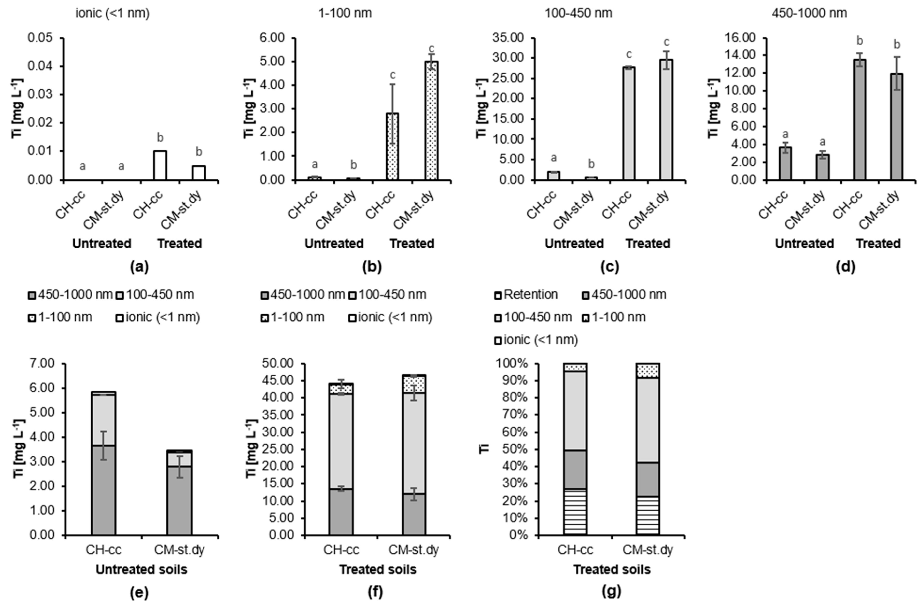
Abstract
1. Introduction
2. Materials and Methods
2.1. Soils Used in the Experiment
2.2. Batch Experiment with Soil
2.3. Cultivation of Fungus Treated with TiO2 Nanoparticles
2.4. Determination of Ti Concentration by ICP-MS
2.5. Statistical Analysis
3. Results
3.1. Batch Experiment with Soil
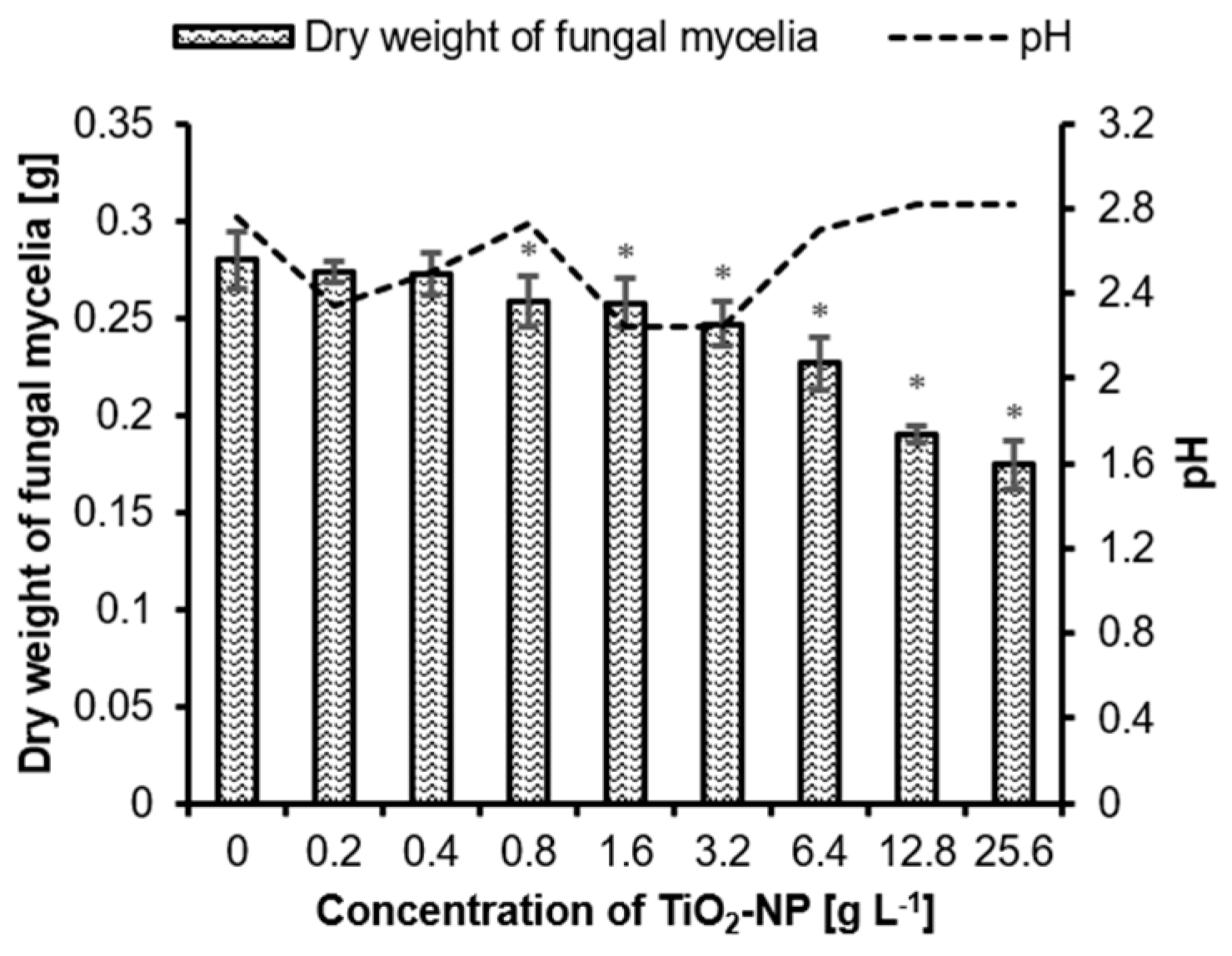
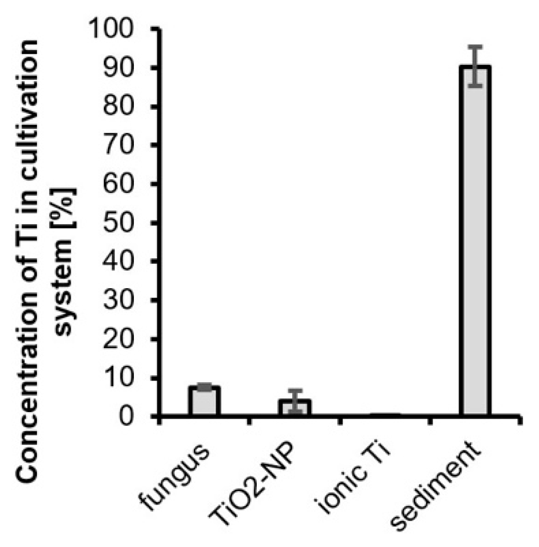
3.2. Mutual Interactions of Fungus and TiO2 NP
4. Discussion
4.1. Batch Experiment with Soils
4.2. Fungal Accumulation of TiO2 Nanoparticles
5. Conclusions
Supplementary Materials
Author Contributions
Funding
Conflicts of Interest
References
- Orians, K.J.; Boyle, E.A.; Bruland, K.W. Dissolved titanium in the open ocean. Nature 1990, 348, 322–325. [Google Scholar] [CrossRef]
- Van Baalen, M.R. Titanium mobility in metamorphic systems: A review. Chem. Geol. 1993, 110, 233–249. [Google Scholar] [CrossRef]
- Cornu, S.; Lucas, Y.; Lebon, E.; Ambrosi, J.P.; Luizão, F.; Rouiller, J.; Bonnay, M.; Neal, C. Evidence of titanium mobility in soil profiles, Manaus, central Amazonia. Geoderma 1999, 91, 281–295. [Google Scholar] [CrossRef]
- Zierden, M.R.; Valentine, A.M. Contemplating a role for titanium in organisms. Metallomics 2016, 8, 9–16. [Google Scholar] [CrossRef]
- Buettner, K.M.; Valentine, A.M. Bioinorganic Chemistry of Titanium. Chem. Rev. 2012, 112, 1863–1881. [Google Scholar] [CrossRef]
- Tan, W.; Peralta-Videa, J.R.; Gardea-Torresdey, J.L. Interaction of titanium dioxide nanoparticles with soil components and plants: Current knowledge and future research needs—A critical review. Environ. Sci. Nano 2018, 5, 257–278. [Google Scholar] [CrossRef]
- Hrubý, M.; Cígler, P.; Kužel, S. Contribution to understanding the mechanism of titanium action in plants. J. Plant Nutr. 2002, 25, 577–598. [Google Scholar] [CrossRef]
- Kužel, S.; Hrubý, M.; Cígler, P.; Tlustoš, P.; Van Nguyen, P. Mechanism of physiological effects of titanium leaf sprays on plants grown on soil. Biol. Trace Elem. Res. 2003, 91, 179–189. [Google Scholar] [CrossRef]
- Kolenčík, M.; Nemček, L.; Šebesta, M.; Urík, M.; Ernst, D.; Kratošová, G.; Konvičková, Z. Effect of TiO2 as Plant-Growth Stimulating Nanomaterial on Crop Production. In Plant Responses to Nanomaterials; Singh, V.P., Singh, S., Prasad, S.M., Chauhan, D.K., Tripathi, D.K., Eds.; Springer Nature: Berlin/Heidelberg, Germany, 2020; In Press. [Google Scholar]
- Šebesta, M.; Illa, R.; Zvěřina, O.; Šeda, M.; Diviš, P.; Kolenčík, M. Positive Effects of TiO2 Nanomaterials on Crop Growth. In Nanotechnology for Plant Diseases; Ingle, A.P., Ed.; Wiley-VCH: Weinheim, Germany, 2020; In Press. [Google Scholar]
- Kolenčík, M.; Ernst, D.; Urík, M.; Ďurišová, Ľ.; Bujdoš, M.; Šebesta, M.; Dobročka, E.; Kšiňan, S.; Illa, R.; Qian, Y.; et al. Foliar Application of Low Concentrations of Titanium Dioxide and Zinc Oxide Nanoparticles to the Common Sunflower under Field Conditions. Nanomaterials 2020, 10, 1619. [Google Scholar] [CrossRef]
- Kořenková, L.; Šebesta, M.; Urík, M.; Kolenčík, M.; Kratošová, G.; Bujdoš, M.; Vávra, I.; Dobročka, E. Physiological response of culture media-grown barley (Hordeum vulgare L.) to titanium oxide nanoparticles. Acta Agric. Scand. Sect. B—Soil Plant Sci. 2017, 67, 285–291. [Google Scholar] [CrossRef]
- Rajwade, J.M.; Chikte, R.G.; Paknikar, K.M. Nanomaterials: New weapons in a crusade against phytopathogens. Appl. Microbiol. Biotechnol. 2020, 104, 1437–1461. [Google Scholar] [CrossRef]
- Yan, J.; Huang, K.; Wang, Y.; Liu, S. Study on anti-pollution nano-preparation of dimethomorph and its performance. Chin. Sci. Bull. 2005, 50, 108–112. [Google Scholar] [CrossRef]
- Guan, H.; Chi, D.; Yu, J.; Li, H. Dynamics of residues from a novel nano-imidacloprid formulation in soyabean fields. Crop Prot. 2010, 29, 942–946. [Google Scholar] [CrossRef]
- Mohamed, M.M.; Khairou, K.S. Preparation and characterization of nano-silver/mesoporous titania photocatalysts for herbicide degradation. Microporous Mesoporous Mater. 2011, 142, 130–138. [Google Scholar] [CrossRef]
- Cartwright, A.; Jackson, K.; Morgan, C.; Anderson, A.; Britt, D.W. A Review of Metal and Metal-Oxide Nanoparticle Coating Technologies to Inhibit Agglomeration and Increase Bioactivity for Agricultural Applications. Agronomy 2020, 10, 1018. [Google Scholar] [CrossRef]
- Priyanka, K.P.; Harikumar, V.S.; Balakrishna, K.M.; Varghese, T. Inhibitory effect of TiO2 nanoparticles on symbiotic arbuscular mycorrhizal fungi in plant roots. IET Nanobiotechnology 2016, 11, 66–70. [Google Scholar] [CrossRef]
- Rafique, R.; Zahra, Z.; Virk, N.; Shahid, M.; Pinelli, E.; Park, T.J.; Kallerhoff, J.; Arshad, M. Dose-dependent physiological responses of Triticum aestivum L. to soil applied TiO2 nanoparticles: Alterations in chlorophyll content, H2O2 production, and genotoxicity. Agric. Ecosyst. Environ. 2018, 255, 95–101. [Google Scholar] [CrossRef]
- Mattiello, A.; Lizzi, D.; Marchiol, L. Influence of Titanium Dioxide Nanoparticles (nTiO2) on Crop Plants: A Systematic Overview. Nanomater. Plants Algae Microorg. 2018, 277–296. [Google Scholar] [CrossRef]
- Šebesta, M.; Kolenčík, M.; Matúš, P.; Kořenková, L. Transport and distribution of engineered nanoparticles in soils and sediments. Chem. List. 2017, 111, 322–328. [Google Scholar]
- Šebesta, M.; Matúš, P. Separation, Determination, and Characterization of Inorganic Engineered Nanoparticles in Complex Environmental Samples. Chem. List. 2018, 112, 583–589. [Google Scholar]
- Ben-Noah, I.; Friedman, S.P. Review and Evaluation of Root Respiration and of Natural and Agricultural Processes of Soil Aeration. Vadose Zone J. 2018, 17, 170119. [Google Scholar] [CrossRef]
- Adeleke, R.; Nwangburuka, C.; Oboirien, B. Origins, roles and fate of organic acids in soils: A review. South Afr. J. Bot. 2017, 108, 393–406. [Google Scholar] [CrossRef]
- Burford, E.P.; Fomina, M.; Gadd, G.M. Fungal involvement in bioweathering and biotransformation of rocks and minerals. Mineral. Mag. 2003, 67, 1127–1155. [Google Scholar] [CrossRef]
- Klaine, S.J.; Alvarez, P.J.J.; Batley, G.E.; Fernandes, T.F.; Handy, R.D.; Lyon, D.Y.; Mahendra, S.; McLaughlin, M.J.; Lead, J.R. Nanomaterials in the environment: Behavior, fate, bioavailability, and effects. Environ. Toxicol. Chem. 2008, 27, 1825–1851. [Google Scholar] [CrossRef]
- Peijnenburg, W.; Praetorius, A.; Scott-Fordsmand, J.; Cornelis, G. Fate assessment of engineered nanoparticles in solids dominated media—Current insights and the way forward. Environ. Pollut. 2016, 218, 1365–1369. [Google Scholar] [CrossRef]
- Welch, S.A.; Christy, A.G.; Kirste, D.; Beavis, S.G.; Beavis, F. Jarosite dissolution I—Trace cation flux in acid sulfate soils. Chem. Geol. 2007, 245, 183–197. [Google Scholar] [CrossRef]
- Polák, F.; Urík, M.; Matúš, P. Low molecular weight organic acids in soil environment. Chem. List. 2019, 113, 307–314. [Google Scholar]
- Urík, M.; Hlodák, M.; Mikušová, P.; Matúš, P. Potential of Microscopic Fungi Isolated from Mercury Contaminated Soils to Accumulate and Volatilize Mercury(II). Water Air Soil Pollut. 2014, 225, 2219. [Google Scholar] [CrossRef]
- Boriová, K.; Urík, M.; Bujdoš, M.; Pifková, I.; Matúš, P. Chemical mimicking of bio-assisted aluminium extraction by Aspergillus niger’s exometabolites. Environ. Pollut. 2016, 218, 281–288. [Google Scholar] [CrossRef]
- Qin, W.; Wang, C.; Ma, Y.; Shen, M.; Li, J.; Jiao, K.; Tay, F.R.; Niu, L. Microbe-Mediated Extracellular and Intracellular Mineralization: Environmental, Industrial, and Biotechnological Applications. Adv. Mater. 2020, 32, 1907833. [Google Scholar] [CrossRef]
- Raliya, R.; Biswas, P.; Tarafdar, J.C. TiO2 nanoparticle biosynthesis and its physiological effect on mung bean (Vigna radiata L.). Biotechnol. Rep. 2015, 5, 22–26. [Google Scholar] [CrossRef]
- Hulkoti, N.I.; Taranath, T.C. Biosynthesis of nanoparticles using microbes—A review. Colloids Surf. B Biointerfaces 2014, 121, 474–483. [Google Scholar] [CrossRef]
- Societas Pedologica Slovaca. Morphogenetic Soil Classification System of Slovakia, Basal Reference Taxonomy, 2nd ed.; Soil Science and Conservation Research Institute: Bratislava, Slovakia, 2014; ISBN 978-80-8163-005-7. [Google Scholar]
- IUSS Working Group. WRB World Reference Base for Soil Resources 2014, Update 2015 International Soil Classification System for Naming Soils and Creating Legends for Soil Maps; World Soil Resources Reports No. 106; FAO: Rome, Italy, 2015. [Google Scholar]
- McKeague, J.A.; Day, J. Dithionite-and oxalate-extractable Fe and Al as aids in differentiating various classes of soils. Can. J. Soil Sci. 1966, 46, 13–22. [Google Scholar] [CrossRef]
- Walkley, A.; Black, I.A. An examination of the Degtjareff method for determining soil organic matter, and a proposed modification of the chromic acid titration method. Soil Sci. 1934, 37, 29–38. [Google Scholar] [CrossRef]
- Fiala, K.; Kobza, J.; Matúšková, Ľ.; Brečková, V.; Makovníková, J.; Barančíková, G.; Búrik, V.; Litavec, T.; Houšková, B.; Chromaničová, A. Záväzné Metódy Rozborov Pôd; VÚPOP: Bratislava, Slovakia, 1999. [Google Scholar]
- FAO. Guidelines for Soil Description, 4th ed.; FAO: Rome, Italy, 2006; ISBN 92-5-105521-1. [Google Scholar]
- Kononova, M.M.; Belčikova, N.P. Uskorennyje metody opredelenija sostava gumusa minerálnych počv. Počvovedenije 1962, 10, 75–87. [Google Scholar]
- Kappen, H. Die Bodenazidität; Springer: Berlin/Heidelberg, Germany, 1929; ISBN 978-3-642-89928-7. [Google Scholar]
- Chowdhury, I.; Hong, Y.; Honda, R.J.; Walker, S.L. Mechanisms of TiO2 nanoparticle transport in porous media: Role of solution chemistry, nanoparticle concentration, and flowrate. J. Colloid Interface Sci. 2011, 360, 548–555. [Google Scholar] [CrossRef]
- Gibbs, R.J.; Matthews, M.D.; Link, D.A. The relationship between sphere size and settling velocity. J. Sediment. Res. 1971, 41, 7–18. [Google Scholar]
- Schwertfeger, D.M.; Velicogna, J.R.; Jesmer, A.H.; Saatcioglu, S.; McShane, H.; Scroggins, R.P.; Princz, J.I. Extracting Metallic Nanoparticles from Soils for Quantitative Analysis: Method Development Using Engineered Silver Nanoparticles and SP-ICP-MS. Anal. Chem. 2017, 89, 2505–2513. [Google Scholar] [CrossRef]
- Šebesta, M.; Nemček, L.; Urík, M.; Kolenčík, M.; Bujdoš, M.; Vávra, I.; Dobročka, E.; Matúš, P. Partitioning and stability of ionic, nano- and microsized zinc in natural soil suspensions. Sci. Total Environ. 2020, 700, 134445. [Google Scholar] [CrossRef]
- Fang, J.; Xu, M.; Wang, D.; Wen, B.; Han, J. Modeling the transport of TiO2 nanoparticle aggregates in saturated and unsaturated granular media: Effects of ionic strength and pH. Water Res. 2013, 47, 1399–1408. [Google Scholar] [CrossRef]
- Loosli, F.; Le Coustumer, P.; Stoll, S. TiO2 nanoparticles aggregation and disaggregation in presence of alginate and Suwannee River humic acids. pH and concentration effects on nanoparticle stability. Water Res. 2013, 47, 6052–6063. [Google Scholar] [CrossRef]
- Chen, G.X.; Liu, X.Y.; Su, C.M. Distinct Effects of Humic Acid on Transport and Retention of TiO2 Rutile Nanoparticles in Saturated Sand Columns. Environ. Sci. Technol. 2012, 46, 7142–7150. [Google Scholar] [CrossRef]
- Zhang, R.; Tu, C.; Zhang, H.; Luo, Y. Stability and transport of titanium dioxide nanoparticles in three variable-charge soils. J. Soils Sediments 2020, 20, 1395–1403. [Google Scholar] [CrossRef]
- Buffle, J.; Wilkinson, K.J.; Stoll, S.; Filella, M.; Zhang, J. A Generalized Description of Aquatic Colloidal Interactions: The Three-colloidal Component Approach. Environ. Sci. Technol. 1998, 32, 2887–2899. [Google Scholar] [CrossRef]
- Fang, J.; Shan, X.; Wen, B.; Lin, J.; Owens, G. Stability of titania nanoparticles in soil suspensions and transport in saturated homogeneous soil columns. Environ. Pollut. 2009, 157, 1101–1109. [Google Scholar] [CrossRef]
- Zehlike, L.; Peters, A.; Ellerbrock, R.H.; Degenkolb, L.; Klitzke, S. Aggregation of TiO2 and Ag nanoparticles in soil solution—Effects of primary nanoparticle size and dissolved organic matter characteristics. Sci. Total Environ. 2019, 688, 288–298. [Google Scholar] [CrossRef]
- Cornelis, G. Fate descriptors for engineered nanoparticles: The good, the bad, and the ugly. Environ. Sci. Nano 2015, 2, 19–26. [Google Scholar] [CrossRef]
- Gadd, G.M. (Ed.) Fungi in Biogeochemical Cycles; Cambridge University Press: Cambridge, UK, 2006; ISBN 9780511550522. [Google Scholar]
- Jonglertjunya, W.; Rubcumintara, T. Titanium and iron dissolutions from ilmenite by acid leaching and microbiological oxidation techniques. Asia-Pac. J. Chem. Eng. 2013, 8, 323–330. [Google Scholar] [CrossRef]
- Polák, F.; Urík, M.; Bujdoš, M.; Matúš, P. Aspergillus niger enhances oxalate production as a response to phosphate deficiency induced by aluminium(III). J. Inorg. Biochem. 2020, 204, 110961. [Google Scholar] [CrossRef]
- Crampon, M.; Hellal, J.; Mouvet, C.; Wille, G.; Michel, C.; Wiener, A.; Braun, J.; Ollivier, P. Do natural biofilm impact nZVI mobility and interactions with porous media? A column study. Sci. Total Environ. 2018, 610, 709–719. [Google Scholar] [CrossRef]
- Jiang, X.; Wang, X.; Tong, M.; Kim, H. Initial transport and retention behaviors of ZnO nanoparticles in quartz sand porous media coated with Escherichia coli biofilm. Environ. Pollut. 2013, 174, 38–49. [Google Scholar] [CrossRef]
- He, J.-Z.; Li, C.-C.; Wang, D.-J.; Zhou, D.-M. Biofilms and extracellular polymeric substances mediate the transport of graphene oxide nanoparticles in saturated porous media. J. Hazard. Mater. 2015, 300, 467–474. [Google Scholar] [CrossRef]
- Tarafdar, J.C.; Agrawal, A.; Raliya, R.; Kumar, P.; Burman, U.; Kaul, R.K. ZnO Nanoparticles Induced Synthesis of Polysaccharides and Phosphatases by Aspergillus Fungi. Adv. Sci. Eng. Med. 2012, 4, 324–328. [Google Scholar] [CrossRef]


Publisher’s Note: MDPI stays neutral with regard to jurisdictional claims in published maps and institutional affiliations. |
© 2020 by the authors. Licensee MDPI, Basel, Switzerland. This article is an open access article distributed under the terms and conditions of the Creative Commons Attribution (CC BY) license (http://creativecommons.org/licenses/by/4.0/).
Share and Cite
Šebesta, M.; Nemček, L.; Urík, M.; Kolenčík, M.; Bujdoš, M.; Hagarová, I.; Matúš, P. Distribution of TiO2 Nanoparticles in Acidic and Alkaline Soil and Their Accumulation by Aspergillus niger. Agronomy 2020, 10, 1833. https://doi.org/10.3390/agronomy10111833
Šebesta M, Nemček L, Urík M, Kolenčík M, Bujdoš M, Hagarová I, Matúš P. Distribution of TiO2 Nanoparticles in Acidic and Alkaline Soil and Their Accumulation by Aspergillus niger. Agronomy. 2020; 10(11):1833. https://doi.org/10.3390/agronomy10111833
Chicago/Turabian StyleŠebesta, Martin, Lucia Nemček, Martin Urík, Marek Kolenčík, Marek Bujdoš, Ingrid Hagarová, and Peter Matúš. 2020. "Distribution of TiO2 Nanoparticles in Acidic and Alkaline Soil and Their Accumulation by Aspergillus niger" Agronomy 10, no. 11: 1833. https://doi.org/10.3390/agronomy10111833
APA StyleŠebesta, M., Nemček, L., Urík, M., Kolenčík, M., Bujdoš, M., Hagarová, I., & Matúš, P. (2020). Distribution of TiO2 Nanoparticles in Acidic and Alkaline Soil and Their Accumulation by Aspergillus niger. Agronomy, 10(11), 1833. https://doi.org/10.3390/agronomy10111833