Article

The Potential of Novel Bacterial Isolates from Natural Soil for the Control of Brown Rot Disease (Monilinia fructigena) on Apple Fruits

1
Phytopathology Unit, Department of Plant Protection, Ecole Nationale d’Agriculture de Meknès, Km10, Rte Haj Kaddour, BP S/40, Meknès 50001, Morocco
2
Laboratory of Functional Ecology and Environmental Engineering, Sidi Mohamed Ben Abdellah University, P.O. Box 2202, Route d’Imouzzer, Fez 30500, Morocco
3
Centre Régional de la Recherche Agronomique (CRRA), Plant Protection Laboratory INRA, Oujda 60000, Qualipole de Berkane, Berkane 63300, Morocco
4
Regional Center of Agricultural Research of Rabat, Biotechnology Unit, INRA of Morocco, Av. Mohamed Belarbi Alaoui, BP. 6356, Rabat-Instituts 10101, Morocco
5
Unité de Recherche Résistance Induite et Bio-Protection des Plantes-EA 4707, Université de Reims Champagne-Ardenne, 51100 Reims, France
*
Authors to whom correspondence should be addressed.
Agronomy 2020, 10(11), 1814; https://doi.org/10.3390/agronomy10111814
Submission received: 18 September 2020 / Revised: 10 November 2020 / Accepted: 16 November 2020 / Published: 18 November 2020

Abstract

:
Monilinia fructigena is one of the most important fungal pathogens causing brown rot on apple and is heavily affecting fruit production. The main objective of this study was to screen for potential bacterial isolates with higher antagonistic activity against M. fructigena. Our study focused on the identification of potential bacterial isolates capable of reducing both the mycelial growth of M. fructigena and the disease severity using in vitro and in planta trials, respectively. To achieve this goal, thirteen bacteria, isolated from natural soil, were evaluated for their abilities to produce lytic enzymes (amylase, cellulase and protease), hydrocyanic acid (HCN) and lipopeptides (bacillomycin, fengycin, iturin and surfactin). Further, results from the dual culture method, volatile and bacterial free-cell filtrate bioassays indicated that tested isolates showed a fungicidal activity against the mycelial growth of M. fructigena. Thus, out of the 13 isolates tested, 12 exhibited significant mycelial inhibition (more than 70%) against M. fructigena, while remaining the last isolate displayed only a partial inhibition (up to 43%). Further, 12 of the bacteria isolates displayed an amylase production, 10 showed cellulase production, 11 revealed protease production, while only 2 displayed HCN production. In addition, most bacterial isolates were found to have genes encoding for different lipopeptides: bacillomycin (10), fengycin (3), iturin (11) and surfactin (1). Interestingly, two bacterial isolates, Bacillus amyloliquefaciens B10W10 and Pseudomonas sp. B11W11 were found to be the most effective and displayed the lowest disease severity in planta trial. These two bacteria reduced the brown rot incidence compared to the synthetic fungicide in a semi-commercial large-scale trial. Therefore, our findings suggest that these two later bacterial isolates provide apple protection against M. fructigena via direct and indirect mechanisms. These isolates may be used, therefore, as potential biological control agents (BCAs) in preventive treatment to control brown rot disease on apple fruits.

1. Introduction

Brown rot is a fungal disease caused by fungi belonging to the genus Monilinia Honey (anamorph Monilia) [1]. This disease has been reported worldwide and causes severe symptoms: wilting of flowers, twigs or brown rot of fruits, according to the affected organs. The pathogen mainly attacks trees belonging to the rosaceous producing stone fruits or pips such as apple, pear, cherry, apricot, peach, etc. [2]. It causes dramatic losses, mainly in postharvest [3].
Several fungal pathogens are reported to trigger brown rot disease, but only three of them cause significant losses: Monilinia laxa, Monilinia fructicola, which are quarantine pathogens in Morocco and Monilinia fructigena [4]. The first two species are mainly confined to stone fruits, while M. fructigena is a major pathogen causing fruit rot disease on apples [5]. Symptoms begin as rounded brownish spots centered at the infection site. M. fructigena has a colony color ranging from white to light beige and large (1.5 mm on average) conidiospores tufts and disposition in concentric circles in the fruit. On the other hand, M. laxa can be distinguished by greenish-gray conidiospores tufts of less than 0.5 mm on average covering the entire infected area [3].
Fruits can be infected by direct penetration of the cuticle, by the production of cutinase [6], stomata or trichomes and by cracks and wounds [7]. Conidia are produced along the growing season and can infect fruits at any stage of their development. Branch and stem cankers, in addition to mummified fruits, ensure the fungal pathogen survival over the year. However, brown rot disease observed during the storage mainly linked to the infection occurring just prior to harvest.
Nowadays, the control of brown rot disease is commonly based on the control of insects that can injure fruits and thus create entrance doors to the pathogenic fungus [2]. Fungicides are used, in addition to prophylactic and sanitation measures deployed against high pressure of insect attacks. As regards attacks on flowers, many active substances are applied, in particular those, which affect fungal sporulation such as dicarboximides, benzimidazoles or triazoles [8]. However, the growing public concerns about fungicidal residues in apple fruit, and even resistance to active ingredients or fungicide families [9,10,11] have forced most fruit-importing countries, and particularly European Union (EU) countries, to adopt very strict legislation regarding the use of certain fungicides to control postharvest diseases. The ban on certain conventional, protective fungicides [12] encouraged scientists and industrials to look for other alternative control methods, which are less harmful to human health and more environmentally friendly. These methods include alternative chemical compounds, postharvest physical treatments and, biological control agents (BCAs) [13,14].
Biological control using microbial antagonists has emerged as an effective strategy to control postharvest diseases of fruits [9]. Therefore, several microorganisms were reported as possible BCAs against pathogens, causing fruit rot under storage conditions [15]. Most of these antagonists are belonging to Bacillus and Pseudomonas genus. Specifically, Bacillus amyloliquefaciens BUZ-14 was found to effectively control apple rot [16]. Furthermore, Epicoccum nigrum, which is a saprophytic fungus, was used as a potential BCAs against Monilinia species [17]. This antagonistic fungus acted against M. laxa by secreting antibiotics such as flavipin, which had a multisite action by inhibiting the cellular respiration and the synthesis of adenosine triphosphate (ATP) and proteins [18]. In addition, De Cal et al. [19] stated that E. nigrum conidia significantly reduced the number of Monilinia spp. conidia on the fruit surface. In addition, bacteria such as B. subtilis and B. amyloliquefaciens strains were able to produce endospores, allowing them to withstand extreme environmental conditions. These bacterial isolates are largely used as BCAs to manage postharvest fruit diseases [20]. Their abilities as BCAs are derived mainly from their high capabilities of producing a high range of antibiotics compounds such as iturin, bacillomycin, surfactin and fengycin [9].
To improve biocontrol strategies for pathogens that affect apple fruits in Morocco, there was an increase of interest among researchers to develop bacterial-based fungicides. The intended purpose associated with these types of pesticides is to ensure the control of fungal diseases and the enhancement of the BCAs efficacy and reliability. Therefore, the main objectives of this study were (i) screening, characterization and identification of novel potential bacterial isolates; (ii) assessing their biocontrol efficacy against M. fructigena, both in vitro and in vivo; (iii) studying their modes of action; and (iv) finally proposing the most effective bacterial isolates for brown rot control.

2. Materials and Methods

2.1. Fungal Pathogen Preparation

Monilinia fructigena (strain VPBG) used in this study was isolated from cherries in Serbia and identified as described previously [21]. The fungal pathogen was subcultured on potato dextrose agar (PDA) medium and incubated 7 days in darkness at 25 °C using an IN30 cultivator instrument (Memmert GmbH Co., Koln, Germany).
The conidial suspension was prepared by flooding a 10–14-day fungal colony with 5 mL of sterile distilled water (SDW) containing Tween-20 (0.05%). The conidial suspension was recovered by scraping the surface of the medium with the tip of a sterile Pasteur pipette. The resulting suspension was filtered through autoclaved tissue layers to remove debris, mycelium and medium. The concentration of conidial suspension (1 × 10⁶ and 1 × 10³ spores/mL) was adjusted determined using a Malassez hematocytometer (Roche, Meylan, France) by adding SDW.

2.2. Bacterial Isolates: Preparation and Molecular Identification

Thirteen bacterial isolates from the culture collection of “Ecole Nationale d’Agriculture of Meknes” (ENA-Meknes), Morocco, and previously isolated from the natural, uncultivated calcimagnesic soil of ENA-Meknes were screened for their potential antagonistic activity against M. fructigena. Before experiments, each bacterial isolate was maintained on Luria–Bertani (LB) medium at 25 °C for 48 h. The bacterial suspension was prepared from a 24 h-old culture grown on PDA medium at 25 °C and adjusted to 1 × 10⁸ CFU/mL concentration using a spectrophotometer at 620 nm wavelength.
Genomic DNA of each bacterial isolate was extracted from fresh bacterial suspensions according to the DNA extraction protocol developed by LIop et al. [22]. Bacteria were identified based on their partial 16S ribosomal DNA gene using the primer pair Fd1/RP2 (Fd1:5′-AGAGTTTGATCCTGGCTCAG-3′ and RP2:5′-ACGGCTACCTTGTTACGACTT-3′). The polymerase chain reaction (PCR) was carried out in a total volume of 25 μL using EnzimaGoTaq DNA polymerase (Bioline, London, UK) according to the manufacturer instructions. Each PCR test consisted of 5 μL (Buffer 5x), 0.25 μL Taq, 1 μL forward Primer (50 μM), 1 μL reverse Primer (50 μM), 15.25 μL of H2O and 2.5 μL of DNA template of each bacterial isolate. The negative control contained only the reaction mixture and SDW. PCR amplifications were carried out in an Eppendorf thermal cycler following conditions: initial denaturation at 96 °C for 4 min followed by 35 cycles of denaturation at 96 °C for 10 s, primers annealing at 52 °C for 40 s and extension at 72 °C for 2 min; with a final extension step at 72 °C for 4 min. PCR products were visualized by agarose gel electrophoresis (1% agarose) and stained with ethidium bromide. Sequencing of PCR-amplified DNA was performed. The obtained sequences were edited and aligned using DNAMAN software (version 6.0, Lynnon Biosoft, Quebec Canada) and compared for homology in the NCBI-BLAST database (National Center for Biotechnology Information http://blast.ncbi.nlm.nih.gov/Blast.cgi). Sequences resulting from this analysis were deposited in GenBank under accession numbers, as mentioned in Table 1.

2.3. Biochemical Traits of Bacterial Isolates

The ability of the thirteen bacterial isolates to produce the amylase activity was carried out on Petri dishes containing the nutrient agar (NA) medium (10 g beef extract, 10 g peptone, 10 g NaCl, 20 g glucose, 15 g agar and 1000 mL of SDW) supplemented with 1% of soluble starch [23]. Briefly, each 48 h-old bacterial culture was spotted (10 μL) onto the center of the Petri dish and incubated for 72 h at 28 °C. Three ml of the iodine solution was added to each Petri plate to visualize the starch hydrolysis. The appearance of a clear zone (halo) around the colony demonstrates a positive amylase activity, whereas a negative activity was characterized by the occurrence of brown color around the colony [24]. For the proteolytic activity, the ability of the thirteen bacterial isolates to produce protease was evaluated on skimmed milk agar medium as previously described by Kumar et al. [25]. An aliquot of 5 µL of each 24 h-old bacterial culture was deposited in the center of each Petri plate. Petri dishes were then incubated for 72 h at 28 °C. The formation of a clear zone around colonies indicated a positive proteolytic activity. In addition, the activity of the cellulase of all bacterial isolates was determined using carboxymethyl cellulose (CMC) solid medium [26]. Briefly, an aliquot (10 μL) of 24 h-old bacterial culture was placed in the center of the Petri plate. The cellulase activity was determined after 8 days of incubation period at 28 °C by adding 0.1% Congo red dye to each Petri dish for 15 min, rinsed three times with 1 M NaCl solution and examined for formation of a clear zone (halo) around colonies. In the last, the ability of the isolates to produce hydrocyanic acid (HCN) was evaluated according to the protocol developed by Bakker and Schipperes [27] with slight modifications. For each bacterial isolate, a 24 h-old culture was prepared and adjusted to a concentration of 1 × 10⁸ CFU/mL using a spectrophotometer. An aliquot (100 µL) of each bacterial suspension was inoculated into Petri dish containing yeast extract peptone dextrose (YEPD) medium (10 g glycose, 5 g peptone, 5 g yeast extract and 15 g agar) amended with glycine (4.4 g/L). A sterile Whatman filter paper no.1 soaked in a picrate solution (2.5% picric acid and 12.5% anhydrous sodium carbonate (Na2CO3)) was placed at the top of each Petri dish. The plates were sealed with Parafilm, inverted, then incubated at 28 °C for 4 days. A control Petri dish only inoculated with SDW served as a negative control. Response was assessed by visual observation of the color of Whatman paper. The change of the color from yellow to orange or brown indicates the production of volatile HCN.

2.4. Polymerase Chain Reaction (PCR) Detection of the Antibiotic Biosynthetic Genes

PCR tests were performed to investigate the presence of the biosynthesis lipopeptides genes (bacillomycin, fengycin, iturin and surfactin) in the DNA of the 13 tested bacterial isolates. Specific primers were used for amplification of those genes: bacillomycin (BACC1F/BACC1R), fengycin (FEND1F/FEND1R), iturin (ITUP1F/ITUP2R) and surfactin (P17/P18) [28]. Each PCR test was achieved in 25 µL mixtures with 5 μL of 5× buffer, 1 μL of each primer (50 μM), 0.25 μL of Taq DNA polymerase 5 U/µL (Bioloine, UK), 2.5 μL of genomic DNA and 15.25 µL of SDW. The amplifications were conducted using the PCR conditions, as previously described by Dimkić et al. [28] (Table 2). PCR products were then visualized on a 1.5% agarose gel by electrophoresis using tris-borate EDTA buffer (TBE) (0.5×), stained with ethidium bromide and visualized using a UV trans-illuminator.

2.5. In Vitro Dual Culture Bioassay

Dual culture plate bioassay was used to screen the best antagonist bacterial isolates against M. fructigena. A twenty-four-hour-old bacterial culture of each isolate was spotted at 4 equidistant streaks (3 to 4 cm length) along the perimeter of the Petri dish containing the PDA medium. A mycelial plug of 7 mm in diameter was cut out from the edge of the 7-day-old actively growing colony, deposited in the center of each Petri dish within bacterium bands and incubated at 28 °C [31]. Petri dishes with only mycelial plugs served as a negative control. The antagonistic activity was determined by calculating the inhibition rate (IR) of mycelial growth as follows:
IR (%) = (DC − DT/DC) × 100
where DC is the diameter of the colony in control treatments while DT corresponds to the diameter of the colony in confrontation treatments. The experiment was repeated twice over time, with 4 Petri plates for each bacterial isolate.

2.6. Volatile Products (VOCs) Bioassay

The antifungal activities of bacterial isolates through the production of VOCs were evaluated by in vitro bioassays. To do this, each bacterium was first subcultured into three parallel streaks on Luria–Bertani medium and incubated at 28 °C for 24 h. Afterward, the lid of the Petri plate was replaced by the bottom of another Petri plate containing a 7 mm-mycelial plug from the edge of the 10-day-old actively growing fungal colony on PDA. Both bottoms plates were sealed together with Parafilm [32]. Control plates were prepared in the same way, without bacterial isolates. The plates were incubated at 28 °C, and inhibitions rates were recorded as described above after 7 days of incubation periods. This experiment was repeated twice over time, with 4 replicates for each treatment.

2.7. In Vitro Bacterial Cell-Free Filtrates Effect

Each bacterial isolate was first subcultured on nutrient broth (NB) medium at 28 °C for 48 h. An aliquot (100 µL) of each bacterial suspension (1 × 10⁸ CFU/mL) was inoculated in an NB culture medium containing 10 g beef extract, 10 g peptone, 10 g NaCl, 20 g glucose and 1000 mL of SDW and then incubated in a rotary shaker (130 rpm) at 30 °C for 3 days. The bacterial cell cultures were centrifuged for 25 min (5.500 rpm), and the supernatant of each bacterium was recovered and filtrated by using a 0.22 µm Millipore filter [33]. The obtained cell-free filtrate was used for in vitro dual culture tests at a concentration of 10% [33]. In addition, a negative control was prepared by adding liquid NA medium to the PDA medium instead of cell-free culture filtrate. Petri plates were then inoculated by 7 mm-mycelial plugs from the edge of the 10-day-old actively growing colony and incubated at 28 °C. The diameter of mycelial growth was determined at 5 and 10 days post-inoculation and used to calculate the inhibition rates as described above. This experiment was repeated twice over time, with four replicate for each bacterium treatment.
To determine the impact of bacterial cell-free filtrates on spore germination, each bacteria-free filtrate (100%) was mixed with conidial suspension of Monilinia (1 × 10⁶) spores/mL at a ratio of 1:1 (v:v). In addition, a bacteria-free NA culture solution mixed with spore suspension at the same volume was served as controls. All mixtures were incubated at 25 °C in Eppendorf tubes. Germination of the spores was observed under the microscope after 24 h of incubation. A spore is considered germinated if the length of the germ tube is greater than its smaller diameter. The inhibition rate of spore germination (Ig) is determined according to the following formula:
Ig = (NC − NT)/NC × 100
with NC: number of germinated spores in culture medium without bacterial cell-free filtrate and NT: number of germinated spores in bacteria cell-free filtrate (100%).

2.8. In Vivo Antagonism Experiment

Apple fruits (Starkrimson) were used for in vivo experiments. Apple fruits with uniform size, a similar maturity stage and without blemishes, injuries or visible rot were stored at 4 °C prior to until use for experiments. Prior to each experiment, apple fruits were surface disinfected by soaking in the sodium hypochlorite solution (1%) for 5 min, washing twice with SDW and drying for 1 h at room temperature under the laminar flow hood.
To monitor the biocontrol efficacy of bacterial isolates on apple fruits, two equidistant wound sites (4 mm in diameter and 3 mm in depth) were performed on the equatorial zone of the fruit using a sterile stainless-steel rod. Each apple wound was treated with 50 μL of each bacteria suspension (1 × 10⁸ CFU/mL) and inoculated 4 h later with 50 μL of fungal suspension of M. fructigena (1 × 10³ and 1 × 10⁶ spores/mL). Wounds that received only SDW were served as controls. Each inoculated apple was deposited in a disinfected cylindrical plastic box (1 L of capacity) where a Whatman paper soaked with SDW was placed to maintain a high humidity level for fungal growth and to prevent moisture loss from the fruit. All boxes were incubated at 24 °C in a humid growth chamber with a 12 h photoperiod. The experiment was repeated twice over time. For each treatment (4 fruits, 8 wounds), the lesion diameters were measured after 5 and 10 days post-inoculation and the disease severity (DS) of pathogenic fungus on apple fruits was calculated according to the following formula:
DS (%) = (average lesion diameter (mm) of treated wounds/average lesion diameter
(mm) of the wounds in the control treatment) × 100
For the semi-practical trials of selected bacteria, out of the 13 tested bacterial isolates, two antagonistic bacteria displaying low disease severity in vivo and the highest inhibition rates of mycelial growth in vitro were selected for a semi-commercial large-scale trial. Apple fruits were wounded (3 mm in diameter and 4 mm-deep) at four equidistant points. They were subsequently dipped in each bacterial suspension (1 × 10⁸ CFU/mL) for 2 min. Apple fruits dipped in SDW served as a control. Apple fruits were placed in plastic bags (10 apple fruits/bag) with three replicates and incubated in the growth chamber at 24 °C. After 24 h of the incubation period, apple fruits were inoculated by spraying with M. fructigena (1 × 10³ spores/mL) and incubated in a growth chamber at 24 °C. The number of infected fruits was monitored every 5 days until apples in control treatments were totally infected. The experiment was repeated twice over time. The disease incidence (%) was calculated for each treatment according to the following formula:
Incidence (%) = number of infected fruits/total number of fruits × 100

2.9. Statistical Analysis

All in vitro and in vivo experiments were carried out twice over time. Analysis of variance (ANOVA) was performed using SPSS statistical software (version 20). When the effect was revealed to be significant, the least significant difference (LSD) test was performed for means separation at p = 0.05.

3. Results

3.1. Bacterial Identification

The bacterial 16S rRNA sequences obtained were deposited in the GenBank database of NCBI with the accession numbers listed in Table 1. Five isolates were identified as B. subtilis. In addition, four bacterial isolates were identified as B. amyloliquefaciens. One isolate was classified as B. siamensis, while the last three isolates were found to belong to Pseudomonas sp.

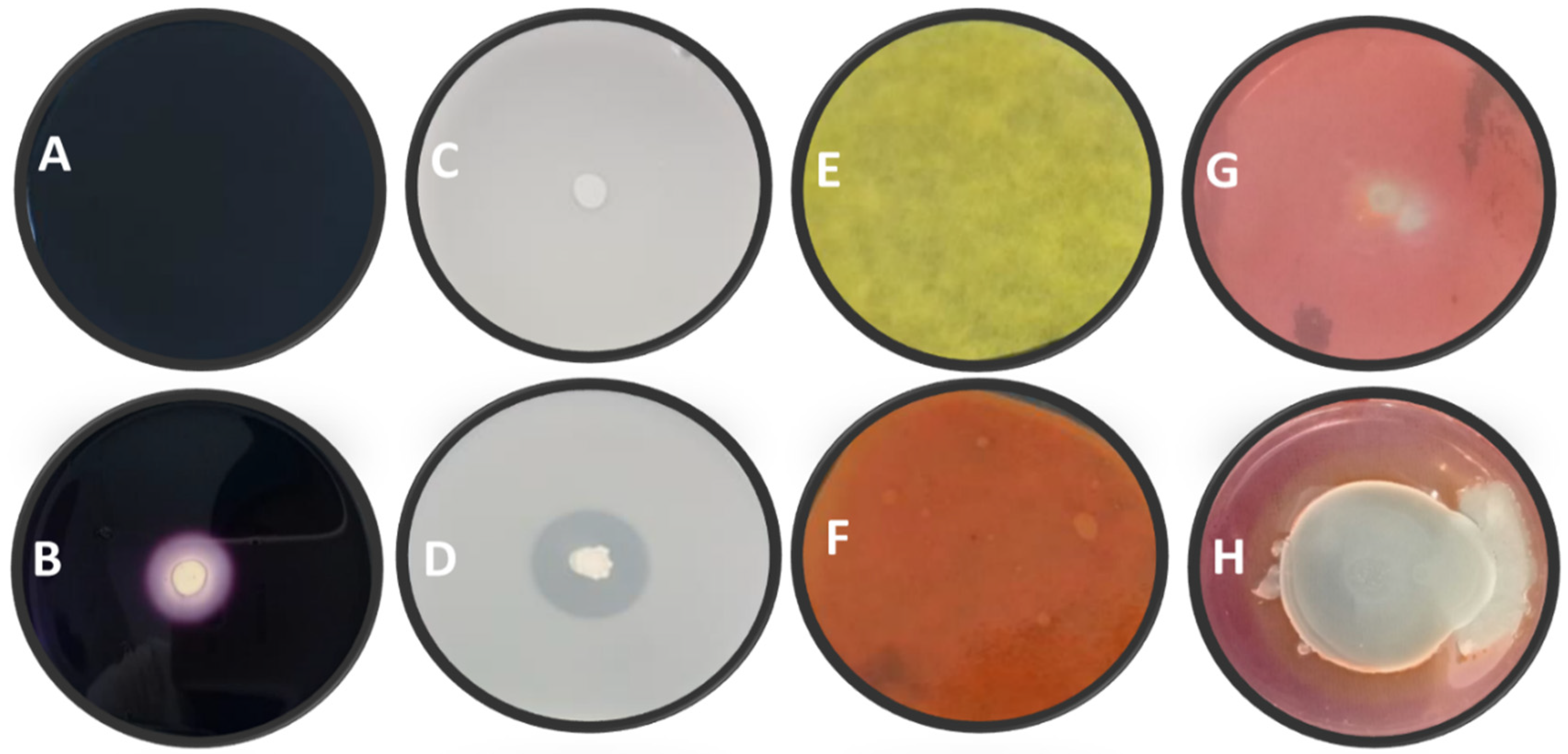
3.2. Biochemical Traits of Bacterial Isolates

The ability of all bacterial isolates to hydrolyze starch showed that 92.31% of isolates exhibited an amylase activity (Figure 1 and Table 3). However, a difference in amylase production between the tested bacterial isolates was observed. Therefore, the bacterial isolate B. amyloliquefaciens B4W4 showed the most important activity (1.4), followed by B. amyloliquefaciens B7W7, while no amylolytic activity was exhibited by Pseudomonas sp. strain FD1-9F.
Results of protease activity showed that out of 13 tested bacteria, 11 (84.62%) were able to exhibit the protease activity (Figure 1 and Table 3). Indeed, isolate B. amyloliquefaciens B4W4 had the highest activity (2.5), whereas both isolates Pseudomonas sp. strain FD1-9F and Pseudomonas sp. strain B11W11 do not show any protease activity.
The evaluation of the ability of isolates to hydrolyze the cellulose revealed that 76.92% of strains have a positive cellulosic activity (Figure 1 and Table 3). However, there was a difference between bacterial isolates in synthesizing cellulase molecules, with then isolates having a positive activity varying between 1 and 1.38. Moreover, only Bacillus subtilis B5W5 had the highest index (1.38), followed by isolate RP2-13F with an index of 1.29. On the other hand, no cellulase activity was recorded for Pseudomonas sp. B6W6 and B. amyloliquefaciens B7W7.
The analysis of HCN production showed that only two bacterial isolates out of 13 (15.38%) were able to produce HCN (Figure 1 and Table 3). However, these two bacterial isolates have only a moderate HCN production.

3.3. Detection of Lipopeptides by PCR

The PCR results for the detection of genes involved in the production of lipopeptides in bacterial antagonists are listed in Table 4.
For bacillomycin production evaluation, two primers, BACC1-F/BACC1-R, were used for the detection of the bamC gene, which is involved in the synthesis of bacillomycin (iturin family). Among the 13 bacteria tested, our results revealed the presence of the expected PCR products (band of 875 bp) for 10 bacterial isolates, including B. amyloliquefaciens B1W1, B. amyloliquefaciens B4W4, B. subtilis B2W2, B. subtilis B3W3, B. subtilis RP2-12F, B. subtilis RP2-13F, Pseudomonas sp. B6W6, Pseudomonas sp. FD1-9F, Pseudomonas sp. B11W11 and B. siamensis B14W14 (Table 4). For the fengycin production, two primers FEND1-F/FEND1-R, were used for the detection of the fenD gene in the genomic DNA of bacterial isolates. The results revealed bands of 964 bp in 3 bacterial isolates out of 13 (Table 4). Bacterial isolates for which the fenD gene was detected were Pseudomonas sp. FD1-9F, Pseudomonas sp. B11W11 and B. subtilis RP2-12F. However, for iturin secretion, the detection of the iturin operon, responsible for the biosynthesis of iturin, two primers were used ITUP1-F and ITUP1-R, which showed 2 kb fragments containing parts of the iturin A and iturin B genes and intergenic sequences between these two genes. Among the 13 tested bacteria, 2 bacterial isolates Pseudomonas sp. B6W6 and Pseudomonas sp. B11W11 lack this gene (Table 4). Lastly, the surfactin production was evaluated using two primers P17/ P18 to detect the presence of the sfp gene encoding for the 4′-phosphopantetheine transferase, which is required for the activation of surfactin synthetase. Results indicated that only isolate Pseudomonas sp. B6W6 had the sfp gene (Table 4).

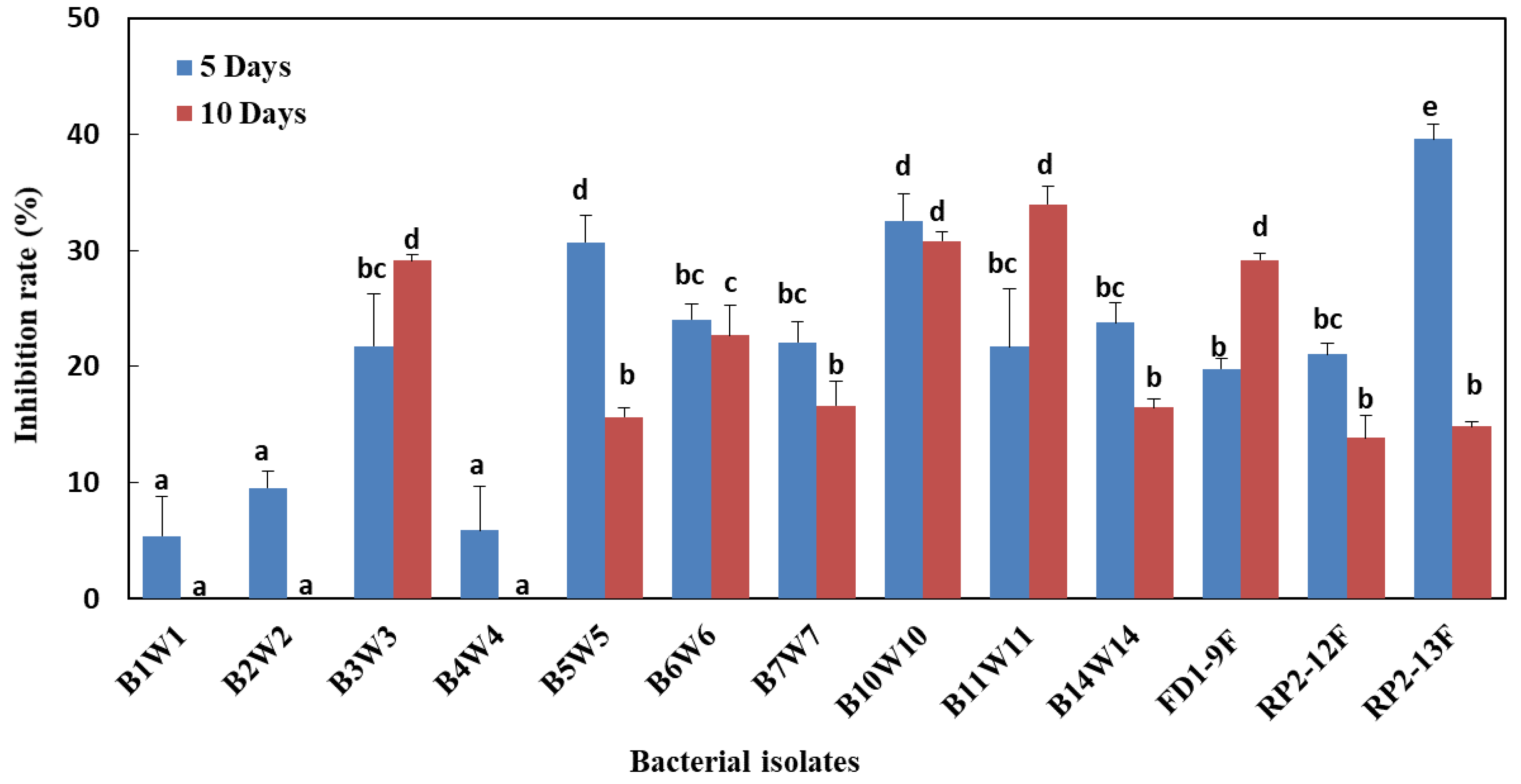
3.4. In Vitro Dual Confrontation Bioassay

A dual culture method was used to investigate the potential antagonistic effect of the 13 bacterial isolates against the pathogenic fungus M. fructigena (Table 1). Results revealed that out of the 13 tested isolates, 12 showed a significant inhibition (more than 70%) against M. fructigena, while the remaining isolate displayed only a partial inhibition (up to 43%) after 10 days of incubation. Further, the statistical analysis highlighted a highly significant difference between isolates. Overall, the mycelial growth in the presence of bacterial isolates was significantly reduced than that of controls. It was noticed, however, that mycelial growth inhibition rates increased with the increasing incubation time from 5 to 10 days (Table 1).

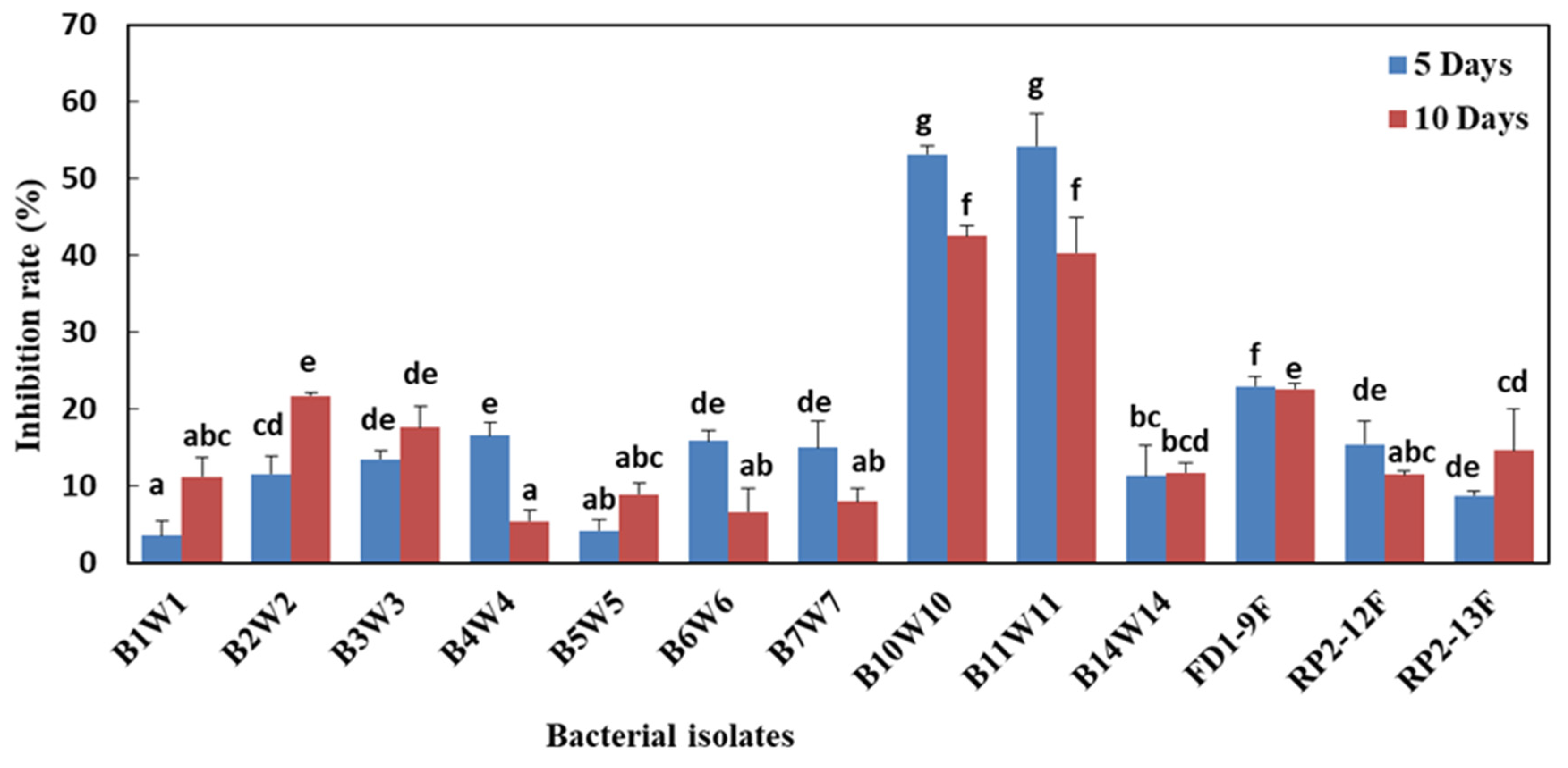
3.5. Effect of Bacteria-Derived VOCs on Mycelial Growth, under In Vitro Conditions

The in vitro tests of VOCs derived from the 13 tested bacterial isolates revealed that 10 isolates have antagonistic activity against M. fructigena by producing volatile antifungal compounds, but with a low IR (35%). In addition, three isolates have an activity of less than 20% after 5 days of incubation; further, their activities disappeared after 10 days post-incubation (Figure 2). Overall, statistical analyses displayed a highly significant difference between isolates.

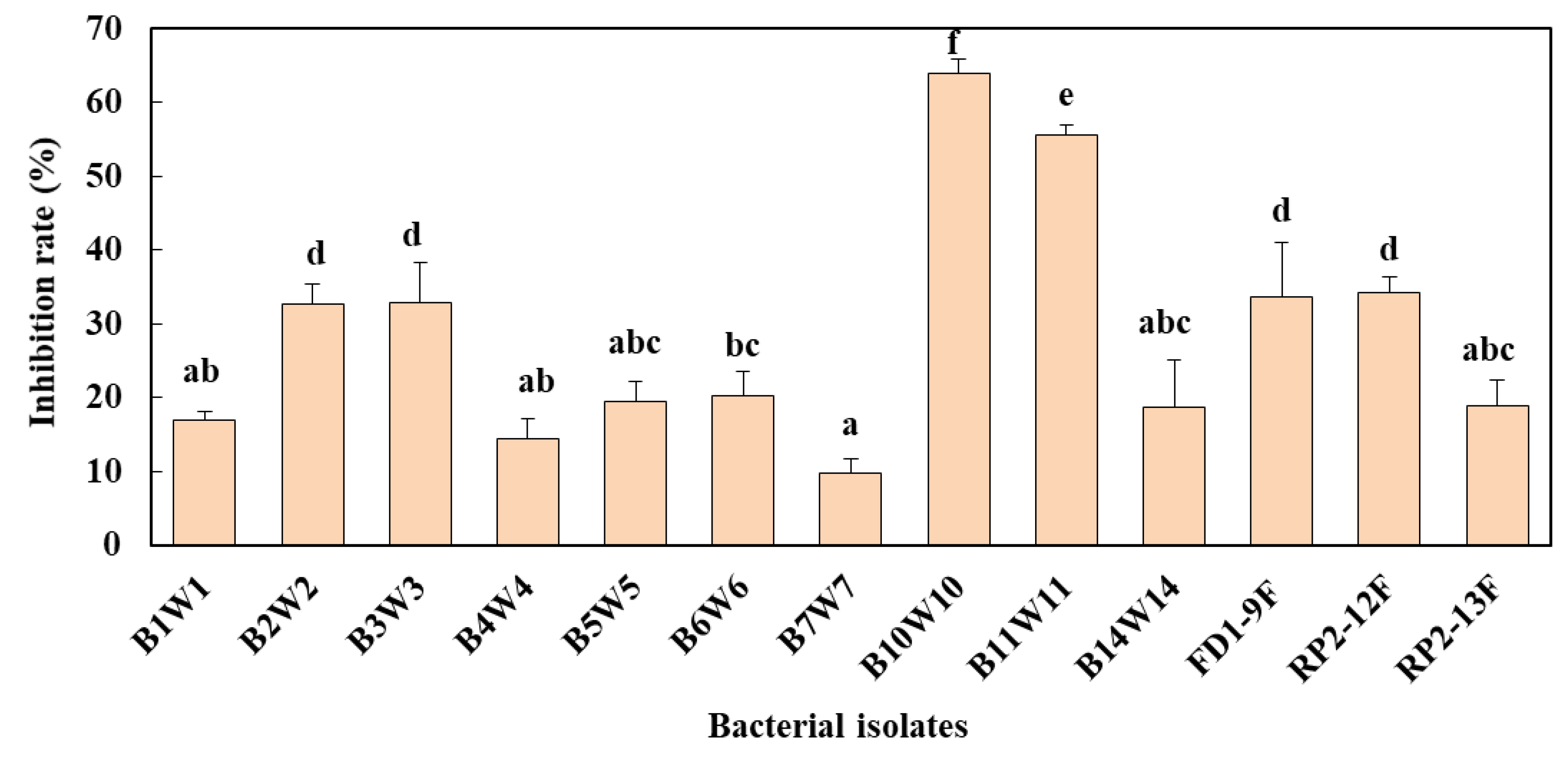
3.6. Effect of Bacterial Cell-Free Filtrates on Mycelial Growth and Spore Germination

Inhibition rates of mycelial growth of M. fructigena obtained with bacterial cell-free filtrates were highly significantly different between all the 13 isolates (Figure 3). e cell-free filtrates of both bacterial isolates B. amyloliquefaciens B10W10 and Pseudomonas sp. B11W11 have the highest reduction of mycelial growth regardless of incubation periods (5 or 10 days). Furthermore, results of cell-free bacterial filtrates showed a significant inhibitory effect on spores germination of M. fructigena, ranging from 10 to 65% (Figure 4). Interestingly, the highest inhibition rates of spore germination were obtained with both bacterial isolates B. amyloliquefaciens B10W10 and Pseudomonas sp. B11W11.

3.7. In Vivo Effectiveness of Bacterial Isolates as Affected by Pathogen Pressure

3.7.1. Lower Pathogen Pressure (1 × 10³ spores/mL)

Two bacterial isolates (B. amyloliquefaciens B10W10 and Pseudomonas sp. B11W11) out of the 13 tested bacteria displayed an antagonistic activity against M. fructigena with 100% inhibition after 5 days. However, their efficiencies decreased slightly after 10 days post-incubation as their disease severity increases from 0–0 to 21.70–29, respectively. However, for other bacterial isolates, the disease severity was decreased with the increase of the incubation period (Table 5). Interestingly, the biocontrol protection obtained with B. amyloliquefaciens B10W10 and Pseudomonas sp. B11W11 against brown rot disease was statistically comparable to that of fungicidal product thiophanate-methyl (Table 5).

3.7.2. Higher Pathogen Pressure (1 × 10⁶ spores/mL)

The same trend of protection described previously was observed with bacterial isolates at higher pathogen pressure (1 × 10⁶ spores/mL). Statistical analyses of disease severity underlined that there was a highly significant difference between bacterial isolates. Surprisingly, both bacterial isolates B. amyloliquefaciens B10W10 and Pseudomonas sp. B11W11allowed higher protection against M. fructigena with fewer disease severities for both incubation periods 5 (0–0%) and 10 days (43.59 and 51.09%). The efficacy of these two isolates was significantly higher (5 days) or comparable (10 days) to that of a commercial fungicidal product (thiophanate-methyl, at 1 ppm) (Table 5).

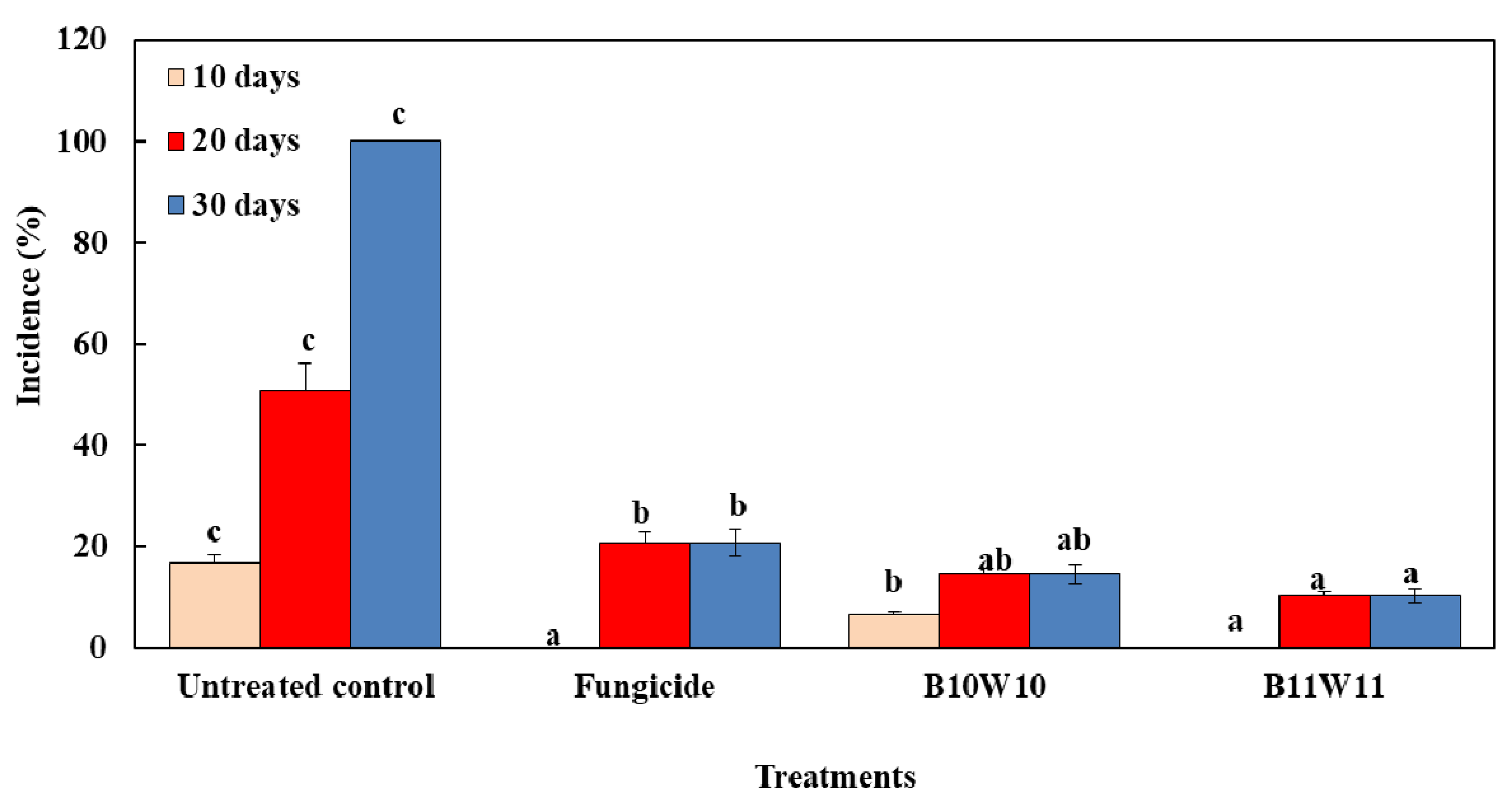
3.8. Semi-Practical Trials of Selected Bacteria

The efficacy of isolates B. amyloliquefaciens B10W10 and Pseudomonas sp. B11W11, displaying the lowest disease severity under in vivo conditions, were tested against brown rot disease in a semi-commercial large-scale trial under conditions used by packinghouses (Figure 5). Their efficacies were compared to a positive control (thiophanate-methyl). Results underlined that all treatments differed significantly from the untreated fruit controls (p < 0.05). Regardless of the treatment, the incidence of brown rot disease was increased gradually with the increase of the incubation period from 10 to 30 days. At 10 days, no infection was detected in apple fruits treated with the bacterial isolate Pseudomonas sp. B11W11 and the fungicide, while treatment with B. amyloliquefaciens B10W10 recorded an incidence of 5%, which was significantly lower than in untreated control (20%). After 20 days of incubation, the incidence of diseased apple fruits treated with Pseudomonas sp. B11W11, B. amyloliquefaciens B10W10, and fungicidal products were 10.37, 14.54 and 20.74%, respectively). However, in untreated control, the disease incidence was much higher, reaching almost 60%. Outstandingly, when untreated apple fruits were completely infected (100%), the incidence of other treatments was the same as that observed at 20 days post-incubation period. Surprisingly, both bacterial isolates offered significantly greater protection against the brown rot disease than that provided by the fungicidal product at 30 days post-incubation period (Figure 5).

4. Discussion

So far, when allowed, chemical fungicides are the principal way to control postharvest diseases. However, to achieve a sustainable lifestyle, the use of bacteria as BCAs for fungal diseases has emerged as one of the most reliable and promising alternatives to synthetic fungicides [9,15,34,35]. In this regard, the present study evaluated the ability of 13 bacteria, isolated from natural soil, to control brown rot diseases on apple fruits.
Our results revealed that most of the tested bacterial isolates displayed a significant inhibition rate (more than 70%) of M. fructigena mycelial growth in dual culture bioassay. These results are in agreement with those previously reported on Monilinia spp. using different species of bacteria [9,36,37]. The results underlined that inhibition rates obtained with VOCs (confrontation in the distance) were significantly lower than those obtained with a direct dual bioassay, which might be explained by the fact that isolates use several modes of action to trigger their antagonistic activities depending on the nature of the pathogen. These results are also consistent with those reported by numerous studies [9,36,38]. Furthermore, cell-free bacterial filtrates demonstrated an inhibitory action both on spore germination and on mycelial growth of M. fructigena under in vitro conditions. Similar results were reported by Zhou et al. [39]. In the same line, Liu et al. [40] have reported an inhibition in vitro conidial germination and mycelium growth of M. fructicola in the presence of cell-free culture filtrates of B. amyloliquefaciens C06, implying that it may contain antifungal compounds.
Bacterial antagonists belonging to Bacillus and Pseudomonas genus are well known to be able to secrete antibiotic substances from the lipopeptide family such as fengycin and bacillomycin, as previously reported in several previous studies [9,28]. Our findings support the hypothesis that the antibiosis was extremely involved in the biocontrol activity of the used bacteria. In accordance, antibiosis has been reported as a major factor for bioactivity of B. subtilis CPA-8 in controlling brown rot disease caused by M. laxa and M. fructicola [37].
Few attempts have been made on the biological control of M. fructigena. Some of them are described by Byrde and Willetts [41], including experiments with Trichoderma viride, which has not been prepared for commercial application. Also, Falconi and Mendgen [42] showed that isolates of Aureobasidium pullulans, Epicoccum nigrum, Sordaria fimicola and Trichoderma polysporum, applied individually or in combination, provided the best protection against M. fructigena. Larena et al. [43] have shown that the antibiotic produced by Epicoccum nigrum and Candida sake are effective against postharvest pathogens infecting apple fruits, including Monilinia spp. In addition, the treatment of apple fruits with Pantoea agglomerans strain EPS125 has significantly reduced the brown rot disease (M. laxa) incidence and the lesion diameter it causes. Generally, our results are in line with our previous findings in which several bacterial isolates having antagonistic activities against fire blight disease were shown effective against both pathogens associated with the brown rot disease; M. laxa and M. fructigena [9]. These bacteria were especially P. agglomerans, Alcaligenes faecalis and B. amyloliquefaciens [9]. In this study, two bacterial isolates, Pseudomonas sp. B11W11, B. amyloliquefaciens B10W10 were highly effective in controlling M. fructigena under both in vitro and in vivo conditions.
Our results indicated highly significant differences between bacterial isolates to control the postharvest brown rot disease caused by M. fructigena. This ability to inhibit the pathogen varies depending on their nature, number and efficacy of the involved mechanisms and metabolites produced by each bacterium as well. Indeed, it is known that bacteria can inhibit the fungal growth via antibiosis by secreting antifungal substances [35,44], VOCs such as HCN or via parasitism by the production of lytic enzymes such as chitinase, glucanase, amylase, protease and cellulase [45]. Our results underlined that the majority of the 13 bacterial isolates had the ability to produce at least one to two lytic enzymes. In most cases, these metabolites are associated with mycelial degradation, vacuolation and release of cytoplasmic content [34,46]. The role of cellulase in plant disease control has been demonstrated in several studies [47,48]. Downer et al. [49] proved the direct inhibitory effect of cellulase on the fungus Phytophthora sp. Richter et al. [48] also mentioned the role of microbial cellulase in the lysis of fungal hyphae walls as a possible biological control mechanism adopted by bacteria. For both efficient bacterial isolates, only Pseudomonas sp. B11W11 was positive for the production of cellulase, confirming findings reported by Sindhu et al. [47]. The latter described that Pseudomonas strains were able to produce appreciable amounts of cellulase in culture-free supernatants and inhibited the mycelial growth of both pathogenic fungi Pythium aphanidermatum and Rhizoctonia solani. In addition, our results are in complete agreement with the findings of Durairaj et al. [50] who reported no cellulase activity in the potent bacterial strains B. stratosphericus FW3 and Pseudomonas aeruginosa D4.
Several authors have studied the ability of antagonistic bacteria to produce amylase [9,51]. They found that more than 50% of the tested bacteria are able to degrade starch in vitro, and most of these isolates are belonging to the Bacillus genus [9]. Interestingly, both potent isolates Pseudomonas sp. B11W11 and B. amyloliquefaciens B10W10 were found able to produce amylase. These results corroborated those of Kim et al. [51], who highlighted that B. subtilis APEC70 produces lytic enzymes. In a recent study, Mardanova et al. [52] reported similar findings for both antagonistic bacteria B. subtilis GM2 and B. subtilis GM5. However, the α-amylase was considered in several studies as an enzyme with a minor role in the control of certain fungi [53].
Our study emphasized a higher score of protease-producing bacteria isolates in comparison with other previous studies, notably those of El-Sayed et al. [54] and Dinesh et al. [23], who found 23.66% and 27% respectively as a percentage of protease-producing bacteria strains. The role that can play this enzyme in the biocontrol of fungal diseases was discussed in several studies [9,44,45]. Indeed, Dunne et al. [55] evidenced that the inhibitory activity of Stenotrophomonas maltophilia W81 against Pythium ultimum is mainly due to proteolytic activity. Similarly, Budi et al. [56] showed that this enzyme induces in vitro inhibition of Fusarium oxysporum and Phytophthora parasitica by cellular interactions. Our study highlights that the potent B. amyloliquefaciens B10W10 was positive for producing the protease, whereas Pseudomonas sp. B11W11 was not. This result is in agreement with Mounia et al. [57] findings. The latter found that most of the Bacillus species can produce the protease suggesting the crucial role that this enzyme certainly played in the antagonism mechanisms through degrading cell walls of the fungal pathogen.
The ability of bacterial isolates to produce HCN, a secondary metabolite of which glycine is the precursor, was investigated. Although cyanide is a general metabolic inhibitor, it is synthesized, excreted and metabolized by some organisms, including bacteria, as a way to prevent predation or competition [58]. The result showed that only two bacterial isolates, B. amyloliquefaciens B4W4 and B. siamensis B14W14, have the ability to produce HCN. In the same context, several studies have been carried out, generally leading to unpromising results in terms of the number of HCN-producing strains among all the tested bacterial strains; Samuel and Muthukkaruppan [59] and Gautam [60] reported 0% and 7.14%. These low percentages may be the result of the absence of genes carrying HCN production in most rhizobacteria or to the absence of an adequate precursor [61]. The role of HCN in controlling plant diseases has been demonstrated by Voisard et al. [62]. In our study, with the exception of B. amyloliquefaciens B4W4 and B. siamensis B14W14, none of the tested bacterial isolates were shown positive for HCN production. This result is consistent with Sethi et al. [39] findings who claimed the absence of HCN production in the majority of bacterial strains of the Bacillus genus. In our previous study, among 18 bacterial isolates, only two bacterial isolates P. agglomerans ACBP1 and B. mojavensis SF16, were capable of producing HCN [9].
In accordance with in vitro bioassay, our in vivo (apple fruits) results underlined that both inhibition rates of mycelial growth of M. fructigena and disease severity of brown rot were significantly affected by bacterial isolates and pathogen pressure. Both bacterial isolates, B. amyloliquefaciens B10W10 and Pseudomonas sp. B11W11 exhibited the highest inhibition rates and the lowest disease severity regardless of the pathogen pressure. These results are similar to those of Mari et al. [63], who found that the antagonistic effect of Aureobasidium pullulans L1 and L8 against three species of Monilinia (M. laxa, M. fructicola and M. fructigena) was higher in vivo bioassays. The two antagonists were selected after preliminary in vitro tests for their ability to reduce mycelial growth. These yeasts have completely inhibited M. laxa, and M. fructicola rots and reduced M. fructigena infections by 70% and 90%, respectively. In our previous study, Lahlali et al. [9] highlighted that B. amyloliquefaciens SF14 and A. faecalis are the most effective bacteria selected among 18 tested bacterial isolates in controlling the brown rot disease.
The effectiveness of both selected bacteria was found to be comparable or even higher to that of fungicidal thiophanate-methyl (1 ppm) and was confirmed in semi-commercial trails. Both selected bacteria have shown an ability to protect apple fruits from M. fructigena attacks through long storage periods (30 days). Finally, the use of these BCAs to control the brown rot disease can be enhanced by their use in combination with other eco-friendly practices. This includes their use, in particular, in mixtures with additives or fungicides at low concentration, as previously demonstrated by previous findings [64,65].

5. Conclusions

In conclusion, results from in vitro dual culture bioassay, VOCs (in distance effect) and cell-free bacterial culture filtrates highlight the significant effect of bacterial isolates in inhibiting the mycelial growth of M. fructigena. This includes particularly the two bacteria B. amyloliquefaciens B10W10 and Pseudomonas sp. B11W11. The results obtained from both in vitro and in vivo trials confirmed the ability of these two bacteria to reduce the brown rot disease severity and incidence on apple fruits. The efficacy of these BCAs depends on their abilities to produce lytic enzymes and lipopeptides, as it was successfully demonstrated herein. However, further studies should be performed before moving into the next phase of marketing of these bacteria as biofungicide products.

Author Contributions

Conceptualization, R.L. and O.M.; methodology, R.L. and O.M.; software, R.L., O.M., N.R., S.E., S.K. and R.M.; validation, R.L., A.T. and E.A.B.; formal analysis, R.L., A.T. and E.A.B.; investigation, R.L.; resources, R.L., A.T. and E.A.B.; data curation, R.L. and O.M.; writing—original draft preparation, R.L.; writing—review and editing, R.L., E.A.B., N.R., S.E., Z.B.; supervision, R.L.; project administration, R.L.; funding acquisition, R.L. and A.T. All authors have read and agreed to the published version of the manuscript.

Funding

This research received no external funding.

Acknowledgments

This work was financially supported by the Phytopathology Unit of the Department of Plant Protection (ENA-Meknes, Morocco). The authors acknowledge the farmers for providing them with apple fruits for the in vivo experiments.

Conflicts of Interest

The authors declare no conflict of interest.

References

  1. Lesik, K. Monilinia species causing fruit brown rot, blossom and twig blight in apple orchards in Belarus. Proc. Latv. Acad. Sci. B Nat. Exact Appl. Sci. 2013, 67, 192–194. [Google Scholar]
  2. Hrustić, J.; Mihajlović, M.; Grahovac, M.; Delibašić, G.; Bulajić, A.; Krstić, B.; Tanovic, B. Genus Monilinia on pome and stone fruit species. Pestic. i Fitomed. 2012, 27, 283–297. [Google Scholar]
  3. De Oliveira Lino, L. Etude de la Variabilité Génétique de la Sensibilité à la Pourriture Brune au cours du Développement du Fruit Chez la Pêche en Lien avec L’évolution des Caractéristiques Physiques et Biochimiques du Fruit. Ph.D. Thesis, Université d’Avignon et des Pays de Vaucluse, Avignon, France, 2016. [Google Scholar]
  4. Dam, D.; Guillaume, R. Identification des Facteurs Intervenant dans le Développement du Monilia sur Fleurs et Rameaux d’Abricotiers et Recherche de Résistances Génétiques, sl: Sn. 2017. Available online: https://www.gisfruits.org/content/download/3802/37487/version/1/file/Rapport+D.+DAM.pdf (accessed on 10 August 2017).
  5. Van Leeuwen, G.C.M. The Brown Rot Fungi of Fruit Crops (Monilinia spp.), with Special Reference to Monilinia Fructigena (Aderh. & Ruhl.) Honey. Ph.D. Thesis, Wageningen University Wageningen, Wageningen, The Netherlands, 2000. [Google Scholar]
  6. Bostock, R.M.; Wilcox, S.M.; Wang, G.; Adaskaveg, J.E. Suppression of Monilinia fructicola cutinase production by peach fruit surface phenolic acids. Physiol. Mol. Plant Pathol. 1999, 54, 37–50. [Google Scholar]
  7. Wade, G.C.; Cruickshank, R.H. Rapid development of resistance of wounds on immature apricot fruit to infection with Monilinia fructicola. J. Phytopathol. 1992, 136, 89–94. [Google Scholar]
  8. Malandrakis, A.A.; Kavroulakis, N.; Chrysikopoulos, C.V. Use of silver nanoparticles to counter fungicide-resistance in Monilinia fructicola. Sci. Total Environ. 2020, 747, 141287. [Google Scholar]
  9. Lahlali, R.; Aksissou, W.; Lyousfi, N.; Ezrari, S.; Blenzar, A.; Tahiri, A.; Ennahli, S.; Hrustić, J.; MacLean, D.; Amiri, S. Biocontrol activity and putative mechanism of Bacillus amyloliquefaciens (SF14 and SP10), Alcaligenes faecalis ACBC1, and Pantoea agglomerans ACBP1 against brown rot disease of fruit. Microb. Pathog. 2020, 139, 103914. [Google Scholar]
  10. Franclet, J. Enquête ANPP-Maladies de conservation des pommes. Arboric. Fruit 1994, 473, 24–26. [Google Scholar]
  11. Lima, G.; De Curtis, F.; Piedimonte, D.; Spina, A.M.; De Cicco, V. Integration of biocontrol yeast and thiabendazole protects stored apples from fungicide sensitive and resistant isolates of Botrytis cinerea. Postharvest Biol. Technol. 2006, 40, 301–307. [Google Scholar]
  12. Wellings, P.W. The role of public policy in biological control: Some global trends. Entomophaga 1996, 41, 435–441. [Google Scholar]
  13. Usall, J.; Casals, C.; Sisquella, M.; Palou, L.; De Cal, A. Alternative technologies to control postharvest diseases of stone fruits. Stewart Postharvest Rev. 2015, 11, 4. [Google Scholar]
  14. Palou, L.; Ali, A.; Fallik, E.; Romanazzi, G. GRAS, plant-and animal-derived compounds as alternatives to conventional fungicides for the control of postharvest diseases of fresh horticultural produce. Postharvest Biol. Technol. 2016, 122, 41–52. [Google Scholar] [CrossRef]
  15. Spadaro, D.; Droby, S. Development of biocontrol products for postharvest diseases of fruit: The importance of elucidating the mechanisms of action of yeast antagonists. Trends Food Sci. Technol. 2016, 47, 39–49. [Google Scholar] [CrossRef]
  16. Calvo, H.; Marco, P.; Blanco, D.; Oria, R.; Venturini, M.E. Potential of a new strain of Bacillus amyloliquefaciens BUZ-14 as a biocontrol agent of postharvest fruit diseases. Food Microbiol. 2017, 63, 101–110. [Google Scholar] [CrossRef] [PubMed] [Green Version]
  17. Zhou, T.; Reeleder, R.D.; Sparace, S.A. Influence of nutrients on growth of Epicoccum nigrum. Can. J. Microbiol. 1996, 42, 647–654. [Google Scholar] [CrossRef]
  18. Madrigal, C.; Melgarejo, P. Mechanisms of action of the antibiotic flavipin on Monilinia laxa and Saccharomyces cerevisiae. Mycol. Res. 1994, 98, 874–878. [Google Scholar] [CrossRef]
  19. De Cal, A.; Larena, I.; Liñán, M.; Torres, R.; Lamarca, N.; Usall, J.; Domenichini, P.; Bellini, A.; De Eribe, X.; Melgarejo, P. Population dynamics of Epicoccum nigrum, a biocontrol agent against brown rot in stone fruit. J. Appl. Microbiol. 2009, 106, 592–605. [Google Scholar] [CrossRef]
  20. Wang, Y.; Yuan, Y.; Liu, B.; Zhang, Z.; Yue, T. Biocontrol activity and patulin-removal effects of Bacillus subtilis, Rhodobacter sphaeroides and Agrobacterium tumefaciens against Penicillium expansum. J. Appl. Microbiol. 2016, 121, 1384–1393. [Google Scholar] [CrossRef] [Green Version]
  21. Hrustić, J.; Delibašić, G.; Stanković, I.; Grahovac, M.; Krstić, B.; Bulajić, A.; Tanović, B. Monilinia spp. causing brown rot of stone fruit in Serbia. Plant Dis. 2015, 99, 709–717. [Google Scholar] [CrossRef] [Green Version]
  22. Llop, P.; Bonaterra, A.; Peñalver, J.; López, M.M. Development of a highly sensitive nested-PCR procedure using a single closed tube for detection of Erwinia amylovora in asymptomatic plant material. Appl. Environ. Microbiol. 2000, 66, 2071–2078. [Google Scholar] [CrossRef] [Green Version]
  23. Dinesh, R.; Anandaraj, M.; Kumar, A.; Bini, Y.K.; Subila, K.P.; Aravind, R. Isolation, characterization, and evaluation of multi-trait plant growth promoting rhizobacteria for their growth promoting and disease suppressing effects on ginger. Microbiol. Res. 2015, 173, 34–43. [Google Scholar] [CrossRef]
  24. Gupta, V.; Peterson, C.B.; Dice, L.T.; Uchiki, T.; Racca, J.; Guo, J.; Xu, Y.; Hettich, R.L.; Zhao, X.; Rothstein, R.; et al. Sml1p is a dimer in solution: Characterization of denaturation and renaturation of recombinant Sml1p. Biochemistry 2004, 43, 8568–8578. [Google Scholar] [CrossRef] [PubMed]
  25. Kumar, R.S.; Ayyadurai, N.; Pandiaraja, P.; Reddy, A.V.; Venkateswarlu, Y.; Prakash, O.; Sakthivel, N. Characterization of antifungal metabolite produced by a new strain Pseudomonas aeruginosa PUPa3 that exhibits broad-spectrum antifungal activity biofertilizing traits. J. Appl. Microbiol. 2005, 98, 145–154. [Google Scholar] [CrossRef] [PubMed]
  26. Cattelan, A.J.; Hartel, P.G.; Fuhrmann, J.J. Screening for plant growth–promoting rhizobacteria to promote early soybean growth. Soil Sci. Soc. Am. J. 1999, 63, 1670–1680. [Google Scholar] [CrossRef]
  27. Bakker, A.W.; Schippers, B. Microbial cyanide production in the rhizosphere in relation to potato yield reduction and Pseudomonas spp-mediated plant growth-stimulation. Soil Biol. Biochem. 1987, 19, 451–457. [Google Scholar] [CrossRef]
  28. Dimkić, I.; Živković, S.; Berić, T.; Ivanović, Ž.; Gavrilović, V.; Stanković, S.; Fira, D. Characterization and evaluation of two Bacillus strains, SS-12.6 and SS-13.1, as potential agents for the control of phytopathogenic bacteria and fungi. Biol. Control 2013, 65, 312–321. [Google Scholar] [CrossRef]
  29. Ramarathnam, R.; Bo, S.; Chen, Y.; Fernando, W.G.D.; Xuewen, G.; De Kievit, T. Molecular and biochemical detection of fengycin-and bacillomycin D-producing Bacillus spp., antagonistic to fungal pathogens of canola and wheat. Can. J. Microbiol. 2007, 53, 901–911. [Google Scholar] [CrossRef] [Green Version]
  30. Hsieh, F.C.; Li, M.C.; Lin, T.C.; Kao, S.S. Rapid detection and characterization of surfactin-producing Bacillus subtilis and closely related species based on PCR. Curr. Microbiol. 2004, 49, 186–191. [Google Scholar] [CrossRef]
  31. Lahlali, R.; Bajii, M.; Jijakli, M. Isolation and evaluation of bacteria and fungi as biological control agents against Rhizoctonia solani. Commun. Agric. Appl. Biol. Sci. 2007, 72, 973–982. [Google Scholar]
  32. Guevara-Avendaño, E.; Carrillo, J.D.; Ndinga-Muniania, C.; Moreno, K.; Méndez-Bravo, A.; Guerrero-Analco, J.A.; Eskalen, A.; Reverchon, F. Antifungal activity of avocado rhizobacteria against Fusarium euwallaceae and Graphium spp., associated with Euwallacea spp. nr. fornicatus, and Phytophthora cinnamomi. Antonie Van Leeuwenhoek 2018, 111, 563–572. [Google Scholar] [CrossRef]
  33. Li, Z.; Guo, B.; Wan, K.; Cong, M.; Huang, H.; Ge, Y. Effects of bacteria-free filtrate from Bacillus megaterium strain L2 on the mycelium growth and spore germination of Alternaria alternata. Biotechnol. Biotechnol. Equip. 2015, 29, 1062–1068. [Google Scholar] [CrossRef] [Green Version]
  34. Barka, E.A.; Vatsa, P.; Sanchez, L.; Gaveau-Vaillant, N.; Jacquard, C.; Klenk, H.-P.; Clément, C.; Ouhdouch, Y.; Van Wezel, G.P. Taxonomy, Physiology, and Natural Products of Actinobacteria. Microbiol. Mol. Biol. Rev. 2016, 80, 1–43. [Google Scholar] [PubMed] [Green Version]
  35. Compant, S.; Duffy, B.; Nowak, J.; Clément, C.; Barka, E.A. Use of plant growth-promoting bacteria for biocontrol of plant diseases: Principles, mechanisms of action, and future prospects. Appl. Environ. Microbiol. 2005, 71, 4951–4959. [Google Scholar] [CrossRef] [PubMed] [Green Version]
  36. Aiello, D.; Restuccia, C.; Stefani, E.; Vitale, A.; Cirvilleri, G. Postharvest biocontrol ability of Pseudomonas synxantha against Monilinia fructicola and Monilinia fructigena on stone fruit. Postharvest Biol. Technol. 2019, 149, 83–89. [Google Scholar] [CrossRef]
  37. Yánez-Mendizábal, V.; Zeriouh, H.; Viñas, I.; Torres, R.; Usall, J.; de Vicente, A.; Pérez-García, A.; Teixidó, N. Biological control of peach brown rot (Monilinia spp.) by Bacillus subtilis CPA-8 is based on production of fengycin-like lipopeptides. Eur. J. Plant Pathol. 2012, 132, 609–619. [Google Scholar] [CrossRef]
  38. Gotor-Vila, A.; Teixidó, N.; Di Francesco, A.; Usall, J.; Ugolini, L.; Torres, R.; Mari, M. Antifungal effect of volatile organic compounds produced by Bacillus amyloliquefaciens CPA-8 against fruit pathogen decays of cherry. Food Microbiol. 2017, 64, 219–225. [Google Scholar] [CrossRef] [PubMed]
  39. Zhou, T.; Schneider, K.E.; Li, X.Z. Development of biocontrol agents from food microbial isolates for controlling post-harvest peach brown rot caused by Monilinia fructicola. Int. J. Food Microbiol. 2008, 126, 180–185. [Google Scholar] [CrossRef] [PubMed]
  40. Liu, J.; Zhou, T.; He, D.; Li, X.Z.; Wu, H.; Liu, W.; Gao, X. Functions of lipopeptides bacillomycin D and fengycin in antagonism of Bacillus amyloliquefaciens C06 towards Monilinia fructicola. J. Mol. Microbiol. Biotechnol. 2011, 20, 43–52. [Google Scholar] [CrossRef]
  41. Willetts, H.J.; Byrde, R.J.W.; Fielding, A.H.; Wong, A.-L. The taxonomy of the brown rot fungi (Monilinia spp.) related to their extracellular cell wall-degrading enzymes. Microbiology 1977, 103, 77–83. [Google Scholar] [CrossRef] [Green Version]
  42. Falconi, C.J.; Mendgen, K. Epiphytic fungi on apple leaves and their value for control of the postharvest pathogens Botrytis cinerea, Monilinia fructigena and Penicillium expansum. J. Plant Dis. Prot. 1994, 101, 38–47. [Google Scholar]
  43. Larena, I.; Linan, M.; Melgarejo, P. Antibiotic production of the biocontrol agents epicoccum nigrum and candida sake. Plant Prot. Sci. 2003, 38, 205–208. [Google Scholar] [CrossRef] [Green Version]
  44. Esmaeel, Q.; Miotto, L.; Rondeau, M.; Leclère, V.; Clément, C.; Jacquard, C.; Sanchez, L.; Ait Barka, E. Paraburkholderia phytofirmans PsJN-Plants Interaction: From Perception to the Induced Mechanisms. Front. Microbiol. 2018, 9, 2093. [Google Scholar] [CrossRef] [PubMed]
  45. Sethi, S.K.; Mukherjee, A.K. Screening of biocontrol potential of indigenous Bacillus spp. isolated from rice rhizosphere against, R. solani, S. oryzae, S. rolfsii and response towards growth of rice. J. Pure Appl. Microbiol. 2018, 12, 41–53. [Google Scholar]
  46. Liu, Y.; Yao, S.; Deng, L.; Ming, J.; Zeng, K. Different mechanisms of action of isolated epiphytic yeasts against Penicillium digitatum and Penicillium italicum on citrus fruit. Postharvest Biol. Technol. 2019, 152, 100–110. [Google Scholar] [CrossRef]
  47. Sindhu, S.S.; Dadarwal, K.R. Chitinolytic and cellulolytic Pseudomonas sp. antagonistic to fungal pathogens enhances nodulation by Mesorhizobium sp. Cicer in chickpea. Microbiol. Res. 2001, 156, 353–358. [Google Scholar] [CrossRef]
  48. Richter, B.S.; Ivors, K.; Shi, W.; Benson, D.M. Cellulase activity as a mechanism for suppression of Phytophthora root rot in mulches. Phytopathology 2011, 101, 223–230. [Google Scholar] [CrossRef] [Green Version]
  49. Downer, A.J.; Menge, J.A.; Pond, E. Effects of cellulytic enzymes on Phytophthora cinnamomi. Phytopathology 2001, 91, 839–846. [Google Scholar] [CrossRef] [Green Version]
  50. Durairaj, K.; Velmurugan, P.; Park, J.H.; Chang, W.S.; Park, Y.J.; Senthilkumar, P.; Choi, K.-M.; Lee, J.H.; Oh, B.-T. Characterization and assessment of two biocontrol bacteria against Pseudomonas syringae wilt in Solanum lycopersicum and its genetic responses. Microbiol. Res. 2018, 206, 43–49. [Google Scholar] [CrossRef]
  51. Kim, Y.S.; Balaraju, K.; Jeon, Y. Effects of rhizobacteria Paenibacillus polymyxa APEC136 and Bacillus subtilis APEC170 on biocontrol of postharvest pathogens of apple fruits. J. Zhejiang Univ. Sci. B 2016, 17, 931–940. [Google Scholar] [CrossRef]
  52. Mardanova, A.M.; Fanisovna Hadieva, G.; Tafkilevich Lutfullin, M.; Valer’evna Khilyas, I.; Farvazovna Minnullina, L.; Gadelevna Gilyazeva, A.; Bogomolnaya, L.M.; Sharipova, M.R. Bacillus subtilis Strains with antifungal activity against the phytopathogenic Fungi. Agric. Sci. 2017, 8, 1–20. [Google Scholar]
  53. Choubane, S.; Cheba, B.A.; Benourrad, A. Screening and phenotypic diversity of amylase producing rhizospheric bacteria from some north african plants. Procedia Technol. 2016, 22, 1197–1204. [Google Scholar] [CrossRef] [Green Version]
  54. El-Sayed, W.S.; Akhkha, A.; El-Naggar, M.Y.; Elbadry, M. In vitro antagonistic activity, plant growth promoting traits and phylogenetic affiliation of rhizobacteria associated with wild plants grown in arid soil. Front. Microbiol. 2014, 5, 651. [Google Scholar] [CrossRef] [PubMed] [Green Version]
  55. Dunne, C.; Crowley, J.J.; Moënne-Loccoz, Y.; Dowling, D.N.; O’Gara, F. Biological control of Pythium ultimum by Stenotrophomonas maltophilia W81 is mediated by an extracellular proteolytic activity. Microbiology 1997, 143, 3921–3931. [Google Scholar] [CrossRef] [Green Version]
  56. Budi, S.W.; van Tuinen, D.; Arnould, C.; Dumas-Gaudot, E.; Gianinazzi-Pearson, V.; Gianinazzi, S. Hydrolytic enzyme activity of Paenibacillus sp. strain B2 and effects of the antagonistic bacterium on cell integrity of two soil-borne pathogenic fungi. Appl. Soil Ecol. 2000, 15, 191–199. [Google Scholar] [CrossRef]
  57. Mounia, Y.-A.; Noreddine, K.C.; Laid, D.; Insaf, B.; Mounira, K.A.; Hlne, C.; Philippe, T. Antifungal activity and bioactive compounds produced by Bacillus mojavensis and Bacillus subtilis. Afr. J. Microbiol. Res. 2014, 8, 476–484. [Google Scholar] [CrossRef] [Green Version]
  58. Cipollone, R.; Ascenzi, P.; Tomao, P.; Imperi, F.; Visca, P. Enzymatic detoxification of cyanide: Clues from Pseudomonas aeruginosa rhodanese. J. Mol. Microbiol. Biotechnol. 2008, 15, 199–211. [Google Scholar] [CrossRef] [PubMed]
  59. Samuel, S.; Muthukkaruppan, S.M. Characterization of plant growth promoting rhizobacteria and fungi associated with rice, mangrove and effluent contaminated soil. Curr Bot. 2011, 2, 22–25. [Google Scholar]
  60. Gautam, S. Characterization of Plant Growth Promoting Rhizobacteria and Evaluation of Their Biocontrol Potential Against Tomato Bacterial Canker. Master’s Thesis, University of Horticulture and Forestry, Nauni Solan, India, 2014; 151p. [Google Scholar]
  61. Laville, J.; Blumer, C.; Von Schroetter, C.; Gaia, V.; Défago, G.; Keel, C.; Haas, D. Characterization of the hcnABC gene cluster encoding hydrogen cyanide synthase and anaerobic regulation by ANR in the strictly aerobic biocontrol agent Pseudomonas fluorescens CHA0. J. Bacteriol. 1998, 180, 3187–3196. [Google Scholar] [CrossRef] [Green Version]
  62. Voisard, C.; Keel, C.; Haas, D.; Dèfago, G. Cyanide production by Pseudomonas fluorescens helps suppress black root rot of tobacco under gnotobiotic conditions. EMBO J. 1989, 8, 351–358. [Google Scholar] [CrossRef]
  63. Mari, M.; Martini, C.; Guidarelli, M.; Neri, F. Postharvest biocontrol of Monilinia laxa, Monilinia fructicola and Monilinia fructigena on stone fruit by two Aureobasidium pullulans strains. Biol. Control 2012, 60, 132–140. [Google Scholar] [CrossRef]
  64. Droby, S.; Wisniewski, M.; El Ghaouth, A.; Wilson, C. Influence of food additives on the control of postharvest rots of apple and peach and efficacy of the yeast-based biocontrol product Aspire. Postharvest Biol. Technol. 2003, 27, 127–135. [Google Scholar] [CrossRef]
  65. Conway, W.S.; Leverentz, B.; Janisiewicz, W.J.; Saftner, R.A.; Camp, M.J. Improving biocontrol using antagonist mixtures with heat and/or sodium bicarbonate to control postharvest decay of apple fruit. Postharvest Biol. Technol. 2005, 36, 235–244. [Google Scholar] [CrossRef]
Figure 1. Capacity of bacterial antagonists to produce lytic enzymes and HCN. Amylase activity assay (A,B: negative and positive responses, respectively). Protease production assay (C,D: negative and positive responses, respectively). HCN production assay (E,F: negative and positive responses, respectively). Cellulase activity assay (G,H: moderate and positive responses, respectively).
Agronomy 10 01814 g001
Figure 2. Inhibition rates of mycelial growth of M. fructigena obtained with volatile organic compounds of bacterial isolates (in dual distance bioassay). Data in the figure represent the mean of two independent trials with 4 replicates. In each incubation period, treatments having the same letter are not significantly different according to the LSD test (p < 0.05).
Agronomy 10 01814 g002
Figure 3. Inhibition rates of mycelial growth of M. fructigena obtained with bacterial cell-free filtrates. Data in the figure represent the mean of two independent trials with 4 replicates. In each incubation period, treatments having the same letter are not significantly different according to the LSD test (p < 0.05).
Agronomy 10 01814 g003
Figure 4. Inhibition rates of spore germination of M. fructigena obtained with cell-free bacterial filtrates (at 100% concentration) after 24 h post-incubation at ambient temperature. Treatments having the same letter are not significantly different according to the LSD test (p < 0.05).
Agronomy 10 01814 g004
Figure 5. Incidence of brown rot disease observed on apple fruits treated by two selected effective bacterial antagonists (Bacillus amyloliquefaciens B10W10 and Pseudomonas sp. B11W11) and infected by M. fructigena at 1 × 10³ spores/mL for different treatments, after incubation at 24 °C for 10, 20 and 30 days. Data in the figure represent the mean of two trials conducted over time with three replicates per treatment (10 apple fruits/replicate). For each incubation period, treatment having the same letter are not significantly different according to the LSD test (p < 0.05).
Agronomy 10 01814 g005
Table 1. Molecular identification of bacterial isolates using subunit 16S rRNA and their potential antagonistic effect on the mycelial growth of the pathogenic fungus Monilinia fructigena under in vitro conditions.
Bacterial IsolatesMolecular IdentificationIn Vitro Inhibition Rate (%) *
Blast Research ResultsAccession5 Days10 Days
B1W1Bacillus amyloliquefaciensMH72753433.16 d80.27 e
B4W4Bacillus amyloliquefaciensMK10615730.01 c78.69 c
B7W7Bacillus amyloliquefaciensMK10666836.92 f82.86 f
B10W10Bacillus amyloliquefaciensMT93966536.37 f82.64 f
B14W14Bacillus siamensisMK12272233.13 d80.11 cd
B2W2Bacillus subtilisMT94064231.92 cd78.95 cd
B3W3Bacillus subtilisMK10603435.81 ef82.11 f
B5W5Bacillus subtilisMK50777735.93 f82.10 f
RP2-12FBacillus subtilisMK51806819.46 b72.70 b
RP2-13FBacillus subtilisMK51836415.50 a43.10 a
B6W6Pseudomonas sp.MT94064435.94 f82.41 f
B11W11Pseudomonas sp.MK10605833.75 de80.62 e
FD1-9FPseudomonas sp.MK51618930.38 c78.57 c
* Data are the main of two independent experiments with four Petri dishes replicates. In the columns, values having the same letter are not statistically different according to the LSD test (p ≤ 0.05).
Table 2. List of specific primers used in this study.
LipopeptidesDesign PrimerPrimer SequenceProduct Length (bp)Annealing T°Reference
BacillomycinBACC1F/BACC1RGAAGGACACGGCAGAGAGTC/
CGCTGATGACTGTTCATGCT
875 bp60 °C[29]
FengycinFEND1F/
FEND1R
TTTGGCAGCAGGAGAAGTT/
GCTGTCCGTTCTGCTTTTTC
964 bp62 °C[29]
IturinITUP1F/
ITUP2R
AGCTTAGGGAACAATTGTCATCGGGGCTTC/
TCAGATAGGCCGCCATATCGGAATGATTCG
2 kb45 °C[28]
SurfactinP17/
P18
ATGAAGATTTACGGAATTTA/
TTATAAAAGCTCTTCGTACG
675 bp53 °C[30]
Table 3. Capacity of bacterial antagonists to produce lytic enzymes involved in the biocontrol mechanisms such as amylase, protease, cellulase and hydrocyanic acid (HCN) production.
Bacterial StrainsAmylaseProteaseCellulaseHCN
Bacillus amyloliquefaciens B1W11.02 b1.26 bc1.02 b
Bacillus amyloliquefaciens B4W41.37 f2.44 f1.05 b+
Bacillus amyloliquefaciens B7W71.31 ef1.17 b0 a
Bacillus amyloliquefaciens B10W101.18 d1.70 d0 a
Bacillus siamensis B14W141.15 cd1.16 b1.21 def+
Bacillus subtilis B2W21.1 bc1.98 e1.23 ef
Bacillus subtilis B3W31.06 b1.12 b1.18 cde
Bacillus subtilis B5W51.1 bc1.14 b1.38 g
Bacillus subtilis RP2-12F1.06 b1.95 e1.29 fg
Bacillus subtilis RP2-13F1.04 b1.26 bc1.11 bcd
Pseudomonas sp. B6W61.29 e1.68 d0 a
Pseudomonas sp. B11W111.17 cd0 a1.07 bc
Pseudomonas sp. FD1-9F0 a0 a1.30 fg
AI: amylolytic index, PI: proteolytic index, CI: cellulosic index were calculated as described by Lahlali et al. [9]; (+): positive reaction; (−): negative reaction. (+): positive reaction; (−): negative reaction. In each column, treatments having the same letter are not significantly different according to the LSD test (p ≤ 0.05).
Table 4. Detection of genes involved in the production of lipopeptides in bacterial antagonists.
StrainBacillomycinFengycinIturinSurfactin
Bacillus amyloliquefaciens B1W1++
Bacillus amyloliquefaciens B4W4++
Bacillus amyloliquefaciens B7W7+
Bacillus amyloliquefaciens B10W10+
Bacillus siamensis B14W14++
Bacillus subtilis B2W2++
Bacillus subtilis B3W3++
Bacillus subtilis B5W5+
Bacillus subtilis RP2-12F+++
Bacillus subtilis RP2-13F++
Pseudomonas sp. B6W6++
Pseudomonas sp. B11W11++
Pseudomonas sp. FD1-9F+++
(+): positive reaction; (−): negative reaction.
Table 5. Disease severity (%) of brown rot disease (Monilinia fructigena) as affected by bacterial isolates and pathogen pressure 5 and 10 days post-incubation periods at 24 °C.
Bacterial IsolatesPathogen Concentration
1 × 10³ spores/mL1 × 106 spores/mL
5 Days10 Days5 Days10 Days
Bacillus amyloliquefaciens B1W161.44 cx37.52 abc71.81 g76.84 c
Bacillus amyloliquefaciens B4W459.91 bc26.22 ab70.43 g73.76 c
Bacillus amyloliquefaciens B7W764.09 c41.75 abcd50.22 de66.84 bc
Bacillus amyloliquefaciens B10W100.00 a21.73 a0.00 a43.59 a
Bacillus siamensis B14W1468.76 c43.85 abcd64.33 fg66.84 bc
Bacillus subtilis B2W260.34 c35.25 abc44.01 cd71.52 bc
Bacillus subtilis B3W363.09 c49.85 bcd69.08 g76.16 c
Bacillus subtilis B5W577.91 c51.23 cd46.93 cde69.78 bc
Bacillus subtilis RP2-12F63.73 c51.83 cd69.81 g70.53 bc
Bacillus subtilis RP2-13F54.19 bc65.34 e66.67 fg70.15 bc
Pseudomonas sp. B6W664.79 c59.37 d69.64 g71.79 bc
Pseudomonas sp. B11W110.00 a28.99 abc0.00 a51.09 ab
Pseudomonas sp. FD1-9F41.01 abc24.54 a36.84 c69.23 bc
Fungicide (TM) y9.24 ab34.95 abc24.86 b40.23 a
x data are the average of two replicated trials over time with four apples per treatment. In each column, treatments with the same letter are not significantly different according to the LSD test (p < 0.05). y thiophanate-methyl (applied at 1 ppm).
Publisher’s Note: MDPI stays neutral with regard to jurisdictional claims in published maps and institutional affiliations.

