Review

Chitosan: Gels and Interfacial Properties

by
Julie Nilsen-Nygaard
*,
Sabina P. Strand
,
Kjell M. Vårum
,
Kurt I. Draget
and
Catherine T. Nordgård
Norwegian Biopolymer Laboratory (NOBIPOL), Department of Biotechnology, Norwegian University of Science and Technology (NTNU), 7491 Trondheim, Norway
*
Author to whom correspondence should be addressed.
Present address: NTNU Technology Transfer AS (TTO), Sem Sælands vei 14, 7491 Trondheim, Norway
Polymers 2015, 7(3), 552-579; https://doi.org/10.3390/polym7030552
Submission received: 19 December 2014 / Revised: 19 February 2015 / Accepted: 5 March 2015 / Published: 13 March 2015
(This article belongs to the Collection Polysaccharides)

Abstract

:
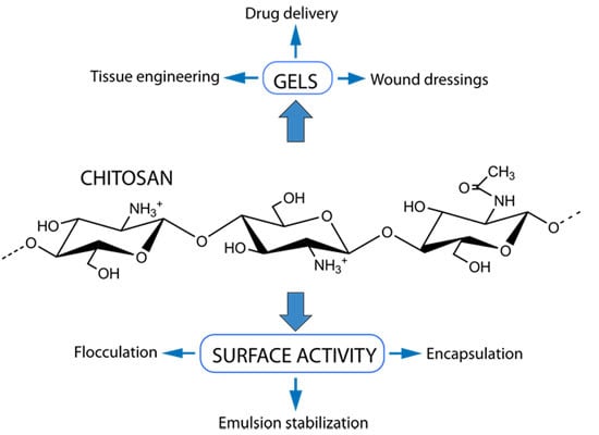
Chitosan is a unique biopolymer in the respect that it is abundant, cationic, low-toxic, non-immunogenic and biodegradable. The relative occurrence of the two monomeric building units (N-acetyl-glucosamine and d-glucosamine) is crucial to whether chitosan is predominantly an ampholyte or predominantly a polyelectrolyte at acidic pH-values. The chemical composition is not only crucial to its surface activity properties, but also to whether and why chitosan can undergo a sol–gel transition. This review gives an overview of chitosan hydrogels and their biomedical applications, e.g., in tissue engineering and drug delivery, as well as the chitosan’s surface activity and its role in emulsion formation, stabilization and destabilization. Previously unpublished original data where chitosan acts as an emulsifier and flocculant are presented and discussed, showing that highly-acetylated chitosans can act both as an emulsifier and as a flocculant.

Graphical Abstract

1. Introduction

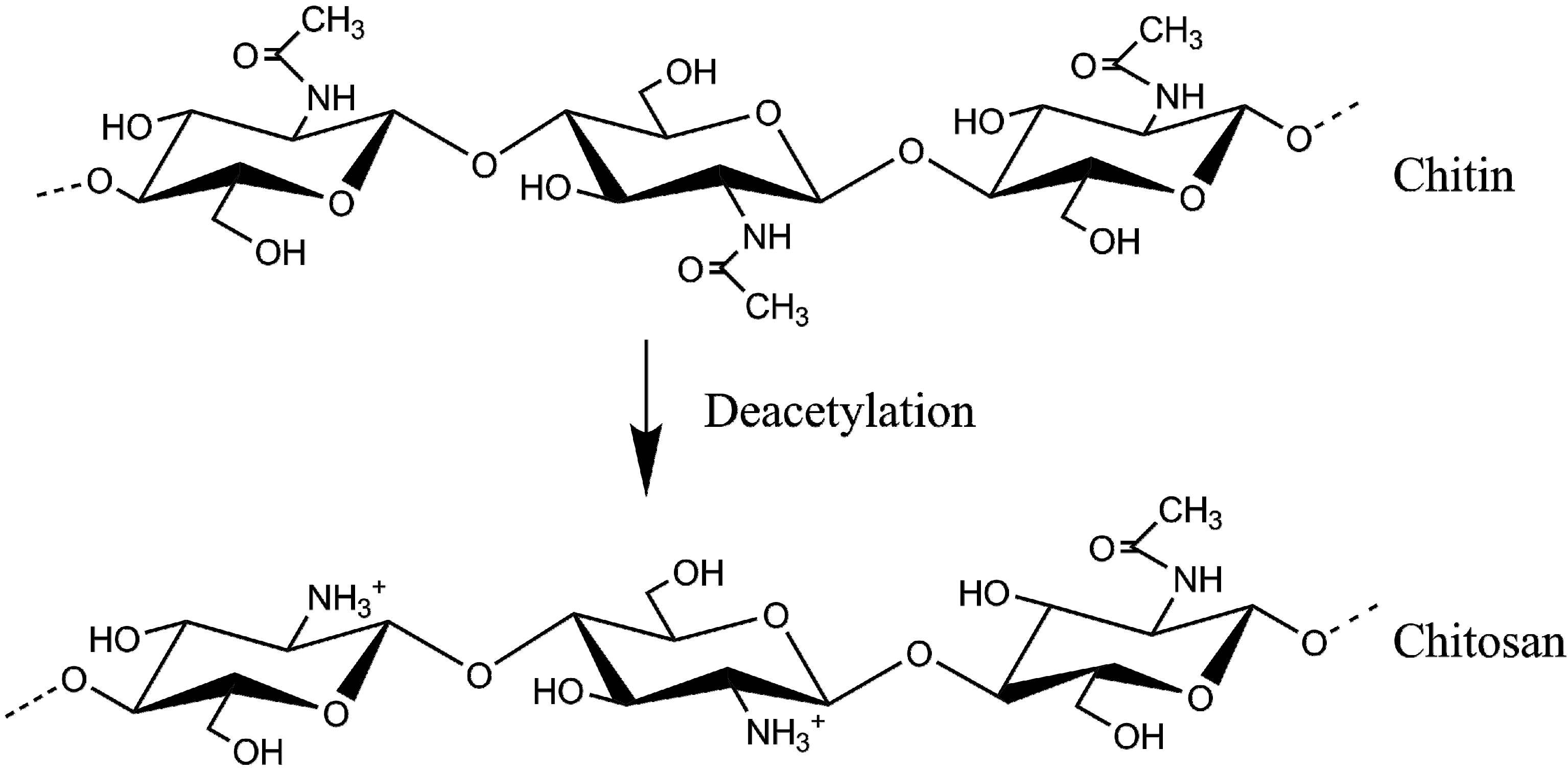
Chitosan is a naturally occurring water-soluble polycation which is today manufactured by alkaline de-acetylation of chitin, and is the only abundant polycationic biopolymer, with a good biocompatibility and biodegradability [1]. Chitosan is a linear polysaccharide of β–(1-4) linked 2-acetamide-2-deoxy-d-glucose (the neutral sugar unit GlcNAc, or A-unit) and 2-amino-2-deoxy-d-glucose (the positively charged sugar unit GlcN, or D-unit), and the biopolymer can be prepared with a wide range of fractions of A-units (FA) and chain lengths (Figure 1).
Figure 1. Structure of chitin and chitosan.
Polymers 07 00552 g001
The FA-value in chitosan can be varied from ca. 0.7 (70% acetylated) to 0 (0% acetylated, meaning that all units are charged) [2]. Thus, the chitosan molecule can be considered to be predominantly an ampholyte (enriched in A-units) or predominantly a polyelectrolyte (enriched in D-units), and chitosan can in this regard be considered as a family of polysaccharides. The amine-groups of D-units (Figure 1) has a rather low pKa-value of 6.5 [3], meaning that the amine-groups are predominantly positively charged at pH-values below 6.5. This also influences the water-solubility of generic chitosans as a function of pH (Vårum et al., 1994), and is a property that has been used to deposit chitosan on electrodes and thereby connect biology to electronics [4]. The possibility to widely vary the chemical composition of chitosan makes the biopolymer rather special and will also determine not only its surface activity properties but also the kinetics of its biodegradation [5]. The presence of a primary amino group of D-units makes chitosan a tempting polymer for chemical modification by covalent attachment of various chemical groups [6] and a variety of different chemical derivatives have indeed been manufactured. In the present paper, we will, however, focus on non-modified chitosan, i.e., chitin that has been 30%–100% deacetylated. Furthermore, we consider it too ambitious to cover all aspects of chitosan applications, and we have therefore chosen to focus on a few established and potential applications of chitosan.

2. Chitosan Hydrogels

It has been said that a gel is more easily observed than described. There seems, however, to be a consensus as to which properties a “true” gel should exhibit: it should be able to capture a large fraction of solvent (water in the case of hydrogels), the dynamic moduli (G' and G'') should be frequency independent over a large range in the plateau zone and the relative difference between the two moduli should preferably be at least two orders of magnitude in favor of the elastic modulus G' [7].
As chitosan is the only abundant low-toxic, biodegradable, and (at acidic pH-values) positively charged biopolymer, it has become a popular device or excipient in many applications involving biological systems. These uses range from compacting nucleic acids for transfection [8,9], acting as scaffolds for cellular and tissue growth [10,11,12] and as a component in drug delivery systems [13]. Such uses require chitosan to undergo a dis-order/order transition, which also is a pre-requisite for gel formation.
As a general approach it should be kept in mind that chitosan is not a common structural polymer in nature. This property belongs to the parent molecule chitin, which is found in the outer skeletons of, e.g., crustaceans and insects. One possible exception to this is chitosan found in the cell wall of some fungi, such as Mucor species [14]. A natural extension of this approach is that, in contrast to other biopolymers like alginates, carrageenans and pectins, there are no “ideas” to gain from nature as how to gel chitosans. Inventive steps will have to be taken in order to obtain a transition from sol to gel of a chitosan solution.
A lot of different covalent crosslinking agents have been proposed [15] but not all of them are biocompatible. It is, however, also possible to play around with the physic–chemical properties of the chitosan molecule itself, as well as in combinations with poly-anions, to obtain non-covalent, physical gels.

2.1. Chitosan Physical Gels

The solubility/insolubility of chitosan when the pH of the solution is changed is determined by the pKa value of the D-units of approximately 6.5, the chemical composition (i.e., FA-value), and the chain length. A polymeric chitosan with approximately equal amounts of A- and D-units will not precipitate even upon increasing the pH values well above the pKa-value [16]. One way to gel a chitosan is to reacetylate the polymer with acetic anhydride thereby shifting the monomer composition in favour of acetylated units [17]. A sol–gel transition occurs through additional intermolecular interactions of a hydrophobic nature, leading to the formation of a turbid chitin gel.
Another way to gel chitosans physically is the use of β-glycerol phosphate combined with temperature [18]. Such combinations gel at around physiological temperature and provide transparent products that can potentially act as injectable pharmaceutical depots (Figure 2). It has been suggested that the driving force for this gelation is a temperature dependent transfer of protons from chitosan to glycerol phosphate, thereby reducing electrostatic repulsion leading to chitosan aggregation [19]. This system may thus be looked upon as having a lower critical solution temperature (LCST).
Figure 2. Changes in laser transmittance (LT, solid line) and pH (dotted line) during a 1 °C/min temperature ramp of a chitosan–glycerol phosphate solution. pH exhibited a decrease at the same temperature as where the chitosan precipitated (Tp). Reprinted with permission from American Chemical Society 2008 [19].
Polymers 07 00552 g002
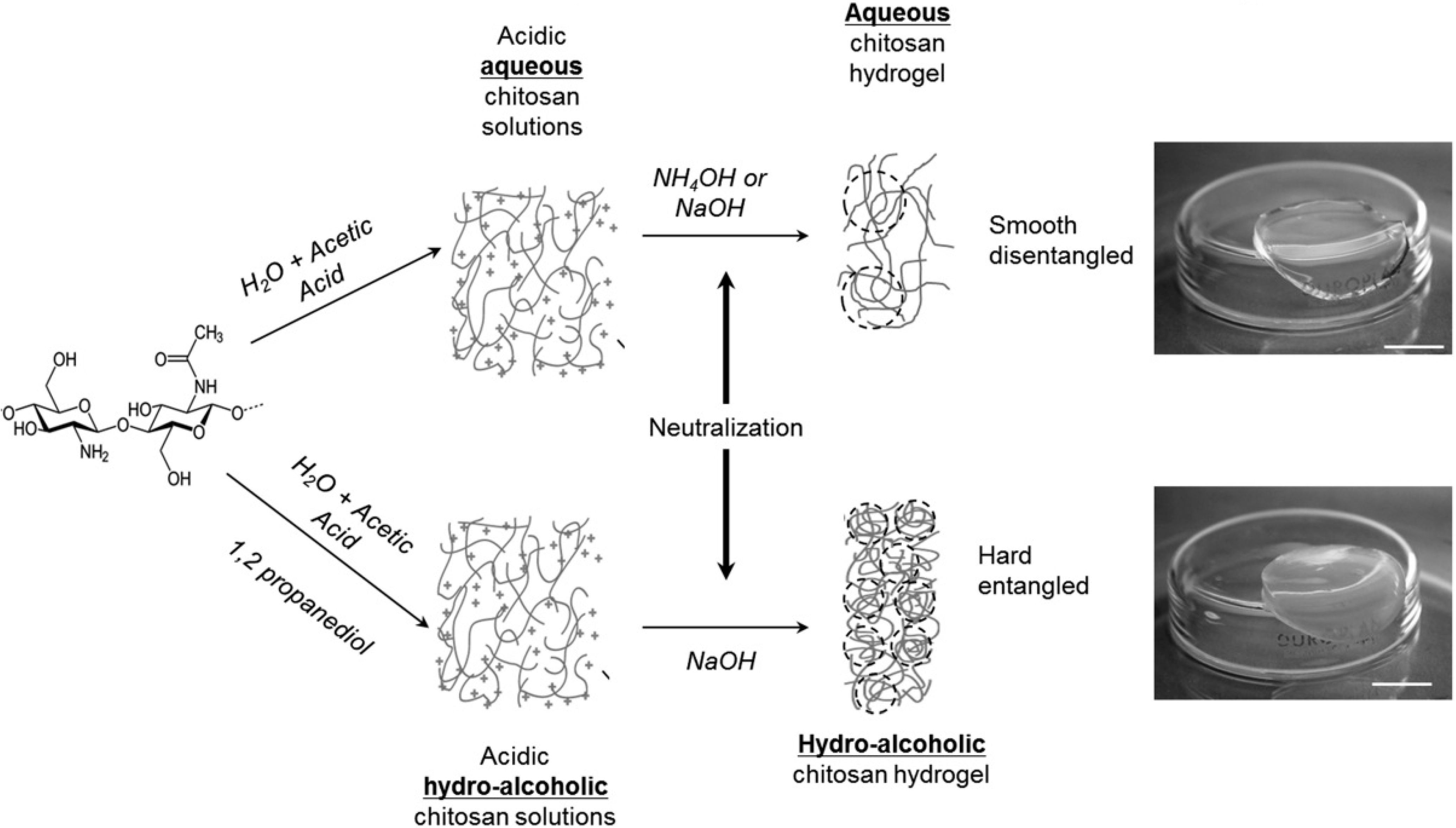
Chitosan can also be gelled without any additions by a direct but controlled exposure to alkali under well-defined conditions [20,21]. Depending on the nature of the solvent and the chitosan’s fraction of acetylated units, different gel morphologies can be obtained with different biological response. In a study by Rami et al., it was shown that soft and easily degraded gels manufactured from a highly acetylated chitosan were not suitable for culture of human mesenchymal stem cells, whereas a highly deacetylated chitosan following the same processing route gave rise to more solid gels with improved cell adhesion (Figure 3) [22].
There are also a large number of ionic cross-linkers and complexing agents that can be used to make physical chitosan gels. Small anionic molecules like phosphates and citrates are able to ionically crosslink chitosans depending on pH and degree of acetylation [23]. Chitosans can also be cross-linked by transition metal ions like molybdate [24]. Differing from citrate and phosphates, where only electrostatic charge plays a role, such transition metal ions work via a more intricate mechanism by forming coordinate-covalent bonds [25].

Polyelectrolyte Complexation

Gelling by complexation represents a borderline case as this mechanism is usually associated with associative phase separation where the main driving force is an entropy gain following release of condensed counter-ions from polyelectrolytes. A direct effect of such processes is a depletion of water from the resulting complexes, and as such, these complexes do not fulfill the previously given definition of a hydrogel; i.e., that a gel should be able to capture large amounts of water. Polyelectrolyte complexation can, however, be controlled by adjusting parameters like relative polymer concentration and molecular weight.
Figure 3. Manufacturing process to obtain chitosan aqueous hydrogels and hydro-alcoholic hydrogel and photographs of the resulting gel products (scale bar = 5 mm). Reprinted with permission from American Chemical Society 2014 [22].
Polymers 07 00552 g003
Chitosan has been shown to form complexes with a large number of different poly-anions, such as DNA, alginates, pectins, xanthan, glucosaminoglycans, carboxymethyl cellulose and gelatin as well as synthetic polymers [26]. The stability of such complexes depends largely on parameters like polymer charge density, molecular weight, net charge ratios, properties of the solvent, ionic strength, pH and temperature [27,28].
One way to avoid precipitation after complex coacervation is to use oligomers as complexing agent instead of polymers. Khong et al. (2013) showed that alginate oligomer mixtures (DPn of 11) were able to crosslink chitosan, and chitosan oligomer mixtures (DPn of 5) to crosslink alginates, to induce transparent gel formation. Furthermore, the importance of identical distance between charges was also proved as mannuronate oligomers with diequatorially linked sugar units (β-1,4 glycosidic linkages as in chitosan) were considerably more potent as cross-linkers compared to guluronate oligomers with diaxially linked sugar units (α-1,4 glyosidic linkages) [29].

2.2. Chitosan Covalently Crosslinked Gels

Covalent crosslinking of chitosan opens up for more controlled gel stability, degradation and porosity compared to physical chitosan gels. It has, however, been observed that additional physical interactions may occur following covalent crosslinking such as additional hydrophobic interactions in highly acetylated chitosan gels crosslinked with glutaraldehyde [30].
The primary amino group of the D-units of the chitosan molecule is the most attractive target for covalent crosslinking and more robust gels [11]. The reaction between a primary amino group and an aldehyde results in Schiff’s base formation, which in turn can be reduced and stabilized a suitable reducing agent [31]. Multiple small bifunctional molecular crosslinkers have been applied in this context, such as glutaraldehyde [32] and diethyl squarate [33]. The main challenge by using such chemical cross-linkers is the removal of e.g., excess toxic glutaraldehyde. Diethyl squarate, on the other hand, is more biocompatible as this reaction only leaves traces of ethanol.
Polymeric, chemical crosslinking of chitosan has been applied using several polysaccharides, such as galactomannan, maltodextrins and methylcellulose [34], xyloglucan [35] and scleroglucan [36,37]. These polysaccharides were all oxidized using periodate, introducing aldehyde functions capable to form Schiff-bases with chitosan’s primary amino groups.
A more biocompatible, and considerably less toxic as compared to glutaraldehyde, covalent crosslinker is genipin; a chemical entity isolated from natural sources [38]. Genipin reacts and crosslinks polymers carrying primary amino groups. The main drawbacks of genipin, which is a reactive chemical, are that it may potentially react with and modify encapsulated drugs and that it is able to self-polymerize.

2.3. Biomedical Applications of Chitosan Gels

The abundance of chitin in nature, and thereby the potential for large scale chitosan production, has given rise to multiple industrial and biotechnological applications for chitosan. Here, we will present an overview of biomedical chitosan applications where the polymer properties of chitosan, including their gel formation capacity, are central to functionality. In these applications, the biocompatibility of chitosan is central, as chitosan is low-toxic, non-immunogenic and potentially biodegradable in vivo, and as such an excellent choice for biomedical applications.

2.3.1. Tissue Engineering

Tissue engineering can be described as the use of a combination of cells, scaffolding materials and suitable biochemical factors to replace non-functioning or poorly functioning tissues within the body. Biopolymer hydrogels are an obvious choice for the scaffolding material as they provide the necessary aqueous environment for cell growth, and are able to provide mechanical properties similar to native tissues [10,22]. Chitosan based hydrogels are of interest in tissue engineering applications, as they have good biocompatibility, are biodegradable in vivo, are low-toxic and non-immunogenic, and have tunable gel properties [39]. Additionally, and rather uniquely, chitosan gels are cationic and are able to interact with structural molecules such as glycosaminoglycans and glycoproteins present in the extracellular matrix [22,39], or indeed with other anionic biopolymers to form mixed biopolymer scaffolds to optimize either the mechanical or biological properties of the biomaterial [40]. Most chitosan scaffolds for tissue engineering are of this mixed type [10]. By using poly-anions such as alginate, hyaluronic acid, collagen, heparin and xanthan, gel formation can be taken advantage of through polyelectrolyte complexation, which avoids the use of any toxic reaction agents that are contraindicated in tissue engineering applications [40]. Another gelation mechanism that avoids the use of (potentially) toxic additives is ionic gelation through controlled exposure to alkali. Depending on the structure of the chitosan (FA) and the nature of the solvent, pure chitosan gels with differing suitability for tissue engineering applications can be obtained [22].

2.3.2. Drug Delivery

Hydrogels are also of interest as drug delivery systems to improve bioavailability or as implantable drug depots, which allow high concentrations of drug to be delivered directly to diseased tissue, overcoming the issue of poor bioavailability and reducing off target effects. In this case, hydrogels offer advantages over solid drug depot implants since they possess physical properties somewhat similar to living tissues, which minimizes irritation to the surrounding tissue after implantation [15]. As in tissue engineering, the biocompatibility of chitosan makes it an attractive choice as a matrix polymer in this instance [40]. Both chemical and physical chitosan gels have been investigated for controlled localized delivery of drugs [13,41].
Chemically cross-linked gels of chitosan offer excellent mechanical properties and good control of network pore size, which enables easy optimization of gel properties for the drug delivery application, with the obvious prerequisite that toxic cross-linkers or reagents are avoided or effectively removed after hydrogel formation [15]. Physical gels have the advantage of avoiding potentially toxic cross-linkers or reagents but are less robust in vivo than chemical gels, having a lifetime which ranges from days to months [15]. Physical gels offer a good solution for short term drug release applications but their relatively poor mechanical strength and uncontrolled dissolution in vivo may limit more widespread use [5]. One particularly interesting application is the use of the thermoreversible gelling of chitosan with β-glycerol phosphate which allows the injection of a liquid polymer-drug solution that gels in vivo when implanted and reaches body temperature thereby passing through its lower critical solution temperature [41]. Another in situ gelling system is based on chitosan’s pH dependent solubility, where a mixture of chitosan and polyacrylic acid is liquid at pH 6.0 but gels rapidly when exposed to physiological pH 7.4 [42]. Such injectable hydrogels avoid the need for surgical gel implantation and can be advantageous for treating irregularly shaped tissue sites within the body [15,43].
Drug loading is central to the function of hydrogel drug depots, and there are three primary mechanisms by which this may be achieved. These are permeation, whereby small drug molecules are allowed to diffuse into the gel after it is formed; entrapment, whereby drugs are mixed into the polymer solution before it is gelled and the drug is incorporated into the hydrogel; and covalent bonding whereby the drug is covalently attached to the chitosan before gel formation [15]. All three methods can be utilized with both physical and chemical chitosan gels although they are not equally suitable for all types of drugs. The entrapment method has broad applicability for small molecules, peptides, proteins, and micro or nanospheres, while the use of covalent crosslinking is limited to small molecules, peptides and proteins, which can easily be attached to the polymer. Permeation has an even more restricted field of use, as it is suitable only for small molecules [15]. Chitosan, which contains both amino and hydroxyl groups that both can be utilized for covalent attachment of the drug, is attractive as a matrix for covalent loading. The structure of the chitosan molecule with both cationic amino-groups (D-units) and hydrophobic acetyl groups (A-units) makes it a good choice for loading both anionic and hydrophobic drugs through entrapment and permeation [15,42].
Once inside the body, the drug release properties are of vital importance, where the release profile depends on both the delivery route/site and the lifetime of the treatment. Chitosan has a number of properties which are of relevance to drug release. The pH sensitivity of chitosan makes it possible to engineer pH responsive hydrogels (for example, polyelectrolyte complex gels) which swell and release their load at specific locations in the gastrointestinal tract dependent on the local pH. The cationic nature of chitosan makes it mucoadhesive by promoting interactions with the negatively charged mucins of the mucosa and this may provide benefits for sustained local delivery of drugs at mucosal surfaces [42,44] and chitosans have also shown promise as penetration enhancers related to their cationic nature [42]. For chitosan based hydrogel implants the lysozyme susceptibility of chitosan can be utilized both to trigger release of the drug and to degrade the implant over time [15,42]. The ability to induce in situ gelation is highly useful for ophthalmic drug delivery, where liquid dosage forms have a very short residence time due to clearance with the tear fluid [15].
It should be noted that whilst complex coacervates with low water content do not fulfill the definition of a hydrogel, chitosan containing complex coacervates may be used for both gene [8] and drug [45] delivery. These applications share a number of the benefits resulting from the molecular nature of chitosan such as mucoadhesion, penetration enhancement and pH sensitive release. Cationic chitosan is particularly useful for delivery applications for anionic drugs [42].
Chitosan also has antimicrobial properties [46], which is potentially very interesting in relation to food applications. The antimicrobial effect can also [47] be potentially useful in drug delivery or implant applications [48] and in wound dressings [49]. Wound dressing applications for chitosan are rather promising as chitosan is bioadhesive, has anti-microbial and anti-fungal properties, allows oxygen permeation and acts as a hemostat [49,50]. These properties are all attributed to the cationic nature of chitosan. The hemostatic effects of chitosan are relevant for acute wounds, and for chronic wounds the ability of chitosan to combine good dressing properties with a suitable matrix to promote cell growth [10] (as seen in tissue engineering applications) as well as its biodegradability are all key properties.

3. Interfacial Properties of Chitosan

3.1. Biopolymer Emulsifiers

Emulsifiers are surface active molecules that, as a result of their amphiphilic nature, have the potential to adsorb at an oil–water interface and decrease the interfacial tension. During homogenization the formation of a kinetically stable oil-in-water emulsion requires the rapid adsorption of emulsifier molecules to the small dispersed oil droplets in order to prevent the droplets from coalescing [51]. The amphiphilic properties of biopolymer emulsifiers are fundamental for their emulsifying capacity [52].
Among the biopolymers, proteins in particular are known for their good emulsifying properties. Important protein biopolymer emulsifiers are soy protein and the different milk proteins (caseins and whey protein) [53]. When it comes to polysaccharide emulsifiers, gum arabic, modified starches, modified cellulose, some pectins and galactomannans are commonly used [52]. In the case of flexible biopolymers such as polysaccharides, adsorption to an i.e., an oil–water interface will only be favorable if the loss of conformational entropy is compensated for by a sufficient interaction between hydrophobic groups on the polysaccharide and the oil phase. The hydrophobic non-polar regions will associate with the oil, while the more hydrophilic segments protrude into the aqueous phase in loops and end segments creating a steric barrier [54]. As most water-soluble polysaccharides carry charge under certain conditions, the emulsion stabilizing mechanisms are both steric and electrostatic (electrosteric stabilization). Upon charge screening, the steric contribution will be dominant [53]. For many of the polysaccharides with emulsifying properties it has been suggested that the origin for the surface active properties lies in the presence of protein, either as a contaminant or as a covalently linked peptide–protein moiety to the polysaccharide [52]. Gum arabic is such a naturally occurring polysaccharide–peptide complex [53].
Chitosan is usually not mentioned in the group of polysaccharides with good emulsification properties, but several studies have shown that chitosan can in fact act as the sole emulsifier and produce emulsions of acceptable stability [55,56,57,58,59,60].

3.2. Surface Activity of Chitosan

Contemplating the molecular structure of chitosan in light of its amphiphilic nature, the D-units of the molecule are strongly hydrophilic while A-units are responsible for the hydrophobic properties of the polysaccharide giving rise to surface activity. The non-polar acetyl groups will associate with the oil phase, while the segments containing polar and (at acidic pH-values) charged D-units will extend into the aqueous phase. One possible reason that can explain the low research activity on the emulsifying properties and surface activity of chitosans is the low surface activity of the commercially available (generic) chitosans, typically with a FA between 0.1 and 0.2. Additionally, current regulatory issues could be a limiting factor in the use of chitosan in food and pharmaceuticals. The regulatory issues are discussed in more detail in Section 4.
The surface (liquid–vapour, i.e., water–air) and/or interfacial (liquid–liquid, i.e., oil-water) tension of chitosan solutions of varying Mw, FA and concentration have been reported in some studies [60,61,62,63,64,65]. Payet et al. studied the interfacial properties of chitosan, both at the air–water surface as well as at the oil–water interface. They observed that the applied chitosan (FA 0.2) had no surface activity at concentrations below 0.1%. At higher concentrations, a gradual lowering of the surface tension was measured indicating slow adsorption kinetics of chitosan at the oil–water interface and a gradual arrangement of the hydrophilic and more hydrophobic parts of the chitosan chains in the interface. However, the overall decrease in surface tension was small compared to that of more commonly applied surface active molecules [60]. These findings are supported by results in a study from 1999. The group measured the surface tension of low-concentration chitosan solutions at pH 4.6 and concluded that no chitosan adsorbed to the interface under the given conditions. However, a slight decrease in interfacial tension was observed at the highest measured concentration. Based on these data, the authors concluded that chitosan does not exhibit surface active properties [64]. Mai-Ngam, on the other hand, reported on classic surfactant behavior and high surface activity of low-molecular weight chitosan hydrochloride (Mw 5000 g/mol, FA 0.035) based on surface tension measurements. Further, it was suggested that the low Mw chitosan formed a monolayer at the air–water interface [62]. The results are in agreement with the ones reported on by Calero et al., through combined surface tension and surface viscoelasticity measurements the group found that chitosan formed a viscoelastic film at the oil–water interface and significantly reduced the surface tension [66]. Pepić and co-workers examined the surface tension of chitosan in water and in acetate buffer. Under addition of chitosan to the air–water interface the surface tension remained constant and close to that of pure water, indicating that chitosan was excluded from the interface. However, in acetate buffer at pH 6 and 6.5 and ionic strengths of 0.1 M and 0.5 M, respectively, a small decrease in the surface tension was documented. The increased adsorption of chitosan to the interface under these conditions can be explained by stronger hydrophobic interactions coming into play at higher ionic strengths when charges are screened. Also, by increasing the pH in the aqueous phase deprotonation of the amino groups in chitosan will progress and this will enhance the hydrophobic interactions, inter- and intra-molecularly as well as with an interface [61]. Geng et al. studied the influence of the acetic acid concentration on the surface tension of chitosan solutions. They found that the surface tension was depressed as the acetic acid concentration was increased, while the viscosity of the solution was unchanged [65]. The authors do not elaborate on their thoughts regarding the underlying mechanisms behind these results, but increased association between hydrophobic acetyl groups as a result of electrostatic screening of the charged amino groups at high ionic strength is a plausible explanation. However, the effect of acetic acid as such cannot be ruled out.
To summarize, the reports in general conclude a low surface activity for chitosan. However, the majority of the studies have focused on very low concentrations. Differences in the reported results can be attributed to a broad distribution in molecular weight of the analyzed chitosans and also possibly in the distributions of the different monomers in the molecules. No clear-cut effect of FA on the surface activity of chitosan can be drawn from the available literature as the chitosans applied for measurements in the abovementioned studies all have a fraction of acetylation in the narrow range of 0.1–0.2, typical for the commercially available chitosan.

3.3. Emulsification Properties of Chitosan

One of the first examples of the use of chitosan as emulsifier is presented in the paper by Schulz and co-workers from 1997. The group prepared W/O/W (water-in-oil-in-water) double emulsions with sunflower oil at a weight fraction of 0.2 and chitosan as the sole emulsifier. The measured FA of the applied chitosan was 0.11, but the authors speculated that the distribution of FA in the sample was broad with a high fraction of low FA chitosan. The chitosan can therefore behave as a mixture of surfactants of varying HLB (hydrophilic-lipophilic balance) values, where the chitosans of low FA (mainly hydrophilic) can stabilize the oil–water emulsion droplets while the more hydrophobic chains of higher FA are assumed to stabilize the water-in-oil inner droplets [56]. In 1999, the same group elaborated on the emulsification capacity of chitosan. Droplet size measurements revealed a unimodal distribution at the highest (0.25) and lowest (0.05) FA-value whilst polymodal distributions were observed for examined chitosans of intermediate FA [57]. A unimodal droplet distribution reflects a more stable emulsion, as the driving force for Ostwald ripening is reduced [53]. In the paper, the authors do not provide an explanation for these results, but one can speculate that a plausible explanation lies in variations in the range of FA in the samples. In 2002, the same group followed up this research with a study focusing on the effect of FA on the emulsification capacity of chitosan. The W/O/W double emulsions were prepared with chitosan of FA between 0.06 and 0.27, however, the results did not show a clear correlation between the FA and the emulsification capacity of the chitosans [55].
In their work from 2008, Payet and Terentjev evaluated the suitability of chitosan (FA 0.2) as an emulsifier without the addition of surfactant. Depending on the oil fraction, different emulsion types were formed; O/W at the lowest oil fractions (>0.60), W/O/W (0.63–0.73) at intermediate and W/O at the highest oil fraction (0.93). The authors argue that the main mechanism for emulsion formation was increase in the viscosity of the continuous phase and not the surface activity of chitosan. Based on chitosan’s low effect on the interfacial tension, the authors suggested that the chitosan chains are associated with the oil droplets only through few hydrophobic anchoring points (acetyl groups), and that the droplets are stabilized by the large hydrophilic loops and end chains of chitosan protruding into the aqueous continuous phase [60].
Liu and co-workers prepared Pickering emulsions stabilized by gelled chitosan nanoparticles. They applied pH regulation for the spontaneous formation of chitosan (FA 0.10) gel nanoparticles and floccular precipitates. At pH values just above the pKa of the amino groups of the chitosan molecules (pKa 6.5), the group reported the formation of chitosan nanoparticles of approximately 100 nm size, which they attributed to weak hydrophobic interactions inter- and intra-molecularly. At pH 9.8, the chitosan chains were insoluble and tightly associated in micro-meter sized floccular precipitates due to strong hydrophobic interactions. The pH responsiveness of chitosan is a result of the deprotonation of the amine groups upon increasing the pH. Oil was added to the chitosan solutions at pH 4 and followed by a pH adjustment to pH > 6 and upon subsequent homogenization O/W Pickering emulsions were formed. One of the most interesting findings in this work is the reversibility of this emulsion system; reversion was repeated more than five times by the alternate addition of HCl and NaOH [59].
In a paper from 2011, Li and Xia studied the effect of FA and molecular weight on the emulsifying properties of chitosan. The chitosans had a relatively broad range of FA, between 0.14 and 0.40. The results showed that the chitosan of FA 0.4 as well as the chitosans with the lower degrees of acetylation (FA 0.23 and 0.14) showed better emulsifying properties and produced more stable emulsions compared to the chitosans of intermediate FA–values (0.3 and 0.35). In the study, the good emulsifying capacity of chitosan of the highest FA is explained by the higher content of hydrophobic acetyl groups causing more of the chitosan to associate with the oil. On the other hand, the emulsification properties of the chitosans of lowest FA was assumed to be a function of the more extended molecular configuration (charge repulsion within the chitosan chains due to high content of charged amino groups) enabling the association between the acetyl groups and the oil droplet surfaces. For the chitosans of intermediate FA, the authors speculate that the poorer emulsifying properties are the combination of electrostatic repulsion and steric hindrance, making the association of the acetyl-groups with the oil phase more difficult [58].
We have studied the forces acting between emulsion droplets stabilized by different surface active biopolymers using the optical tweezers. In a very recently published work, we presented data for optical tweezers force measurements on sugar beet pectin stabilized emulsion droplets, reporting on a signature force profile observed only for emulsions stabilized by macromolecules [67]. Here, we present new data for the corresponding phenomenon for chitosan, i.e., the assumed reorganization of the polymer layer in the oil–water interface when a single pair of emulsion droplets is compressed under controlled conditions.
The optical tweezers is an emerging tool in the study of colloidal interactions and unique in the respect that single pairs of emulsion droplets can be trapped in the continuous phase and led into proximity without mechanical contact. The optical traps are formed in the focal point of a tightly focused Gaussian laser beam and particles in the size range of 1–3 μm can be trapped due to the radiation pressure of light. The diluted emulsion is contained in a closed liquid chamber and single droplet pairs are trapped in separate optical traps and visualized in the microscope during force measurements, as displayed in Figure 4.
Figure 4. Emulsion droplets held in the optical traps of the JPK Nanotracker. The emulsion droplet in trap 1 (red) is moved toward the emulsion droplet in trap 2 (blue) at a specified speed and distance, the images display how droplet 1 gradually approaches droplet 2 (left to right). Reproduced with permission from The Royal Society of Chemistry 2014 [67].
Polymers 07 00552 g004
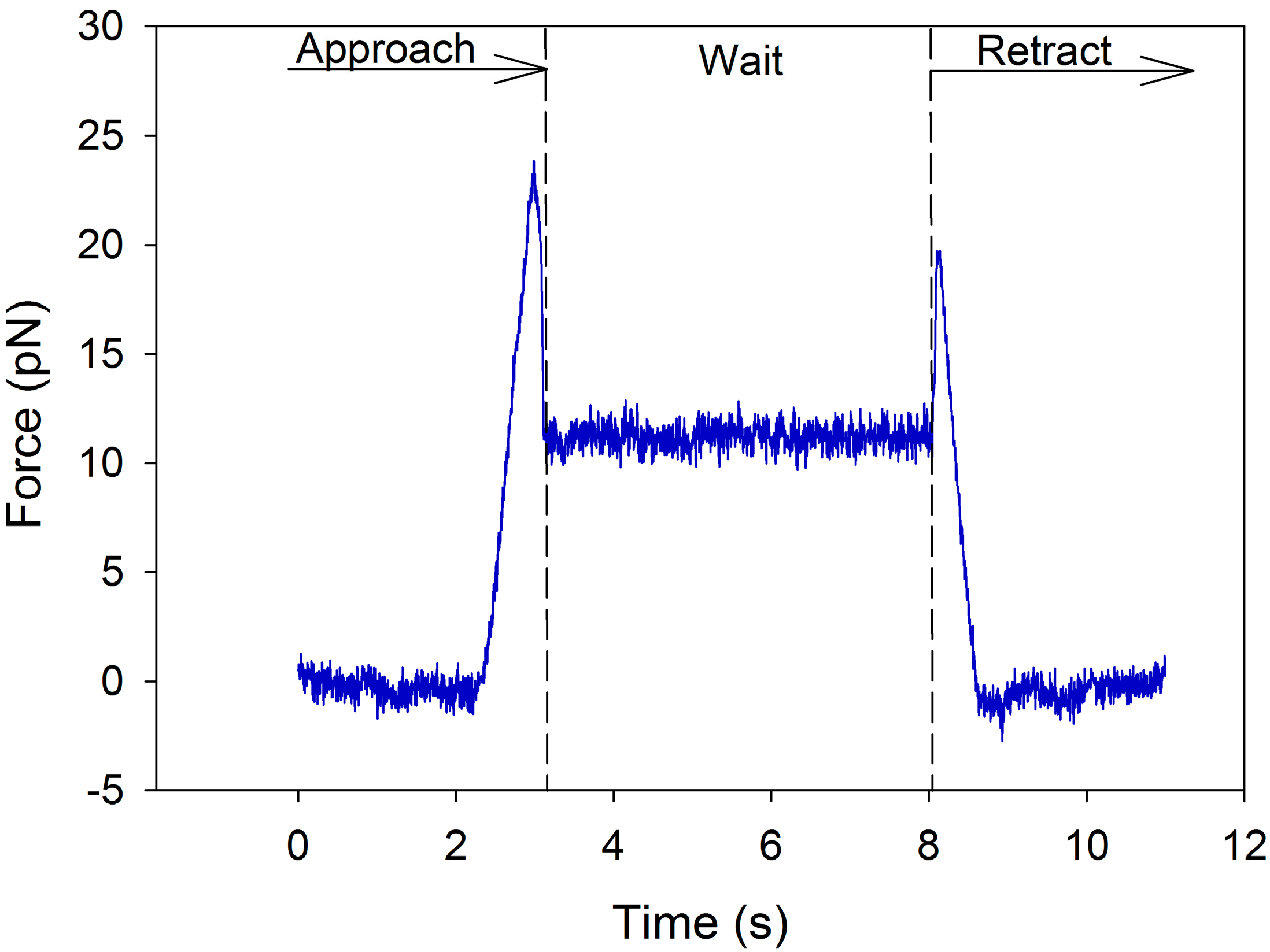
In the measurements, the chitosan stabilized corn oil droplets were led into contact at a speed of 0.5 μm/s, held together at maximum contact for 5 s and then pulled apart at the same speed of retraction. (The method is described in detail in Section 5.1). The curve displayed in Figure 5 shows the characteristic and reproducible response of the force signal for droplet 2 in the x-direction for this system. When the droplets approach, there is a steady increase in the repulsive force, due at first to overlap of the electrical double layers and subsequently to direct contact between the polymer layers. However, at a certain point in the approach segment of the curve, the force suddenly drops to a lower level and stabilizes there during the last part of the approach as well as during the hold time at this position (5 s). Upon retraction, the force is re-established at close to the maximum level before it drops as a result of the increased separation and then stabilizes at baseline level at large separation.
The fact that the force reduction is reversible upon retraction demonstrates that the changes occurring in the interface are restored when the strain on the system is reduced. One plausible explanation for the characteristic course of progress for these measurements is the rearrangement of the polymer layer at the interface as the droplets are compressed. When the strain on the system is increased as the separation reaches its minimum, the polymer layer is assumed to partially collapse, though not followed by coalescence of the droplets. This could be the result of a partial reorganization of the chitosan layer. The shift in the system to a lower energy level causes the force to drop as the static potential that has built up during compression is released when the entanglements relax. No further structural changes in the interface are observed until the retraction of the droplets starts. The force is reestablished at a slightly lower level as prior to the entanglement relaxation indicating that some degree of entangled structure in the emulsifier layer is re-established as space is made available.
Figure 5. Force vs. time curve for the approach and retract portion of an optical tweezers measurement with emulsion droplets stabilized by chitosan as the sole emulsifier in 10 mM acetate buffer at pH 5. The approach and retract segments of the measurement are separated by a holding time of 5 s at maximum contact between the emulsion droplets. Approach/retract speed: 0.5 μm/s.
Polymers 07 00552 g005
The emulsion droplets stabilized by chitosan as emulsifier displayed good stability during the work with the system on the optical tweezers. At a high degree of compression of several independent droplet pairs, flocculation and coalescence was only rarely observed indicating a good coverage of chitosan on the oil–water interface. In future work, with chitosan stabilized emulsion droplets on the optical tweezers it would be desirable to obtain force measurements when the pH in the system is set well above the pKa of the amino groups, e.g., pH 8. At this pH most of the amino groups will be deprotonated and the emulsion droplets practically uncharged, making it possible to look at the effect of steric stabilization separately, as opposed to the electrosteric stabilization at pH 5.

3.4. Chitosan as Emulsion Stabilizer

In emulsion stabilization, the most evident application of chitosans are in texture modification, i.e., as thickening and gelling agent. By increasing the viscosity of the continuous phase, chitosan will increase the emulsions’ creaming stability. The other important function of chitosan in emulsion stabilization is related to the electrostatically driven adsorption of the polymer to emulsion droplets stabilized by surfactants, proteins or polymers of opposite charge, creating a multilayer interfacial membrane [53,68]. Chitosan is unique in this regard as it is an abundant and cationic biopolymer, making it the biopolymer of choice for combination with the diversity of existing anionic surfactants, anionic biopolymers and proteins. In the following, a brief summary of chitosan’s role in multilayer stabilized emulsions will be given.

Chitosan in Multilayer Emulsions

Emulsions stabilized by a single surfactant or surface active biopolymer, may display limited stability to changes in, e.g., pH, ionic strength, heating and freezing. The formation of multilayer stabilized emulsions is a commonly applied strategy for emulsion scientists, e.g., in the food industry, to enhance the stability of the resulting product [69]. The emulsion droplets will be prevented from coalescence by a strong steric barrier. The interfacial layers are formed using a layer-by-layer (LBL) electrostatic deposition technique [70,71,72,73,74,75,76,77,78,79,80]. The first emulsifier added is a potent ionic emulsifier that rapidly adsorbs to the oil droplet surfaces during homogenization, forming an emulsion of small dispersed droplets. The addition of an oppositely charged polyelectrolyte results in the formation of the second layer; the polymer adheres to the emulsion droplet surfaces through electrostatic attraction. Subsequent layers can be obtained by adding polyelectrolytes of opposite charge to the previous layer, creating three or more layers covering the emulsion droplets [69].
In many multilayer emulsion systems, chitosan is the polymer of choice for interaction with anionic surfactants (primary layer) such as sodium dodecyl sulphate (SDS) [75,76], sodium stearoyl lactylate (SSL) [81], lecithin [72,73,78,79,82], and CITREM (citric acid ester of mono-and diglycerides) [71]. Chitosan interacts strongly through electrostatic interaction forming stable chitosan-surfactant interfacial membranes and emulsion droplets of net positive charge below the pKa of the chitosan amino group [68]. Surfactant-chitosan layers can also be formed with non-ionic surfactants [76] such as polysorbate 80, due to the small negative charge of the surfactant, attributed to the adsorption of OH ions from water or potentially from free fatty acids or phospholipids in the oil phase [68]. Many studies can be found on protein–chitosan stabilized emulsions, e.g., with whey protein isolate (β-lactoglobulin) [83,84,85,86] and casein [87] as commonly applied proteins. Chitosan can be added to emulsion droplets primarily stabilized by protein through electrostatic deposition. In order for the protein to carry a net negative charge and the chitosan a net positive charge, the pH during the emulsion preparation must be higher than the isoelectric point (IEP) of the protein and lower than the pKa of the amino group in chitosan, facilitating the electrostatic interaction. It should be noted that the hydrophobicity of chitosan (acetyl groups) may also contribute to the adsorption of chitosan to protein coated emulsion droplets. Anionic polysaccharides often play a part together with chitosan in multilayer emulsions; examples are pectin [80,88] and alginate [71].

3.5. Emulsion Flocculation

Chitosan has been applied as a flocculant in different colloidal systems, including bacteria [89,90,91], algea [92], latex particles [93] and emulsions [94,95].
In the existing literature, there are few studies focusing on describing the flocculation efficiency of chitosan in emulsions. In particular, there is a lack of information regarding the emulsion destabilizing properties of chitosans of a wide range of FA where also highly acetylated chitosans are included. This has motivated us to study chitosan with a wider range of FA. We have investigated the effect of different parameters on the amount of chitosan required to flocculate an oil-in-water emulsion stabilized by sodium dodecyl sulphate (SDS). The effect on flocculation of FA (0–0.6) and molecular weight of the chitosan, the ionic strength of the continuous phase, oil content and amount of emulsifier (SDS) was determined. Degree of flocculation (%) was measured as a relative decrease in the turbidity of the emulsion upon addition of chitosan (refer to Materials and Methods Section). Due to the increase in effective size, aggregated droplets cream faster and form an oily layer on top of the sample tube, thereby reducing the turbidity of the emulsion [53].
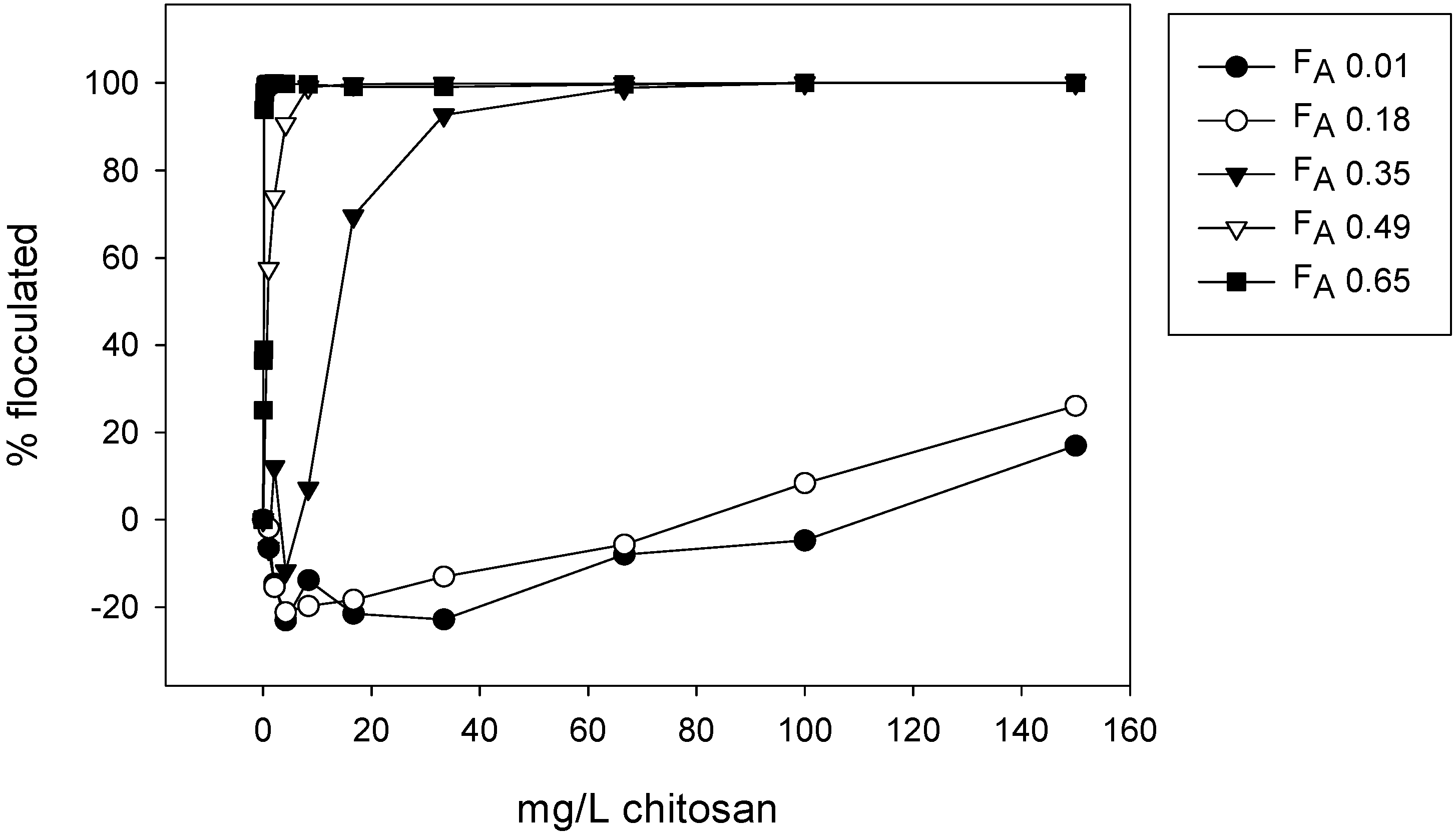
Chitosans with FA ranging from 0 to 0.6 and similar molecular weights were evaluated with respect to their flocculation efficiency (see Table 1 for details of the FA, intrinsic viscosities and molecular weights of the chitosans). Figure 6 displays the effect of FA of chitosan on the degree of flocculation in the emulsion system. A clear-cut effect can be observed; with increasing FA-values the flocculation of the SDS-stabilized droplets occurs at lower concentrations of added chitosan.
Figure 6. Flocculation of SDS stabilized O/W emulsion (3% w/w) by chitosans of different FA. Conditions: HAc/NaAC buffer pH 5, ionic strength 100 mM, 0.9 g/L SDS.
Polymers 07 00552 g006
The emulsion droplets carry a net negative charge due to the anionic SDS aligned in the oil–water interface, while the chitosan carry a positive charge at chosen pH value of 5. Thus, the potential electrostatic association between chitosan in the continuous phase and the anionic surfactant layer was facilitated. However, the ionic strength was 100 mM in the experiments, implying charge screening of the amino-groups. As the electrical double layer surrounding the SDS coated emulsion droplets can be assumed to be heavily compressed at this ionic strength, it is likely that the main underlying mechanism for flocculation of emulsion droplets in these experiments is not electrostatic, but rather hydrophobic interactions. The chitosans ability to induce flocculation increases as the fraction of hydrophobic acetyl groups in the chains increases. The acetyl-rich areas of the chitosan chains are assumed to associate with the emulsion droplets and create polymer bridges connecting the droplets resulting in bridging flocculation and accelerated creaming. A similar mechanism was previously reported for chitosan flocculating bacteria [90].
In a previous study, it was also suggested that interaction between the hydrophobic part of the surfactant and the emulsion destabilizing polymer plays a part in bridging flocculation [96]. Also, the length of the hydrocarbon tail of the applied surfactants have been shown to have an effect on the association of polyelectrolyte to the emulsion droplet surfaces; longer hydrocarbon tail of the surfactant resulted in a stronger association with chitosan [94]. In their study of the formation of O/W emulsions stabilized by surfactant-chitosan membranes, Mun et al. reported on flocculation occurring within a wider concentration range for the chitosan of highest FA (0.6) in a SDS stabilized emulsion. The results were explained based on the assumption that the mechanism was electrostatically mediated bridging flocculation and that more high FA chitosan was required to neutralize the negative emulsion droplet surfaces, resulting in the observation of flocculation at higher concentrations [76]. However, it cannot be ruled out that hydrophobic interactions, due to their higher acetyl content, also contributed to the increased flocculation tendency for this chitosan.
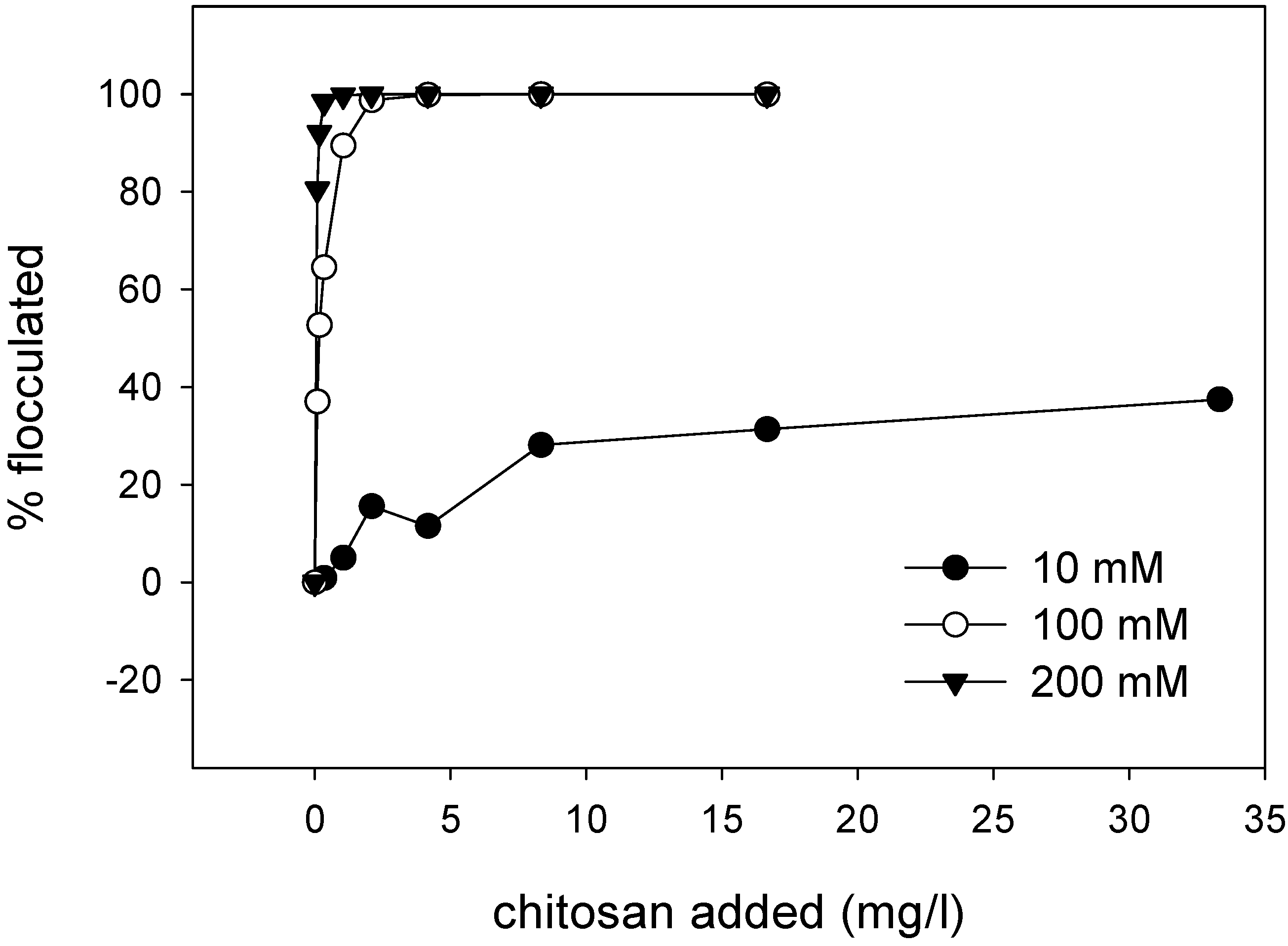
The hypothesis of hydrophobically mediated bridging flocculation is strengthened when considering the effect of varying the ionic strength of the continuous phase, presented in Figure 7, showing increased flocculation efficiency with increasing ionic strength. This indicates that the flocculation is more efficient when the charges on both the chitosan and the emulsion droplets are screened, ruling out electrostatic association as a flocculation mechanism in this emulsion-flocculant system.
Figure 7. Flocculation of SDS stabilized O/W emulsion (3% w/w) by chitosan (FA 0.49, [η] = 900 mL/g) at pH 5 and different ionic strengths. Conditions: HAc/NaAC buffer pH 5, 0.9 g/L SDS.
Polymers 07 00552 g007
Pinotti and co-workers examined the effect of ionic strength on the flocculation efficiency of chitosan (FA 0.23) on SDS- and STS (sodium tetradecyl sulphate)-stabilized emulsions, with results in agreement with our, i.e., the amount of chitosan necessary for complete flocculation decreased as the ionic strength increased. The authors attributed the ionic strength effect to screening of the electrical double layer surrounding the emulsion droplets, allowing the droplets surfaces to come into closer proximity and through this facilitate flocculation [94]. Screening of the electrical double layer at high ionic strength results in a short Debye screening length, possibly even shorter than the range of the attractive van der Waals interaction [97]. This can give rise to a net attractive interaction during short separations. Seen in light of the results presented here, the effect is assumed to be two-fold; the increase in ionic strength facilitates the association between acetyl groups in chitosan and the emulsion droplets, but also has a more general effect in lowering the potential energy barrier that has to be overcome in order for the emulsion droplets to flocculate.
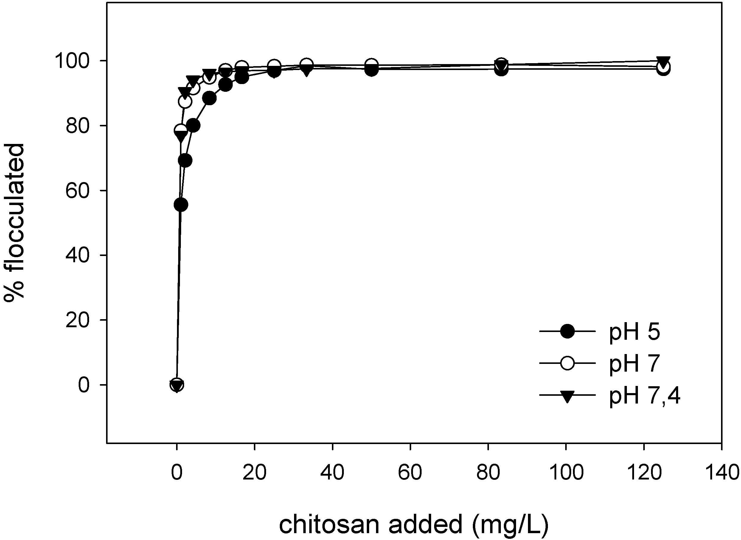
The effect of pH on the degree of flocculation was examined and the results are presented in Figure 8. The effect of increasing the pH of the continuous phase from 5 (well below the pKa of 6.5 of the amino-groups of chitosan) to 7.4 (basically uncharged amino-groups) was insignificant. Only a small reduction in the measured degree of flocculation at pH 5 was observed, thus reflecting that the chitosan amount required for emulsion destabilization were practically independent of pH at the chosen conditions (ionic strength of 100 mM). Bratskaya et al. evaluated the flocculation efficiency of chitosan (FA 0.16) as well as hydrophobically modified chitosan derivatives in SDS stabilized emulsions. Flocculation was measured for pH values spanning the pKa of the amino-group (pH 4–9). Their results are in agreement with our results; flocculation efficiencies were practically independent of pH [95].
A comparison of the flocculation efficiency of a chitosan (FA 0.49) with varying intrinsic viscosities from 220 mL/g (Mw 220,000 g/mol) to 970 mL/g (Mw 270,000 g/mol) was performed under the same conditions (pH 7, ionic strength 100 mM, oil fraction 3% w/w and 0.9 g/L SDS), and the dose-response curves were practically superimposed (results not shown). Thus, the molecular weight of the chitosans (within the range investigated here) is not a determinant parameter for the effectiveness of chitosan as a flocculant. The results are supported by previous findings of Mun et al. In their study from 2006 they observed that large flocs were formed in SDS and polysorbate 20 stabilized emulsions upon addition of low concentrations of chitosan (0.01%–0.04%). The degree of flocculation was independent of the molecular weights of the chitosans (FA in the range 0.15–0.25 for all three samples). However, at higher concentrations of chitosan assumed depletion flocculation was observed, an interaction arising as a result of non-adsorbed polymer in the continuous phase. The effect was more pronounced at higher Mw of the chitosans and this is in accordance with theory [76].
Figure 8. Flocculation of SDS stabilized O/W emulsion (3% w/w) by chitosan (FA 0.49, [η] = 900 mL/g) at pH 5, 7 and 7.4. Conditions: HAc/NaAC buffer pH 5, HEPES buffer pH 7 and 7.4, ionic strength 100 mM, 0.9 g/L SDS.
Polymers 07 00552 g008
The effect of varying the amount of emulsifier (SDS) when preparing the emulsions was examined (results not shown). The results showed no clear effect on the flocculation efficiency and the lower SDS concentrations were only marginally less effective. We speculate that this reflects that the association between the hydrophobic tail of the surfactant and the hydrophobic acetyl groups in chitosan is an underlying mechanism for flocculation, as also postulated by Pinotti et al. [94].
Variable oil content has also been evaluated as a parameter for flocculation efficiency of chitosans of FA 0.49 and 0.01. For the chitosan of FA 0.49 measurements on emulsions with an oil content in the range of 1.5%–10% (w/w) were performed at pH 5 (results not included). Only small differences were observed, but the results display a tendency towards smaller doses of chitosan being required for complete flocculation at higher oil content in the emulsion, possibly due to a higher collision frequency and degree of association between the emulsion droplets at higher oil content which cause flocculation to proceed faster with lower amounts of chitosan. On the contrary, for highly deacetylated chitosan (FA 0.01) with a comparable molecular weight and under the same conditions, flocculation occurred more efficiently at lower oil content. The oil fractions tested for this low FA were all low; 0.5%, 1.5% and 2%. The fact that only the emulsion of lowest oil fraction is completely flocculated can be explained by the very low amount of acetyl groups present. The very low amount of acetyl groups is sufficient for flocculation of the oil droplets in the emulsion of lowest oil fraction, while it apparently is not for flocculation of emulsions with higher oil fractions.
Bridging flocculation is assumed to be the predominant flocculation mechanism in this system. The clear effect of FA on the flocculation efficiency as well as the non-significant effect of molecular weight (intrinsic viscosity) further support this assumption. However, based on the present results presented here, it cannot be ruled out that a depletion interaction contribution is present at some concentrations of chitosan, giving rise to an osmotic pressure gradient and an attractive interaction between the droplets. Further support to the bridging mechanism could be achieved through the effect of diluting the flocculated emulsion. In the case of bridging flocculation, the network of flocculated droplets will not dissolve upon dilution, while in the case of depletion the droplets would disconnect once the osmotic pressure gradient (concentration of chitosan) is lowered to below a threshold value.

3.6. Applications

In this section, we briefly consider some of the potential uses of chitosan with respect to its interfacial activity.
In addition to the possibilities for applying chitosan as an emulsifying, stabilizing and thickening agent, as discussed in the previous sections, an arising application area currently receiving attention is in the encapsulation of lipophilic functional components, e.g., nutraceuticals such as fish oil. The aim is to protect the nutraceuticals for example within a food product, to limit oxidation and ensure bioavailability in the gastrointestinal tract. Basically, the oil is emulsified and stabilized by a multilayered interfacial membrane through a LBL-technique. In many of these systems, chitosan is commonly applied being a cationic biopolymer able to electrostatically interact with anionic surfactant, proteins and/or biopolymer. Then, a wall material is added, forming an outer coating and a continuous matrix between droplets; maltodextrin or corn syrup solids are commonly applied wall materials. Encapsulation is followed by spray or freeze-drying to remove the water, resulting in a powder of encapsulated oil. The powder can for example be administered together with food, masking taste and simplifying the intake, e.g., for children and senior citizens. Several studies focus on different variations of the encapsulation technique in which chitosan is included as a stabilizer [72,73,98]. A different approach is the hydrogel based oil encapsulation in which gelation of the stabilizing biopolymer layer forms a protective coating around the oil droplets prior to freeze drying. Examples of gelled chitosan layers in encapsulation can be found in literature [99,100,101].
Another discussed potential application of chitosan that has been in focus in several studies is related to reduction of the absorption and digestion of dietary lipids [102,103,104,105,106,107]. Some of these focus on the potential ability of chitosan to inhibit the lipase activity in the stomach. However, chitosan has also been reported to cause aggregation of emulsified lipids in an in vitro model simulating the conditions in the gastrointestinal tract and through this reducing the absorption of fat. The underlying mechanism was assumed to be bridging flocculation [103]. Scientists are divided in their view on chitosan’s ability to reduce lipid absorption, but the ambiguous results may be due to the heterogeneity of the chitosans used in the experiments (e.g. with regard to FA and Mw) as well as differences in the experimental set-up. A study from 2009 using well-defined chitosans in flocculation experiments concluded that the flocculation efficiency of chitosan in the in vitro model was highly dependent on parameters such as molecular weight and charge [102].
Finally, without going into detail, applications of chitosan in industry is by far the most utilized today; including in purification of water and waste water treatment [108,109], treatment of oil spills [110] and treatment of oily waste from the food industry and oil industry [111,112], as well as the potential use in enhanced oil recovery [113].

4. Regulatory Status

As previously mentioned, the applications of chitosan in foods and pharmaceuticals are limited by regulatory regulations, and the present commercial use of chitosan within this area is limited. The biopolymer is not presently on the GRAS (generally recognized as safe) list, nor is it on the list of European Union (EU) approved additives. However, in Japan and Korea, chitosan has been approved as a food additive (Ministry of Health, Labour and Welfare, MHLW, and Korean Food and Drug Administration, KFDA, respectively). Major regulatory issues include the structural heterogeneity of chitosans, large batch variations, potential impurities in the samples and that the polymer originates from potentially allergenic raw materials (crustaceans shells). Currently, there are no chitosan-containing drug products, biopharmaceuticals or food products approved by the FDA (US Food and Drug Administration). However, FDA has cleared several chitosan based medical devices and combination products; topical wound dressings and wound dressings containing drugs (antimicrobials). In the European Union (EU), chitosan is approved for biomedical use as an excipient in nasal drug delivery and vaccine delivery [114].

5. Materials and Methods

5.1. Emulsion Droplet Measurements on Optical Tweezers

A total of 1% w/w chitosan solution (Viscosan™, Viscogel AB, Solna, Sweden) with FA 0.49 was prepared by dissolution during stirring overnight. The O/W emulsions were prepared using an Ultra turrax homogenizer (IKA, Staufen, Germany). The initial weight fraction of the corn oil (Sigma-Aldrich, St. Louis, MO, USA) in the emulsions was 20% (w/w) and the 1% (w/w) chitosan solution was the aqueous phase. The emulsions were diluted 1:5000–1:10000 in 10 mM acetate buffer at pH 5 to a droplet concentration appropriate for work on the optical tweezers.
The optical tweezers instrument Nanotracker from JPK Instruments (Berlin, Germany) was applied for measurements on pairs of emulsion droplets. The instrument is mounted on an inverted light microscope (Zeiss Axio Observer A1, Carl Zeiss AG, Oberkuchen, Germany). The accessible force of the instrument is between 1 and 100 pN and the position sensitivity of the trapped object is in the nanometer range. The instrument is a dual beam optical tweezers with a built in beam splitter enabling the emulsion droplets to be trapped in separate optical traps and led into proximity at a controlled speed. The sample chambers utilized consisted of a circular borosilicate glass (35 mm diameter, VWR International, Radnor, PA, USA) as the bottom glass, two pieces of double sided tape as side walls and a regular square cover glass (22 mm × 22 mm, VWR International) for attachment on top. In order to prevent evaporation of the continuous phase of the diluted emulsion, the sample chamber was sealed with nail polish. Immersion oil (Carl Zeiss AG) was applied to both sides of the sample chamber before approach of the objectives. Prior to sample chamber assembly, the top cover glass was treated with a solution of 1 mg/mL of bovine serum albumin (BSA, Sigma-Aldrich) in order to delay adhesion of the droplets to the sample chamber surface. All measurements were performed at room temperature. The optimal droplet size for stable trapping in the optical traps on the Nanotracker has been suggested to be between 1 and 3 μm (oral communication with JPK Instruments) and effort was made to find droplets of comparable size. The measurements were performed by moving the emulsion droplet in trap 1 towards the emulsion droplet in trap 2, which was held steady (Figure 4). At contact, the droplets were held together at fixed positions for 5 s (while continuously recording the force) before the retract portion of the measurement started. Both the approach and retract speeds were 0.5 μm/s. For a more in depth methodical description and instrument specifications, the authors refer to the paper by Nilsen-Nygaard et al. [67].

5.2. Flocculation of SDS Coated Emulsion Droplets by Chitosan

Five chitosans of different FA, but comparable molecular weights were applied in the flocculation experiments. The chemical composition of the chitosans (FA) was determined by 1H-NMR spectroscopy [115] and their intrinsic viscosities (at an ionic strength of 100 mM) were determined as previously described [24]. The FA, Mw and intrinsic viscosity of the chitosans applied as flocculants are summarized in Table 1.
Table 1. Characteristics of the chitosans applied in the emulsion flocculation experiments.
ChitosanFraction of Acetylation (FA)Intrinsic Viscosity ([η])Molecular Weight (Mw) *
10.001800 mL/g220,000
20.18800 mL/g220,000
30.35760 mL/g210,000
40.49900 mL/g260,000
50.65950 mL/g270,000
* Determined from the Mark Houwink Sakurada equation with K = 0.00843 and a = 0.93 [116].
Stock solutions of chitosans (1 mg/mL) were prepared by gentle shaking in Milli-Q water (Merck Millipore, Billerica, MA, USA) at 5 °C overnight. The ionic strength was adjusted to 100 mM with NaCl for all samples. A concentration series (1–1000 mg/L) was then prepared by dilution with 100 mM NaCl.
O/W emulsions with sunflower oil as the oil phase and sodium dodecyl sulphate (SDS) as an emulsifier were prepared by homogenization with an Ultra Turrax (IKA) at 24,000 rpm for 2 min. The sunflower oil content was varied from 0.5% to 10% (w/w) and the total amount of SDS was 3% (w/w) of the oil phase. Emulsions with three different pH values of 5, 7 and 7.4 were prepared using 50 mM acetate buffer (pH 5) or HEPES (4-(2-hydroxyethyl)-piperazine-1-ethanesulfonic acid) buffer (pH 7 and 7.4) as the continuous aqueous phase. The ionic strength of all buffers was adjusted to 0.1 M or 0.2 M with NaCl.
The flocculation assay was performed in 13 mL polypropylene tubes (Sarstedt AG, Nümbrecht, Germany). Five-milliliter aliquots of freshly prepared emulsion were pipetted into the tubes and 1 mL of chitosan solution was added under stirring on a Vortex mixer (1800 rpm, 10 s) to ensure proper mixing. A corresponding blank was prepared with 1 mL of 0.1 M NaCl. After the preparation of the entire concentration series, the tubes were once again mixed on a Vortex mixer (1400 rpm, 5 s) and placed on a rotating stand for 5 min. The samples were then allowed to stand for 120 min before the sample for optical density measurement was taken from the middle of the tube. The optical densities of the samples were measured at 620 nm on a spectrophotometer (Lambda 25 UV/VIS spectrometer, Perkin Elmer Instruments, Waltham, MA, USA), zero-set against the buffer solution. Flocculation was expressed as a decrease in turbidity relative to the blank (referred to as% flocculated), calculated as (1 − (ODsample/ODblank)) × 100. All samples were run in duplicates.

6. Conclusions

A major challenge with respect to chitosan applications for food and pharmaceuticals is its regulatory status. Chitosan is also a rather complex biopolymer by way of its varying chemical composition/molecular weight which means chitosan be tailored for specific purposes. Simple facts like, e.g., a pKa value close to the physiological pH, where generic and highly deacetylated chitosans will not be soluble, and rate of biodegradation highly dependent on FA are rather demanding properties that put serious constraints on optimization of a product. If not properly understood and dealt with, suboptimal or even failed products will emerge. If properly appreciated and treated, as in the many examples put forth in this review, these properties can be exploited to obtain inventive biomaterials. The uniqueness of chitosan as a biopolymer, a remarkable low-toxic and biodegradable cationic biopolymer exhibiting surface active properties, points towards both an increased basic research activity as well as an increased number of chitosan based applications.

Acknowledgments

We would like to thank Viscogel AB for kindly providing us with Viscosan™, as well as associate professor Marit Sletmoen for assistance in experimental design and result interpretation in the work on optical tweezers.

Author Contributions

Julie Nilsen-Nygaard, Kurt I. Draget and Catherine T. Nordgård wrote the paper. Kjell M. Vårum and Sabina P. Strand designed the emulsion flocculation experiments, Sabina P. Strand performed the experiments. Kurt I. Draget and Julie Nilsen-Nygaard designed the emulsion droplet experiments on optical tweezers, Julie Nilsen-Nygaard performed the experiments.

Conflicts of Interest

The authors declare no conflict of interest.

References

  1. Peter, M.G. Chitin and chitosan from animal sources. In Polysaccharides and Polyamides in the Food Industry; Steinbüchel, A., Rhee, S.K., Eds.; Wiley-VCH Verlag GmbH: Weinheim, Germany, 2005; pp. 115–208. [Google Scholar]
  2. Vårum, K.M.; Smidsrød, O. Structure–property relationship in chitosans. In Polysaccharides: Structural Diversity and Functional Versatility; Dimitriu, S., Ed.; Marcel Decker: New York, NY, USA, 2005; pp. 625–642. [Google Scholar]
  3. Strand, S.P.; Tømmeraas, K.; Vårum, K.M.; Østgaard, K. Electrophoretic light scattering studies of chitosans with different degrees of N-acetylation. Biomacromolecules 2001, 2, 1310–1314. [Google Scholar] [CrossRef] [PubMed]
  4. Kim, E.; Xioung, Y.; Cheng, Y.; Wu, H.C.; Liu, Y.; Morrow, B.H.; Ben-Yoav, H.; Ghodssi, R.; Rubloff, G.W.; Shen, J.; et al. Chitosan to connect biology to electronics: Fabricating the bio-device interface and communicating across this interface. Polymers 2015, 7, 1–46. [Google Scholar] [CrossRef]
  5. Vårum, K.M.; Holme, H.K.; Izume, M.; Torger Stokke, B.; Smidsrød, O. Determination of enzymatic hydrolysis specificity of partially N-acetylated chitosans. Biochim. Biophys. Acta 1996, 1291, 5–15. [Google Scholar] [CrossRef] [PubMed]
  6. Nordtveit, R.J.; Vårum, K.M.; Smidsrød, O. Degradation of partially N-acetylated chitosans with hen egg white and human lysozyme. Carbohydr. Polym. 1996, 29, 163–167. [Google Scholar] [CrossRef]
  7. Ross-Murphy, S. Rheological methods. In Biophysical Methods in Food Research; Chan, H.W.-S., Ed.; Blackwell Scientific Publications: Oxford, UK, 1984; pp. 137–199. [Google Scholar]
  8. Strand, S.P.; Lelu, S.; Reitan, N.K.; Davies, C.D.; Artursson, P.; Varum, K.M. Molecular design of chitosan gene delivery systems with an optimized balance between polyplex stability and polyplex unpacking. Biomaterials 2010, 31, 975–987. [Google Scholar] [CrossRef] [PubMed]
  9. Mumper, R.; Wang, J.; Claspell, J.; Rolland, A.P. Novel polymeric condensing carriers for gene delivery. Proc. Int. Symp. Control Release Bioact. Mater. 1995, 22, 178–179. [Google Scholar]
  10. Croisier, F.; Jerome, C. Chitosan-based biomaterials for tissue engineering. Eur. Polym. J. 2013, 49, 780–792. [Google Scholar] [CrossRef]
  11. Berger, J.; Reist, M.; Mayer, J.M.; Felt, O.; Peppas, N.A.; Gurny, R. Structure and interactions in covalently and ionically crosslinked chitosan hydrogels for biomedical applications. Eur. J. Pharm. Biopharm. 2004, 57, 19–34. [Google Scholar] [CrossRef] [PubMed]
  12. Yao, K.D.; Yao, F.L.; Li, J.J.; Yin, Y.J.; Jarry, C.; Shive, M.S. Chitosan-based gels and hydrogels. In Smart Materials; CRC Press: Boca Raton, FL, USA, 2008. [Google Scholar]
  13. Ta, H.T.; Dass, C.R.; Dunstan, D.E. Injectable chitosan hydrogels for localised cancer therapy. J. Control. Release 2008, 126, 205–216. [Google Scholar] [CrossRef] [PubMed]
  14. Kaur, S.; Dhillon, G.S. The versatile biopolymer chitosan: Potential sources, evaluation of extraction methods and applications. Crit. Rev. Microbiol. 2014, 40, 155–175. [Google Scholar] [CrossRef] [PubMed]
  15. Bhattarai, N.; Gunn, J.; Zhang, M.Q. Chitosan-based hydrogels for controlled, localized drug delivery. Adv. Drug. Deliv. Rev. 2010, 62, 83–99. [Google Scholar] [CrossRef] [PubMed]
  16. Vårum, K.M.; Ottøy, M.H.; Smidsrød, O. Water-solubility of partially N-acetylated chitosans as a function of pH: Effect of chemical composition and depolymerisation. Carbohydr. Polym. 1994, 25, 65–70. [Google Scholar] [CrossRef]
  17. Vachoud, L.; Zydowicz, N.; Domard, A. Physicochemical behaviour of chitin gels. Carbohydr. Res. 2000, 326, 295–304. [Google Scholar] [CrossRef] [PubMed]
  18. Chenite, A.; Buschmann, M.; Wang, D.; Chaput, C.; Kandani, N. Rheological characterisation of thermogelling chitosan/glycerol-phosphate solutions. Carbohydr. Polym. 2001, 46, 39–47. [Google Scholar] [CrossRef]
  19. Lavertu, M.; Filion, D.; Buschmann, M.D. Heat-induced transfer of protons from chitosan to glycerol phosphate produces chitosan precipitation and gelation. Biomacromolecules 2008, 9, 640–650. [Google Scholar] [CrossRef] [PubMed]
  20. Montembault, A.; Viton, C.; Domard, A. Rheometric study of the gelation of chitosan in aqueous solution without cross-linking agent. Biomacromolecules 2005, 6, 653–662. [Google Scholar] [CrossRef] [PubMed]
  21. Ladet, S.; David, L.; Domard, A. Multi-membrane hydrogels. Nature 2008, 452, 76–79. [Google Scholar] [CrossRef] [PubMed]
  22. Rami, L.; Malaise, S.; Delmond, S.; Fricain, J.-C.; Siadous, R.; Schlaubitz, S.; Laurichesse, E.; Amédéé, J.; Montembault, A.; David, L.; et al. Physicochemical modulation of chitosan-based hydrogels induces different biological responses: Interest for tissue engineering. J. Biomed. Mater. Res. A 2014, 102, 3666–3676. [Google Scholar] [CrossRef] [PubMed]
  23. Shu, X.Z.; Zhu, K.J. Controlled drug release properties of ionically cross-linked chitosan beads: The influence of anion structure. Int. J. Pharm. 2002, 233, 217–225. [Google Scholar] [CrossRef] [PubMed]
  24. Draget, K.I.; Värum, K.M.; Moen, E.; Gynnild, H.; Smidsrød, O. Chitosan cross-linked with Mo (VI) polyoxyanions: A new gelling system. Biomaterials 1992, 13, 635–638. [Google Scholar] [CrossRef] [PubMed]
  25. Dambies, L.; Vincent, T.; Domard, A.; Guibal, E. Preparation of chitosan gel beads by ionotropic molybdate gelation. Biomacromolecules 2001, 2, 1198–1205. [Google Scholar] [CrossRef] [PubMed]
  26. Luo, Y.; Wang, Q. Recent development of chitosan-based polyelectrolyte complexes with natural polysaccharides for drug delivery. Int. J. Biol. Macromol. 2014, 64, 353–367. [Google Scholar] [CrossRef] [PubMed]
  27. Tsuchida, E.; Abe, K. Interactions between macromolecules in solution and intermolecular complexes. Adv. Polym. Sci. 1982, 45, 1–119. [Google Scholar]
  28. Sæther, H.V.; Holme, H.K.; Maurstad, G.; Smidsrød, O.; Stokke, B.T. Polyelectrolyte complex formation using alginate and chitosan. Carbohydr. Polym. 2008, 74, 813–821. [Google Scholar] [CrossRef]
  29. Khong, T.T.; Aarstad, O.A.; Skjåk-Bræk, G.; Draget, K.I.; Vårum, K.M. Gelling concept combining chitosan and alginate—Proof of principle. Biomacromolecules 2013, 14, 2765–2771. [Google Scholar] [CrossRef] [PubMed]
  30. Draget, K.I. Associating phenomena in highly acetylated chitosan gels. Polym. Gels Netw. 1996, 4, 143–151. [Google Scholar] [CrossRef]
  31. Tømmeraas, K.; Strand, S.P.; Christensen, B.E.; Smidsrød, O.; Vårum, K.M. Preparation and characterization of branched chitosans. Carbohydr. Polym. 2011, 83, 1558–1564. [Google Scholar] [CrossRef]
  32. Argüelles-Monal, W.; Goycoolea, F.M.; Peniche, C.; Higuera-Ciapara, I. Rheological study of the chitosan/glutaraldehyde chemical gel system. Polym. Gels Netw. 1998, 6, 429–440. [Google Scholar] [CrossRef]
  33. Neimert-Andersson, T.; Hällgren, A.-C.; Andersson, M.; Langebäck, J.; Zettergren, L.; Nilsen-Nygaard, J.; Draget, K.I.; van Hage, M.; Lindberg, A.; Gafvelin, G.; et al. Improved immune responses in mice using the novel chitosan adjuvant ViscoGel, with a Haemophilus influenzae type b glycoconjugate vaccine. Vaccine 2011, 29, 8965–8973. [Google Scholar] [CrossRef] [PubMed]
  34. Rinaudo, M. New way to crosslink chitosan in aqueous solution. Eur. Polym. J. 2010, 46, 1537–1544. [Google Scholar] [CrossRef]
  35. Simi, C.K.; Abraham, T.E. Transparent xyloglucan–chitosan complex hydrogels for different applications. Food Hydrocoll. 2010, 24, 72–80. [Google Scholar] [CrossRef]
  36. Guo, B.; Elgsaeter, A.; Stokke, B.T. Gelation kinetics of scleraldehyde–chitosan co-gels. Polym. Gels Netw. 1998, 6, 113–135. [Google Scholar] [CrossRef]
  37. Christensen, B.E.; Aasprong, E.; Stokke, B.T. Gelation of periodate oxidised scleroglucan (scleraldehyde). Carbohydr. Polym. 2001, 46, 241–248. [Google Scholar] [CrossRef]
  38. Hennink, W.E.; van Nostrum, C.F. Novel crosslinking methods to design hydrogels. Adv. Drug. Deliv. Rev. 2012, 64, 223–236. [Google Scholar] [CrossRef]
  39. Kim, I.Y.; Seo, S.J.; Moon, H.S.; Yoo, M.K.; Park, I.Y.; Kim, B.C.; Cho, C.S. Chitosan and its derivatives for tissue engineering applications. Biotech. Adv. 2008, 26, 1–21. [Google Scholar] [CrossRef]
  40. Anitha, A.; Sowmya, S.; Kumar, P.T.S.; Deepthi, S.; Chennazhi, K.P.; Ehrlich, H.; Tsurkan, M.; Jayakumar, R. Chitin and chitosan in selected biomedical applications. Prog. Polym. Sci. 2014, 39, 1644–1667. [Google Scholar] [CrossRef]
  41. Chenite, A.; Chaput, C.; Wang, D.; Combes, C.; Buschmann, M.D.; Hoemann, C.D.; Leroux, J.C.; Atkinson, B.L.; Binette, F.; Selmani, A. Novel injectable neutral solutions of chitosan form biodegradable gels in situ. Biomaterials 2000, 21, 2155–2161. [Google Scholar] [CrossRef] [PubMed]
  42. Bernkop-Schnurch, A.; Dunnhaupt, S. Chitosan-based drug delivery systems. Eur. J. Pharm. Biopharm. 2012, 81, 463–469. [Google Scholar] [CrossRef] [PubMed]
  43. Agarwal, P.; Rupenthal, I.D. Injectable implants for the sustained release of protein and peptide drugs. Drug Discov. Today 2013, 18, 337–349. [Google Scholar] [CrossRef] [PubMed]
  44. Luppi, B.; Bigucci, F.; Cerchiara, T.; Zecchi, V. Chitosan-based hydrogels for nasal drug delivery: From inserts to nanoparticles. Exp. Opin. Drug Deliv. 2010, 7, 811–828. [Google Scholar] [CrossRef]
  45. Bhise, K.S.; Dhumal, R.S.; Paradkar, A.R.; Kadam, S.S. Effect of drying methods on swelling, erosion and drug release from chitosan-naproxen sodium complexes. AAPS PharmSciTech 2008, 9, 1–12. [Google Scholar] [CrossRef] [PubMed]
  46. Dutta, P.K.; Tripathi, S.; Mehrotra, G.K.; Dutta, J. Perspectives for chitosan based antimicrobial films in food applications. Food Chem. 2009, 114, 1173–1182. [Google Scholar] [CrossRef]
  47. Xia, W.S.; Liu, P.; Zhang, J.L.; Chen, J. Biological activities of chitosan and chitooligosaccharides. Food Hydrocoll. 2011, 25, 170–179. [Google Scholar] [CrossRef]
  48. Norowski, P.A.; Bumgardner, J.D. Biomaterial and antibiotic strategies for peri-implantitis. J. Biomed. Mater. Res. 2009, 88B, 530–543. [Google Scholar] [CrossRef]
  49. Ong, S.Y.; Wu, J.; Moochhala, S.M.; Tan, M.H.; Lu, J. Development of a chitosan-based wound dressing with improved hemostatic and antimicrobial properties. Biomaterials 2008, 29, 4323–4332. [Google Scholar] [CrossRef] [PubMed]
  50. Jayakumar, R.; Prabaharan, M.; Kumar, P.T.S.; Nair, S.V.; Tamura, H. Biomaterials based on chitin and chitosan in wound dressing applications. Biotech. Adv. 2011, 29, 322–337. [Google Scholar] [CrossRef]
  51. Walstra, P. Physical Chemistry of Foods; Marcel Dekker: New York, NY, USA, 2003. [Google Scholar]
  52. Dickinson, E. Hydrocolloids at interfaces and the influence on the properties of dispersed systems. Food Hydrocoll. 2003, 17, 25–39. [Google Scholar] [CrossRef]
  53. McClements, D.J. Food Emulsions: Principles, Practices and Techniques; CRC Press: Boca Raton, FL, USA, 2005; p. 609. [Google Scholar]
  54. Norde, W. Colloids and Interfaces in Life Sciences; Marcel Dekker: New York, NY, USA, 2003. [Google Scholar]
  55. Del Blanco, L.F.; Rodriguez, M.S.; Schulz, P.C.; Agullo, E. Influence of the deacetylation degree on chitosan emulsification properties. Colloid Polym. Sci. 1999, 277, 1087–1092. [Google Scholar]
  56. Schulz, P.C.; Rodríguez, M.S.; del Blanco, L.F.; Pistonesi, M.; Agulló, E. Emulsification properties of chitosan. Colloid Polym. Sci. 1998, 276, 1159–1165. [Google Scholar] [CrossRef]
  57. Rodrı́guez, M.S.; Albertengo, L.A.; Agulló, E. Emulsification capacity of chitosan. Carbohydr. Polym. 2002, 48, 271–276. [Google Scholar] [CrossRef]
  58. Li, X.K.; Xia, W.S. Effects of concentration, degree of deacetylation and molecular weight on emulsifying properties of chitosan. Int. J. Biol. Macromol. 2011, 48, 768–772. [Google Scholar] [CrossRef] [PubMed]
  59. Liu, H.; Wang, C.Y.; Zou, S.W.; Wei, Z.J.; Tong, Z. Simple, reversible emulsion system switched by pH on the basis of chitosan without any hydrophobic modification. Langmuir 2012, 28, 11017–11024. [Google Scholar] [CrossRef] [PubMed]
  60. Payet, L.; Terentjev, E.M. Emulsification and stabilization mechanisms of O/W emulsions in the presence of chitosan. Langmuir 2008, 24, 12247–12252. [Google Scholar] [CrossRef] [PubMed]
  61. Pepic, I.; Filipovic-Grcic, J.; Jalsenjak, I. Interactions in a nonionic surfactant and chitosan mixtures. Colloid Surface A 2008, 327, 95–102. [Google Scholar] [CrossRef]
  62. Mai-Ngam, K. Comblike poly(ethylene oxide)/hydrophobic C6 branched chitosan surfactant polymers as anti-infection surface modifying agents. Colloid Surface B 2006, 49, 117–125. [Google Scholar] [CrossRef]
  63. Qun, G.; Ajun, W. Effects of molecular weight, degree of acetylation and ionic strength on surface tension of chitosan in dilute solution. Carbohydr. Polym. 2006, 64, 29–36. [Google Scholar] [CrossRef]
  64. Babak, V.; Lukina, I.; Vikhoreva, G.; Desbrières, J.; Rinaudo, M. Interfacial properties of dynamic association between chitin derivatives and surfactants. Colloid Surface A 1999, 147, 139–148. [Google Scholar] [CrossRef]
  65. Geng, X.; Kwon, O.-H.; Jang, J. Electrospinning of chitosan dissolved in concentrated acetic acid solution. Biomaterials 2005, 26, 5427–5432. [Google Scholar] [CrossRef] [PubMed]
  66. Calero, N.; Munoz, J.; Ramirez, P.; Guerrero, A. Flow behaviour, linear viscoelasticity and surface properties of chitosan aqueous solutions. Food Hydrocoll. 2010, 24, 659–666. [Google Scholar] [CrossRef]
  67. Nilsen-Nygaard, J.; Sletmoen, M.; Draget, K.I. Stability and interaction forces of oil-in-water emulsions as observed by optical tweezers—A proof-of-concept study. RSC Adv. 2014, 4, 52220–52229. [Google Scholar] [CrossRef]
  68. Klinkesorn, U. The role of chitosan in emulsion formation and stabilization. Food Rev. Int. 2013, 29, 371–393. [Google Scholar] [CrossRef]
  69. Guzey, D.; McClements, D.J. Formation, stability and properties of multilayer emulsions for application in the food industry. Adv. Colloid Interface Sci. 2006, 128–130, 227–248. [Google Scholar] [CrossRef] [PubMed]
  70. Guzey, D.; McClements, D.J. Influence of environmental stresses on O/W emulsions stabilized by β-lactoglobulin-pectin and β-lactoglobulin-pectin-chitosan membranes produced by the electrostatic layer-by-layer deposition technique. Food Biophys. 2006, 1, 30–40. [Google Scholar] [CrossRef]
  71. Gudipati, V.; Sandra, S.; McClements, D.J.; Decker, E.A. Oxidative stability and in vitro digestibility of fish oil-in-water emulsions containing multilayered membranes. J. Agric. Food Chem. 2010, 58, 8093–8099. [Google Scholar] [CrossRef] [PubMed]
  72. Klinkesorn, U.; Sophanodora, P.; Chinachoti, P.; Decker, E.A.; McClements, D.J. Encapsulation of emulsified tuna oil in two-layered interfacial membranes prepared using electrostatic layer-by-layer deposition. Food Hydrocoll. 2005, 19, 1044–1053. [Google Scholar] [CrossRef]
  73. Klinkesorn, U.; Sophanodora, P.; Chinachoti, P.; Decker, E.A.; McClements, D.J. Characterization of spray-dried tuna oil emulsified in two-layered interfacial membranes prepared using electrostatic layer-by-layer deposition. Food Res. Int. 2006, 39, 449–457. [Google Scholar] [CrossRef]
  74. Calvo, P.; RemunanLopez, C.; VilaJato, J.L.; Alonso, M.J. Development of positively charged colloidal drug carriers: Chitosan coated polyester nanocapsules and submicron-emulsions. Colloid Polym. Sci. 1997, 275, 46–53. [Google Scholar] [CrossRef]
  75. Mun, S.; Decker, E.A.; McClements, D.J. Influence of droplet characteristics on the formation of oil-in-water emulsions stabilized by surfactant-chitosan layers. Langmuir 2005, 21, 6228–6234. [Google Scholar] [CrossRef] [PubMed]
  76. Mun, S.; Decker, E.A.; McClements, D.J. Effect of molecular weight and degree of deacetylation of chitosan on the formation of oil-in-water emulsions stabilized by surfactant-chitosan membranes. J. Colloid Interface Sci. 2006, 296, 581–590. [Google Scholar] [CrossRef] [PubMed]
  77. Svensson, O.; Lindh, L.; Cardenas, M.; Arnebrant, T. Layer-by-layer assembly of mucin and chitosan—Influence of surface properties, concentration and type of mucin. J. Colloid Interface Sci. 2006, 299, 608–616. [Google Scholar] [CrossRef] [PubMed]
  78. Ogawa, S.; Decker, E.A.; McClements, D.J. Production and characterization of O/W emulsions containing cationic droplets stabilized by lecithin-chitosan membranes. J. Agric. Food Chem. 2003, 51, 2806–2812. [Google Scholar] [CrossRef] [PubMed]
  79. Ogawa, S.; Decker, E.A.; McClements, D.J. Influence of environmental conditions on the stability of oil in water emulsions containing droplets stabilized by lecithin-chitosan membranes. J. Agric. Food Chem. 2003, 51, 5522–5527. [Google Scholar] [CrossRef] [PubMed]
  80. Ogawa, S.; Decker, E.A.; McClements, D.J. Production and characterization of O/W emulsions containing droplets stabilized by lecithin-chitosan-pectin mutilayered membranes. J. Agric. Food Chem. 2004, 52, 3595–3600. [Google Scholar] [CrossRef] [PubMed]
  81. Zinoviadou, K.G.; Moschakis, T.; Kiosseoglou, V.; Biliaderis, C.G. Impact of emulsifier–polysaccharide interactions on the stability and rheology of stabilised oil-in-water emulsions. Proced. Food Sci. 2011, 1, 57–61. [Google Scholar] [CrossRef]
  82. Klinkesorn, U.; McClements, D.J. Influence of chitosan on stability and lipase digestibility of lecithin-stabilized tuna oil-in-water emulsions. Food Chem. 2009, 114, 1308–1315. [Google Scholar] [CrossRef]
  83. Speiciene, V.; Guilmineau, F.; Kulozik, U.; Leskauskaite, D. The effect of chitosan on the properties of emulsions stabilized by whey proteins. Food Chem. 2007, 102, 1048–1054. [Google Scholar] [CrossRef]
  84. Laplante, S.; Turgeon, S.L.; Paquin, P. Emulsion stabilizing properties of various chitosans in the presence of whey protein isolate. Carbohydr. Polym. 2005, 59, 425–434. [Google Scholar] [CrossRef]
  85. Laplante, S.; Turgeon, S.L.; Paquin, P. Effect of pH, ionic strength, and composition on emulsion stabilising properties of chitosan in a model system containing whey protein isolate. Food Hydrocoll. 2005, 19, 721–729. [Google Scholar] [CrossRef]
  86. Laplante, S.; Turgeon, S.L.; Paquin, P. Emulsion-stabilizing properties of chitosan in the presence of whey protein isolate: Effect of the mixture ratio, ionic strength and pH. Carbohydr. Polym. 2006, 65, 479–487. [Google Scholar] [CrossRef]
  87. Zinoviadou, K.G.; Scholten, E.; Moschakis, T.; Biliaderis, C.G. Properties of emulsions stabilised by sodium caseinate–chitosan complexes. Int. Dairy. J. 2012, 26, 94–101. [Google Scholar] [CrossRef]
  88. Aoki, T.; Decker, E.A.; McClements, D.J. Influence of environmental stresses on stability of O/W emulsions containing droplets stabilized by multilayered membranes produced by a layer-by-layer electrostatic deposition technique. Food Hydrocoll. 2005, 19, 209–220. [Google Scholar] [CrossRef]
  89. Strand, S.P.; Nordengen, T.; Ostgaard, K. Efficiency of chitosans applied for flocculation of different bacteria. Water Res. 2002, 36, 4745–4752. [Google Scholar] [CrossRef] [PubMed]
  90. Strand, S.P.; Vandvik, M.S.; Varum, K.M.; Ostgaard, K. Screening of chitosans and conditions for bacterial flocculation. Biomacromolecules 2001, 2, 126–133. [Google Scholar] [CrossRef] [PubMed]
  91. Hughes, J.; Ramsden, D.K.; Symes, K.C. The flocculation of bacteria using cationic synthetic flocculatants and chitosan. Biotechnol. Tech. 1990, 4, 55–60. [Google Scholar] [CrossRef]
  92. Divakaran, R.; Pillai, V.N.S. Flocculation of algae using chitosan. J. Appl. Phycol. 2002, 14, 419–422. [Google Scholar] [CrossRef]
  93. Ashmore, M.; Hearn, J. Flocculation of model latex particles by chitosans of varying degrees of acetylation. Langmuir 2000, 16, 4906–4911. [Google Scholar] [CrossRef]
  94. Pinotti, A.; Bevilacqua, A.; Zaritzky, N. Optimization of the flocculation stage in a model system of a food emulsion waste using chitosan as polyelectrolyte. J. Food Eng. 1997, 32, 69–81. [Google Scholar] [CrossRef]
  95. Bratskaya, S.; Avramenko, V.; Schwarz, S.; Philippova, I. Enhanced flocculation of oil-in-water emulsions by hydrophobically modified chitosan derivatives. Colloid Surface A 2006, 275, 168–176. [Google Scholar] [CrossRef]
  96. Axberg, C.; Wennerburg, A.M.; Stenius, P. Flocculation of waste emulsion using polyelectrolytes. Progr. Water Tech. 1980, 12, 371–384. [Google Scholar]
  97. Israelachvili, J.N. Intermolecular and Surface Forces, 3rd ed.; Academic Press: Burlington, MA, USA, 2011; p. 674. [Google Scholar]
  98. Klaypradit, W.; Huang, Y.W. Fish oil encapsulation with chitosan using ultrasonic atomizer. LWT-Food Sci. Technol. 2008, 41, 1133–1139. [Google Scholar] [CrossRef]
  99. Hosseini, S.F.; Zandi, M.; Rezaei, M.; Farahmandghavi, F. Two-step method for encapsulation of oregano essential oil in chitosan nanoparticles: Preparation, characterization and in vitro release study. Carbohydr. Polym. 2013, 95, 50–56. [Google Scholar] [CrossRef] [PubMed]
  100. Abreu, F.O.M.S.; Oliveira, E.F.; Paula, H.C.B.; de Paula, R.C.M. Chitosan/cashew gum nanogels for essential oil encapsulation. Carbohydr. Polym. 2012, 89, 1277–1282. [Google Scholar] [CrossRef] [PubMed]
  101. Yoksan, R.; Jirawutthiwongchai, J.; Arpo, K. Encapsulation of ascorbyl palmitate in chitosan nanoparticles by oil-in-water emulsion and ionic gelation processes. Colloid Surface B 2010, 76, 292–297. [Google Scholar] [CrossRef]
  102. Helgason, T.; Gislason, J.; McClements, D.J.; Kristbergsson, K.; Weiss, J. Influence of molecular character of chitosan on the adsorption of chitosan to oil droplet interfaces in an in vitro digestion model. Food Hydrocoll. 2009, 23, 2243–2253. [Google Scholar] [CrossRef]
  103. Beysseriat, M.; Decker, E.A.; McClements, D.J. Preliminary study of the influence of dietary fiber on the properties of oil-in-water emulsions passing through an in vitro human digestion model. Food Hydrocoll. 2006, 20, 800–809. [Google Scholar] [CrossRef]
  104. Fäldt, P.; Bergenståhl, B.; Claesson, P.M. Stabilization by chitosan of soybean oil emulsions coated with phospholipid and glycocholic acid. Colloid Surface A 1993, 71, 187–195. [Google Scholar] [CrossRef]
  105. Yao, H.-T.; Huang, S.-Y.; Chiang, M.-T. A comparative study on hypoglycemic and hypocholesterolemic effects of high and low molecular weight chitosan in streptozotocin-induced diabetic rats. Food Chem. Toxicol. 2008, 46, 1525–1534. [Google Scholar] [CrossRef] [PubMed]
  106. Zhang, J.; Liu, J.; Li, L.; Xia, W. Dietary chitosan improves hypercholesterolemia in rats fed high-fat diets. Nutr. Res. 2008, 28, 383–390. [Google Scholar] [CrossRef] [PubMed]
  107. Han, L.K.; Kimura, Y.; Okuda, H. Reduction in fat storage during chitin-chitosan treatment in mice fed a high-fat diet. Int. J. Obes. 1999, 23, 174–179. [Google Scholar] [CrossRef]
  108. Renault, F.; Sancey, B.; Badot, P.M.; Crini, G. Chitosan for coagulation/flocculation processes—An eco-friendly approach. Eur. Polym. J. 2009, 45, 1337–1348. [Google Scholar] [CrossRef]
  109. Guibal, E.; Roussy, J. Coagulation and flocculation of dye-containing solutions using a biopolymer (chitosan). React. Funct. Polym. 2007, 67, 33–42. [Google Scholar] [CrossRef]
  110. Venkataraman, P.; Tang, J.J.; Frenkel, E.; McPherson, G.L.; He, J.B.; Raghavan, S.R.; Kolesnichenko, V.; Bose, A.; John, V.T. Attachment of a hydrophobically modified biopolymer at the oil-water interface in the treatment of oil spills. ACS Appl. Mater. Inter. 2013, 5, 3572–3580. [Google Scholar] [CrossRef]
  111. Ahmad, A.L.; Sumathi, S.; Hameed, B.H. Coagulation of residue oil and suspended solid in palm oil mill effluent by chitosan, alum and PAC. Chem. Eng. J. 2006, 118, 99–105. [Google Scholar] [CrossRef]
  112. Ahmad, A.L.; Sumathi, S.; Hameed, B.H. Residual oil and suspended solid removal using natural adsorbents chitosan, bentonite and activated carbon: A comparative study. Chem. Eng. J. 2005, 108, 179–185. [Google Scholar] [CrossRef]
  113. Khachatoorian, R.; Petrisor, I.G.; Kwan, C.C.; Yen, T.F. Biopolymer plugging effect: Laboratory-pressurized pumping flow studies. J. Pet. Sci. Eng. 2003, 38, 13–21. [Google Scholar] [CrossRef]
  114. Dornish, M.; Kaplan, D.S.; Arepalli, S.R. Regulatory status of chitosan and derivatives. In Chitosan-Based Systems for Biopharmaceuticals: Delivery, Targeting and Polymer Therapeutics; Sarmento, B., das Neves, J., Eds.; John Wiley and Sons, Ltd.: Chichester, UK, 2012. [Google Scholar]
  115. Vårum, K.M.; Antohonsen, M.W.; Grasdalen, H.; Smidsrød, O. Determination of the degree of N-acetylation and the distribution of N-acetyl groups in partially N-deacetylated chitins (chitosans) by high-field NMR spectroscopy. Carbohyd. Res. 1991, 211, 17–23. [Google Scholar] [CrossRef]
  116. Berth, G.; Dautzenberg, H. The degree of acetylation of chitosans and its effect on the chain conformation in aqueous solution. Carbohydr. Polym. 2002, 47, 39–51. [Google Scholar] [CrossRef]