Article

2-(Dimethylamino)ethyl Methacrylate/(2-Hydroxyethyl) Methacrylate/α-Tricalcium Phosphate Cryogels for Bone Repair, Preparation and Evaluation of the Biological Response of Human Trabecular Bone-Derived Cells and Mesenchymal Stem Cells

1
Biomaterials Laboratory, Federal University of Rio Grande do Sul, 9500 Bento Gonçalves Av., 91509-900 Porto Alegre, RS, Brazil
2
Ceramic Laboratory, Materials Engineering Department, Franciscan University Center, 1323 Silva Jardim St., 97010-491 Santa Maria, RS, Brazil
3
Biomedical Research Networking Center in Bioengineering, Biomaterials and Nanomedicine (CIBER-BBN), 28006 Madrid, Spain
4
Tissue Engineering and Cellular Therapy Group (CBTTC-CHUAC), Servício de Reumatología, Instituto de Investigación Biomédica de A Coruña (INIBIC), Complexo Hospitalario Universitario de A Coruña (CHUAC), Sergas, Universidade da Coruña (UDC), C/As Xubias, 15006 A Coruña, Spain
5
Department Polymeric Nanomaterials and Biomaterials, Instituto de Ciencia y Tecnología de Polímeros (ICTP-CSIC), C/Juan de la Cierva, 3, 28006 Madrid, Spain
6
Polymeric Biomaterials Group (GBP-CSIC), CIBER-BBN, 28006 Madrid, Spain
*
Author to whom correspondence should be addressed.
Polymers 2014, 6(10), 2510-2525; https://doi.org/10.3390/polym6102510
Submission received: 24 June 2014 / Revised: 19 August 2014 / Accepted: 15 September 2014 / Published: 29 September 2014
(This article belongs to the Special Issue Biomimetic Polymers)

Abstract

:
The aim of this work is to evaluate the potential of cryogels to be used as scaffolds in tissue engineering. Scaffolds based on the α-tricalcium phosphate reinforced PDMAEMA (Poly(dimethyl aminoethyl methacrylate))/PHEMA (poly(hydroxyethyl methacrylate)) system were prepared and human trabecular bone-derived cells (HTBs) and bone marrow derived-mesenchymal stem cells (BM-MSCs) cultured on them. Several features, such as porosity, pore shape, molecular weight between crosslinks and mesh size, are studied. The most suitable PDMAEMA/PHEMA ratio for cell proliferation has been assessed and the viability, adhesion, proliferation and expression of osteoblastic biochemical markers are evaluated. The PDMAEMA/PHEMA ratio influences the scaffolds porosity. Values between 53% ± 5.7% for a greater content in PHEMA and 75% ± 5.5% for a greater content in PDMAEMA have been obtained. The polymer ratio also modifies the pore shape. A greater content in PDMAEMA leads also to bigger network mesh size. Each of the compositions were non-cytotoxic, the seeded cells remained viable for both BM-MSCs and HTBs. Thus, and based on the structural analysis, specimens with a greater content in PDMAEMA seem to provide a better structural environment for their use as scaffolds for tissue engineering. The α-tricalcium phosphate incorporation into the composition seems to favor the expression of the osteogenic phenotype.

Graphical Abstract

1. Introduction

Cryogels have considerable potential for applications as scaffolds for tissue engineering since they may provide with the necessary support for cells to proliferate and maintain their differentiated function [1,2]. Cryotopic gelation is a specific type of gelation that takes place upon cryogenic treatment of initial systems potentially capable of forming a gel [3]. The cryogel structure is mainly controlled by the freezing conditions because the formed solvent crystals acts as the porogenic factor [4]. After melting the solvent crystals, a system of large interconnected pores is formed. The formation of interconnections occurs when ice crystals from the solvent grow enough to connect with another ice crystal [5]. Concomitant with the solvent crystallization, there is an increase in the monomers concentration in the remaining liquid solvent that leads to the polymerization, by a phenomenon called cryoconcentration. Subsequently, dense pore walls are formed, resulting in a cryogel with enhanced elasticity and sponginess [1]. Furthermore, materials obtained with this technique possess an elevated chemical and mechanical stability. This technique has been successfully employed for the creation of porous systems used as chromatographic columns to capture enzymes [6], as bioreactors [7], in water purification [8] bioengineering [9] and tissue engineering applications [10,11]. Recently, its application in the preparation of scaffolds for bone regeneration by including bioactive ceramic particles to a poly(hydroxyethyl methacrylate) (PHEMA)/polyacrylic acid system has been reported [12].
Poly(dimethyl aminoethyl methacrylate) (PDMAEMA) is a polymer that possess pH sensitivity [13], widely used for gene transfection [14,15]. PDMAEMA can be easily synthesized by radical polymerization and is therefore relatively simple to be modified by copolymerization. PHEMA is a non-ionic polymer, suitable for implantation due to its good biocompatibility, haemocompatibility [13,16,17] and high chemical and hydrolytic stability, which makes it resistant to degradation [13].
Thus, the utilization of PDMAEMA in a cryopolimerization reaction offers the potential of combining a scaffold-based bone tissue repair approach of regenerating damaged tissues, by culturing cells ex vivo on biocompatible scaffolds, with the possibility of incorporating genetically modified cells. In addition, ceramic loading of cryogels has proven to provide appropriate biological cues to promote calcified matrix deposition for the regeneration of calcified tissues [12]. However, before assessing the potential for cell encapsulation in the proposed system some parameters, such as adequate polymer ratio or need for a ceramic component have to be established.
The aims of this work are the preparation of cryogels based on α-tricalcium phosphate (α-TCP) reinforced PDMAEMA/PHEMA system; the assessment of the most suitable PDMAEMA/PHEMA ratio and to determine the viability of human trabecular bone-derived cells and human mesenchymal stem cells when cultured on these scaffolds.

2. Experimental Section

2.1. Materials

2-(Dimethylamino) ethyl methacrylate (DMAEMA, 98%), hydroxyethyl methacrylate (HEMA, 99%), and N,Nꞌ-methylenebisacrylamide (MBAM, 99%) were purchased from Sigma-Aldrich (St. Louis, MO, USA). Triethylenglycol dimethacrylate (TEGDMA, 90%), sodium persulfate (SPS, 99%) and gelatin were bought from Fluka (St. Gallen, Switzerland). The α-tricalcium phosphate (α-TCP) used in this work was synthesized in laboratory by solution combustion solution (SCS) [18].

2.2. Preparation of Cryogels

The monomers were mixed and added to deionized water in three HEMA:DMAEMA weight:weight ratios; 25:75, 50:50 and 75:25, named 25 HD, 50 HD and 75 HD. A second set of reactions were prepared by incorporating a 5% (w/w) of α-TCP for the same monomer ratios, and the samples were named as 25 HDT, 50 HDT and 75 HDT. The viscosity of the media was increased by adding a 1% weight of gelatin. TEGDMA was used as crosslinker in a crosslinker:monomer ratio 1:300 (w:w). The catalyst to monomer ratio selected were 300:1 (w:w), respectively. Total monomer content was 10%. The mixtures were stirred for 5 min, in an ice bath, under nitrogen atmosphere, and poured into 13 mm diameter plastic molds that were immediately introduced into a bath at −20 °C. After 20 h, the ice of the specimens was extracted by freeze-drying. Specimens were then washed by immersion, in distilled water at room temperature, for 3 h.

2.3. Characterization

Infrared spectroscopy was performed with an Attenuated Total Reflection-Fourier Transformed Infrared (ATR-FTIR) Spectrum 100—Perkin Elmer with the Universal (Waltham, MA, USA) ATR sampling accessory from Perkin Elmer. Spectra were registered in the 600 to 4000 cm−1 range, performing 16 scans for each measurement, with a 2 cm−1 resolution. Polymers thermal behaviour was assessed by thermogravimetric analysis in a TA TGAQ500 (New Castle, DE, USA). A temperature range from 50 to 500 °C and a ramp of 10 °C per minute in a nitrogen atmosphere were used. Apparent and real density of specimens were measured by the Arquimedes method and by helium pycnometry (Micromeritics Accupyc 1330, Norcross, GA, USA) respectively. Total porosity was calculated from Equation (1):
P = ( ρ ap ρ r ) 100
where P is the Porosity, ρap is the apparent porosity and ρr is the real porosity. The microstructure of porous scaffolds was analyzed by Scanning Electronic Microscopy (SEM) (Philips XL30, Amsterdam, The Netherlands). Prior analysis, samples were coated with a thin gold layer by sputtering. The software Image Tool (UTHSCSA, San Antonio, TX, USA) was used to measure the pore size distribution (PSD) of the scaffolds. At least 50 pores were measured for each composition. Energy-dispersive X-ray spectroscopy (EDX) (EDAX SUTW with software Genesis, EDAX, Mahwah, NJ, USA) was used to observe the incorporation of the α-TCP component onto the polymeric matrix. Rheological properties of the cryogels were studied with a TA AR2000 rheomether (TA instruments, New Castle, DE, USA), using steel 20 mm parallel plates. Strain sweeps were performed and the frequency was fixed in 0.5 Hz and the gap was adjusted by the normal force, which was adjusted to be around 1 N. Swelling experiments were carried out by incubating 1 mm height disk cut from the prepared specimens in PBS, pH 7.4, at 37 °C for 21 days. The Equilibrium Swelling Ratio (Q) was calculated from Equation (2):
Q = m s m d
where ms is the mass of the swollen cryogel after reaching the equilibrium state and md is the mass of the dried cryogel. Swelling experiments in pH 2, 7 and 10 buffers were carried out for testing the pH sensitivity of the system. The samples were immersed in water for 24 h before each measurement in order to reach equilibrium.

2.4. Biological Analysis

2.4.1. Isolation and Culture of Human Trabecular Bone-Derived Cells (HTB)

Trabecular bone was obtained from the femoral head of one donor (77 years, female), with no history of joint disease, undergoing total hip replacement, and processed using a protocol previously established [19]. This study was approved by the Ethics Committee of Clinical Research of Galicia (CEIC) and informed consent was obtained from the patient. Culture-processed trabecular bone fragments were subsequently plated in Minimum essential medium alpha (αMEM), supplemented with 10% foetal bovine serum (FBS), 1% penicillin/streptomycin (P/S), 1% l-Glutamine and 1 ng/mL of fibroblast growth factor 2 (FGF-2). Medium was changed every 3–4 days and cells were expanded until 90% confluence.

2.4.2. Isolation and Culture of Human Bone Marrow Derived-Mesenchymal Stem Cells (BM-MSC)

Human mesenchymal stem cells were isolated from the bone marrow stroma of one patient (77 years, female), with no history of joint disease, undergoing total hip replacement. Isolated bone marrow cells were cultured in monolayer in Dulbecco’s modified Eagles medium (DMEM) supplemented with 20% FBS and 1% P/S until 90% confluent. Pre-plating for 15 min in the first two passages eliminated any fibroblasts remaining in the culture [20]. Cells were expanded until passage 3.

2.4.3. Cell Seeding and Osteogenic Differentiation

HTB and BM-MSCs were cultured on a selection of the described systems. Cryogels were sterilised overnight under U.V. light irradiation. Then, they were washed several times with a phosphate buffered saline solution (PBS) and kept in cell culture medium for 24 h prior to cell seeding. Cryogels were placed individually (n = 3) in a 24-well plate and HTBs or MSCs were seeded at a concentration of 3 × 10⁵ cells per well, being fully covered by cell culture medium, composed of α-MEM supplemented with 10% FBS in HTBs culture and supplemented with 1% penicillin-streptomycin, 50 μM ascorbic acid phosphate, 1% l-Glutamine and 2 mM β-glycerolphosphate, which are well-documented supplements for promoting the osteogenic differentiation of hMSCs seeded on materials [21,22]. Cell-constructs culture was maintained for 14 days.

2.4.4. HTB and BM-MSC Metabolic Activity

The viability of the cultured cells (HTB and BM-MSC) was assessed after 96 h, using the Alamar Blue® (Invitrogen, Barcelona, Spain) assay. Alamar Blue® is a non-toxic aqueous dye used in the measurement of cell viability and proliferation. As cells grow, innate metabolic activity results in a chemical reduction of dye. Continued growth will maintain a reduced environment whilst inhibition of growth will lead to an oxidized environment [23,24]. To perform this assay the manufacturer’s instructions were followed (Invitrogen). Briefly, cell-constructs and culture wells containing cells-only were incubated with a 10% solution (v/v) of the commercial dye and the amount of reduced dye was determined by measuring the optical density of the dye solution at 570 nm, using 600 nm as reference.

2.4.5. Histological and Immunohistochemical Analysis

Cell distribution and morphology of the MSCs were analysed after 14 days. Cell-constructs were fixed and parafinized. Staining with hematoxylin and eosin (HE) was carried out according to standard histological techniques. For osteogenic evaluation, 4 µm sections were incubated with a primary antibody to detect the expression of type-I collagen (Abcam, Cambridge, UK) and alkaline phosphatase (R&D Systems, Minneapolis, MN, USA). The peroxidase/diaminobenzidine ChemMate™ DAKO EnVision™ detection kit (Dako, Barcelona, Spain) was used to determine antigen-antibody interactions.

2.4.6. Statistical Analysis

Statistical analyses were performed using a one-way analysis of variance (Kruskal–Wallis one-way analysis of variance ANOVA). Differences were considered statistically significant for p < 0.05.

3. Results and Discussion

The most suitable ratio of DMAEMA/HEMA and the consequences of introducing α-tricalcium phosphate will be evaluated based on the resulting structural features and on the response of cultured human trabecular bone-derived cells and human mesenchymal stem cells. Several features, such as porosity, pore shape, elastic modulus, cell viability, adhesion and proliferation, are studied, together with two biochemical parameters of osteoblastic phenotype expression, namely type I collagen and alkaline phosphatase production to perform a preliminary study of the potential of the system for applications in bone tissue repair.

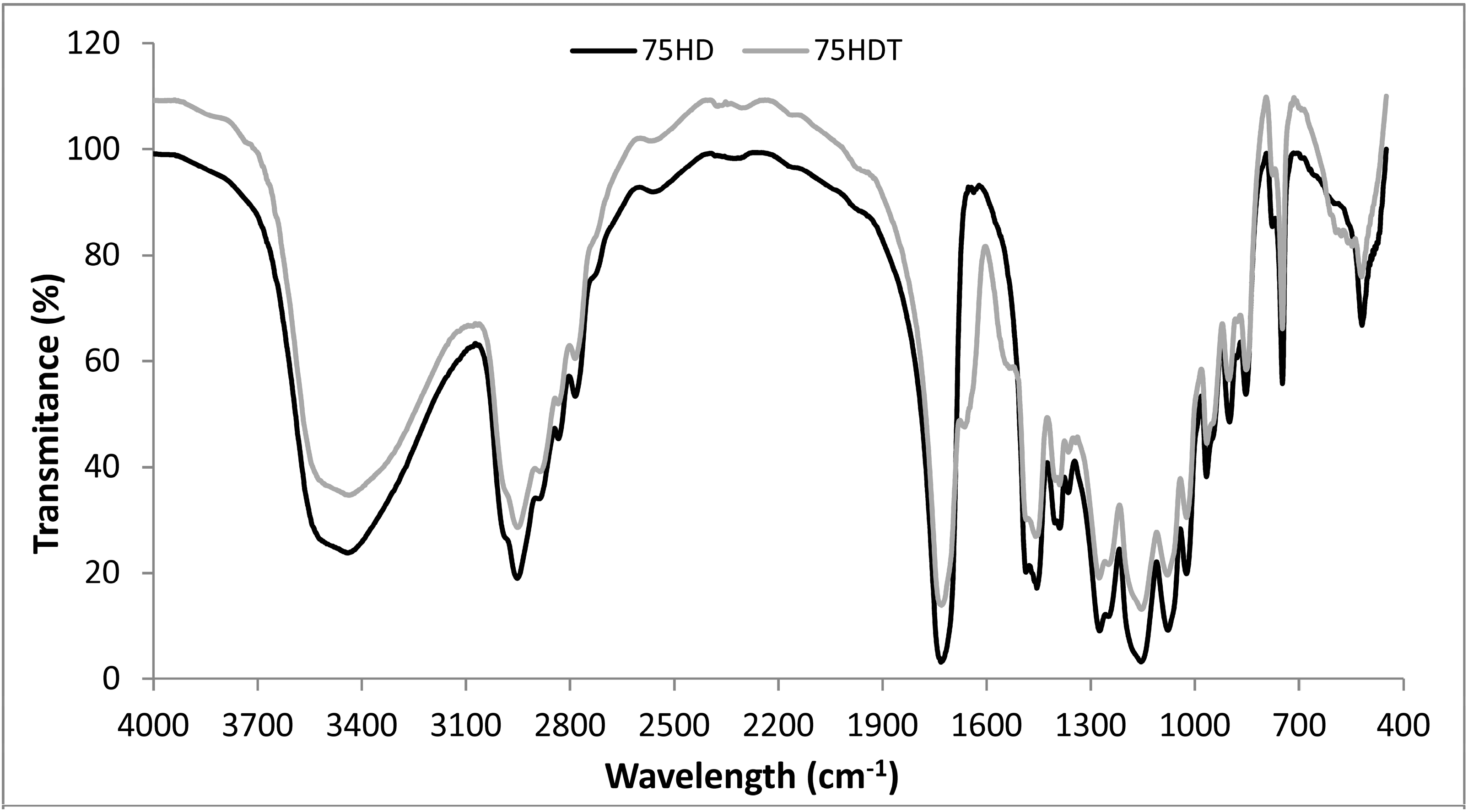
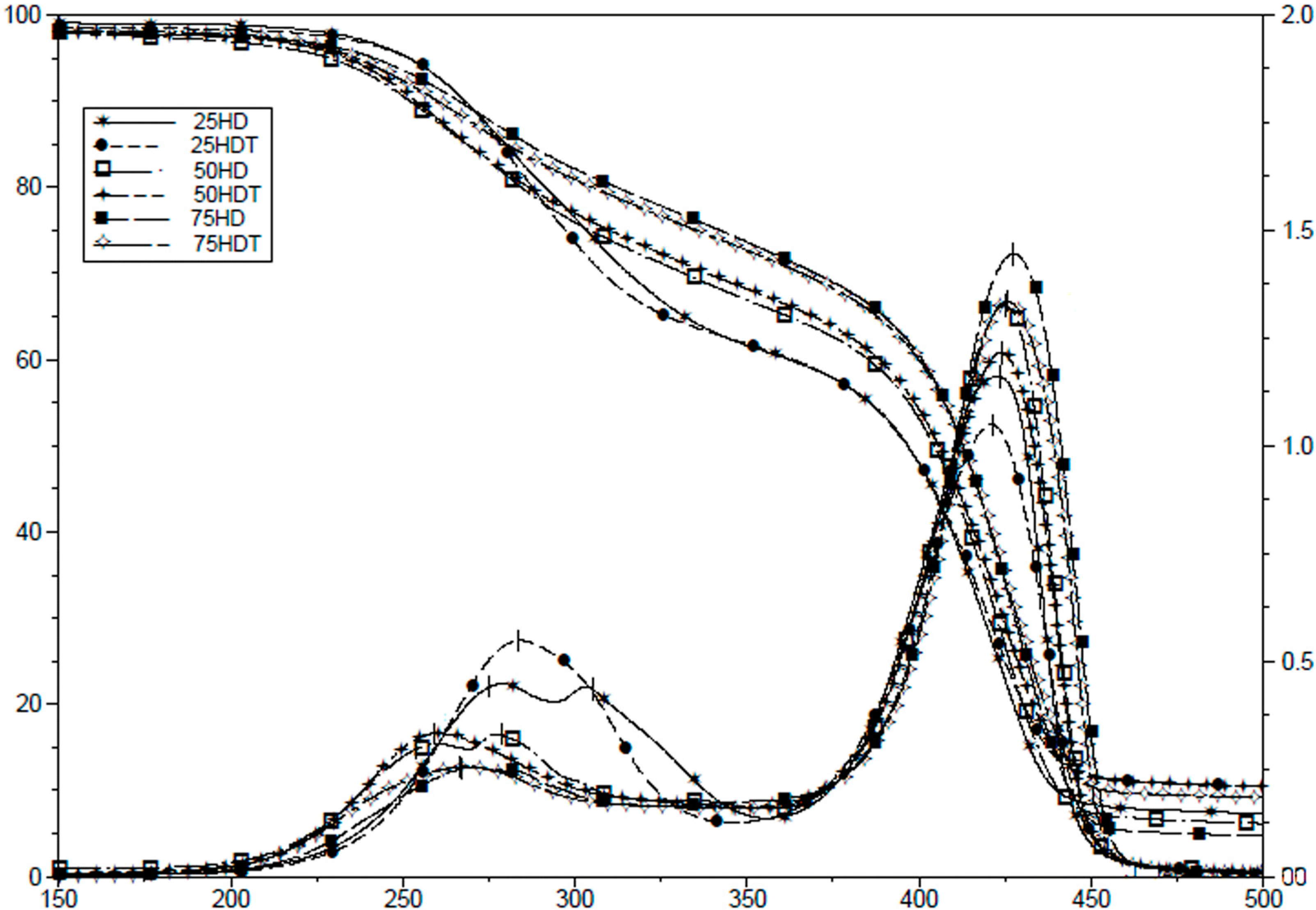
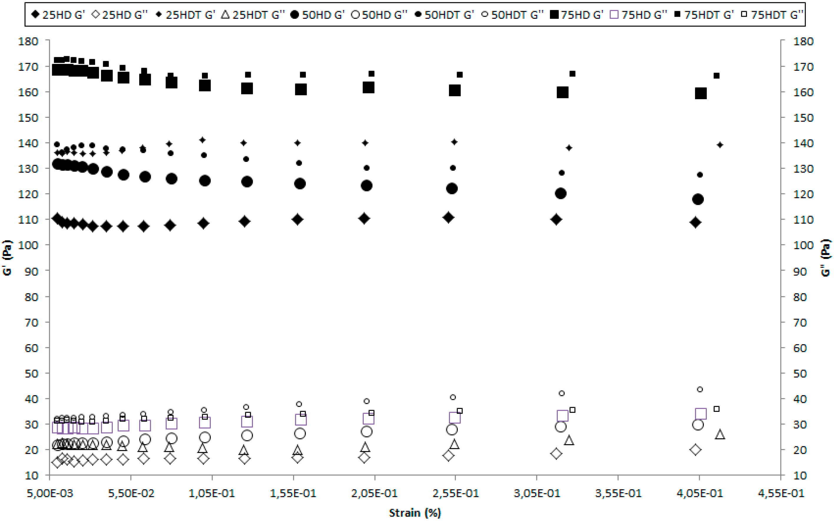
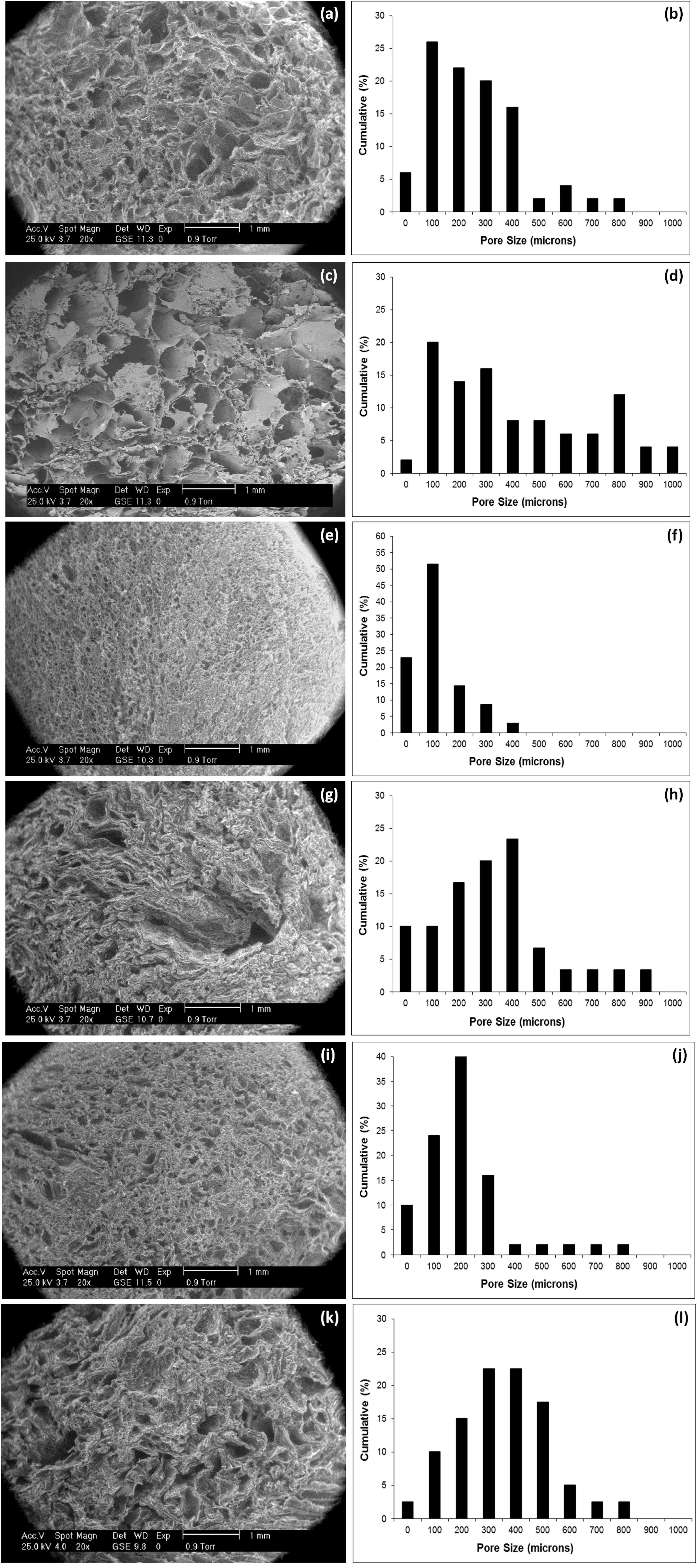
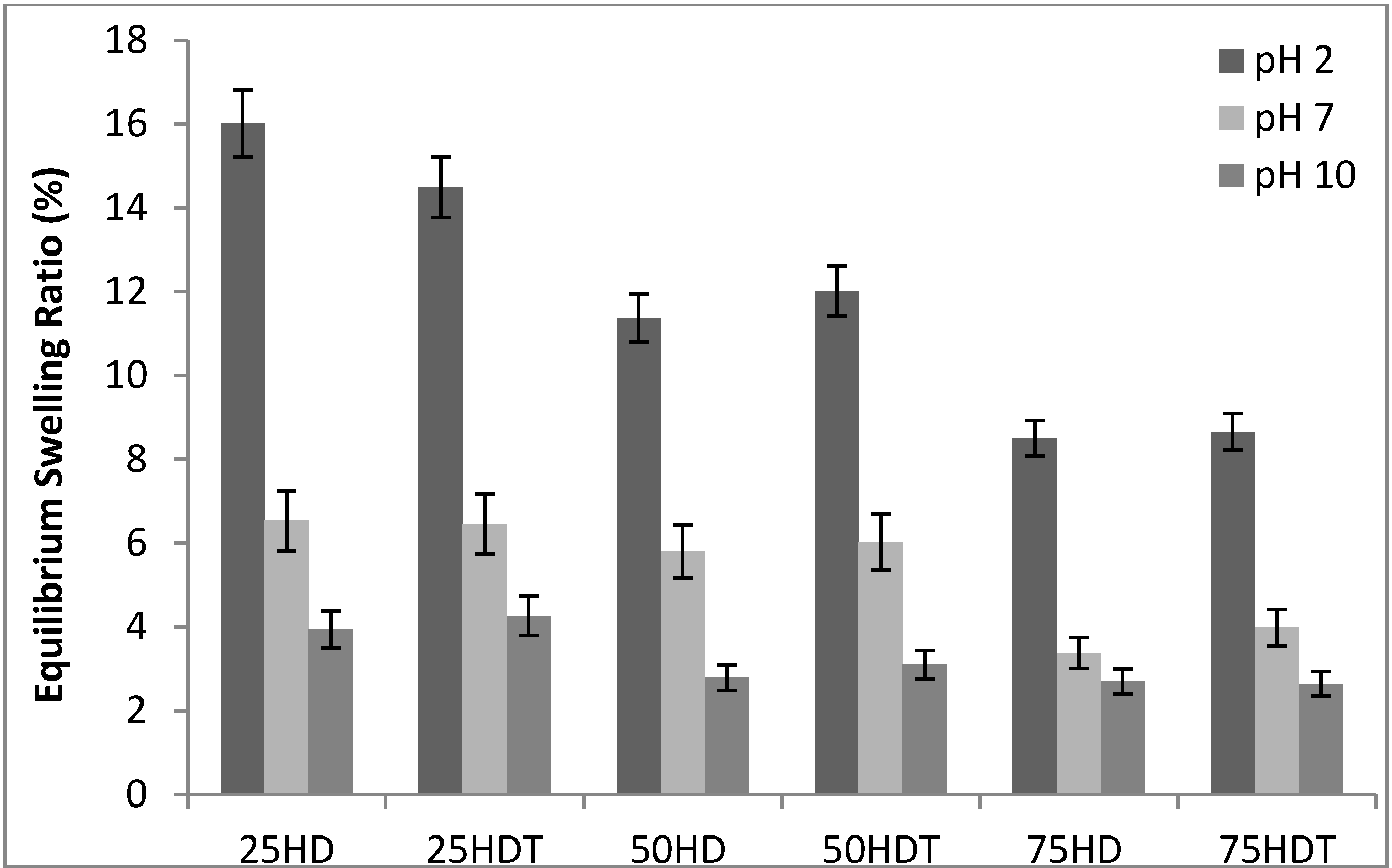
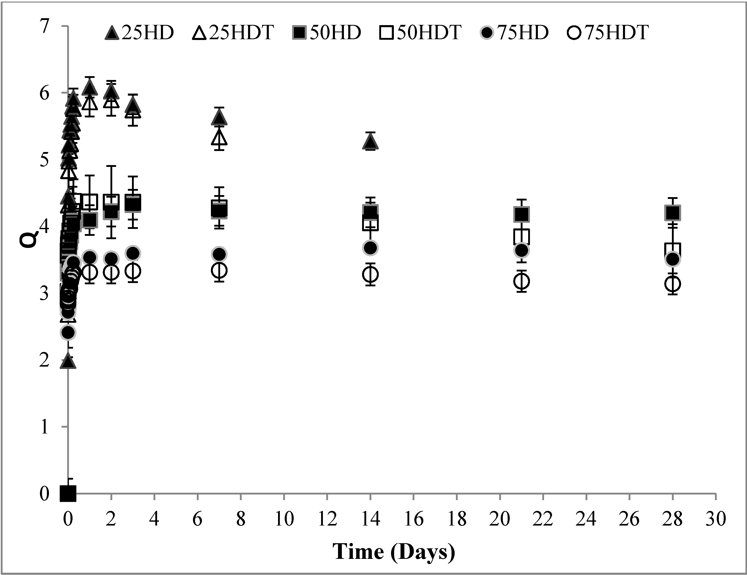
3.1. Scaffold Characterization

The obtained dry scaffolds are rigid bodies with a 13 mm diameter and up to 20 mm height. They are easily handled and can be sliced into the required size for testing.
FTIR spectra of the obtained compositions are displayed in Figure 1. Compositions with a greater DMAEMA percentage show stronger stretching vibration modes from the methyl group at 2940 cm−1 and from the dimethylamino group at 2819 and 2767 cm−1. These bands can only proceed from the DMAEMA component, (–N(CH3)2) [25]. The incorporation of α-TCP, resulted in the appearance of a band at 1139 cm−1 that can be assigned to the υ3 vibration mode from TCP [26] and hides the band around 1150 cm−1 assigned to –CN from the DMAEMA. Also bands at 1664 and 1524 cm−1 may be caused by electrostatic interactions between the tricalcium phosphate and free electrons on the DMAEMA’s carbonyl bond, suggesting a possible interaction between the ceramic and the polymeric components.
Figure 1. Fourier Transformed Infrared Spectroscopy for samples 75 HD and 75 HDT.
Polymers 06 02510 g001
The thermal stability of the cryogels and the influence of the incorporation of the ceramic phase have been evaluated by TGA as showed in Figure 2. Two weight loss steps can be appreciated for each composition. The first one centered between 254 and 305 °C depending on the composition and the second one centered between 421 and 425 °C, also depending on the composition. A greater weight loss can be observed for the first peak for greater percentages of DMAEMA. This step corresponds to the decomposition of the lateral chains of the copolymer and the greater weight loss can be attributed to the longer lateral chains of dimethylamino ethyl groups from PDMAEMA in comparison with hydroxyl ethyl groups from PHEMA. For 25 HD and 50 HD, this first step is split in two parts. The first one can be related to the DMAEMA’s amino group [25] and the second to the rest of both DMAEMA’s and HEMA’s lateral chains; For the α-TCP containing compositions, the lateral group decomposition occurred in only one step at 267 °C which seems to indicate an interaction between the ceramic and the dimethylamino groups from PDMAEMA. This is reinforced with the lack of influence of the ceramic phase in the decomposition temperature of the lateral chain for specimens with greater content in PHEMA. From the weight of the samples after heating at 500 °C the final ceramic contents of the specimens were calculated. Ceramic contents obtained for 25 HDT, 50 HDT and 75 HDT were 3.1%, 4.3% and 4.4% referring to the ceramic content. Theoretical percentage is 5%, in each case, related to initial monomer mass. The deviation from the initial content can be attributed to partial evaporation of the monomers during the mixing and homogenization procedures, but the variation is <2%, indicating that the ceramic particles do not interfere with the polymerization process and suggesting a reliable preparation procedure.
Figure 2. Thermogravimetric analysis of all studied compositions.
Polymers 06 02510 g002
The porosity of the prepared cryogels is displayed on Table 1. Porosities between 53% ± 5.7% and 75% ± 5.5% were observed. The incorporation of α-TCP seems to produce an increase in the porosity that can be related with the simultaneous addition of gelatin that is washed out on the late washing step [27,28].
Table 1. Porosity, and modal pore size of the specimens. Standard deviation is displayed on brackets.
SamplePorosity (%)Modal Size (μm)
25 HD66.7 (9.5)100–200
25 HDT75.2 (5.5)100–200
50 HD53.8 (5.7)100–200
50 HDT59.0 (7.3)400–500
75 HD59.3 (8.2)200–300
75 HDT73.4 (5.5)300 and 400
Figure 3 shows SEM pictures obtained from transversal cuts for each composition. Cryogels with a greater content on PDMAEMA, 25 HD and 25 HDT, display spherical pores, whereas cryogels with a greater content in PHEMA, 75 HD and 75 HDT, display pores with elongated shapes. PSD is also displayed in Figure 3. Pore sizes up to 900 μm were found for 25 HD and 75 HD and up to 500 μm for 50 HD. The modal size is smaller for 25 HD and 50 HD than for 75 HD as it can be seen in Table 1. The incorporation of tricalcium phosphate yields a wider PSD and pore modal size than for the TCP free specimens. The pore size ranges between 139 and 481 μm for each of the prepared compositions, which seems appropriate for a bone-engineering scaffold [29].
The equilibrium-swelling ratio at pH 7.4 is included in Figure 4. The greater Q observed for 25 HD in comparison with the observed for 75 HD can be attributed to the greater presence of protonable amino groups that induce a greater hydrophilic character to the specimens [30].
Figure 4 shows the Q of the cryogels at different pHs. The figure shows that all the specimens maintain the pH sensitivity whatever the content of the PDMAEMA component is [13].
The PDMAEMA is a well-known pH sensitive polymer [31,32,33] Yangfeng and Min studied the swelling kinetic and stimuli response of hydrogels containing DMAEMA as the main monomer and found that for a pH as low as 3.0, a drastic increase in the Q can be observed. The same behavior was found for the cryogels obtained in this work. The composition 25 HDT shows an equilibrium-swelling ratio around 16 at pH 2.0, whereas the Q was 8.5 for the composition 75 HDT. However, at pH 10.0, the Q value was 4.2 and 2.6 for samples 25 HDT and 75 HDT, respectively.
Rheology measurements are displayed in Figure 5. The gel quality, of the hydrated specimens, is confirmed by the greater Gꞌ value when it is compared with the Gꞌꞌ value at the same frequency. A greater Gꞌ can be observed for 75 HD than for 50 HD and 25 HD. The addition of the ceramic component does not induce any significant change on the Gꞌ values of the cryogels from the value of the analogous α-TCP free composition.
Figure 3. Scanning Electronic Microscopy and Pore size distribution graphics of samples (a) and (b) 25 HD, (c) and (d) 25 HDT, (e) and (f) 50 HD, (g) and (h) 50 HDT, (i) and (j) 75 HD and (k) and (l) 75 HDT, respectively.
Polymers 06 02510 g003
Figure 4. Equilibrium-swelling ratio (Q) of the cryogels at different pHs.
Polymers 06 02510 g004
In order to study the long-term behavior of the synthesized cryogels, samples were incubated for 28 days in PBS (Phosphate Buffer Saline, pH 7.4) at 37 °C and the results are displayed in Figure 6.
After 28 days immersed in PBS, α-TCP containing samples showed a higher degradation process, probably due the degradation process of the calcium phosphate [34]. Since the α-TCP used as reinforcement as a particle, it is expected a higher degradation rate due to its high specific surface area compared to its volume. Sample 25 HDT lost the mechanical integrity after 7 days of immersion, while 25 HD after 14 days. While samples containing 50% and 75% of HEMA started to show a tendency to loss mass after 21 days. Despite cryogels with a greater content in PDMAEMA, (25 HD) provide with a greater Q, that may favor cell penetration [12], samples containing higher DMAEMA amounts showed poorer mechanical properties and degraded after 7 or 14 days, for samples 25 HDT and 25 HD, respectively, showing that samples containing higher amounts of DMAEMA are not suitable for bone repair applications.
Figure 5. Rheology measurements of cryogels.
Polymers 06 02510 g005
Figure 6. Swelling behavior after 28 days of immersion in PBS at 37 °C.
Polymers 06 02510 g006

3.2. Biological Analysis

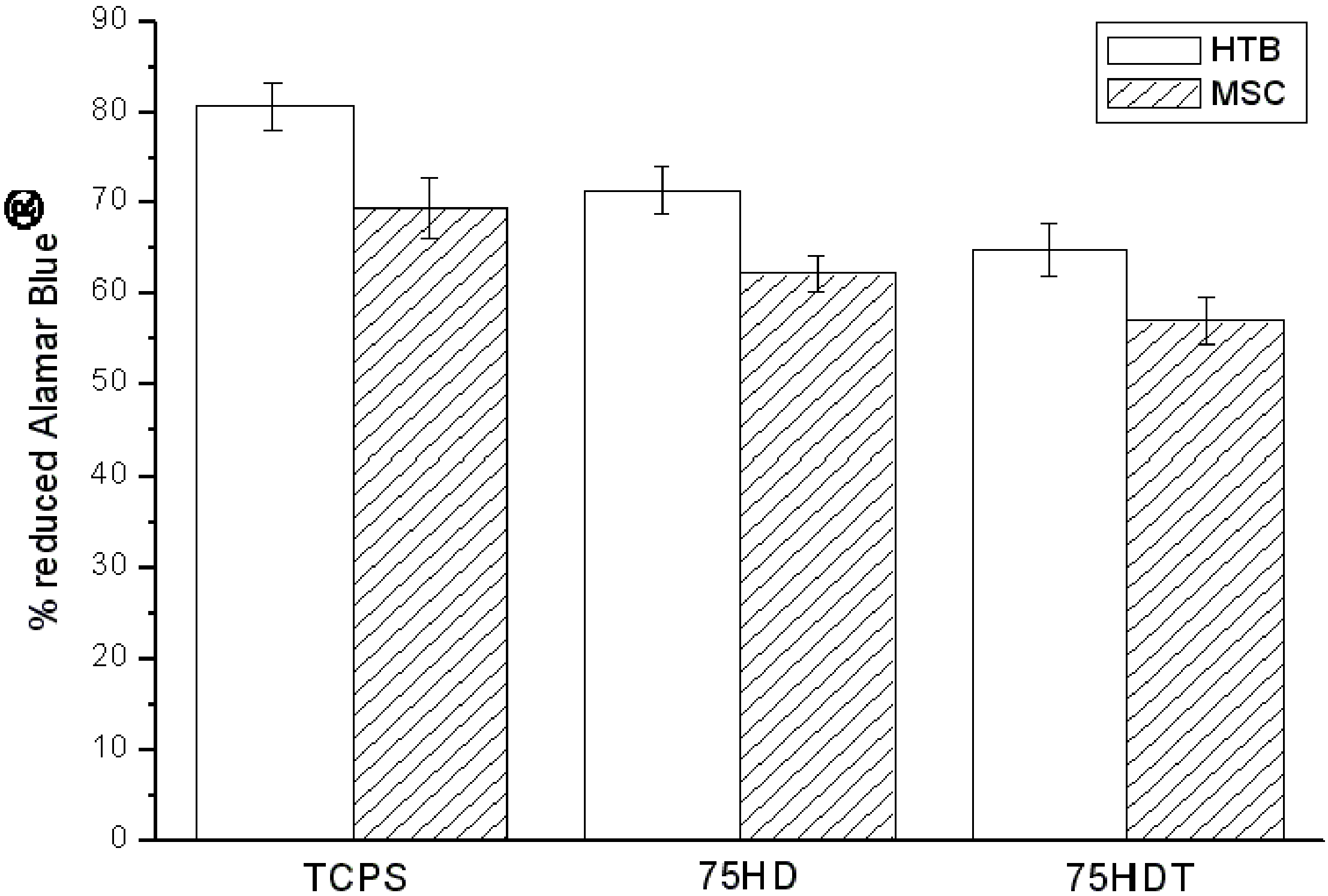
3.2.1. HTB and BM-MSC Metabolic Activity

HTB and BM-MSCs were cultured on 75 HD and 75 HDT. The viability of the different cell types was quantitatively assessed, through the Alamar Blue® assay. Figure 7 represents the metabolic activity of the cells, after 96 h seeding. The percentage of the reduced dye was not significantly altered (p > 0.05) by the presence of the different materials in comparison with the cells grown on tissue culture polystyrene (TCPS, positive control). For both BM-MSCs and HTBs, a similar behaviour was observed. In addition, the introduction of a 5% α-TCP did not induce a significant decrease in the cells metabolic activity, (p > 0.05). Nevertheless both 75 HD and 75 HDT cryogels were non-toxic and the seeded cells remained viable.
Figure 7. Metabolic activity of human trabecular bone-derived cells (HTB) and bone marrow derived mesenchymal stem cells (BM-MSC) through the Alamar Blue® assay.
Polymers 06 02510 g007

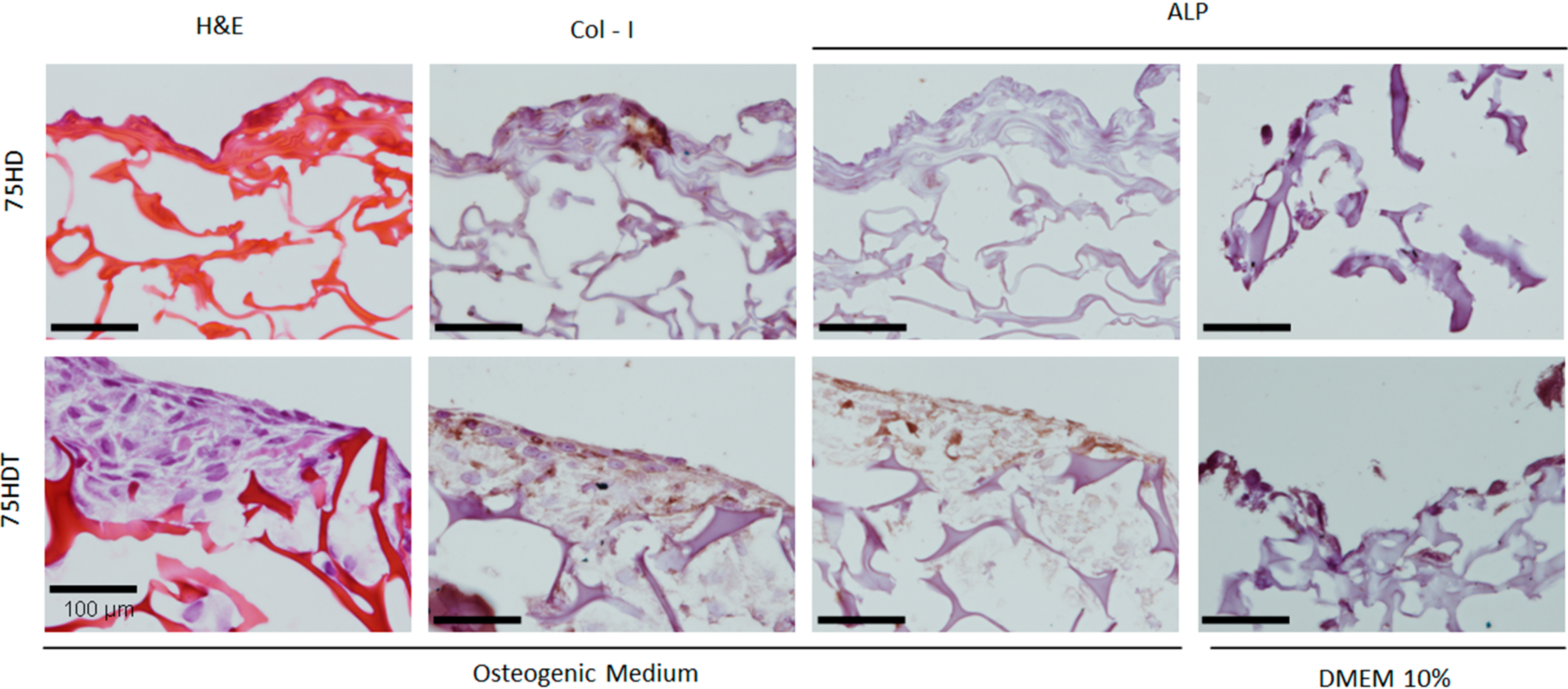
3.2.2. Histological and Immunohistochemical Analysis

The histological observations of the cryogels seeded with BM-MSCs are shown in Figure 8. The HE staining revealed differences in cell distribution between the different compositions. For cryogels with no α-TCP (75 HD), cells were mainly found at the surface. 75 HDT cryogels showed MSCs proliferating on the surface and into the bulk of the materials, as well as the formation of an extracellular matrix. We believe that the different pore sizes and porosity presented by 75 HD and 75 HDT may have influenced the different cell response on both the materials. Thus, higher porosity (734% ± 5.5%, Table 1) and pore size (300–400 μm, Table 1) presented by 75 HDT, have supported better cell infiltration and seems appropriate for a bone engineering scaffold [33]. Other studies have focused on the cellular response to pore structure. Spherical pore shapes will act as ideal surfaces for the ridges by providing a surface for ease of cell spreading, angular pores, similar to the ones 75 HD and 75 HDT present, will suspend cell movement within the scaffold [35].
Figure 8. Human MSCs on 75 HD and 75 HDT scaffolds, after 21 days culture on osteogenic or control medium (DMEM (Dulbecco’s Modified Eagles Medium) 10%): hematoxylin and eosin histologic staining (H&E) and immunolocalization of type-I collagen (col-I) and alkaline phosphatase (ALP), (magnification 20×).
Polymers 06 02510 g008
The differentiation of bone marrow-derived mesenchymal stem cells was assessed by immunohistochemistry. The osteogenic differentiation process comprises three main biological phases: cellular proliferation, cellular maturation and matrix mineralization. Type I collagen is expressed during the initial period of proliferation and extracellular matrix (ECM) biosynthesis, whereas alkaline phosphatase is expressed during the post-proliferative period of ECM maturation and other proteins occur later during the third period of mineralization. As shown in Figure 8, col-I was present in both materials, under osteogenic inducing conditions. Additionally, ALP could be visualized, although not on many cells, only on the α-TCP-containing cryogels, and it seems that the presence of α-TCP may have incited a higher expression of this protein. This relation was also observed when non-osteogenic differentiation medium was used.
The tricalcium phosphate incorporation on cryogels does not affect significantly the Gꞌ value of the cryogels, indicating that new crosslinks are not induced, as it has been described for other systems [12,36]. Despite this fact, an improvement in the biological response of the cryogels has been found as a result of the ceramic incorporation.
In summary, α-TCP incorporation to PDMAEMA/PHEMA cryogels had a positive effect on a biological level, after 14 days in culture, as it allowed for a better cell infiltration and matrix secretion, as well as the expression of bone related proteins. The specific role of α-TCP and the mechanisms involved in the observed cell response should be further evaluated.

4. Conclusions

Porous scaffolds based on the system Poly 2-(dimethylamino)ethyl Methacrylate/(2-Hydroxyethyl) Methacrylate MAEMA/PHEMA reinforced with α-tricalcium phosphate can be prepared by cryopolymerization. The properties of the scaffolds can be tuned by modifying the PDMAEMA/PHEMA ratio and by the incorporation of α-TCP. Specimens with a greater content in PHEMA seem to provide with a better structural environment for their use as scaffolds for bone engineering repair and the incorporation of α-TCP seems to favor the post-proliferative period of MSCs osteogenic differentiation, during the maturation of the extracellular cell matrix.

Acknowledgments

Financial support from projects MAT2010-18155, from Comision Interministerial de Ciencia y Tecnología (CICYT), Spain and PLE (2009-0144), ACI-PROMOCIONA (ACI2010-1095), from the Ministry of Science and Education (MICINN), Spain are gratefully acknowledged. CIBER-BBN is an initiative from ISCIII. The authors also gratefully acknowledged the Scholarship from the Brazilian National Council for Scientific and Technological Development (CNPq), process 202018/2008-3.

Author Contributions

Tiago Volkmer worked on the cryogels synthesis and characterization and wrote a draft of the paper. Vania Sousa worked on the synthesis of the α-tricalcium phosphate and in the data analysis. Luis M. Rodríguez-Lorenzo worked on the data analysis and designed the experiments with Julio San Román. Joana Magalhães and Francisco J. Blanco designed the biological characterization. Elena F. Burguera worked on data acquisition. Joana Magalhães, Elena F. Burguera and Francisco J. Blanco analyzed and interpreted the data. Luis A. Santos, Luis M. Rodríguez-Lorenzo and Joana Magalhães critically reviewed the manuscript.

Conflicts of Interest

The authors declare no conflict of interest.

References

  1. Kirsebom, H.; Aguilar, M.R.; Roman, J.S.; Fernandez, M.; Prieto, M.A.; Bondar, B. Macroporous scaffolds based on chitosan and bioactive molecules. J. Bioact. Compat. Polym. 2007, 22, 621–636. [Google Scholar]
  2. Dispinar, T.; van Camp, W.; de Cock, L.J.; de Geest, B.G.; du Prez, F.E. Redox-responsive degradable PEG cryogels as potential cell scaffolds in tissue engineering. Macromol. Biosci. 2012, 12, 383–394. [Google Scholar] [PubMed]
  3. Lozinski, V.I. Cryogels on teh basis of natural and synthetic polymers: Preparation, properties and applications. Russ. Chem. Rev. 2002, 71, 489–511. [Google Scholar]
  4. Yao, K.J.; Shen, S.C.; Yun, J.X.; Wang, L.H.; He, X.J.; Yu, X.M. Preparation of polyacrylamide-based supermacroporous monolithic cryogel beds under freezing-temperature variation conditions. Chem. Eng. Sci. 2006, 61, 6701–7608. [Google Scholar]
  5. Kirsebom, H.; Rata, G.; Topgaard, D.; Mattiasson, B.; Galaev, I.Y. Mechanism of Cryopolymerization: Diffusion-controlled polymerization in a nonfrozen microphase. An NMR study. Macromolecules 2009, 42, 5208–5214. [Google Scholar]
  6. Arvidsson, P.; Plieva, F.M.; Lozinsky, V.I.; Galaev, I.Y.; Mattiasson, B. Direct chromatographic capture of enzyme from crude homogenate using immobilized metal affinity chromatography on a continuous supermacroporous adsorbent. J. Chromatogr. A 2003, 986, 275–290. [Google Scholar] [PubMed]
  7. Kumar, A.; Bansal, V.; Nandakumar, K.S.; Galaev, I.Y.; Roychoudhury, P.K.; Holmdahl, R.; Mattiasson, B. Integrated bioprocess for the production and isolation of urokinase from animal cell culture using supermacroporous cryogel matrices. Biotechnol. Bioeng. 2006, 93, 636–646. [Google Scholar] [PubMed]
  8. Le Noir, M.; Plieva, F.; Hey, T.; Guieysse, B.; Mattiasson, B. Macroporous molecularly imprinted polymer/cryogel composite systems for the removal of endocrine disrupting trace contaminants. J. Chromatogr. A 2007, 1154, 158–164. [Google Scholar]
  9. Tripathi, A.; Kumar, A. Multi-featured macroporous agarose-alginate cryogel: Synthesis and characterization for bioengineering applications. Macromol. Biosci. 2011, 11, 22–35. [Google Scholar] [PubMed]
  10. Bolgen, N.; Plieva, F.; Galaev, I.Y.; Mattiasson, B.; Piskin, E. Cryogelation for preparation of novel biodegradable tissue-engineering scaffolds. J. Biomater. Sci. Polym. Ed. 2007, 18, 1165–1179. [Google Scholar] [PubMed]
  11. Kirsebom, H.; Elowsson, L.; Berillo, D.; Cozzi, S.; Inci, I.; Piskin, E.; Galaev, I.Y.; Mattiasson, B. Enzyme-catalyzed crosslinking in a partly frozen state: A new way to produce supermacroporous protein structures. Macromol. Biosci. 2013, 13, 67–76. [Google Scholar] [PubMed]
  12. Rodriguez-Lorenzo, L.M.; Saldana, L.; Benito-Garzon, L.; García-Carrodeguas, R.; de Aza, S.; Vilaboa, N.; Román, J.S. Feasibility of ceramic-polymer composite cryogels as scaffolds for bone tissue engineering. J. Tissue Eng. Regen. Med. 2012, 6, 421–433. [Google Scholar] [PubMed]
  13. Brahim, S.; Narinesingh, D.; Guiseppi-Elie, A. Synthesis and hydration properties of pH-sensitive p(HEMA)-based hydrogels containing 3-(trimethoxysilyl)propyl methacrylate. Biomacromolecules 2003, 4, 497–503. [Google Scholar] [PubMed]
  14. Cherng, J.Y.; vandeWetering, P.; Talsma, H.; Crommelin, D.J.A.; Hennink, W.E. Effect of size and serum proteins on transfection efficiency of poly((2-dimethylamino)ethyl methacrylate)-plasmid nanoparticles. Pharm. Res. 1996, 13, 1038–1042. [Google Scholar] [PubMed]
  15. Maji, S.; Mitschang, F.; Chen, L.N.; Jin, Q.; Wang, Y.X.; Agarwal, S. Functional poly(dimethyl aminoethyl methacrylate) by combination of radical ring-opening polymerization and click chemistry for biomedical applications. Macromol. Chem. Phys. 2012, 213, 1643–1654. [Google Scholar]
  16. Aguilar, M.R.; Gallardo, A.; Lechuga, L.M.; Calle, A.; San Roman, J. Modulation of proteins adsorption onto the surface of chitosan complexed with anionic copolymers. real time analysis by surface plasmon resonance. Macromol. Biosci. 2004, 4, 631–638. [Google Scholar] [PubMed]
  17. Bosetti, M.; Boccafoschi, F.; Calarco, A.; Leigheb, M.; Gatti, S.; Piffanelli, V.; Peluso, G.; Cannas, M. Behaviour of human mesenchymal stem cells on a polyelectrolyte-modified HEMA hydrogel for silk-based ligament tissue engineering. J. Biomater. Sci. Polym. Ed. 2008, 19, 1111–1123. [Google Scholar] [CrossRef] [PubMed]
  18. Volkmer, T.M.; Lengler, F.; Barreiro, O.; Sousa, V.C.; dos Santos, L.A. Novel method for the obtainment of nanostructured calcium phosphate cements: Synthesis, mechanical strength and cytotoxicity. Powder Technol. 2013, 235, 599–605. [Google Scholar]
  19. Gallagher, J.A. Human osteoblast culture. Methods Mol. Med. 2003, 80, 3–18. [Google Scholar] [PubMed]
  20. Richler, C.; Yaffe, D. The in vitro cultivation and differentiation capacities of myogenic cell lines. Dev. Biol. 1970, 23, 1–22. [Google Scholar] [PubMed]
  21. Jaiswal, N.; Haynesworth, S.E.; Caplan, A.I.; Bruder, S.P. Osteogenic differentiation of purified, culture-expanded human mesenchymal stem cells in vitro. J. Cell. Biochem. 1997, 64, 295–312. [Google Scholar] [PubMed]
  22. Anderson, J.M.; Vines, J.B.; Patterson, J.L.; Chen, H.; Javed, A.; Jun, H.W. Osteogenic differentiation of human mesenchymal stem cells synergistically enhanced by biomimetic peptide amphiphiles combined with conditioned medium. Acta Biomater. 2011, 7, 675–682. [Google Scholar] [PubMed]
  23. Hollander, A.P.; Hatton, P.V. Biopolymer Methods in Tissue Engineering Methods in Molecular Biology; Humana Press: New York, NY, USA, 2006; pp. 147–157. [Google Scholar]
  24. Magalhaes, J.; Sousa, R.A.; Mano, J.F.; Reis, R.L.; Blanco, F.J.; San Roman, J. Synthesis and characterization of sensitive hydrogels based on semi-interpenetrated networks of poly 2-ethyl-(2-pyrrolidone) methacrylate and hyaluronic acid. J. Biomed. Mater. Res. Part A 2013, 101A, 157–166. [Google Scholar]
  25. Roy, D.; Guthrie, J.T.; Perrier, S. Synthesis of natural-synthetic hybrid materials from cellulose via the RAFT process. Soft Matter 2008, 4, 145–155. [Google Scholar]
  26. Fahami, A.; Nasiri-Tabrizi, B.; Ebrahimi-Kahrizsangi, R. Synthesis of calcium phosphate-based composite nanopowders by mechanochemical process and subsequent thermal treatment. Ceram. Int. 2012, 38, 6729–6738. [Google Scholar]
  27. Jain, E.; Srivastava, A.; Kumar, A. Macroporous interpenetrating cryogel network of poly(acrylonitrile) and gelatin for biomedical applications. J. Mater. Sci. Mater. Med. 2009, 20, 173–179. [Google Scholar]
  28. Dainiak, M.B.; Allan, I.U.; Savina, I.N.; Cornelio, L.; James, E.S.; James, S.L.; Mikhalovsky, S.V.; Jungvid, H.; Galaev, I.Y. Gelatin-fibrinogen cryogel dermal matrices for wound repair: Preparation, optimisation and in vitro study. Biomaterials 2010, 31, 67–76. [Google Scholar] [PubMed]
  29. Hutmacher, D.W. Scaffolds in tissue engineering bone and cartilage. Biomaterials 2000, 21, 2529–2543. [Google Scholar] [PubMed]
  30. You, J.O.; Auguste, D.T. The effect of swelling and cationic character on gene transfection by pH-sensitive nanocarriers. Biomaterials 2010, 31, 6859–6866. [Google Scholar] [PubMed]
  31. Munoz-Bonilla, A.; Fernandez-Garcia, M.; Haddleton, D.M. Synthesis and aqueous solution properties of stimuli-responsive triblock copolymers. Soft Matter 2007, 3, 725–731. [Google Scholar]
  32. Sui, X.; Yuan, J.; Zhou, M.; Zhang, J.; Yang, H.; Yuan, W.; Wei, Y.; Pan, C. Synthesis of cellulose-graft-poly(N,N-dimethylamino-2-ethyl methacrylate) copolymers via homogeneous ATRP and their aggregates in aqueous media. Biomacromolecules 2008, 9, 2615–2620. [Google Scholar] [PubMed]
  33. Chen, Y.; Yi, M. Swelling kinetics and stimuli-responsiveness of poly(DMAEMA) hydrogels prepared by UV-irradiation. Radiat. Phys. Chem. 2001, 61, 65–68. [Google Scholar]
  34. Grandi, G.; Heitz, C.; Santos, L.A.; Silva, M.L.; Filho, M.S.; Pagnocelli, R.M.; Silva, D.N. Comparative histomorphometric analysis between α-TCP and β-TCPHA granules in the bone repair of rat calvaria. Mater. Res. 2011, 14, 11–16. [Google Scholar]
  35. Gross, K.A.; Rodríguez-Lorenzo, L.M. Biodegradable composite scaffolds with an interconnected spherical network for bone tissue engineering. Biomaterials 2004, 25, 4955–4962. [Google Scholar] [PubMed]
  36. Rodriguez-Lorenzo, L.M.; Garcia-Carrodeguas, R.; Rodriguez, M.A.; de Aza, S.; Jiménez, J.; López-Bravo, A.; Fernandez, M.; San Román, J. Synthesis, characterization, bioactivity and biocompatibility of nanostructured materials based on the wollastonite-poly(ethylmethacrylate-co-vinylpyrrolidone) system. J. Biomed. Mater. Res. Part A 2009, 88A, 53–64. [Google Scholar]

