Article

Phase Transformation of Adefovir Dipivoxil/Succinic Acid Cocrystals Regulated by Polymeric Additives

1
Department of Chemical Engineering, Soongsil University, Seoul 156-743, Korea
2
Clean Energy Research Center, Korea Institute of Science and Technology, Seoul 136-791, Korea
*
Author to whom correspondence should be addressed.
Polymers 2014, 6(1), 1-11; https://doi.org/10.3390/polym6010001
Submission received: 11 November 2013 / Revised: 13 December 2013 / Accepted: 17 December 2013 / Published: 20 December 2013
(This article belongs to the Special Issue Biomimetic Polymers)

Abstract

:
The polymorphic phase transformation in the cocrystallization of adefovir dipivoxil (AD) and succinic acid (SUC) was investigated. Inspired by biological and biomimetic crystallization, polymeric additives were utilized to control the phase transformation. With addition of poly(acrylic acid), the metastable phase newly identified through the analysis of X-ray diffraction was clearly isolated from the previously reported stable form. Without additives, mixed phases were obtained even at the early stage of cocrystallization. Also, infrared spectroscopy analysis verified the alteration of the hydrogen bonding that was mainly responsible for the cocrystal formation between AD and SUC. The hydrogen bonding in the metastable phase was relatively stronger than that in the stable form, which indicated the locally strong AD/SUC coupling in the initial stage of cocrystallization followed by the overall stabilization during the phase transformation. The stronger hydrogen bonding could be responsible for the faster nucleation of the initially observed metastable phase. The present study demonstrated that the polymeric additives could function as effective regulators for the polymorph-selective cocrystallization.

1. Introduction

Cocrystal formation of active pharmaceutical ingredients (APIs) is a method to control the physical properties of the solid phase APIs through the intermolecular interactions with the cocrystal formers (coformers) [1]. Some examples of the API cocrystals with improved properties are as follows. Hygroscopic behavior of theophylline and caffeine was lessened by forming cocrystals with dicarboxylic acids [2]. Stability against degradation of adefovir dipivoxil was increased by cocrystallization with saccharin [3]. Dissolution behavior and bioavailability of a development candidate API was improved when cocrystallized with glutaric acid [4]. In addition, the cocrystallization was also employed between two different APIs for the combined treatment as exemplified in the case of isoniazid and 4-aminosalicylic acid [5].
Polymorphism in crystallization is a ubiquitous phenomenon critically affecting the crystal properties. For example, cases of polymorphism in the molecular crystals have been widely studied to understand the changes in the physical properties of commercial and industrial importance [6]. Those in the inorganic crystals have been important problems especially in biomineralization to understand the unusual behavior of biological crystallization [7]. In contrast, the studies on the polymorphism in cocrystallization appear relatively at the early stage. While the intermolecular interactions within the cocrystals are usually designed for reliable cocrystal formation, subtle changes of the interactions and the subsequent differences in the molecular packing could result in the polymorphism of cocrystallization [8,9,10].
Macromolecules have been discovered as important regulators of the polymorphs in both natural and synthetic crystallization. For example, biological crystallization of calcium carbonate in mollusks is well known in the utilization of biomacromolecules to stabilize the metastable polymorphs [11,12,13]. Industrial crystallization of diverse inorganic and organic compounds, including pharmaceutical materials, has been guided using polymeric additives [14,15,16,17]. In contrast, similar control of the polymorphs (or phases) of cocrystals is yet to be thoroughly explored [18,19].
In the present study, we report an explicit case of using polymeric additives to regulate the polymorphism of pharmaceutical cocrystallization. The cocrystal of adefovir dipivoxil (AD) and succinic acid (SUC) was previous reported in its stable crystal structure of AD/SUC = 2:1 [20], and we herein investigated the effects of poly(acrylic acid) (PAA) and poly(ethylene glycol) (PEG) on the formation and stabilization of a metastable phase.

2. Experimental Section
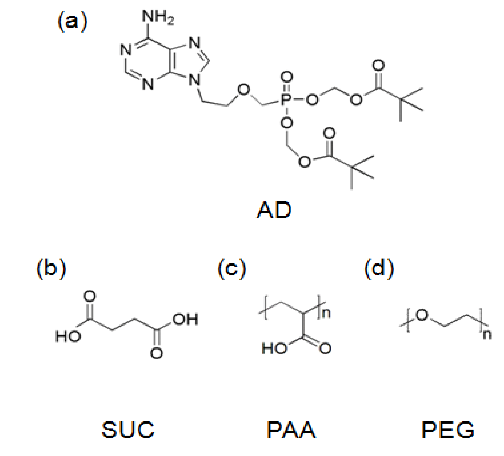
2.1. Materials

AD (L-form, > 99%) was provided by Amore Pacific Co. (Yongin, Korea) SUC (>99%), PAA (Mw 1800 g/mol), and PEG (Mw 8000 g/mol) were purchased from Sigma-Aldrich (Milwaukee, WI, USA). The chemical structures of AD, SUC, PAA, and PEG were shown in Figure 1. Ethanol (HPLC grade) was from Samchun Chemical (Pyeongtaek, Korea). Deionized water was obtained from Direct-Q (Millipore) (Billerica, MA, USA) with its resistivity over 18.2 MΩ·cm.
Figure 1. Chemical structures: (a) adefovir dipivoxil (AD); (b) succinic acid (SUC); (c) poly(acrylic acid) (PAA); (d) poly(ethylene glycol) (PEG).
Polymers 06 00001 g001

2.2. Formation of AD/SUC Cocrystals

Cocrystals of AD and SUC were produced by cooling homogeneous ethanol solutions of AD and SUC. The stoichiometric ratio of AD (0.4 mmol, 200 mg) and SUC (0.2 mmol, 23.6 mg) were dissolved in ethanol (3.0 mL) at 40–50 °C for 1 h. Glass vials were used with magnetic stir bars for mixing. The solution vials were then moved to a 25 °C incubator (BF-150LI; BioFree, Bucheon, Korea). The crystal formation was clearly noticeable after 1 day. The crystals were filtered (pore size 1 μm, cellulose acetate; Hyundae Micro, Seoul, Korea), thoroughly washed with deionzed water, and dried for 24 h in a 40 °C vacuum oven (J-DVO1; Jisico, Seoul, Korea). The filtration was either after 1 day for initially formed crystals or 7 days for mature crystals.
Cocrystal formation with polymeric additives was the same as the previous procedures except that the polymers were added at the same time when AD and SUC were dissolved in ethanol. The amount of the polymers were (AD + SUC)/polymer = 100:1 by weight. The resulting AD/SUC crystals were collected after 7 days.

2.3. Characterization

Crystal morphology was observed through optical microscopy (OM) using a BX51 microscope from Olympus (Tokyo, Japan). Reflectance mode was employed with cross polarization. The morphology of fine crystals was observed through scanning electron microscopy (SEM) using a field emission microscope (JSM-6700F) from JEOL (Tokyo, Japan). Thin platinum coating was applied to minimize charging during SEM observation.
Crystal structures were monitored with X-ray diffraction (XRD). XRD was performed using diffractometers from Bruker AXS (Billerica, MA, USA) with CuKα radiation (λ = 1.5406 Å) in the 2θ range of 6°–40°. D8 Discover diffractometer was operated at the conditions of 40 kV, 40 mA, and 1°/min; D2 Phaser was used at the conditions of 30 kV, 10 mA, and 1.2°/min.
Additional analyses were as follows. Fourier transformation infrared spectroscopy (FT-IR; Excalibur FTS 3000, Bio-Rad, Philadelphia, PA, USA) was performed using an accessory of diamond attenuated total reflectance without further grinding or additive. The IR spectra were obtained in the range of 3500–700 cm−1 with a spectral resolution of 4 cm−1. Differential scanning calorimetry (DSC; DSC 821e, Mettler-Toledo, Columbus, OH, USA) was used with a scanning rate of 10 °C/min. The DSC instrument was calibrated for enthalpy and temperature using indium. Nuclear magnetic resonance spectroscopy (NMR; Avance 500, Bruker, Billerica, MA, USA) was done for proton at 298 K. Solvent for NMR was hexadeuterodimethyl sulfoxide (DMSO-d6; 99.9%, Cambridge Isotope Laboratories, Inc., Tewksbury, MA, USA).

3. Results and Discussion

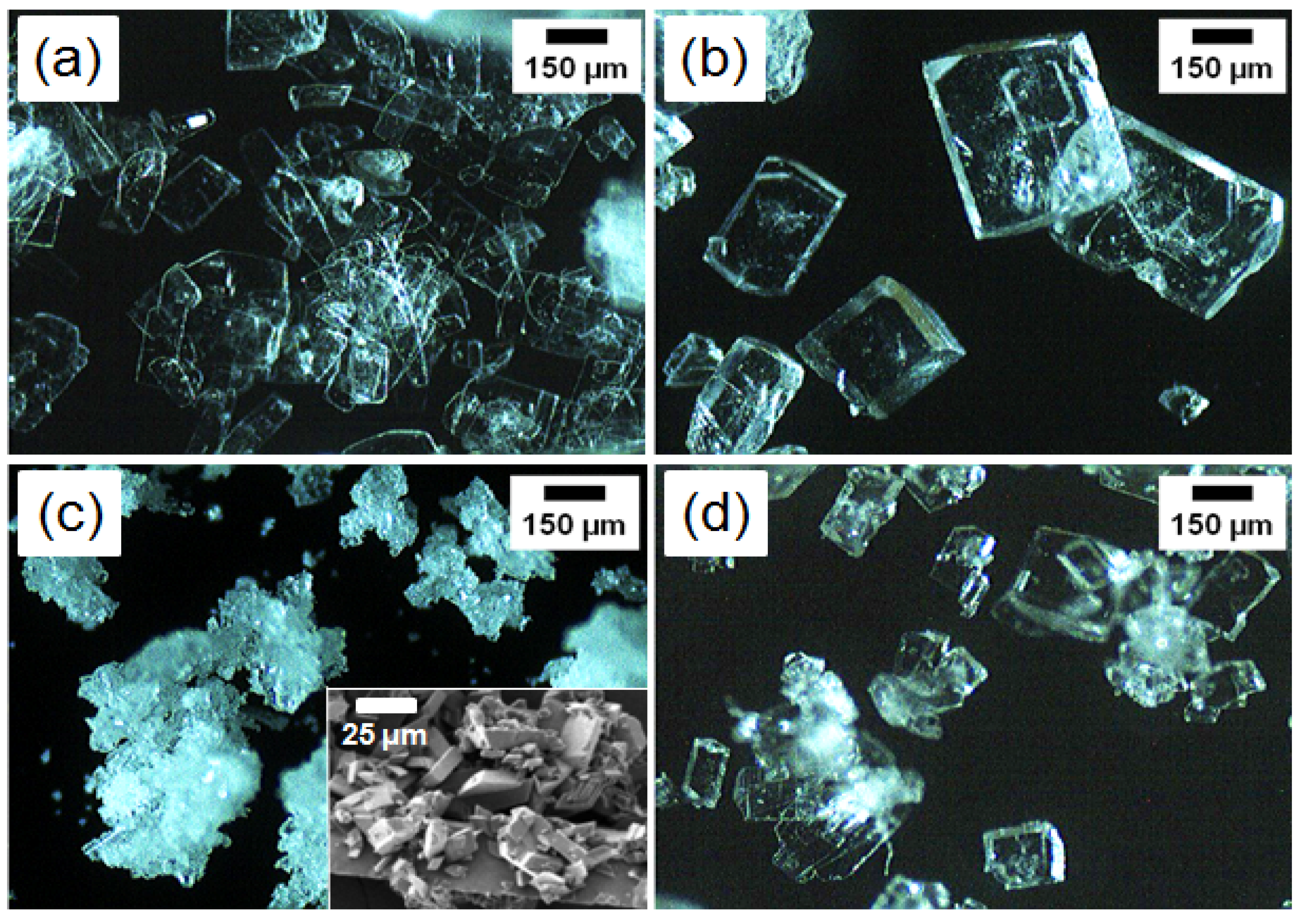
3.1. Microscopic Observation
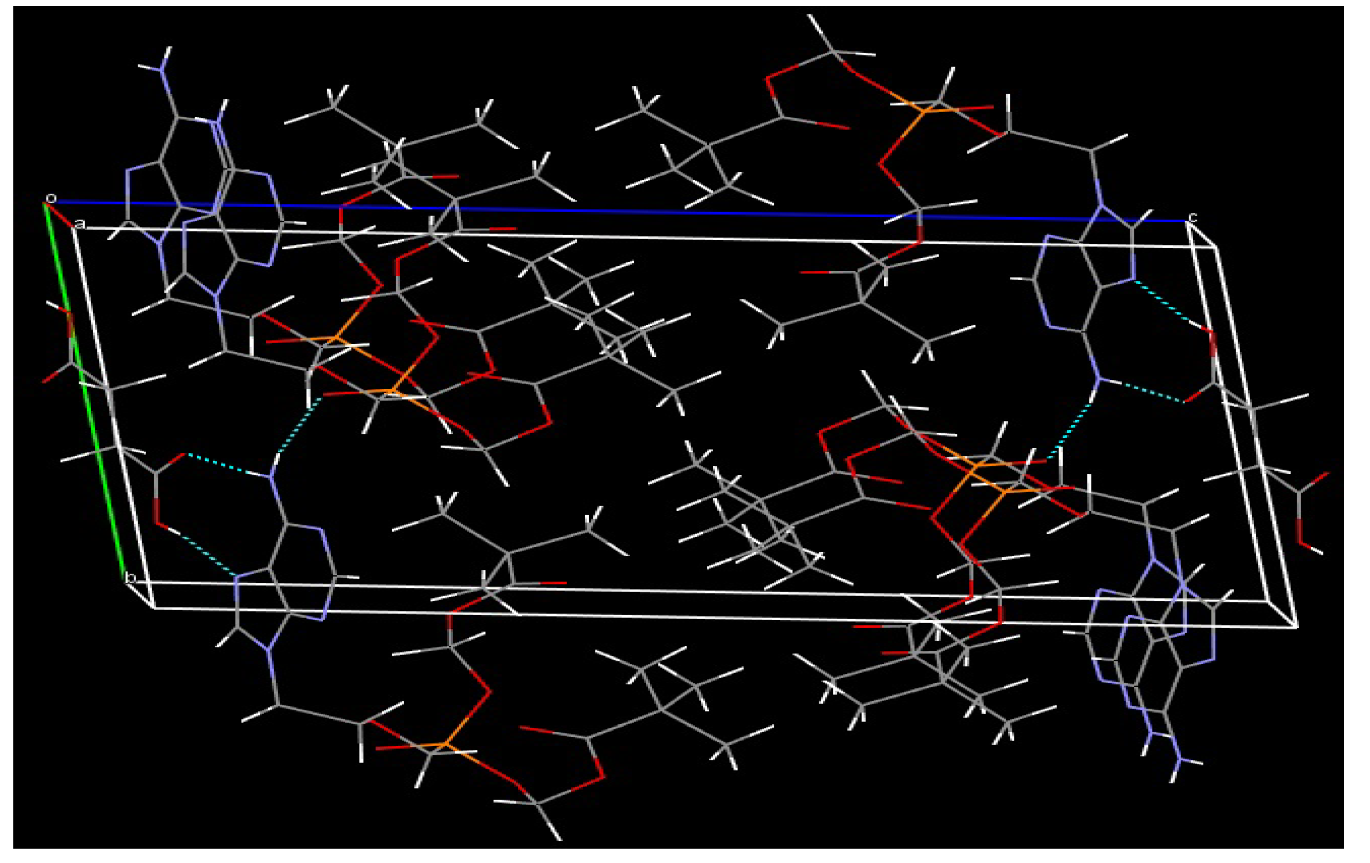
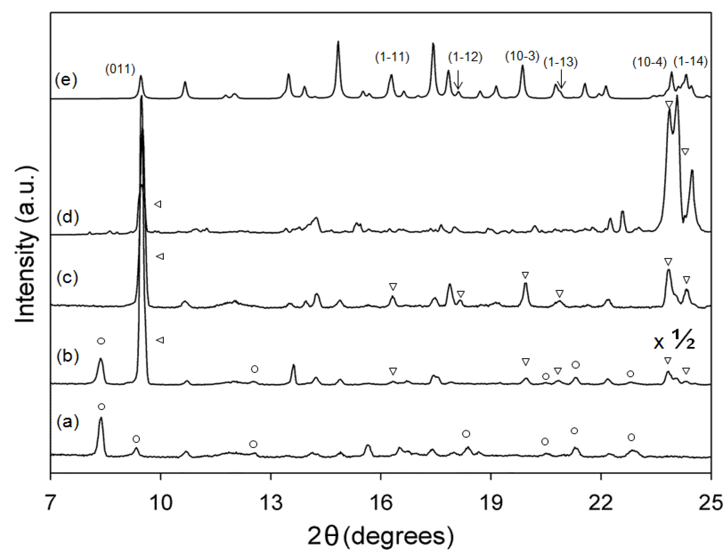
We previously reported the stable form II of AD/SUC cocrystal [20]. It has the AD/SUC = 2:1 composition, where the two AD molecules form hydrogen bonding with one SUC molecule via N–H---O=C (AD-SUC) and N---H–O (AD–SUC) hydrogen bonding (Figure 2). In addition, AD–AD intermolecular stabilization exists through N–H---O=P hydrogen bonding and π–π stacking. We decided to perform the regulated growth of the AD/SUC cocrystals with polymeric additives, because the previously reported form of the AD/SUC cocrystal appeared to stabilize after several days of crystal growth indicating the probable presence of the metastable form during the process.
Figure 2. The previously reported structure of the stable form II of AD/SUC cocrystal: hydrogen bonds between molecules are indicated in turquoise dotted lines [20].
Polymers 06 00001 g002
Microscopic observations of the morphology of the AD/SUC cocrystals were summarized in Figure 3. The initial formation of AD/SUC cocrystals without any additive started as thin plates as shown in the crystals of 1-day growth (Figure 3a), although a small amount of thicker crystals also appeared to exist at even earlier stages. After 7 days, the thin platelet crystals completely disappeared, and thicker cuboid crystals formed (Figure 3b). When PAA was present in the growth solution in the weight ratio of (AD + SUC)/PAA = 100:1, much smaller crystals were obtained in the aggregated fashion after 7 days (Figure 3c). The reduced size of the cocrystals in the presence of the polymeric additives, that had the similar functional groups to the coformer, was previously reported for the caffeine/oxalic acid system [21]. Closer observation, as shown in the inset of Figure 3c, revealed that the size distribution of the crystals seemed wider than the case without additive, although they were devoid of thicker ones. When PEG was present in the growth solution in the weight ratio of (AD + SUC)/PEG = 100:1, overall crystal morphology (7 days) was more or less the same as those obtained without any additives, but the crystal size was slightly smaller (Figure 3d). We designated the crystals of Figure 3a–c as form I + II, form II, and form I based on the microscopic observations and XRD analysis in the following section.
Figure 3. Optical microscopy (OM) and SEM micrographs of AD/SUC cocrystals, prepared by the precipitation from the AD/SUC = 2:1 solutions: (a) form I + II collected after 1 day; (b) form II collected after 7 day; (c) form I prepared with PAA (7 day); (d) crystals prepared with PEG (7 day). When polymers were added, the weight ratio in the growth solution was (AD + SUC)/polymer = 100:1.
Polymers 06 00001 g003

3.2. XRD Analysis

XRD analysis was performed to clearly identify the polymorph (or form) of the AD/SUC cocrystals obtained at various growth conditions (Figure 4). The sequence of the multiple XRD patterns of Figure 4 was arranged in such a way that the different forms could be easily discerned: cocrystals prepared with PAA (7 day) (Figure 4a: form I), collected after 1 day without additive (Figure 4b: form I + II), collected after 7 days without additive (Figure 4c: form II), and prepared with PEG (7 day) (Figure 4d). Also shown as Figure 4e was the XRD pattern calculated from the single crystal X-ray data of the previously reported stable form [20].
Figure 4. XRD patterns of AD/SUC cocrystals, prepared by the precipitation from the AD/SUC = 2:1 solutions: (a) form I prepared with PAA; (b) form I + II collected after 1 day without additive (Intensity was reduced to half (×1/2) for easy comparison.); (c) form II collected after 7 days without additive; (d) mostly form II prepared with PEG; (e) form II calculated from the single crystal X-ray data [20]. Some diffraction peaks from form I were marked as circles, and those from form II were identified with triangles.
Polymers 06 00001 g004
The diffraction positions of the XRD pattern calculated from the previously reported structure corresponded well with those of the form II cocrystals (Figure 4e vs. Figure 4c). Differences in the intensities of the specific peaks were probably from the preferred morphology and non-random orientation during solution growth and XRD measurement [22,23]. In contrast, several new diffraction peaks (marked as circles) were found for the form I + II cocrystals (Figure 4b), although a number of peaks were still in common with the form II samples (marked as triangles). When crystals were prepared with addition of PAA (form I), most diffraction peaks in common with form II samples disappeared while those newly found in the form I + II samples were retained (marked as circles) (Figure 4a). When crystals were prepared with addition of PEG, the overall diffraction pattern was more or less similar to the form II samples, although some minor new peaks appeared (Figure 4e).
Overall, the XRD results strongly indicated that the AD/SUC cocrystals experienced the phase transformation during crystal growth, which was in accordance with the morphology observation. The metastable form I started to transform to the stable form II (the previously reported structure) in less than 1 day, since the 1-day sample (form I + II) exhibited the diffraction pattern from both forms (marked as triangles and circles). The addition of PAA seemed to kinetically suppress the phase transformation to stabilize the metastable form I, while the effect of PEG appeared modest to generate inhomogeneous forms of crystals. Without PAA, the isolation of the metastable form I was difficult, probably because of the diverse nucleation time and ensuing phase transformation [23,24].

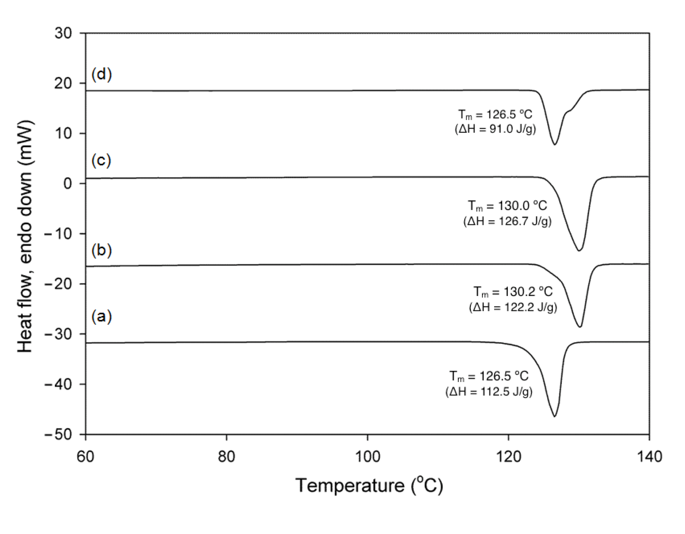
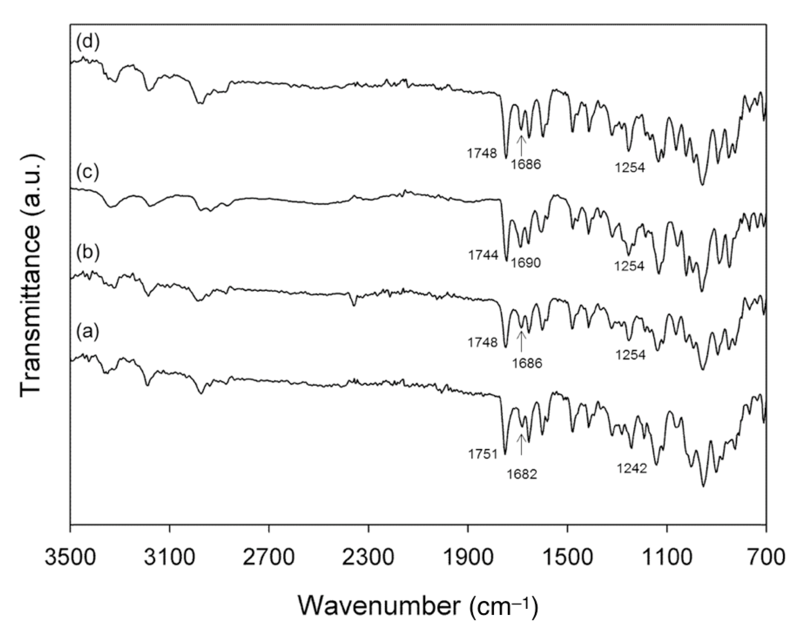
3.3. NMR, DSC and IR Analyses

NMR study showed that the form I and form II were identical in there composition. The composition was studied using the proton signal ratio between the alkane parts (–CH2–CH2–) of SUC (2.42 ppm) and the CH2 of AD (4.31–4.33 ppm) adjacent to the adenine group. The intensity ratio was 1.03:1 and 1.00:1 for form I and form II, respectively, which indicated that the AD/SUC = 2:1 composition was unaltered during the phase transformation. In fact, the NMR spectra of form I prepared with PAA was nearly indistinguishable from those of form II (data now shown), which also suggested that the amount of polymers adsorbed on the AD/SUC crystals and/or trapped between the crystal domains was very small (probably less than 2%) [25,26], and the polymers were not an integral part of the cocrystal.
DSC thermograms also displayed the subtle difference between the polymorphs of AD/SUC cocrystals as well as the effects of polymers (Figure 5). The difference in the melting behavior between the form I + II and II crystals is minute in that the melting enthalpy only slightly increased from ca. 122 J/g (1 day, form I + II) to ca. 127 J/g (7 days, form II) with the melting temperature about the same at 130 °C. The similar result was probably partly because the structural differences between the forms were only subtle, and partly because the form I+II sample was already fairly occupied by the stable form II as was shown in the XRD patterns. With addition of both PAA and PEG, the melting temperature decreased to ca. 126 °C, and the enthalpy decreased to 112 J/g with PAA (form I) and 91 J/g with PEG, respectively. The decreases of the melting temperature and enthalpy were difficult to analyze at this point because of the combined effects of polymorph change and probable mesocrystal formation that could lead imperfect crystallization caused by adventitious polymer inclusion [17,27,28]. Further study would be necessary to differentiate those effects. Note that the melting behavior of the crystals with PEG was distinctive in that inhomogeneous melting was observed, which indicated that the effect of PEG was not as defined as that of PAA. Overall, DSC results suggested that the differences between the polymorphs of AD/SUC cocrystals were subtle in their nature.
Figure 5. DSC thermograms showing the melting behaviors of AD/SUC cocrystals: (a) form I prepared with PAA; (b) form I + II collected after 1 day without additive; (c) form II collected after 7 days without additive; (d) mostly form II prepared with PEG.
Polymers 06 00001 g005
Figure 6. IR spectra of AD/SUC cocrystals, prepared by the precipitation from the AD/SUC = 2:1 solutions: (a) form I prepared with PAA; (b) form I + II collected after 1 day without additive; (c) form II collected after 7 days without additive; (d) mostly form II prepared with PEG.
Polymers 06 00001 g006
IR spectroscopy was performed to understand the changes in the intermolecular interactions accompanied by the cocrystal polymorphism (Figure 6). The vibrational wavenumbers of the functional groups in the strong hydrogen bonding (N–H---O=C (AD–SUC) and N–H---O=P (AD–AD), (Figure 2) showed progressive increases along the phase transformation [3,29,30,31] Form I (Figure 6a) showed the C=O stretching of SUC at 1682 cm−1, and P=O stretching at 1242 cm−1. Form I + II (Figure 6b) showed the C=O stretching of SUC at 1686 cm−1, and P=O stretching at 1254 cm−1. Form II (Figure 6c) exhibited the stretching of C=O at 1690 cm−1, and P=O at 1254 cm−1. Note that the N–H stretching peaks at about 3200 and 3300 cm−1 were too broad to accurately assess the differences, and the effects of hydrogen bonding on the N–H region were known to be weak and sometimes not very noticeable [32]. In addition, the wavenumber of C=O stretching of AD, which only weakly interacted with C–H of another AD molecule (Figure 2), decreased along the phase transformation: 1751 (form I), 1748 (form I+II), and 1744 (form II) cm−1 [3]. Also note that the IR spectra for the crystals obtained with PEG were in general similar to those of form I + II (Figure 6d).
Overall, the IR analysis indicated that the hydrogen bonds that sustained the AD/SUC cocrystal formation were adjusted as the cocrystal polymorphs changed. Specifically, the strong hydrogen bonding between the adjacent molecules became weaker as the metastable form I transformed to the stable form II, as evidenced by the increased vibrational frequency of C=O (SUC) and P=O (AD) groups [3,20,33]. It could be interpreted as that the initially AD and SUC formed locally strong interactions in the metastable form I, which seemed preferable for the preparatory AD–SUC complex formation. Then, the strong hydrogen bonds were attenuated with the globally stabilized interactions during the transformation to the stable form II. The increased interactions during the stabilization were in part indicated by the decreased vibrational frequency of C=O (AD) participating in the weak C=O---H–C (AD–AD) interactions [3,20,33].

4. Conclusions

In summary, the AD/SUC cocrystals exhibited polymorphic phase transformation, and the metastable form was successfully isolated with addition of PAA. It appeared to positively contribute to the regulation of the transformation that PAA possessed the functional group of carboxylic acid in common with the coformer, SUC. In addition, the analysis of the intermolecular interactions indicated that AD and SUC formed strong hydrogen bonding in the initially obtained metastable form I, which was relaxed in the globally stabilized form II. This could be the underlying reason for the faster nucleation of form I since the structures of strong hydrogen bonding could be favorable in overcoming activation energy of nucleation, where ethanol solvation had to be overcome. Further efforts, starting from the crystal structure solving of the metastable form, would be needed to fully understand the process of phase transformation. Finally, this study demonstrates that using the polymeric additives are useful for the polymorphic control of the pharmaceutical cocrystals, as have been shown in the various biological and biomimetic crystallization. We also believe that more studies with polymeric additives would contribute to the understanding of the nucleation and growth of cocrystallization.

Acknowledgments

This work was supported by the Soongsil University Research Fund of 2010.

Conflicts of Interest

The authors declare no conflict of interest.

References

  1. Jones, W.; Motherwell, W.D.S.; Trask, A.V. Pharmaceutical cocrystals: An emerging approach to physical property enhancement. MRS Bull. 2006, 31, 875–879. [Google Scholar] [CrossRef]
  2. Trask, A.V.; Motherwell, W.D.S.; Jones, W. Physical stability enhancement of theophylline via cocrystallization. Int. J. Pharm. 2006, 320, 114–123. [Google Scholar] [CrossRef]
  3. Gao, Y.; Gao, J.; Liu, Z.; Kan, H.; Zu, H.; Sun, W.; Zhang, J.; Qian, S. Coformer selection based on degradation pathway of drugs: A case study of adefovir dipivoxil-saccharin and adefovirdipivoxil-nicotinamide cocrystals. Int. J. Pharm. 2012, 438, 327–335. [Google Scholar] [CrossRef]
  4. McNamara, D.P.; Childs, S.L.; Giordano, J.; Jarriccio, A.; Cassidy, J.; Shet, M.S.; Mannion, R.; O’Donnel, E.; Park, A. Use of a glutaric acid cocrystal to improve oral bioavailability of a low solubility API. Pharm. Res. 2012, 23, 1888–1897. [Google Scholar]
  5. Grobelny, P.; Mukherjee, A.; Desiraju, G.R. Drug-drug co-crystals: Temperature-dependent proton mobility in the molecular complex of isoniazid with 4-aminosalicylic acid. CrystEngComm 2011, 13, 4358–4364. [Google Scholar] [CrossRef]
  6. Bernstein, J. Polymorphism in Molecular Crystals; Oxford University Press: New York, NY, USA, 2002; pp. 1–28. [Google Scholar]
  7. Lowenstam, H.A.; Weiner, S. On Biomineralization; Oxford University Press: New York, NY, USA, 1989; pp. 25–49. [Google Scholar]
  8. Trask, A.V.; Motherwell, W.D.S.; Jones, W. Solvent-drop grinding: Green polymorph control of cocrystallisation. Chem. Commun. 2004, 2004, 890–891. [Google Scholar]
  9. Babu, N.J.; Reddy, L.S.; Aitipamula, S.; Nangia, A. Polymorphs and polymorphic cocrystals of temozolomide. Chem. Asian J. 2008, 3, 1122–1133. [Google Scholar] [CrossRef]
  10. Eddleston, M.D.; Sivachelvam, S.; Jones, W. Screening for polymorphs of cocrystals: A case study. CrystEngComm 2013, 15, 175–181. [Google Scholar] [CrossRef]
  11. Belcher, A.M.; Wu, X.H.; Christensen, R.J.; Hansma, P.K.; Stucky, G.D.; Morse, D.E. Control of crystal phase switching and orientation by soluble mollusc-shell proteins. Nature 1996, 381, 56–58. [Google Scholar] [CrossRef]
  12. Weiner, S.; Addadi, L. Design strategies in mineralized biological materials. J. Mater. Chem. 1997, 7, 689–702. [Google Scholar] [CrossRef]
  13. Kim, I.W.; Robertson, R.E.; Zand, R. Effects of some nonionic polymeric additives on the crystallization of calcium carbonate. Cryst. Growth Des. 2005, 5, 513–522. [Google Scholar] [CrossRef]
  14. Lang, M.; Grzesiak, A.L.; Matzger, A.J. The use of polymer heteronuclei for crystalline polymorph selection. J. Am. Chem. Soc. 2002, 124, 14834–14835. [Google Scholar] [CrossRef]
  15. Yin, Y.; Alivisatos, A.P. Colloidal nanocrystal synthesis and the organic-inorganic interface. Nature 2005, 437, 664–669. [Google Scholar] [CrossRef]
  16. Warren, D.B.; Benameur, H.; Porter, C.J.H.; Pouton, C.W. Using polymeric precipitation inhibitors to improve the absorption of poorly water-soluble drugs: A mechanistic basis for utility. J. Drug Target. 2010, 18, 704–731. [Google Scholar] [CrossRef]
  17. Lee, M.K.; Lee, H.; Kim, I.W.; Lee, J. Novel polymorphic form of adefovir dipivoxil derived from polymer-directed crystallization. Pharmazie 2011, 66, 766–770. [Google Scholar]
  18. Jung, S.; Kim, I.W. Effects of polymers on the cocrystallization of adefovir dipivoxil and suberic acid. Polymer (Korea) 2013, 37, 663–668. [Google Scholar]
  19. Porter, W.W., III; Elie, S.C.; Matzger, A.J. Polymorphism in carbamazepine cocrystals. Cryst. Growth Des. 2008, 8, 14–16. [Google Scholar] [CrossRef]
  20. Jung, S.; Ha, J.-M.; Kim, I.W. Bis[(2,2-dimethylpropanoyloxy)methyl] {[2-(6-amino-9H-purin-9-yl)ethoxy]methyl}phosphonate–succinic acid (2/1). Acta Crystallogr. E 2012, 68, o809–o810. [Google Scholar] [CrossRef]
  21. Chung, J.; Kim, I.W. Effects of some polymeric additives on the cocrystallization of caffeine. J. Cryst. Growth 2011, 335, 106–109. [Google Scholar] [CrossRef]
  22. Cullity, B.D.; Stock, S.R. Elements of X-ray Diffraction, 3rd ed.; Prentice Hall: Upper Saddle River, NJ, USA, 2001; pp. 347–361. [Google Scholar]
  23. Mullin, J.W. Crystallization, 4th ed.; Butterworth-Heinemann: Oxford, UK, 2001; pp. 181–314. [Google Scholar]
  24. Sangwal, K. Additives and Crystallization Processes: From Fundamentals to Applications; John Wiley & Sons: Chichester, UK, 2007; pp. 1–176. [Google Scholar]
  25. Curran, D.P.; Hadida, S. Tris(2-(perfluorohexyl)ethyl)tin hydride: A new fluorous reagent for use in traditional organic synthesis and liquid phase combinatorial synthesis. J. Am. Chem. Soc. 1996, 118, 2531–2532. [Google Scholar] [CrossRef]
  26. Robins, M.J.; Sarker, S.; Wnuk, S.F. What are the practical limits for detection of minor nucleoside reaction products with HPLC (UV detection), H NMR, and TLC (UV detection)? Nucleo. Nucleot. 1998, 17, 785–790. [Google Scholar] [CrossRef]
  27. Cölfen, H.; Antonietti, M. Mesocrystals: Inorganic superstructures made by highly parallel crystallization and controlled alignment. Angew. Chem. Int. Ed. 2005, 44, 5576–5591. [Google Scholar] [CrossRef]
  28. Kim, R.; Kim, C.; Lee, S.; Kim, J.; Kim, I.W. In situ atomic force microscopy study on the crystallization of calcium carbonate modulated by poly(vinyl alcohol)s. Cryst. Growth Des. 2009, 9, 4584–4587. [Google Scholar] [CrossRef]
  29. Suzuki, M.; Shimanouchi, T. Infrared and Raman spectra of succinic acid crystal. J. Mol. Spectrosc. 1968, 28, 394–410. [Google Scholar] [CrossRef]
  30. Bullen, H.A.; Oehrle, S.A.; Bennett, A.F.; Taylor, N.M.; Barton, H.A. Use of attenuated total reflectance Fourier transform infrared spectroscopy to identify microbial metabolic products on carbonate mineral surfaces. Appl. Environ. Microbiol. 2008, 74, 4553–4559. [Google Scholar] [CrossRef]
  31. Sigma-Aldrich Catalog. Available online: http://www.sigmaaldrich.com/spectra/rair/RAIR001162.PDF (accessed on 10 November 2013).
  32. Coates, J. Interpretation of Infrared Spectra, A Practical Approach. In Encyclopedia of Analytical Chemistry; Meyers, R.A., Ed.; John Wiley & Sons: Chichester, UK, 2000; pp. 10815–10837. [Google Scholar]
  33. Pavia, D.L.; Lampman, G.M.; Kriz, G.S.; Vyvyan, J.R. Introduction to Spectroscopy, 4th ed.; Brooks/Cole: Belmont, CA, USA, 2008; p. 56. [Google Scholar]

