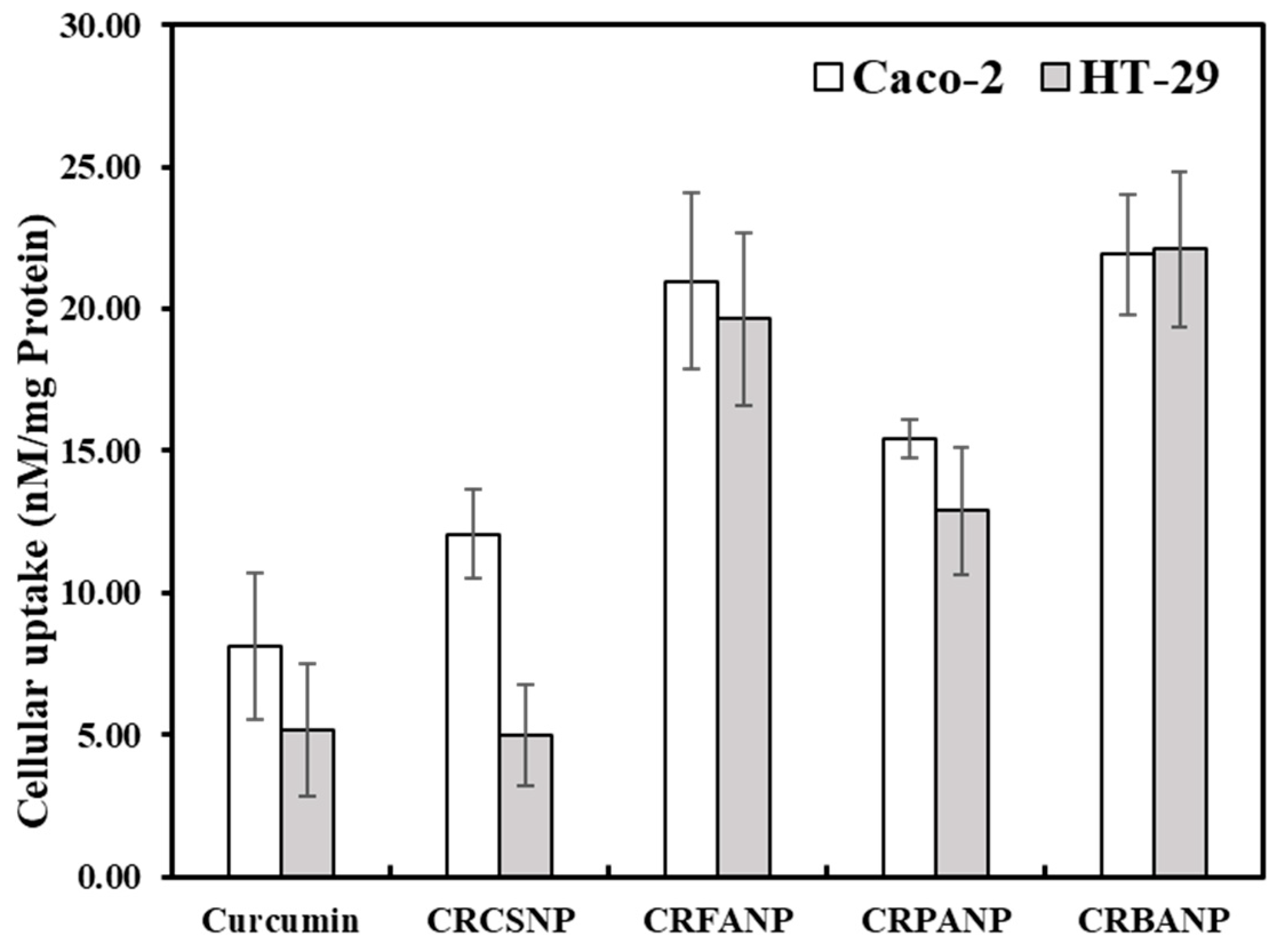
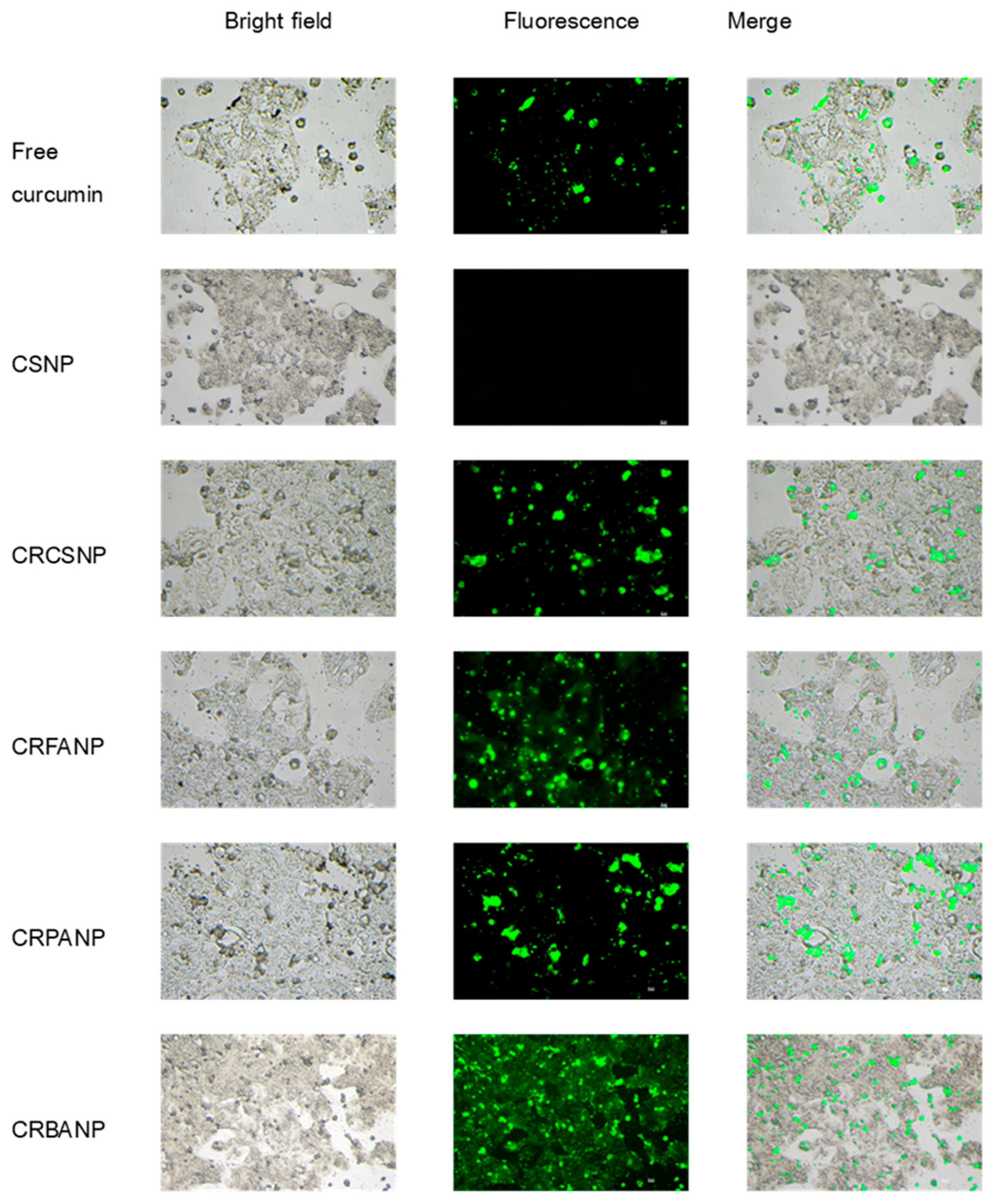
Ligand structural features influenced the physicochemical properties of the functionalized nanoparticles, cellular internalization efficiency, and overall therapeutic efficacy of the functionalized chitosan nanoparticles, and the relationships between these factors are evaluated below. FA, PA, and BA modifications are compared directly using identical polymer parameters. Previous studies evaluated these ligands under varying experimental conditions, making direct comparison of their delivery efficiency difficult. The data confirms that butyric acid modification provides potent cytotoxicity to enhance the therapeutic response of the released curcumin. The correlation between these verified structural profiles and the subsequent physicochemical properties is evaluated systematically in the following sections.
4.1. Synthesis and Characterization of Functionalized Polymers
The functionalization of chitosan is achieved by grafting specific targeting ligands, including folic acid, butyric acid, and phenylalanine, onto the polymer backbone to adjust the nanocarrier properties for biological evaluation. Quantitative assessment in
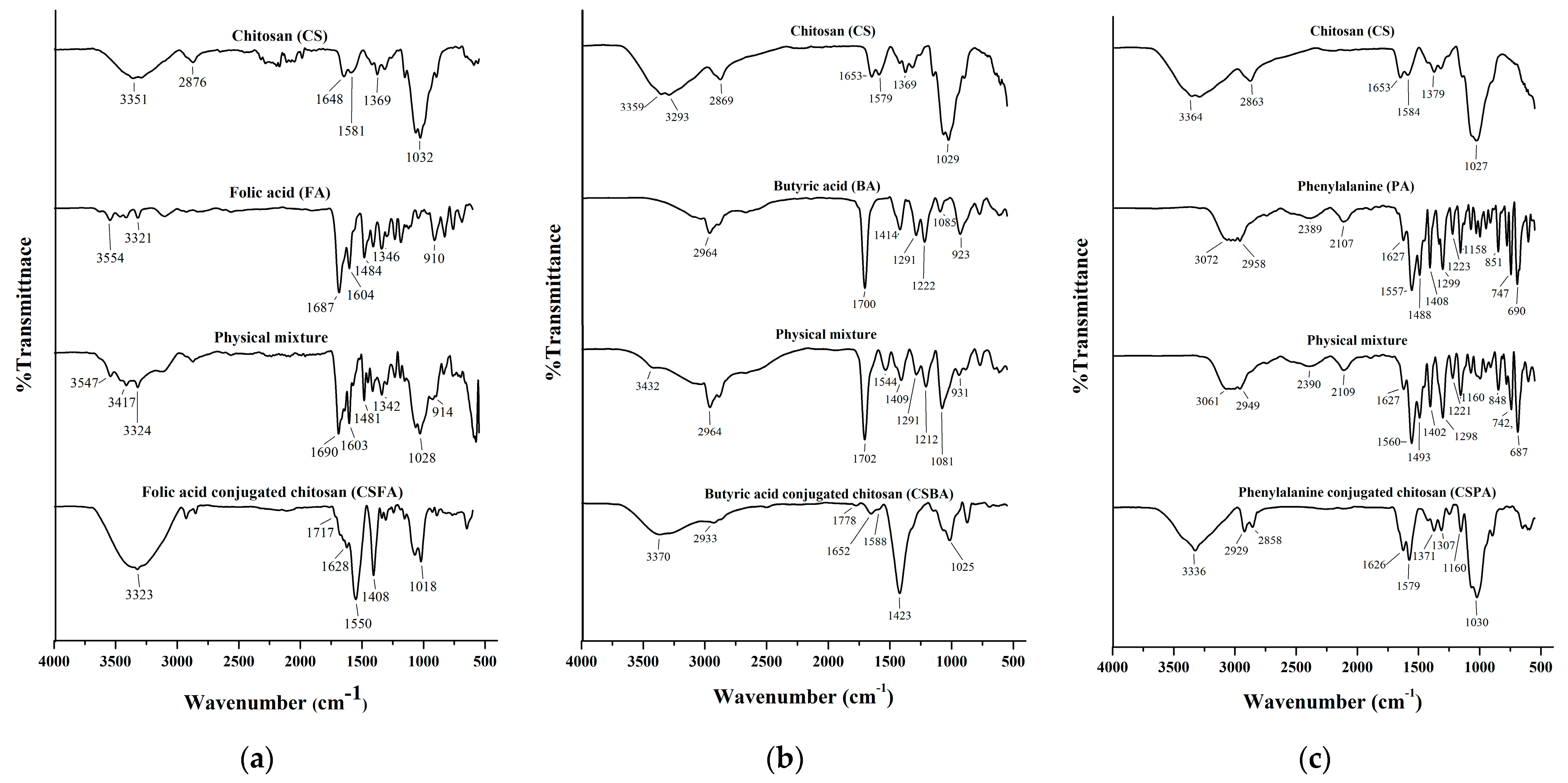
Table 2 demonstrates that this chemical modification is regulated by the initial ligand feeding ratios. Under lower feeding conditions, from 1:20 to 1:100 molar ratios, the actual bound ligand density increases proportionally, maintaining a controlled modification that preserves a fraction of the primary amine groups on the chitosan backbone. For the 1:100 ratio utilized in biological studies, the actual bound ligand reaches its optimal density while leaving a functional fraction of free amine groups available. This primary modification is substantiated by the FTIR profiles presented in
Figure 1, confirming the successful amide formation on the chitosan backbone. However, increasing the feeding ratio to 1:300 leads to an over-saturation of the primary amines, rendering the remaining free amines negligible in
Table 2.
Under these maximum feeding parameters, the high concentration of the acylating agent promotes a shift in chemical reactivity, initiating esterification at the available hydroxyl sites in addition to the primary amide formation [
16]. This dual-functionalization pathway explains the high density of bound ligands recorded at elevated ratios without indicating polymer chain degradation.
4.2. Structural Influence on Nanoparticle Formation and Physicochemical Properties
The specific structure of the ligands significantly impacted the formation process during ionic gelation, thus influencing particle size and surface charge. Folic acid (FA), characterized by its rigid aromatic pteridine ring structure, functioned as a bulky, branched scaffold when conjugated to the chitosan backbone. This structural rigidity restricted the rotation and extension of the polymer chains, physically hindering tight packing [
17,
18] and resulting in the largest average particle size of 263.5 nm. In contrast, butyric acid (BA), a short-chain fatty acid (C4), possesses high flexibility and low steric hindrance. The degree of deacetylation (DDA) and molecular weight (MW) of the chitosan used affect the final nanoparticle parameters. A DDA of 82.5% was utilized to provide primary amine groups (-NH
2) along the polymer chain, ensuring reactive sites for covalent attachment with FA, PA, and BA ligands while leaving free positive charges to react with TPP during ionic gelation. Low molecular weight chitosan (50–190 kDa) was used because shorter polymer chains lower solution viscosity and reduce chain entanglement, allowing the molecules to pack together during TPP addition. Its conjugation maintained a more linear polymer arrangement, promoting efficient fabrication into compact nanoparticles [
19] with the smallest size (128.2 nm). The observed influence of ionic strength and pH on resulting morphology is consistent with other reports on doxorubicin-loaded chitosan nanoparticles [
20,
21].
Surface charge analysis via zeta potential revealed that electron-withdrawing aromatic groups present in FA and PA decreased the electron density of the nearby amino groups on the chitosan chain, consequently leading to lower surface charges (17.05 mV and 15.59 mV, respectively). In contrast, BA induced no significant electron-withdrawing effect, maintaining a high positive zeta potential. (23.27 mV). This high positive charge is a critical factor, enhancing initial cellular contact and adhesion via robust electrostatic interactions with the negatively charged components of the cell membrane [
22,
23].
The reported PDI values for CRCSNP (0.162), CRFANP (0.174), CRBANP (0.349), and CRPANP (0.340) demonstrate a physical shift following ligand conjugation. While the unmodified CRCSNP displays a narrow size distribution, ligand conjugation induces steric hindrance on the polymer surface, causing a broader hydrodynamic size distribution across the functionalized configurations. This physical polydispersity presents a technical challenge for batch-to-batch consistency during large-scale clinical translation and regulatory approval. Although the current biological profiles exhibit reproducible antitumor efficacy (
Table 5) and stable cellular uptake (
Table 6) with narrow standard deviations, further optimization of the fabrication method is required for downstream clinical translation. Future scaling up will utilize advanced manufacturing techniques, such as microfluidic mixing or high-pressure homogenization, to control particle size distribution and minimize PDI variance before clinical evaluation.
4.3. Encapsulation Efficiency and Drug Loading Optimization
The evaluation of encapsulation efficiency was based on theoretical calculation and protocols adapted from verified literature, where hydroalcoholic solvent extraction combined with sonication demonstrated high recovery rates (>95%) for encapsulated hydrophobic drugs [
20]. Based on this established methodology, the current formulation was strategically designed using a 5% (
w/
w) initial curcumin-to-chitosan ratio to maintain an optimal physicochemical equilibrium. Preliminary optimization indicated that while higher initial drug feeding could slightly increase the absolute drug loading (%LC), it induced a sharp reduction in entrapment efficiency (%EE) due to polymer core saturation. By maintaining a 5% initial load, a high %EE (77–80%) was achieved, preventing curcumin waste outside the particles while ensuring a stable formulation. This specific loading level provides sufficient therapeutic efficiency, as evidenced by the significantly lower IC
50 values in
Table 5 and enhanced cellular internalization in
Table 6 of the functionalized systems compared to free curcumin. These results confirm that targeted delivery and improved intracellular accumulation eliminate the requirement for a high curcumin content to enhance localized antitumor cytotoxicity. The analysis of encapsulation efficiency was based on protocols adapted from verified literature, where hydroalcoholic solvent extraction combined with sonication demonstrated high recovery rates (>95%) for encapsulated hydrophobic drugs [
20].
4.4. Mechanisms of Enhanced Cellular Uptake
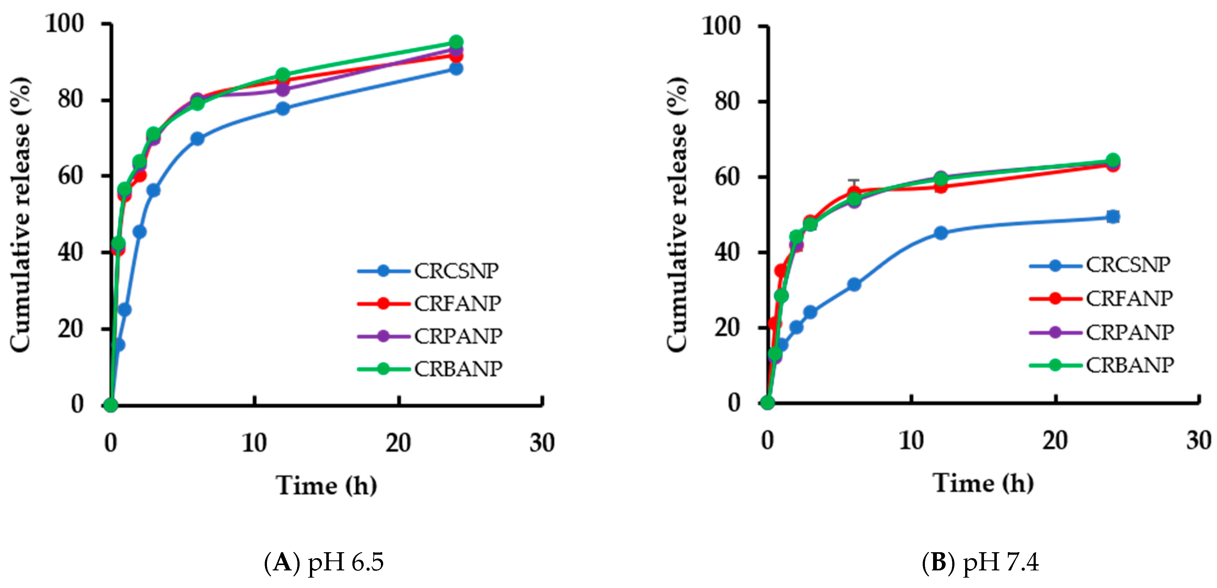
To ensure the analytical reliability of the intracellular quantitative data, chromatographic evaluation via HPLC requires target analytes to exist in a fully dissolved solution phase. The extraction protocol utilizing 70% methanol with sonication forces a total drug release from the nanocarriers before quantification. This extraction behavior is supported by the in vitro dissolution profiles in
Figure 3A, which demonstrate rapid polymer matrix swelling and 70% to 80% curcumin release within 4 h. Considering this high dissolution susceptibility in standard buffer solutions, subsequent exposure to a concentrated organic solvent combined with mechanical sonication breaks down the polymer matrix, releasing the remaining entrapped drug into the liquid medium. This extraction process transforms the internalized formulations into a uniform solution of free curcumin, confirming that the observed uptake differences reflect biological internalization instead of extraction efficiency differences.
The observed cellular accumulation was a synergistic result of physical properties and specific biochemical recognition events. While CRBANP achieved maximum uptake efficiency primarily due to its strong positive charge and smaller size, CRFANP displayed comparable accumulation despite its larger size. This is attributed to folate receptor-mediated endocytosis—a well-established targeting pathway in many cancer cells [
24,
25]. The specific recognition of the pterin moiety by the overexpressed folate receptors (FRs) facilitates active transport into the cell via clathrin-coated pits. This approach has consistently demonstrated enhanced accumulation compared to passive targeting strategies [
26]. For CRPANP, the intermediate uptake suggests that the LAT-1 transporter pathway, although a potential target, may be less efficiently utilized by Caco-2 cells for this specific conjugate compared to the FR pathway [
27,
28]. Furthermore, previous studies have confirmed that chitosan nanoparticles inherently enhance cellular permeability through non-specific mechanisms such as clathrin-mediated endocytosis [
29] and micropinocytosis [
30], further contributing to the enhanced delivery observed in this study.
4.5. Synergistic Cytotoxicity and pH-Responsive Release
A pivotal finding was that the magnitude of cellular uptake did not exclusively determine the level of cytotoxicity. Despite similar uptake levels in Caco-2 cells, CRBANP was significantly more potent than CRFANP. The enhanced efficacy is caused by the intrinsic histone deacetylase (HDAC) inhibitory activity of butyric acid [
30]. The role of butyrate as an HDAC inhibitor is well documented in References [
31,
32]. By inhibiting HDAC enzymes, BA induces hyperacetylation of histone proteins, leading to chromatin remodeling into an “open” conformation, facilitating the access and expression of pro-apoptotic genes (e.g., BAX, BIM, PUMA) [
33], providing a synergistic effect with curcumin’s action on key signaling pathways like NF-κB [
34]. This potentiation of chemotherapy by HDAC inhibitors is a known strategy in cancer therapy [
35].
The enhanced cytotoxicity of CRBANP is attributed to the nanoparticle-mediated delivery rather than a simple additive effect of the separate components. Free curcumin exhibits poor aqueous solubility and rapid degradation, which limits its cellular internalization in an unencapsulated form. Nanoparticle encapsulation bypasses these solubility limitations through receptor-mediated endocytosis. As confirmed by the FTIR analysis, butyric acid is covalently conjugated to the chitosan backbone, ensuring that the internalization of the nanoparticles drives the co-delivery of both chemical components into the intracellular environment to support the observed synergistic anticancer activity. Although the drug loading is approximately 5% relative to the chitosan mass, this curcumin content is highly sufficient for therapeutic applications. The lower IC
50 values in
Table 5 and the enhanced internalization in
Table 6 confirm that this loading configuration delivers an effective intracellular dose, as receptor-mediated targeting and the combined antitumor activity minimize the required curcumin load for selective cytotoxicity.
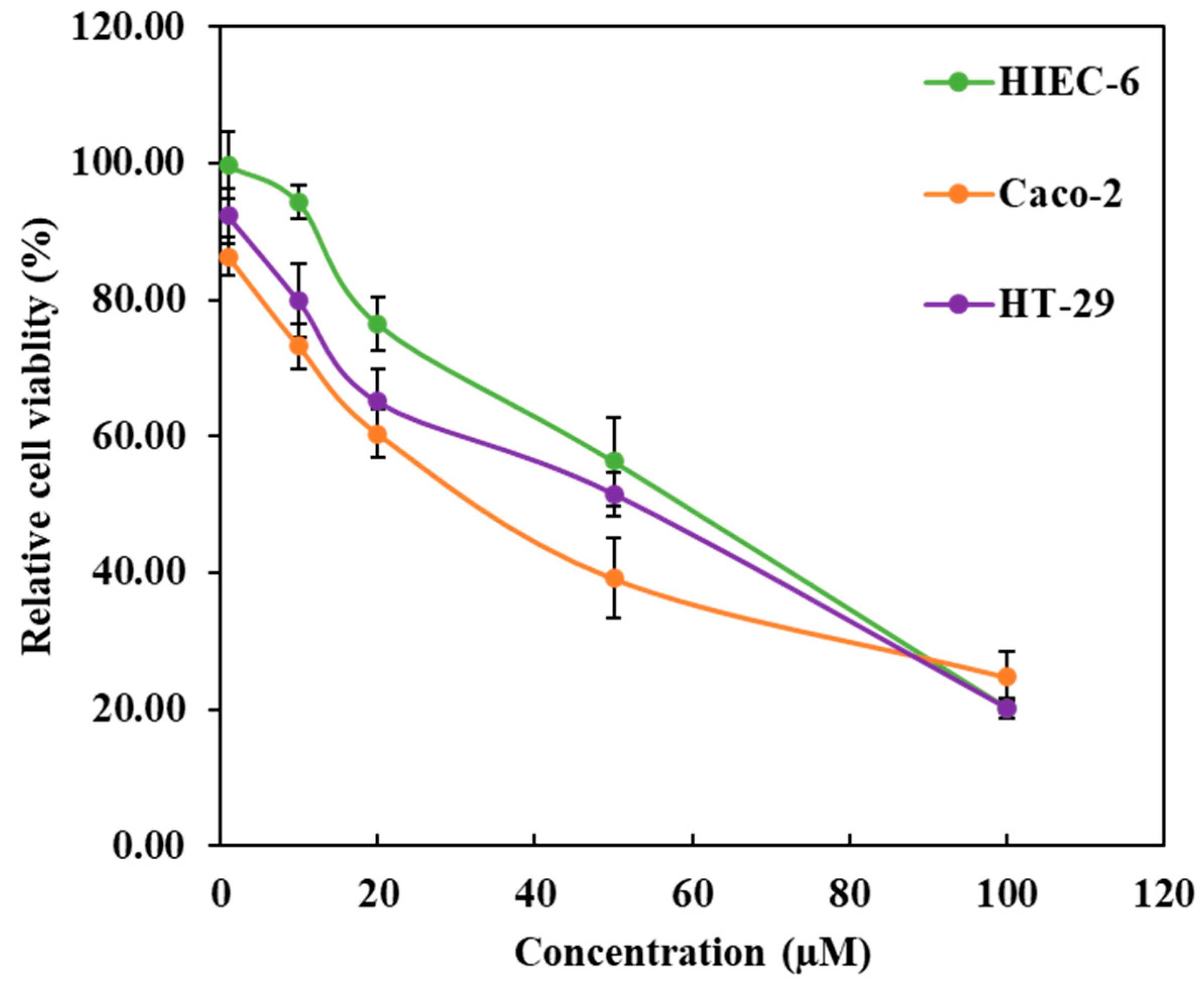
The biological safety of the functionalized carriers was also evaluated alongside these formulation parameters. Blank chitosan nanoparticles and unconjugated ligands exhibit no significant cytotoxicity at the concentrations utilized in this study, maintaining cell viability above 90% as these baseline components are biocompatible. This confirms that the observed anticancer activity is derived from the encapsulated curcumin and its synergistic interaction with the modified backbone, rather than the intrinsic toxicity of the carriers or free ligands themselves.
The in vitro release data presented in
Section 3.3 and
Figure 3 confirm the pH-responsive properties of the functionalized nanoparticles. At an acidic pH of 6.5, protonation of the remaining free amino groups of chitosan combined with the partial deprotonation of the butyric acid carboxyl groups (pKa ~ 4.8) induces strong intra- and intermolecular electrostatic repulsion [
36,
37]. This repulsion drives structural loosening and swelling of the polymer network to accelerate curcumin release, while the neutral environment at pH 7.4 maintains matrix stability and minimizes premature drug leakage. The initial burst release at pH 6.5 rapidly increases localized curcumin concentrations to induce immediate cytotoxicity in cancer cells [
34]. This pH-dependent behavior limits drug exposure to healthy tissues at pH 7.4, preventing toxic side effects during circulation [
36,
37].
The cellular internalization of these functionalized nanoparticles is suggested to involve a combination of passive and active targeting pathways. Initial cellular uptake is partially mediated by non-specific endocytosis, facilitated by the submicron diameter and surface charge detailed in
Table 1. However, these physicochemical attributes of nanoparticles are comparable across all formulations; therefore, they cannot explain the distinct variations in cellular accumulation and cytotoxicity observed in
Table 3. While these ligand-dependent variations suggest a trend, it is important to note that specific receptor participation remains a preliminary hypothesis. Without definitive competitive inhibition studies, the exact contribution of receptor-mediated pathways cannot be conclusively confirmed. These distinct profiles instead suggest a potential role of ligand-specific interactions that warrant future mechanistic verification.
The variance in the selectivity index (SI) between Caco-2 (46.5) and HT-29 (5.1) cells relates to differential receptor density and HDAC isoform profiles. Baseline expression data confirm that Caco-2 cells possess higher baseline densities of free fatty acid receptors (FFAR2 and FFAR3) than HT-29 cells, promoting increased targeted binding and internalization of the butyric acid formulation [
38]. Additionally, HT-29 cells maintain elevated endogenous levels of class I HDACs, particularly HDAC1 and HDAC3, which reduce cell susceptibility to butyric acid-mediated inhibition compared to the response profile in Caco-2 cells [
39]. These baseline biological differences cause the lower selectivity index observed in the HT-29 cell line.
4.6. Selective Targeting and Safety Profile
The safety of the delivery system was confirmed by the Selectivity Index (SI), which measured the preference for cancer cells over normal HIEC-6 cells. The calculated SI values for all formulations are presented in
Table 7. CRBANP achieved the highest SI of 46.5 against Caco-2 cells, showing a higher selectivity profile than the other formulations. The nanoparticle formulations did not reduce normal HIEC-6 cell viability to 50%. As reported in
Table 5, the exact IC50 values were not detected within the tested range. Therefore, the maximum tested concentration of 100 µM was used in the SI formula. This calculation results in the greater-than values (>) shown in
Table 7. This high selectivity is attributed to the oncogenic overexpression of specific HDAC isoforms (e.g., HDAC 1, 6, and 8) in colorectal adenocarcinoma cells [
40,
41]. BA induces apoptosis in malignant cells while exhibiting low toxicity toward healthy HIEC-6 cells with normal HDAC levels [
42]. In contrast, the lower SI of CRFANP (17.3 for Caco-2) suggests that folate receptors, which are also expressed on some normal tissues, may lead to less specific targeting [
43]. This selective toxicity is a highly desirable attribute for targeted cancer therapies. Preliminary screening confirms that a 1% (
v/
v) DMSO vehicle control and individual free ligands (butyric acid, folic acid, and phenylalanine) from 5 to 50 µM show no intrinsic cytotoxicity, maintaining high cell viability over 93% across all tested groups.
In summary, the specific structural characteristics of the conjugated ligands directly influenced the physicochemical properties and biological performance of the nanoparticles. The CRBANP formulation provided the best results, as its small size and high positive charge worked together with the anti-cancer activity of butyric acid. By acting as an HDAC inhibitor, the BA-modified system not only delivered the drug effectively but also added a synergistic effect that increased cancer cell death. Most importantly, this system showed a clear preference for Caco-2 cells, meaning it could kill cancer cells while leaving normal cells mostly unharmed. Overall, these results suggest that CRBANP is a strong candidate for further development in targeted colorectal cancer treatment.
4.7. Limitations and Future Perspectives
Establishing a baseline understanding of each individual ligand under controlled conditions is necessary before moving toward combination systems. This systematic evaluation isolated the specific effects of FA, PA, and BA modifications within a single controlled system, providing the detailed data needed for future formulation designs. Although this study focused on single-ligand systems without multi-ligand configurations, the synthesis of multi-ligand systems is planned for future investigations to optimize co-conjugation ratios. However, this study is currently limited to in vitro models, meaning that in vivo validation is required to confirm safety and performance. Regarding nanoparticle parameters, the high polydispersity index (PDI) of CRBANP indicates a need to optimize synthesis conditions for better size uniformity. The absence of a control group combining free curcumin and free butyric acid also represents a study limitation, as potential additive effects in the assay solvent were not evaluated. Future investigations must incorporate in vivo models, free drug combination controls, receptor-mediated pathways, and exact extraction recovery rates.