Polyhydroxyalkanoates in Bone Alloplastic Materials: State of the Art and Future Perspectives



Abstract
1. Introduction
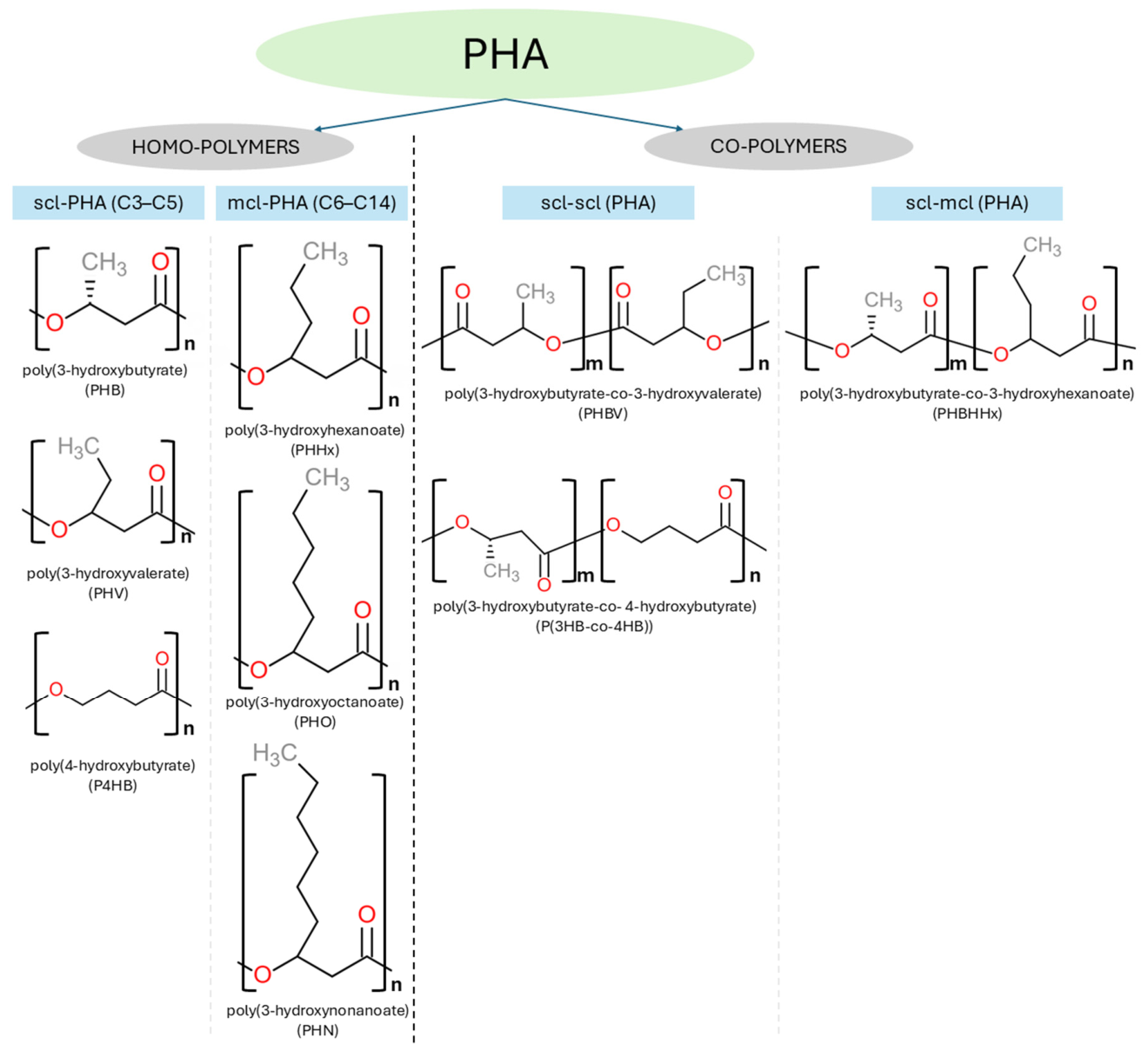
2. Classification of PHAs
3. Production, Extraction and Purification of PHA
4. Solvent Casting Methods for PHA-Based Scaffolds
5. Electrospun Materials Mimicking Bone Extracellular Matrix and Promoting Vascularization
6. 3D Printing Technologies for PHA-Based BTE Scaffolds
7. Discussion
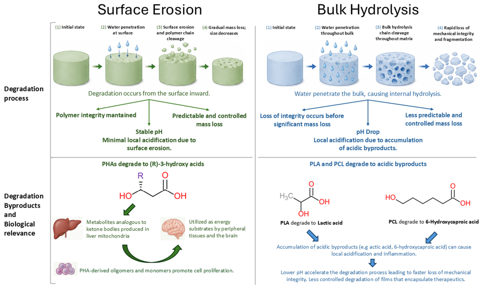
7.1. Degradation for BTE
7.2. Mechanical Properties for BTE
7.3. Functionalization for BTE
7.4. Barriers to the Clinical Use of PHA Scaffolds
8. Conclusions
Author Contributions
Funding
Data Availability Statement
Conflicts of Interest
Abbreviations
| ALG | Alginate |
| ALP | Alkaline phosphatase |
| AM | Additive manufacturing |
| BG | Bioglass |
| BMSC | Bone marrow stromal cell |
| BR | Bredigite |
| BTE | Bone tissue engineering |
| CAM | Chorioallantoic membrane |
| CAWS | Computer-aided wet-spinning |
| Dex | Dextran |
| DLCS | Drug-loaded compo-site scaffold |
| DMOG | Dimethyloxallyl glycine |
| DSDiatom shellEGM | Poly(ethylene) glycidyl methacrylate |
| ELO | Epoxidized linseed oil |
| EMAG | Poly(ethylene) methyl acrylate-co-glycidyl methacrylate |
| FDM | Fused deposition modeling |
| FG | Fibrinogen |
| GBR | Guided bone regeneration |
| GO | Graphene oxide |
| HA | Hydroxyapatite |
| HB | 3-hydroxybutyrate |
| hFOB | Human fetal osteoblast |
| HIF-1α | Hypoxia-inducible factor-1α |
| hUC-MSC | Human umbilical cord-derived mesenchymal stem cell |
| HV | 3-hydroxyvalerate |
| iPSC | Induced pluripotent stem cell |
| MBG | Mesoporous bioactive glass |
| MC3T3-E1 | Mouse calvarian osteoblast cells |
| mcl-PHA | Medium-chain-length PHA |
| MEX | Material extrusion |
| MMC | Mixed microbial culture |
| mPG | Mineralized fibers |
| MRSA | Methicillin-resistant Staphylococcus aureus |
| MSC | Mesenchymal stromal cell |
| MWCNT | Multi-walled carbon nanotube |
| nHA | Hydroxyapatite nanoparticle |
| OCP | Octacalcium phosphate |
| OGP | Osteogenic growth peptide |
| P(3HB-co-3HV) | Poly(3-hydroxybutyrrate-co-3-hydroxyvalerate) |
| P(3HO-co-3HD-co-3HDD) | Poly(3-hydroxyoctanoate-co-hydroxydecanoate-co-hydroxydodecanoate) |
| P(4HB) | Poly(4-hydroxybutyrate) |
| P34HB | Poly(3-hydroxybutyric acid-co-4-hydroxybutyric acid) |
| PAA | Polyaspartic acid |
| PBVHx | Poly(3-hydroxybutyrate-co-3-hydroxyvalerate-co-3-hydroxyhexanoate) |
| PC | Poly-3 hydroxybutyrate-chitosan |
| PCL | Polycaprolactone |
| PCM | Pure-culture microorganism |
| PDEP | Phosphodiesterase-5 |
| PEG | Poly(ethylene glycol) |
| PG | Gelatin fibers |
| PHA | Polyhydroxyalkanoate |
| PHB | Poly(3-hydroxybutyrate) |
| PHB-CTS | Polyhydroxybutyrate-chitosan |
| PHBHHx | Poly(3-hydroxybutyrate-co-3-hydroxyhexanoate) |
| PHB-K | Polyhydroxybutyrate-keratin |
| PHBV | Poly(3-hydroxybutyrate-co-3-hydroxyvalerate) |
| PHHx | Poly(3-hydroxyhexanoate) |
| PHO | Poly(3-hydroxyoctanoate) |
| PHV | Poly(3-hydroxyvalerate) |
| PLA | Polylactic acid |
| PLGA | Poly(lactide-co-glycolide) |
| PLLA | Poly(l-lactide) |
| PPy | Polypyrrole fibers |
| PUL | Pullulan |
| PVA | Polyvinyl alcohol |
| Saos-2 | Human primary sarcoma cell line |
| scl-PHA | Short-chain-length PHA |
| SEM | Scanning electron microscope |
| SF | Silk fibroin |
| SLS | Selective laser sintering |
| TCP | Tricalcium phosphate |
| WL | Whitlockite |
| Xibond | Poly(styrene-co-maleic anhydride) |
| β-TCP | β-Tricalcium phosphate |
References
- Wang, W.; Yeung, K.W.K. Bone Grafts and Biomaterials Substitutes for Bone Defect Repair: A Review. Bioact. Mater. 2017, 2, 224–247. [Google Scholar] [CrossRef] [Scilit] [PubMed]
- Yu, X.; Tang, X.; Gohil, S.V.; Laurencin, C.T. Biomaterials for Bone Regenerative Engineering. Adv. Healthc. Mater. 2015, 4, 1268–1285. [Google Scholar] [CrossRef] [Scilit] [PubMed]
- Tang, D.; Tare, R.S.; Yang, L.-Y.; Williams, D.F.; Ou, K.-L.; Oreffo, R.O.C. Biofabrication of Bone Tissue: Approaches, Challenges and Translation for Bone Regeneration. Biomaterials 2016, 83, 363–382. [Google Scholar] [CrossRef] [Scilit] [PubMed]
- Ho-Shui-Ling, A.; Bolander, J.; Rustom, L.E.; Johnson, A.W.; Luyten, F.P.; Picart, C. Bone Regeneration Strategies: Engineered Scaffolds, Bioactive Molecules and Stem Cells Current Stage and Future Perspectives. Biomaterials 2018, 180, 143–162. [Google Scholar] [CrossRef] [Scilit] [PubMed]
- Culbreath, C.J.; Taylor, M.S.; McCullen, S.D.; Mefford, O.T. A Review of Additive Manufacturing in Tissue Engineering and Regenerative Medicine. Biomed. Mater. Devices 2025, 3, 237–258. [Google Scholar] [CrossRef] [Scilit]
- Kuttappan, S.; Mathew, D.; Jo, J.; Tanaka, R.; Menon, D.; Ishimoto, T.; Nakano, T.; Nair, S.V.; Nair, M.B.; Tabata, Y. Dual Release of Growth Factor from Nanocomposite Fibrous Scaffold Promotes Vascularisation and Bone Regeneration in Rat Critical Sized Calvarial Defect. Acta Biomater. 2018, 78, 36–47. [Google Scholar] [CrossRef] [Scilit] [PubMed]
- Dou, D.D.; Zhou, G.; Liu, H.W.; Zhang, J.; Liu, M.L.; Xiao, X.F.; Fei, J.J.; Guan, X.L.; Fan, Y.B. Sequential Releasing of VEGF and BMP-2 in Hydroxyapatite Collagen Scaffolds for Bone Tissue Engineering: Design and Characterization. Int. J. Biol. Macromol. 2019, 123, 622–628. [Google Scholar] [CrossRef] [Scilit] [PubMed]
- Nifant’ev, I.; Tavtorkin, A.; Komarov, P.; Kretov, E.; Korchagina, S.; Chinova, M.; Gavrilov, D.; Ivchenko, P. Dispersant and Protective Roles of Amphiphilic Poly(Ethylene Phosphate) Block Copolymers in Polyester/Bone Mineral Composites. Int. J. Mol. Sci. 2023, 24, 11175. [Google Scholar] [CrossRef] [Scilit] [PubMed]
- Wang, H.-W.; Chang, C.-M.; Cheng, Y.; Šimoliūnas, E.; Lee, P.-W.; Tsai, W.-C.; Lin, C.-L. Early-Stage Stability and Degradation of Patient-Specific Biodegradable 3D-Printed Implants for Critical Mandibular Reconstruction. Dent. Mater. 2026, 42, 790–806. [Google Scholar] [CrossRef] [Scilit] [PubMed]
- Kang, L.; Ma, C.; Hu, X.; Wu, X.; Shan, R.; Song, F. Digitally Customized 3D PCL/β-TCP Scaffold for Precise Reconstruction of Alveolar Crest Defects. ACS Appl. Bio Mater. 2026, 9, 1403–1411. [Google Scholar] [CrossRef] [Scilit] [PubMed]
- Dedeloudi, A.; Bertelli, P.M.; Martinez-Marcos, L.; Quinten, T.; Lengyel, I.; Andersen, S.K.; Lamprou, D.A. Development of Bioceramic Bone-Inspired Scaffolds Through Single-Step Melt-Extrusion 3D Printing for Segmental Defect Treatment. J. Funct. Biomater. 2025, 16, 358. [Google Scholar] [CrossRef] [Scilit] [PubMed]
- Lv, X.; Liu, Y.; Wang, L.; Liu, X.; Wang, S.; Xu, Z.; Ma, Y. Research on 3D-Printed Scaffolds with Microstructure Bio-Inspired Optimization for Orbital Bone Defect Repair. J. Mater. Sci. Mater. Med. 2025, 36, 95. [Google Scholar] [CrossRef] [Scilit] [PubMed]
- Sowislok, A.; Gruber, G.; Kaschani, F.; Kaiser, M.; Papaeleftheriou, E.; Jäger, M. Intraoperative Biologization of β-TCP and PCL-TCP by Autologous Proteins. J. Funct. Biomater. 2025, 16, 340. [Google Scholar] [CrossRef] [Scilit] [PubMed]
- Hu, J.; Yang, X.; Yang, X.; Xu, H. Clinical Trial of 3D-Printed PCL/β-TCP Composite Scaffold for Craniofacial Reconstruction in China. J. Craniofac. Surg. 2025, 36, 2158–2160. [Google Scholar] [CrossRef] [Scilit] [PubMed]
- Wong, W.-S.-Y.; Koh, H.Y.; Lee, K.H.; Bin Abd Razak, H.R. The Novel Osteopore® Wedge in Medial Opening Wedge High Tibial Osteotomy: A Technical Note. Cureus 2025, 17, e86272. [Google Scholar] [CrossRef] [Scilit] [PubMed]
- Nizamuddin, S.; Baloch, A.J.; Chen, C.; Arif, M.; Mubarak, N.M. Bio-Based Plastics, Biodegradable Plastics, and Compostable Plastics: Biodegradation Mechanism, Biodegradability Standards and Environmental Stratagem. Int. Biodeterior. Biodegrad. 2024, 195, 105887. [Google Scholar] [CrossRef] [Scilit]
- Yang, X.; Zhao, K.; Chen, G.-Q. Effect of Surface Treatment on the Biocompatibility of Microbial Polyhydroxyalkanoates. Biomaterials 2002, 23, 1391–1397. [Google Scholar] [CrossRef] [Scilit] [PubMed]
- Ye, X.; Zhang, Y.; Liu, T.; Chen, Z.; Chen, W.; Wu, Z.; Wang, Y.; Li, J.; Li, C.; Jiang, T.; et al. Beta-Tricalcium Phosphate Enhanced Mechanical and Biological Properties of 3D-Printed Polyhydroxyalkanoates Scaffold for Bone Tissue Engineering. Int. J. Biol. Macromol. 2022, 209, 1553–1561. [Google Scholar] [CrossRef] [Scilit] [PubMed]
- Lemoigne, M. Products of Dehydration and of Polymerization of β-Hydroxybutyric Acid. Bull. Soc. Chem. Biol. 1926, 8, 770–782. [Google Scholar]
- Li, Z.; Yang, J.; Loh, X.J. Polyhydroxyalkanoates: Opening Doors for a Sustainable Future. NPG Asia Mater. 2016, 8, e265. [Google Scholar] [CrossRef] [Scilit]
- Howells, E. Opportunities in Biotechnology for the Chemical Industry. Chem. Ind. 1982, 7, 5008–5012. [Google Scholar]
- King, P.P. Biotechnology. An Industrial View. J. Chem. Technol. Biotechnol. 1982, 32, 2–8. [Google Scholar] [CrossRef] [Scilit]
- Peng, S.-W.; Guo, X.-Y.; Shang, G.-G.; Li, J.; Xu, X.-Y.; You, M.-L.; Li, P.; Chen, G.-Q. An Assessment of the Risks of Carcinogenicity Associated with Polyhydroxyalkanoates through an Analysis of DNA Aneuploid and Telomerase Activity. Biomaterials 2011, 32, 2546–2555. [Google Scholar] [CrossRef] [Scilit] [PubMed]
- Chiulan, I.; Frone, A.; Brandabur, C.; Panaitescu, D. Recent Advances in 3D Printing of Aliphatic Polyesters. Bioengineering 2017, 5, 2. [Google Scholar] [CrossRef] [Scilit] [PubMed]
- Ramachandran, S.; Prakash, P.; Raman, S.; Dheyab, M.A.; Sudesh, K.; Moses, E.J.; Parumasivam, T. Optimisation of Verapamil Hydrochloride Loaded Polyhydroxyalkanoate Nano and Microparticles Using Response Surface Methodology. Sci. Rep. 2026, 16, 12288. [Google Scholar] [CrossRef] [Scilit] [PubMed]
- Yuan, S.; Shen, Y.; Li, Z. Injectable Cell- and Growth Factor-Free Poly(4-Hydroxybutyrate) (P4HB) Microspheres with Open Porous Structures and Great Efficiency of Promoting Bone Regeneration. ACS Appl. Bio Mater. 2021, 4, 4432–4440. [Google Scholar] [CrossRef] [Scilit] [PubMed]
- Alfano, S.; Ceparano, L.; Brugnoli, B.; Forcina, G.; Pellegrino, L.; Lauta, F.C.; Rusconi, R.; Francolini, I.; Piozzi, A.; Martinelli, A. Self-Surfactant Poly-3hydroxybutyrate-Co-3hydroxyhexanoate (PHBHHx) for the Preparation of Usnic Acid Loaded Antimicrobial Nanoparticles Using Nontoxic Chemicals. ACS Appl. Bio Mater. 2025, 8, 6109–6120. [Google Scholar] [CrossRef] [Scilit] [PubMed]
- Sanhueza, C.; Acevedo, F.; Rocha, S.; Villegas, P.; Seeger, M.; Navia, R. Polyhydroxyalkanoates as Biomaterial for Electrospun Scaffolds. Int. J. Biol. Macromol. 2019, 124, 102–110. [Google Scholar] [CrossRef] [Scilit] [PubMed]
- Lim, J.; You, M.; Li, J.; Li, Z. Emerging Bone Tissue Engineering via Polyhydroxyalkanoate (PHA)-Based Scaffolds. Mater. Sci. Eng. C 2017, 79, 917–929. [Google Scholar] [CrossRef] [Scilit] [PubMed]
- Wu, C.-S.; Liao, H.-T. The Mechanical Properties, Biocompatibility and Biodegradability of Chestnut Shell Fibre and Polyhydroxyalkanoate Composites. Polym. Degrad. Stab. 2014, 99, 274–282. [Google Scholar] [CrossRef] [Scilit]
- Mehrpouya, M.; Vahabi, H.; Barletta, M.; Laheurte, P.; Langlois, V. Additive Manufacturing of Polyhydroxyalkanoates (PHAs) Biopolymers: Materials, Printing Techniques, and Applications. Mater. Sci. Eng. C 2021, 127, 112216. [Google Scholar] [CrossRef] [Scilit] [PubMed]
- Rai, R.; Keshavarz, T.; Roether, J.A.; Boccaccini, A.R.; Roy, I. Medium Chain Length Polyhydroxyalkanoates, Promising New Biomedical Materials for the Future. Mater. Sci. Eng. R Rep. 2011, 72, 29–47. [Google Scholar] [CrossRef] [Scilit]
- Naser, A.Z.; Deiab, I.; Darras, B.M. Poly(Lactic Acid) (PLA) and Polyhydroxyalkanoates (PHAs), Green Alternatives to Petroleum-Based Plastics: A Review. RSC Adv. 2021, 11, 17151–17196. [Google Scholar] [CrossRef] [Scilit] [PubMed]
- Grigore, M.E.; Grigorescu, R.M.; Iancu, L.; Ion, R.-M.; Zaharia, C.; Andrei, E.R. Methods of Synthesis, Properties and Biomedical Applications of Polyhydroxyalkanoates: A Review. J. Biomater. Sci. Polym. Ed. 2019, 30, 695–712. [Google Scholar] [CrossRef] [Scilit] [PubMed]
- Cichoń, E.; Guzik, M. Bacterial-Derived Polyhydroxyalkanoate/Bioceramic Composites in Clinical Practice: State of the Art and Future Perspectives. ACS Biomater. Sci. Eng. 2025, 11, 4653–4670. [Google Scholar] [CrossRef] [Scilit] [PubMed]
- Shrivastav, A.; Kim, H.-Y.; Kim, Y.-R. Advances in the Applications of Polyhydroxyalkanoate Nanoparticles for Novel Drug Delivery System. BioMed Res. Int. 2013, 2013, 581684. [Google Scholar] [CrossRef] [Scilit] [PubMed]
- Pachiyappan, S.; Shanmuganatham Selvanantham, D.; Kuppa, S.S.; Chandrasekaran, S.; Samrot, A.V. Surfactant-Mediated Synthesis of Polyhydroxybutyrate (PHB) Nanoparticles for Sustained Drug Delivery. IET Nanobiotechnol. 2019, 13, 416–427. [Google Scholar] [CrossRef] [Scilit] [PubMed]
- Steinbüchel, A.; Valentin, H.E. Diversity of Bacterial Polyhydroxyalkanoic Acids. FEMS Microbiol. Lett. 1995, 128, 219–228. [Google Scholar] [CrossRef] [Scilit]
- Steinbüchel, A.; Füchtenbusch, B. Bacterial and Other Biological Systems for Polyester Production. Trends Biotechnol. 1998, 16, 419–427. [Google Scholar] [CrossRef] [Scilit] [PubMed]
- Sudesh, K.; Abe, H.; Doi, Y. Synthesis, Structure and Properties of Polyhydroxyalkanoates: Biological Polyesters. Prog. Polym. Sci. 2000, 25, 1503–1555. [Google Scholar] [CrossRef] [Scilit]
- Koller, M.; Maršálek, L.; De Sousa Dias, M.M.; Braunegg, G. Producing Microbial Polyhydroxyalkanoate (PHA) Biopolyesters in a Sustainable Manner. New Biotechnol. 2017, 37, 24–38. [Google Scholar] [CrossRef] [Scilit] [PubMed]
- Beccari, M.; Majone, M.; Massanisso, P.; Ramadori, R. A Bulking Sludge with High Storage Response Selected Under Intermittent Feeding. Water Res. 1998, 32, 3403–3413. [Google Scholar] [CrossRef] [Scilit]
- Satoh, H.; Iwamoto, Y.; Mino, T.; Matsuo, T. Activated Sludge as a Possible Source of Biodegradable Plastic. Water Sci. Technol. 1998, 38, 103–109. [Google Scholar] [CrossRef] [Scilit]
- van Loosdrecht, M.C.M.; Pot, M.A.; Heijnen, S.J. Importance of Bacterial Storage Polymers in Bioprocesses. Water Sci. Technol. 1997, 35, 41–47. [Google Scholar] [CrossRef] [Scilit]
- Kim, B.S. Production of Poly(3-Hydroxybutyrate) from Inexpensive Substrates. Enzym. Microb. Technol. 2000, 27, 774–777. [Google Scholar] [CrossRef] [Scilit] [PubMed]
- Koller, M.; Bona, R.; Braunegg, G.; Hermann, C.; Horvat, P.; Kroutil, M.; Martinz, J.; Neto, J.; Pereira, L.; Varila, P. Production of Polyhydroxyalkanoates from Agricultural Waste and Surplus Materials. Biomacromolecules 2005, 6, 561–565. [Google Scholar] [CrossRef] [Scilit] [PubMed]
- Yellore, V.; Desai, A. Desai Production of Poly-3-hydroxybutyrate from Lactose and Whey by Methylobacterium Sp. ZP24. Lett. Appl. Microbiol. 1998, 26, 391–394. [Google Scholar] [CrossRef] [Scilit] [PubMed]
- Halami, P.M. Production of Polyhydroxyalkanoate from Starch by the Native Isolate Bacillus Cereus CFR06. World J. Microbiol. Biotechnol. 2008, 24, 805–812. [Google Scholar] [CrossRef] [Scilit]
- Obruca, S.; Marova, I.; Snajdar, O.; Mravcova, L.; Svoboda, Z. Production of Poly(3-Hydroxybutyrate-Co-3-Hydroxyvalerate) by Cupriavidus Necator from Waste Rapeseed Oil Using Propanol as a Precursor of 3-Hydroxyvalerate. Biotechnol. Lett. 2010, 32, 1925–1932. [Google Scholar] [CrossRef] [Scilit] [PubMed]
- Taniguchi, I.; Kagotani, K.; Kimura, Y. Microbial Production of Poly(Hydroxyalkanoate)s from Waste Edible Oils. Green Chem. 2003, 5, 545–548. [Google Scholar] [CrossRef] [Scilit]
- Alva Munoz, L.E.; Riley, M.R. Utilization of Cellulosic Waste from Tequila Bagasse and Production of Polyhydroxyalkanoate (PHA) Bioplastics by Saccharophagus degradans. Biotechnol. Bioeng. 2008, 100, 882–888. [Google Scholar] [CrossRef] [Scilit] [PubMed]
- Silva, L.F.; Taciro, M.K.; Michelin Ramos, M.E.; Carter, J.M.; Pradella, J.G.C.; Gomez, J.G.C. Poly-3-Hydroxybutyrate (P3HB) Production by Bacteria from Xylose, Glucose and Sugarcane Bagasse Hydrolysate. J. Ind. Microbiol. Biotechnol. 2004, 31, 245–254. [Google Scholar] [CrossRef] [Scilit] [PubMed]
- Yu, J.; Stahl, H. Microbial Utilization and Biopolyester Synthesis of Bagasse Hydrolysates. Bioresour. Technol. 2008, 99, 8042–8048. [Google Scholar] [CrossRef] [Scilit] [PubMed]
- Gouda, M.K.; Swellam, A.E.; Omar, S.H. Production of PHB by a Bacillus Megaterium Strain Using Sugarcane Molasses and Corn Steep Liquor as Sole Carbon and Nitrogen Sources. Microbiol. Res. 2001, 156, 201–207. [Google Scholar] [CrossRef] [Scilit] [PubMed]
- Jiang, Y.; Song, X.; Gong, L.; Li, P.; Dai, C.; Shao, W. High Poly(β-Hydroxybutyrate) Production by Pseudomonas Fluorescens A2a5 from Inexpensive Substrates. Enzym. Microb. Technol. 2008, 42, 167–172. [Google Scholar] [CrossRef] [Scilit] [PubMed]
- Liu, F.; Li, W.; Ridgway, D.; Gu, T.; Shen, Z. Production of Poly-β-Hydroxybutyrate on Molasses by Recombinant Escherichia coli. Biotechnol. Lett. 1998, 20, 345–348. [Google Scholar] [CrossRef] [Scilit]
- Cavalheiro, J.M.B.T.; De Almeida, M.C.M.D.; Grandfils, C.; Da Fonseca, M.M.R. Poly(3-Hydroxybutyrate) Production by Cupriavidus Necator Using Waste Glycerol. Process Biochem. 2009, 44, 509–515. [Google Scholar] [CrossRef] [Scilit]
- Du, G.; Yu, J. Green Technology for Conversion of Food Scraps to Biodegradable Thermoplastic Polyhydroxyalkanoates. Environ. Sci. Technol. 2002, 36, 5511–5516. [Google Scholar] [CrossRef] [Scilit] [PubMed]
- Rhu, D.H.; Lee, W.H.; Kim, J.Y.; Choi, E. Polyhydroxyalkanoate (PHA) Production from Waste. Water Sci. Technol. 2003, 48, 221–228. [Google Scholar] [CrossRef] [Scilit]
- Liu, H.; Hall, P.V.; Darby, J.L.; Coats, E.R.; Green, P.G.; Thompson, D.E.; Loge, F.J. Production of Polyhydroxyalkanoate During Treatment of Tomato Cannery Wastewater. Water Environ. Res. 2008, 80, 367–372. [Google Scholar] [CrossRef] [Scilit] [PubMed]
- Dionisi, D.; Carucci, G.; Papini, M.P.; Riccardi, C.; Majone, M.; Carrasco, F. Olive Oil Mill Effluents as a Feedstock for Production of Biodegradable Polymers. Water Res. 2005, 39, 2076–2084. [Google Scholar] [CrossRef] [Scilit] [PubMed]
- Villano, M.; Beccari, M.; Dionisi, D.; Lampis, S.; Miccheli, A.; Vallini, G.; Majone, M. Effect of pH on the Production of Bacterial Polyhydroxyalkanoates by Mixed Cultures Enriched Under Periodic Feeding. Process Biochem. 2010, 45, 714–723. [Google Scholar] [CrossRef] [Scilit]
- Beccari, M.; Dionisi, D.; Giuliani, A.; Majone, M.; Ramadori, R. Effect of Different Carbon Sources on Aerobic Storage by Activated Sludge. Water Sci. Technol. 2002, 45, 157–168. [Google Scholar] [CrossRef] [Scilit]
- Albuquerque, M.G.E.; Eiroa, M.; Torres, C.; Nunes, B.R.; Reis, M.A.M. Strategies for the Development of a Side Stream Process for Polyhydroxyalkanoate (PHA) Production from Sugar Cane Molasses. J. Biotechnol. 2007, 130, 411–421. [Google Scholar] [CrossRef] [Scilit] [PubMed]
- Albuquerque, M.G.E.; Concas, S.; Bengtsson, S.; Reis, M.A.M. Mixed Culture Polyhydroxyalkanoates Production from Sugar Molasses: The Use of a 2-Stage CSTR System for Culture Selection. Bioresour. Technol. 2010, 101, 7112–7122. [Google Scholar] [CrossRef] [Scilit] [PubMed]
- Albuquerque, M.G.E.; Torres, C.A.V.; Reis, M.A.M. Polyhydroxyalkanoate (PHA) Production by a Mixed Microbial Culture Using Sugar Molasses: Effect of the Influent Substrate Concentration on Culture Selection. Water Res. 2010, 44, 3419–3433. [Google Scholar] [CrossRef] [Scilit] [PubMed]
- Bengtsson, S.; Pisco, A.R.; Reis, M.A.M.; Lemos, P.C. Production of Polyhydroxyalkanoates from Fermented Sugar Cane Molasses by a Mixed Culture Enriched in Glycogen Accumulating Organisms. J. Biotechnol. 2010, 145, 253–263. [Google Scholar] [CrossRef] [Scilit] [PubMed]
- Dias, J.M.L.; Lemos, P.C.; Serafim, L.S.; Oliveira, C.; Eiroa, M.; Albuquerque, M.G.E.; Ramos, A.M.; Oliveira, R.; Reis, M.A.M. Recent Advances in Polyhydroxyalkanoate Production by Mixed Aerobic Cultures: From the Substrate to the Final Product. Macromol. Biosci. 2006, 6, 885–906. [Google Scholar] [CrossRef] [Scilit] [PubMed]
- Bengtsson, S.; Werker, A.; Welander, T. Production of Polyhydroxyalkanoates by Glycogen Accumulating Organisms Treating a Paper Mill Wastewater. Water Sci. Technol. 2008, 58, 323–330. [Google Scholar] [CrossRef] [Scilit] [PubMed]
- Morgan-Sagastume, F.; Hjort, M.; Cirne, D.; Gérardin, F.; Lacroix, S.; Gaval, G.; Karabegovic, L.; Alexandersson, T.; Johansson, P.; Karlsson, A.; et al. Integrated Production of Polyhydroxyalkanoates (PHAs) with Municipal Wastewater and Sludge Treatment at Pilot Scale. Bioresour. Technol. 2015, 181, 78–89. [Google Scholar] [CrossRef] [Scilit] [PubMed]
- Guzik, M.; Witko, T.; Steinbüchel, A.; Wojnarowska, M.; Sołtysik, M.; Wawak, S. What Has Been Trending in the Research of Polyhydroxyalkanoates? A Systematic Review. Front. Bioeng. Biotechnol. 2020, 8, 959. [Google Scholar] [CrossRef] [Scilit] [PubMed]
- Dalton, B.; Bhagabati, P.; De Micco, J.; Padamati, R.B.; O’Connor, K. A Review on Biological Synthesis of the Biodegradable Polymers Polyhydroxyalkanoates and the Development of Multiple Applications. Catalysts 2022, 12, 319. [Google Scholar] [CrossRef] [Scilit]
- Yu, J. Microbial Production of Bioplastics from Renewable Resources. In Bioprocessing for Value-Added Products from Renewable Resources; Elsevier: Amsterdam, The Netherlands, 2007; pp. 585–610. [Google Scholar]
- Nonato, R.; Mantelatto, P.; Rossell, C. Integrated Production of Biodegradable Plastic, Sugar and Ethanol. Appl. Microbiol. Biotechnol. 2001, 57, 1–5. [Google Scholar] [CrossRef] [Scilit] [PubMed]
- Bedade, D.K.; Edson, C.B.; Gross, R.A. Emergent Approaches to Efficient and Sustainable Polyhydroxyalkanoate Production. Molecules 2021, 26, 3463. [Google Scholar] [CrossRef] [Scilit] [PubMed]
- Singh Saharan, B.; Grewal, A.; Kumar, P. Biotechnological Production of Polyhydroxyalkanoates: A Review on Trends and Latest Developments. Chin. J. Biol. 2014, 2014, 802984. [Google Scholar] [CrossRef] [Scilit]
- Kurian, N.S.; Das, B. Comparative Analysis of Various Extraction Processes Based on Economy, Eco-Friendly, Purity and Recovery of Polyhydroxyalkanoate: A Review. Int. J. Biol. Macromol. 2021, 183, 1881–1890. [Google Scholar] [CrossRef] [Scilit] [PubMed]
- Kosseva, M.R.; Rusbandi, E. Trends in the Biomanufacture of Polyhydroxyalkanoates with Focus on Downstream Processing. Int. J. Biol. Macromol. 2018, 107, 762–778. [Google Scholar] [CrossRef] [Scilit] [PubMed]
- López-Abelairas, M.; García-Torreiro, M.; Lú-Chau, T.; Lema, J.M.; Steinbüchel, A. Comparison of Several Methods for the Separation of Poly(3-Hydroxybutyrate) from Cupriavidus Necator H16 Cultures. Biochem. Eng. J. 2015, 93, 250–259. [Google Scholar] [CrossRef] [Scilit]
- Tamer, I.M.; Moo-Young, M.; Chisti, Y. Optimization of Poly(β-Hydroxybutyric Acid) Recovery from Alcaligenes Latus: Combined Mechanical and Chemical Treatments. Bioprocess Eng. 1998, 19, 459–468. [Google Scholar] [CrossRef] [Scilit]
- Wampfler, B.; Ramsauer, T.; Rezzonico, S.; Hischier, R.; Köhling, R.; Thöny-Meyer, L.; Zinn, M. Isolation and Purification of Medium Chain Length Poly(3-Hydroxyalkanoates) (Mcl-PHA) for Medical Applications Using Nonchlorinated Solvents. Biomacromolecules 2010, 11, 2716–2723. [Google Scholar] [CrossRef] [Scilit] [PubMed]
- Williams, S.F.; Martin, D.P.; Gerngross, T.; Horowitz, D.M. Medical Device Comprising Polyhydroxyalkanoate Having Pyrogen Removed Using Oxidizing Agent. U.S. Patent 6,738,537, 8 July 2014. [Google Scholar]
- Dobrovolskaia, M.A. Pre-Clinical Immunotoxicity Studies of Nanotechnology-Formulated Drugs: Challenges, Considerations and Strategy. J. Control. Release 2015, 220, 571–583. [Google Scholar] [CrossRef] [Scilit] [PubMed]
- ISO 10993-1:2018; Biological Evaluation of Medical Devices—Part 1: Evaluation and Testing Within a Risk Management Process. International Organization for Standardization (ISO): Geneva, Switzerland, 2018.
- Misra, S.K.; Philip, S.E.; Chrzanowski, W.; Nazhat, S.N.; Roy, I.; Knowles, J.C.; Salih, V.; Boccaccini, A.R. Incorporation of Vitamin E in Poly(3hydroxybutyrate)/Bioglass Composite Films: Effect on Surface Properties and Cell Attachment. J. R. Soc. Interface 2009, 6, 401–409. [Google Scholar] [CrossRef] [Scilit] [PubMed]
- Misra, S.K.; Ansari, T.I.; Valappil, S.P.; Mohn, D.; Philip, S.E.; Stark, W.J.; Roy, I.; Knowles, J.C.; Salih, V.; Boccaccini, A.R. Poly(3-Hydroxybutyrate) Multifunctional Composite Scaffolds for Tissue Engineering Applications. Biomaterials 2010, 31, 2806–2815. [Google Scholar] [CrossRef] [Scilit] [PubMed]
- Wang, N.; Zhou, Z.; Xia, L.; Dai, Y.; Liu, H. Fabrication and Characterization of Bioactive β-Ca2SiO4/PHBV Composite Scaffolds. Mater. Sci. Eng. C 2013, 33, 2294–2301. [Google Scholar] [CrossRef] [Scilit] [PubMed]
- Xia, L.; Li, Y.; Zhou, Z.; Dai, Y.; Liu, H.; Liu, H. Icariin Delivery Porous PHBV Scaffolds for Promoting Osteoblast Expansion In Vitro. Mater. Sci. Eng. C 2013, 33, 3545–3552. [Google Scholar] [CrossRef] [Scilit] [PubMed]
- Xu, C.; Liu, Y. Osteosarcoma Cells/Cell Lines Are Not Appropriate for Studies on Bone Regeneration In Vitro. Bone Jt. Res. 2023, 12, 311–312. [Google Scholar] [CrossRef] [Scilit] [PubMed]
- Ozaki, T.; Neumann, T.; Wai, D.; Schäfer, K.-L.; Van Valen, F.; Lindner, N.; Scheel, C.; Böcker, W.; Winkelmann, W.; Dockhorn-Dworniczak, B.; et al. Chromosomal Alterations in Osteosarcoma Cell Lines Revealed by Comparative Genomic Hybridization and Multicolor Karyotyping. Cancer Genet. Cytogenet. 2003, 140, 145–152. [Google Scholar] [CrossRef] [Scilit] [PubMed]
- Lajeunesse, D.; Frondoza, C.; Schoffield, B.; Sacktor, B. Osteocalcin Secretion by the Human Osteosarcoma Cell Line MG-63. J. Bone Miner. Res. 1990, 5, 915–922. [Google Scholar] [CrossRef] [Scilit] [PubMed]
- Wu, X.; Wang, Z.; Teng, J.; Yang, L.; Xu, S.; Luo, S.; Wu, Z.; Ye, C. Electrospun Microfiber Composite Scaffolds of Polyvinyl Alcohol, Polyhydroxybutyrate, and Multiwalled Carbon Nanotubes for Enhancing the Osteogenic Differentiation of Stem Cells to Promote Bone Regeneration. Int. J. Biol. Macromol. 2025, 309, 142988. [Google Scholar] [CrossRef] [Scilit] [PubMed]
- Asl, M.A.; Karbasi, S.; Beigi-Boroujeni, S.; Benisi, S.Z.; Saeed, M. Polyhydroxybutyrate-Starch/Carbon Nanotube Electrospun Nanocomposite: A Highly Potential Scaffold for Bone Tissue Engineering Applications. Int. J. Biol. Macromol. 2022, 223, 524–542. [Google Scholar] [CrossRef] [Scilit] [PubMed]
- Sriram, M.; Priya, S.; Katti, D.S. Polyhydroxybutyrate-Based Osteoinductive Mineralized Electrospun Structures That Mimic Components and Tissue Interfaces of the Osteon for Bone Tissue Engineering. Biofabrication 2024, 16, 025036. [Google Scholar] [CrossRef] [Scilit] [PubMed]
- Ghasemi, S.; Alibabaie, A.; Saberi, R.; Esmaeili, M.; Semnani, D.; Karbasi, S. Evaluation of the Effects of Zein Incorporation on Physical, Mechanical, and Biological Properties of Polyhydroxybutyrate Electrospun Scaffold for Bone Tissue Engineering Applications. Int. J. Biol. Macromol. 2023, 253, 126843. [Google Scholar] [CrossRef] [Scilit] [PubMed]
- Zonari, A.; Novikoff, S.; Electo, N.R.P.; Breyner, N.M.; Gomes, D.A.; Martins, A.; Neves, N.M.; Reis, R.L.; Goes, A.M. Endothelial Differentiation of Human Stem Cells Seeded onto Electrospun Polyhydroxybutyrate/Polyhydroxybutyrate-Co-Hydroxyvalerate Fiber Mesh. PLoS ONE 2012, 7, e35422. [Google Scholar] [CrossRef] [Scilit] [PubMed]
- Li, J.; Chen, J.-N.; Peng, Z.-X.; Chen, N.-B.; Liu, C.-B.; Zhang, P.; Zhang, X.; Chen, G.-Q. Multifunctional Electrospinning Polyhydroxyalkanoate Fibrous Scaffolds with Antibacterial and Angiogenesis Effects for Accelerating Wound Healing. ACS Appl. Mater. Interfaces 2023, 15, 364–377. [Google Scholar] [CrossRef] [Scilit] [PubMed]
- Motiee, E.-S.; Karbasi, S.; Bidram, E.; Sheikholeslam, M. Investigation of Physical, Mechanical and Biological Properties of Polyhydroxybutyrate-Chitosan/Graphene Oxide Nanocomposite Scaffolds for Bone Tissue Engineering Applications. Int. J. Biol. Macromol. 2023, 247, 125593. [Google Scholar] [CrossRef] [Scilit] [PubMed]
- Ghafari, F.; Karbasi, S.; Baghaban Eslaminejad, M.; Sayahpour, F.A.; Kalantari, N. Biological Evaluation and Osteogenic Potential of Polyhydroxybutyrate-Keratin/Al2O3 Electrospun Nanocomposite Scaffold: A Novel Bone Regeneration Construct. Int. J. Biol. Macromol. 2023, 242, 124602. [Google Scholar] [CrossRef] [Scilit] [PubMed]
- Mohammadalipour, M.; Asadolahi, M.; Mohammadalipour, Z.; Behzad, T.; Karbasi, S. Plasma Surface Modification of Electrospun Polyhydroxybutyrate (PHB) Nanofibers to Investigate Their Performance in Bone Tissue Engineering. Int. J. Biol. Macromol. 2023, 230, 123167. [Google Scholar] [CrossRef] [Scilit] [PubMed]
- Tahmasebi, A.; Shapouri Moghadam, A.; Enderami, S.E.; Islami, M.; Kaabi, M.; Saburi, E.; Daei Farshchi, A.; Soleimanifar, F.; Mansouri, V. Aloe Vera–Derived Gel-Blended PHBV Nanofibrous Scaffold for Bone Tissue Engineering. ASAIO J. 2020, 66, 966–973. [Google Scholar] [CrossRef] [Scilit] [PubMed]
- Wang, X.; Yan, H.; Shen, Y.; Tang, H.; Yi, B.; Qin, C.; Zhang, Y. Shape Memory and Osteogenesis Capabilities of the Electrospun Poly(3-Hydroxybutyrate-Co-3-Hydroxyvalerate) Modified Poly(l-Lactide) Fibrous Mats. Tissue Eng. Part A 2021, 27, 142–152. [Google Scholar] [CrossRef] [Scilit] [PubMed]
- Wang, Z.; Ma, K.; Jiang, X.; Xie, J.; Cai, P.; Li, F.; Liang, R.; Zhao, J.; Zheng, L. Electrospun Poly(3-Hydroxybutyrate-Co-4-Hydroxybutyrate)/Octacalcium Phosphate Nanofibrous Membranes for Effective Guided Bone Regeneration. Mater. Sci. Eng. C 2020, 112, 110763. [Google Scholar] [CrossRef] [Scilit]
- Kara, A.; Gunes, O.C.; Albayrak, A.Z.; Bilici, G.; Erbil, G.; Havitcioglu, H. Fish Scale/Poly(3-Hydroxybutyrate-Co-3-Hydroxyvalerate) Nanofibrous Composite Scaffolds for Bone Regeneration. J. Biomater. Appl. 2020, 34, 1201–1215. [Google Scholar] [CrossRef] [Scilit] [PubMed]
- Ang, S.L.; Shaharuddin, B.; Chuah, J.-A.; Sudesh, K. Electrospun Poly(3-Hydroxybutyrate-Co-3-Hydroxyhexanoate)/Silk Fibroin Film Is a Promising Scaffold for Bone Tissue Engineering. Int. J. Biol. Macromol. 2020, 145, 173–188. [Google Scholar] [CrossRef] [Scilit] [PubMed]
- Dalgic, A.D.; Atila, D.; Karatas, A.; Tezcaner, A.; Keskin, D. Diatom Shell Incorporated PHBV/PCL-Pullulan Co-Electrospun Scaffold for Bone Tissue Engineering. Mater. Sci. Eng. C 2019, 100, 735–746. [Google Scholar] [CrossRef] [Scilit] [PubMed]
- Fu, N.; Meng, Z.; Jiao, T.; Luo, X.; Tang, Z.; Zhu, B.; Sui, L.; Cai, X. P34HB Electrospun Fibres Promote Bone Regeneration In Vivo. Cell Prolif. 2019, 52, e12601. [Google Scholar] [CrossRef] [Scilit] [PubMed]
- Toloue, E.B.; Karbasi, S.; Salehi, H.; Rafienia, M. Potential of an Electrospun Composite Scaffold of Poly (3-Hydroxybutyrate)-Chitosan/Alumina Nanowires in Bone Tissue Engineering Applications. Mater. Sci. Eng. C 2019, 99, 1075–1091. [Google Scholar] [CrossRef] [Scilit]
- Kouhi, M.; Jayarama Reddy, V.; Fathi, M.; Shamanian, M.; Valipouri, A.; Ramakrishna, S. Poly (3-hydroxybutyrate-co-3-hydroxyvalerate)/Fibrinogen/Bredigite Nanofibrous Membranes and Their Integration with Osteoblasts for Guided Bone Regeneration. J. Biomed. Mater. Res. A 2019, 107, 1154–1165. [Google Scholar] [CrossRef] [Scilit] [PubMed]
- Zhou, T.; Li, G.; Lin, S.; Tian, T.; Ma, Q.; Zhang, Q.; Shi, S.; Xue, C.; Ma, W.; Cai, X.; et al. Electrospun Poly(3-Hydroxybutyrate-Co-4-Hydroxybutyrate)/Graphene Oxide Scaffold: Enhanced Properties and Promoted In Vivo Bone Repair in Rats. ACS Appl. Mater. Interfaces 2017, 9, 42589–42600. [Google Scholar] [CrossRef] [Scilit] [PubMed]
- Gorodzha, S.N.; Muslimov, A.R.; Syromotina, D.S.; Timin, A.S.; Tcvetkov, N.Y.; Lepik, K.V.; Petrova, A.V.; Surmeneva, M.A.; Gorin, D.A.; Sukhorukov, G.B.; et al. A Comparison Study between Electrospun Polycaprolactone and Piezoelectric Poly(3-Hydroxybutyrate-Co-3-Hydroxyvalerate) Scaffolds for Bone Tissue Engineering. Colloids Surf. B Biointerfaces 2017, 160, 48–59. [Google Scholar] [CrossRef] [Scilit] [PubMed]
- Chen, Z.; Song, Y.; Zhang, J.; Liu, W.; Cui, J.; Li, H.; Chen, F. Laminated Electrospun nHA/PHB-Composite Scaffolds Mimicking Bone Extracellular Matrix for Bone Tissue Engineering. Mater. Sci. Eng. C 2017, 72, 341–351. [Google Scholar] [CrossRef] [Scilit] [PubMed]
- Zhang, S.; Jiang, G.; Prabhakaran, M.P.; Qin, X.; Ramakrishna, S. Evaluation of Electrospun Biomimetic Substrate Surface-Decorated with Nanohydroxyapatite Precipitation for Osteoblasts Behavior. Mater. Sci. Eng. C 2017, 79, 687–696. [Google Scholar] [CrossRef] [Scilit] [PubMed]
- Unalan, I.; Colpankan, O.; Albayrak, A.Z.; Gorgun, C.; Urkmez, A.S. Biocompatibility of Plasma-Treated Poly(3-Hydroxybutyrate-Co-3-Hydroxyvalerate) Nanofiber Mats Modified by Silk Fibroin for Bone Tissue Regeneration. Mater. Sci. Eng. C 2016, 68, 842–850. [Google Scholar] [CrossRef] [Scilit] [PubMed]
- Paşcu, E.I.; Cahill, P.A.; Stokes, J.; McGuinness, G.B. Towards Functional 3D-Stacked Electrospun Composite Scaffolds of PHBV, Silk Fibroin and Nanohydroxyapatite: Mechanical Properties and Surface Osteogenic Differentiation. J. Biomater. Appl. 2016, 30, 1334–1349. [Google Scholar] [CrossRef] [Scilit] [PubMed]
- Zhang, S.; Prabhakaran, M.P.; Qin, X.; Ramakrishna, S. Poly-3-Hydroxybutyrate-Co-3-Hydroxyvalerate Containing Scaffolds and Their Integration with Osteoblasts as a Model for Bone Tissue Engineering. J. Biomater. Appl. 2015, 29, 1394–1406. [Google Scholar] [CrossRef] [Scilit] [PubMed]
- Ramier, J.; Bouderlique, T.; Stoilova, O.; Manolova, N.; Rashkov, I.; Langlois, V.; Renard, E.; Albanese, P.; Grande, D. Biocomposite Scaffolds Based on Electrospun Poly(3-Hydroxybutyrate) Nanofibers and Electrosprayed Hydroxyapatite Nanoparticles for Bone Tissue Engineering Applications. Mater. Sci. Eng. C 2014, 38, 161–169. [Google Scholar] [CrossRef] [Scilit] [PubMed]
- Paşcu, E.I.; Stokes, J.; McGuinness, G.B. Electrospun Composites of PHBV, Silk Fibroin and Nano-Hydroxyapatite for Bone Tissue Engineering. Mater. Sci. Eng. C 2013, 33, 4905–4916. [Google Scholar] [CrossRef] [Scilit] [PubMed]
- Paula, A.C.C.; Carvalho, P.H.; Martins, T.M.M.; Boeloni, J.N.; Cunha, P.S.; Novikoff, S.; Correlo, V.M.; Reis, R.L.; Goes, A.M. Improved Vascularisation but Inefficient In Vivo Bone Regeneration of Adipose Stem Cells and Poly-3-Hydroxybutyrate-Co-3-Hydroxyvalerate Scaffolds in Xeno-Free Conditions. Mater. Sci. Eng. C 2020, 107, 110301. [Google Scholar] [CrossRef] [Scilit] [PubMed]
- Berger, M.; Probst, F.; Schwartz, C.; Cornelsen, M.; Seitz, H.; Ehrenfeld, M.; Otto, S. A Concept for Scaffold-Based Tissue Engineering in Alveolar Cleft Osteoplasty. J. Cranio-Maxillofac. Surg. 2015, 43, 830–836. [Google Scholar] [CrossRef] [Scilit] [PubMed]
- Saska, S.; Pires, L.C.; Cominotte, M.A.; Mendes, L.S.; De Oliveira, M.F.; Maia, I.A.; Da Silva, J.V.L.; Ribeiro, S.J.L.; Cirelli, J.A. Three-Dimensional Printing and In Vitro Evaluation of Poly(3-Hydroxybutyrate) Scaffolds Functionalized with Osteogenic Growth Peptide for Tissue Engineering. Mater. Sci. Eng. C 2018, 89, 265–273. [Google Scholar] [CrossRef] [Scilit] [PubMed]
- Nazar, L.A.; Al-salman, S.S.; Torki, S.H.; Al-Musawi, M.H.; Najafinezhad, A.; Noory, P.; Rajab, E.S.; Khosravi, N.; Talebi, S.; Azamian, F.; et al. 3D Printed PHB-Dextran-Whitlockite Porous Construct Coated with Sildenafil-Loaded Nanofibers: A Hybrid Scaffold for Craniofacial Reconstruction. Int. J. Biol. Macromol. 2025, 314, 144352. [Google Scholar] [CrossRef] [Scilit] [PubMed]
- Pecorini, G.; Domingos, M.A.N.; Richardson, S.M.; Carmassi, L.; Vecchi, D.L.; Parrini, G.; Puppi, D. 3D Printing of Bacterial Poly(3-Hydroxybutyrate-Co-3-Hydroxyvalerate)/Poly(Lactide-Co-Glycolide) Blend Loaded with β-Tricalcium Phosphate for the Development of Scaffolds to Support Human Mesenchymal Stromal Cell Proliferation. Int. J. Biol. Macromol. 2025, 288, 138744. [Google Scholar] [CrossRef] [Scilit] [PubMed]
- Pecorini, G.; Braccini, S.; Simoni, S.; Corti, A.; Parrini, G.; Puppi, D. Additive Manufacturing of Wet-Spun Poly(3-hydroxybutyrate-Co-3-hydroxyvalerate)-Based Scaffolds Loaded with Hydroxyapatite. Macromol. Biosci. 2024, 24, 2300538. [Google Scholar] [CrossRef] [Scilit] [PubMed]
- Dominguez-Candela, I.; Sempere-José, L.; Sandoval-Perez, I.; Martínez-García, A. Evaluation of Additives on the Cell Metabolic Activity of New PHB/PLA-Based Formulations by Means of Material Extrusion 3D Printing for Scaffold Applications. Polymers 2024, 16, 2784. [Google Scholar] [CrossRef] [Scilit] [PubMed]
- Marcello, E.; Nigmatullin, R.; Basnett, P.; Maqbool, M.; Prieto, M.A.; Knowles, J.C.; Boccaccini, A.R.; Roy, I. 3D Melt-Extrusion Printing of Medium Chain Length Polyhydroxyalkanoates and Their Application as Antibiotic-Free Antibacterial Scaffolds for Bone Regeneration. ACS Biomater. Sci. Eng. 2024, 10, 5136–5153. [Google Scholar] [CrossRef] [Scilit] [PubMed]
- Shishatskaya, E.I.; Demidenko, A.V.; Sukovatyi, A.G.; Dudaev, A.E.; Mylnikov, A.V.; Kisterskij, K.A.; Volova, T.G. Three-Dimensional Printing of Poly(3-Hydroxybutyrate-Co-3-Hydroxyvalerate) [P(3HB-Co-3HV)] Biodegradable Scaffolds: Properties, In Vitro and In Vivo Evaluation. Int. J. Mol. Sci. 2023, 24, 12969. [Google Scholar] [CrossRef] [Scilit] [PubMed]
- Zhang, J.; Ye, X.; Li, W.; Lin, Z.; Wang, W.; Chen, L.; Li, Q.; Xie, X.; Xu, X.; Lu, Y. Copper-Containing Chitosan-Based Hydrogels Enabled 3D-Printed Scaffolds to Accelerate Bone Repair and Eliminate MRSA-Related Infection. Int. J. Biol. Macromol. 2023, 240, 124463. [Google Scholar] [CrossRef] [Scilit] [PubMed]
- Pecorini, G.; Braccini, S.; Parrini, G.; Chiellini, F.; Puppi, D. Additive Manufacturing of Poly(3-Hydroxybutyrate-Co-3-Hydroxyvalerate)/Poly(D,L-Lactide-Co-Glycolide) Biphasic Scaffolds for Bone Tissue Regeneration. Int. J. Mol. Sci. 2022, 23, 3895. [Google Scholar] [CrossRef] [Scilit] [PubMed]
- Volkov, A.V.; Muraev, A.A.; Zharkova, I.I.; Voinova, V.V.; Akoulina, E.A.; Zhuikov, V.A.; Khaydapova, D.D.; Chesnokova, D.V.; Menshikh, K.A.; Dudun, A.A.; et al. Poly(3-Hydroxybutyrate)/Hydroxyapatite/Alginate Scaffolds Seeded with Mesenchymal Stem Cells Enhance the Regeneration of Critical-Sized Bone Defect. Mater. Sci. Eng. C 2020, 114, 110991. [Google Scholar] [CrossRef] [Scilit] [PubMed]
- Min, Z.; Shichang, Z.; Chen, X.; Yufang, Z.; Changqing, Z. 3D-Printed Dimethyloxallyl Glycine Delivery Scaffolds to Improve Angiogenesis and Osteogenesis. Biomater. Sci. 2015, 3, 1236–1244. [Google Scholar] [CrossRef] [Scilit] [PubMed]
- Li, K.; Zhu, M.; Xu, P.; Xi, Y.; Cheng, Z.; Zhu, Y.; Ye, X. Three-Dimensionally Plotted MBG/PHBHHx Composite Scaffold for Antitubercular Drug Delivery and Tissue Regeneration. J. Mater. Sci. Mater. Med. 2015, 26, 102. [Google Scholar] [CrossRef] [Scilit] [PubMed]
- Yang, S.; Wang, J.; Tang, L.; Ao, H.; Tan, H.; Tang, T.; Liu, C. Mesoporous Bioactive Glass Doped-Poly (3-Hydroxybutyrate-Co-3-Hydroxyhexanoate) Composite Scaffolds with 3-Dimensionally Hierarchical Pore Networks for Bone Regeneration. Colloids Surf. B Biointerfaces 2014, 116, 72–80. [Google Scholar] [CrossRef] [Scilit] [PubMed]
- Gregory, D.A.; Fricker, A.T.R.; Mitrev, P.; Ray, M.; Asare, E.; Sim, D.; Larpnimitchai, S.; Zhang, Z.; Ma, J.; Tetali, S.S.V.; et al. Additive Manufacturing of Polyhydroxyalkanoate-Based Blends Using Fused Deposition Modelling for the Development of Biomedical Devices. J. Funct. Biomater. 2023, 14, 40. [Google Scholar] [CrossRef] [Scilit] [PubMed]
- Kenawy, H.I.; Boral, I.; Bevington, A. Complement-Coagulation Cross-Talk: A Potential Mediator of the Physiological Activation of Complement by Low pH. Front. Immunol. 2015, 6, 215. [Google Scholar] [CrossRef] [Scilit] [PubMed]
- Silver, I. Microelectrode Studies on the Acid Microenvironment beneath Adherent Macrophages and Osteoclasts*1. Exp. Cell Res. 1988, 175, 266–276. [Google Scholar] [CrossRef] [Scilit] [PubMed]
- Félix Lanao, R.P.; Leeuwenburgh, S.C.G.; Wolke, J.G.C.; Jansen, J.A. Bone Response to Fast-Degrading, Injectable Calcium Phosphate Cements Containing PLGA Microparticles. Biomaterials 2011, 32, 8839–8847. [Google Scholar] [CrossRef] [Scilit] [PubMed]
- Gould, P.; Holland, S.; Tighe, B. Polymers for Biodegradable Medical Devices. IV. Hydroxybutyrate-Valerate Copolymers as Non-Disintegrating Matrices for Controlled-Release Oral Dosage Forms. Int. J. Pharm. 1987, 38, 231–237. [Google Scholar] [CrossRef] [Scilit]
- Shishatskaya, E.I.; Volova, T.G.; Gordeev, S.A.; Puzyr, A.P. Degradation of P(3HB) and P(3HB-Co-3HV) in Biological Media. J. Biomater. Sci. Polym. Ed. 2005, 16, 643–657. [Google Scholar] [CrossRef] [Scilit] [PubMed]
- Żur-Pińska, J.; Gładysz, M.Z.; Ubels, D.; Siebring, J.; Włodarczyk-Biegun, M.K. Smart and Sustainable: Exploring the Future of PHAs Biopolymers for 3D Printing in Tissue Engineering. Sustain. Mater. Technol. 2023, 38, e00750. [Google Scholar] [CrossRef] [Scilit]
- Rossi, S.; Azghani, A.O.; Omri, A. Antimicrobial Efficacy of a New Antibiotic-Loaded Poly(Hydroxybutyric-Co-Hydroxyvaleric Acid) Controlled Release System. J. Antimicrob. Chemother. 2004, 54, 1013–1018. [Google Scholar] [CrossRef] [Scilit] [PubMed]
- Haraźna, K.; Cichoń, E.; Skibiński, S.; Witko, T.; Solarz, D.; Kwiecień, I.; Marcello, E.; Zimowska, M.; Socha, R.; Szefer, E.; et al. Physicochemical and Biological Characterisation of Diclofenac Oligomeric Poly(3-Hydroxyoctanoate) Hybrids as β-TCP Ceramics Modifiers for Bone Tissue Regeneration. Int. J. Mol. Sci. 2020, 21, 9452. [Google Scholar] [CrossRef] [Scilit] [PubMed]
- Skibiński, S.; Cichoń, E.; Haraźna, K.; Marcello, E.; Roy, I.; Witko, M.; Ślósarczyk, A.; Czechowska, J.; Guzik, M.; Zima, A. Functionalized Tricalcium Phosphate and Poly(3-Hydroxyoctanoate) Derived Composite Scaffolds as Platforms for the Controlled Release of Diclofenac. Ceram. Int. 2021, 47, 3876–3883. [Google Scholar] [CrossRef] [Scilit]
- Dwivedi, R.; Pandey, R.; Kumar, S.; Mehrotra, D. Poly Hydroxyalkanoates (PHA): Role in Bone Scaffolds. J. Oral Biol. Craniofacial Res. 2020, 10, 389–392. [Google Scholar] [CrossRef] [Scilit] [PubMed]
- Dedkova, E.N.; Blatter, L.A. Role of Î2-Hydroxybutyrate, Its Polymer Poly-Î2-Hydroxybutyrate and Inorganic Polyphosphate in Mammalian Health and Disease. Front. Physiol. 2014, 5, 260. [Google Scholar] [CrossRef] [Scilit] [PubMed]
- Volova, T.; Kiselev, E.; Nemtsev, I.; Lukyanenko, A.; Sukovatyi, A.; Kuzmin, A.; Ryltseva, G.; Shishatskaya, E. Properties of Degradable Polyhydroxyalkanoates with Different Monomer Compositions. Int. J. Biol. Macromol. 2021, 182, 98–114. [Google Scholar] [CrossRef] [Scilit] [PubMed]
- Wang, Y.; Wang, X.; Wei, K.; Zhao, N.; Zhang, S.; Chen, J. Fabrication, Characterization and Long-Term In Vitro Release of Hydrophilic Drug Using PHBV/HA Composite Microspheres. Mater. Lett. 2007, 61, 1071–1076. [Google Scholar] [CrossRef] [Scilit]
- Qu, Y.; Shang, Y.; Luo, S.; Pei, X.; Xiao, Y.; Zhao, J.; Zheng, L.; Liao, C.; Liang, R. Biodegradable Piezoelectric PHB-BT Nanofiber Scaffolds Combined with Ultrasound Stimulation to Accelerate Bone Regeneration by Regulating Ca2+/CaN/NFAT. Theranostics 2026, 16, 4283–4303. [Google Scholar] [CrossRef] [Scilit] [PubMed]
- Sharma, V.; Sehgal, R.; Gupta, R. Polyhydroxyalkanoate (PHA): Properties and Modifications. Polymer 2021, 212, 123161. [Google Scholar] [CrossRef] [Scilit]
- Wong, J.X.; Rehm, B.H.A. Design of Modular Polyhydroxyalkanoate Scaffolds for Protein Immobilization by Directed Ligation. Biomacromolecules 2018, 19, 4098–4112. [Google Scholar] [CrossRef] [Scilit] [PubMed]
- Ke, Y.; Liu, C.; Zhang, X.; Xiao, M.; Wu, G. Surface Modification of Polyhydroxyalkanoates toward Enhancing Cell Compatibility and Antibacterial Activity. Macromol. Mater. Eng. 2017, 302, 1700258. [Google Scholar] [CrossRef] [Scilit]
- Raza, Z.A.; Riaz, S.; Banat, I.M. Polyhydroxyalkanoates: Properties and Chemical Modification Approaches for Their Functionalization. Biotechnol. Prog. 2018, 34, 29–41. [Google Scholar] [CrossRef] [Scilit] [PubMed]
- Zhao, C.; Li, J.; He, B.; Zhao, L. Fabrication of Hydrophobic Biocomposite by Combining Cellulosic Fibers with Polyhydroxyalkanoate. Cellulose 2017, 24, 2265–2274. [Google Scholar] [CrossRef] [Scilit]
- Zhang, D.M.; Cui, F.Z.; Luo, Z.S.; Lin, Y.B.; Zhao, K.; Chen, G.Q. Wettability Improvement of Bacterial Polyhydroxyalkanoates via Ion Implantation. Surf. Coat. Technol. 2000, 131, 350–354. [Google Scholar] [CrossRef] [Scilit]
- Lou, Q.; Ma, Y.; Che, X.; Zhong, J.; Sun, X.; Zhang, H. Preparation and characterization of polyhydroxyalkanoate bioplastics with antibacterial activity. J. Biotechnol. 2016, 32, 1052–1059. [Google Scholar] [CrossRef]
- Huang, X.; Tao, X.; Zhang, Z.; Chen, P. Properties and Performances of Fabrics Made from Bio-Based and Degradable Polylactide Acid/Poly (Hydroxybutyrate-Co-Hydroxyvalerate) (PLA/PHBV) Filament Yarns. Text. Res. J. 2017, 87, 2464–2474. [Google Scholar] [CrossRef] [Scilit]
- Ma, L.; Zhang, Z.; Li, J.; Yang, X.; Fei, B.; Leung, P.H.M.; Tao, X. A New Antimicrobial Agent: Poly (3-hydroxybutyric Acid) Oligomer. Macromol. Biosci. 2019, 19, 1800432. [Google Scholar] [CrossRef] [Scilit] [PubMed]
- Zhang, Z.; Li, J.; Ma, L.; Yang, X.; Fei, B.; Leung, P.H.M.; Tao, X. Mechanistic Study of Synergistic Antimicrobial Effects between Poly (3-Hydroxybutyrate) Oligomer and Polyethylene Glycol. Polymers 2020, 12, 2735. [Google Scholar] [CrossRef] [Scilit] [PubMed]
- Yagmurlu, M.F.; Korkusuz, F.; Gürsel, I.; Korkusuz, P.; Örs, Ü.; Hasirci, V. Sulbactam-Cefoperazone Polyhydroxybutyrate-Co- Hydroxyvalerate (PHBV) Local Antibiotic Delivery System: In Vivo Effectiveness and Biocompatibility in the Treatment of Implant-Related Experimental Osteomyelitis. J. Biomed. Mater. Res. 1999, 46, 494–503. [Google Scholar] [CrossRef] [Scilit]
- Ribeiro, M.E.A.; Huaman, N.R.C.; Folly, M.M.; Gomez, J.G.C.; Sánchez Rodríguez, R.J. A Potential Hybrid Nanocomposite of Poly(3-Hydroxybutyrate-Co-3-Hydroxyvalerate) and Fullerene for Bone Tissue Regeneration and Sustained Drug Release Against Bone Infections. Int. J. Biol. Macromol. 2023, 251, 126531. [Google Scholar] [CrossRef] [Scilit] [PubMed]
- Genova, T.; Munaron, L.; Carossa, S.; Mussano, F. Overcoming Physical Constraints in Bone Engineering: ‘The Importance of Being Vascularized’. J. Biomater. Appl. 2016, 30, 940–951. [Google Scholar] [CrossRef] [Scilit] [PubMed]
- Huang, X.-Y.; Zhou, X.-X.; Yang, H.; Xu, T.; Dao, J.-W.; Bian, L.; Wei, D.-X. Directed Osteogenic Differentiation of Human Bone Marrow Mesenchymal Stem Cells via Sustained Release of BMP4 from PBVHx-Based Nanoparticles. Int. J. Biol. Macromol. 2024, 265, 130649. [Google Scholar] [CrossRef] [Scilit] [PubMed]
- Rosenboom, J.-G.; Langer, R.; Traverso, G. Bioplastics for a Circular Economy. Nat. Rev. Mater. 2022, 7, 117–137. [Google Scholar] [CrossRef] [Scilit] [PubMed]
- Shukla, P.; Rout, A.P.; Banerjee, A.; Jamornsuriya, S.; Sakurai, S.; Katiyar, V. Poly(L-Lactic Acid)/Poly(3-Hydroxybutyrate-Co-3-Hydroxyhexanoate)/Hydroxyapatite Based Composites for Bone Tissue Engineering Applications. Int. J. Biol. Macromol. 2026, 358, 151591. [Google Scholar] [CrossRef] [Scilit] [PubMed]
- Palmieri, S.; Tittarelli, F.; Sabbatini, S.; Cespi, M.; Bonacucina, G.; Eusebi, A.L.; Fatone, F.; Stipa, P. Effects of Different Pre-Treatments on the Properties of Polyhydroxyalkanoates Extracted from Sidestreams of a Municipal Wastewater Treatment Plant. Sci. Total Environ. 2021, 801, 149633. [Google Scholar] [CrossRef] [Scilit] [PubMed]
- Matos, M.; Cruz, R.A.P.; Cardoso, P.; Silva, F.; Freitas, E.B.; Carvalho, G.; Reis, M.A.M. Sludge Retention Time Impacts on Polyhydroxyalkanoate Productivity in Uncoupled Storage/Growth Processes. Sci. Total Environ. 2021, 799, 149363. [Google Scholar] [CrossRef] [Scilit] [PubMed]
- Valentino, F.; Morgan-Sagastume, F.; Campanari, S.; Villano, M.; Werker, A.; Majone, M. Carbon Recovery from Wastewater Through Bioconversion into Biodegradable Polymers. New Biotechnol. 2017, 37, 9–23. [Google Scholar] [CrossRef] [Scilit] [PubMed]



| Microbial System | Substrate | References |
|---|---|---|
| PCM | Whey | Kim et al., Koller et al., Yellore et al. [45,46,47] |
| PCM | Starch | Kim et al., Halami et al. [45,48] |
| PCM | Vegetable oils | Obruca et al., Taniguchi et al. [49,50] |
| PCM | Bagasse and cellulosic materials | Alva Munoz et al., Silva et al., Yu et al. [51,52,53] |
| PCM | Molasses | Gouda et al., Jiang et al., Liu et al. [54,55,56] |
| PCM | Glycerol | Koller et al., Cavalheiro et al. [46,57] |
| PCM | Fermented food waste | Du et al. [58] |
| MMC | Food waste | Rhu et al. [59] |
| MMC | Tomato cannery wastewater | Liu et al. [60] |
| MMC | Olive oil mill wastewater | Dionisi et al., Villano et al. [61,62] |
| MMC | Domestic wastewater | Beccari et al. [63] |
| MMC | Fermented molasses | Albuquerque et al., Bengtsson et al., Dias et al. [64,65,66,67,68] |
| MMC | Paper mill wastewater | Bengtsson et al. [69] |
| MMC | Municipal wastewater | Morgan-Sagastume et al. [70] |
| Authors | Scaffold Material | Mechanical Features | Level of Evidence | |
|---|---|---|---|---|
| In Vitro | In Vivo | |||
| Wu et al. [92] | Poly (3-hydroxybutyric acid-co-4-hydroxybutyric acid) (P34HB) and polyvinyl alcohol (PVA) were employed as matrix materials, with multi-walled carbon nanotubes (MWCNTs) | Pure PVA scaffolds exhibited superior Young’s modulus (7.63 ± 0.99 MPa) compared to pure P34HB (0.43 ± 0.03 MPa) and hybrid PVA-P34HB scaffolds (2.74 ± 0.22 MPa). This enhancement correlated directly with MWCNT concentration | Human BMSC viability, proliferation, osteogenic differentiation | White rabbits with femoral condyle loosening. Hybrid electrospun scaffolds incorporating 1% MWCNTs demonstrated superior osteogenic potential and enhanced pull-out resistance |
| Sriram et al. [94] | Polyhydroxybutyrate (PHB) + gelatin fibers (PG) to fabricate mineralized fibers (mPG). A bi-layered tubular construct that had PG in the outer layer and 7%PHB/0.5%Polypyrrole fibers (PPy) in the inner layer | Not assessed | Mouse BMSC viability, proliferation, osteogenic differentiation | Not assessed |
| Ghasemi et al. [95] | Zein/poly(hydroxybutyrate) (PHB) electrospun scaffolds | Zein increased tensile stress (from 2.94 ± 0.23 MPa of PHB only to 4.49 ± 0.25 MPa) and elongation at break (from 11.8 ± 0.8% of PHB only to 82.6 ± 2.3%) | Human MG-63 cell line used to evaluate adhesion, viability, osteogenic differentiation | Not assessed |
| Zonari et al. [96] | PHB and PHB-co-3-hydroxyvalerate (PHB-HV) | Not assessed | Human adipose tissue-derived stem cells (hASCs) to test viability, adhesion, morphology and their endothelial differentiation | Nude mouse model of the critical-sized calvarial defect showed hASCs cultured on PHB-HV scaffolds were ineffective to promote bone regeneration it improved scaffold vascularization |
| Li et al. [97] | P34HB + ciprofloxacin (CIP) + dimethyloxalylglycine (DMOG) | Increased tensile stress to 4.08 ± 0.18 MPa and elongation break to 354.8 ± 18.4% | Mouse L929 cells used to evaluate cell proliferation, spreading and migration | Mouse full thickness excisional wound model showed a complete skin regeneration and hair follicles with increased re-epithelialization, collagen formation, angiogenesis and remodeling |
| Motiee et al. [98] | Poly-3 hydroxybutyrate-chitosan (PC) scaffolds + graphene oxide (GO) | Increasing percentage of GO improved tensile strength (from 0.84 ± 1.18 MPa to 8.175 ± 0.59 MPa with 0.5% GO), Young’s modulus of about 4.5-fold and elongation of about 3-fold | Human MG-63 cell line used to evaluate cellular adhesion and viability | Not assessed |
| Ghafari et al. [99] | Polyhydroxybutyrate-keratin (PHB-K) + alumina nanowire | Alumina nanowire increased tensile strength (from 2.31 ± 0.11 to 6.72 ± 0.12 MPa), Young’s modulus (from 101.32 ± 17 MPa to 168.45 ± 4 MPa) and elongation (from 3.84 ± 0.02% to 23.02 ± 0.25%) | Rat adipose tissue-derived mesenchymal stem cells (MSCs) used to test viability, adhesion, morphology, osteogenic differentiation | Not assessed |
| Mohammadalipour et al. [100] | PHB nanofibers with oxygen/argon plasma | Plasma surface modification of PHB nano fibers reduced the scaffolds’ tensile modulus (around 1.4-fold, reaching 99.85 ± 23.87 MPa) and strength (around 1.3-fold reaching 2.89 ± 0.44 MPa) | Human MG-63 cell line used to evaluate adhesion, viability, osteogenic differentiation | Not assessed |
| Asl et al. [93] | PHB-starch-multiwalled carbon nanotubes (MWCNTs) | MWCNT increased tensile strength (24.37 ± 0.22 MPa) | Human MG-63 cell line used to evaluate adhesion, viability, osteogenic differentiation | Not assessed |
| Tahmasebi et al. [101] | Aloe vera-derived gel-blended poly(3-hydroxybutyrate-co-3-hydroxyvalerate) (PHBV) | Strain and elongation at break of PHBV scaffold, after coating with gel, increased (from 67.38 ± 5.1% to 77.28 ± 2.89%), while PHBV Young’s modulus decreased (from 1.6 ± 0.35 to 1.2 ± 0.45 MPa) | Human-induced pluripotent stem cells (iPSC) tested for cell adhesion, viability and osteodifferentiation | Not assessed |
| Wang et al. [102] | Poly(l-lactide) (PLLA) + poly(3-hydroxybutyrate-co-3-hydroxyvalerate) (PHBV) | The Young’s modulus increased for PLLA-PHBV blends (from 41.5 ± 3.3 of PLLA to 73.5 ± 3.2 MPa) | Mouse bone mesenchymal stem cells tested for osteodifferentiation | Not assessed |
| Wang et al. [103] | Poly(3-hydroxybutyrate-co-4-hydroxybutyrate) (P34HB)/octacalcium phosphate (OCP) (P34HB)/(OCP) | OCP enhanced tensile strength of (P34HB) reaching 2.73 ± 0.03 MPa | Rat mesenchymal stem cells tested for cell adhesion, viability and osteodifferentiation | Bone repair of a calvarial bone defect in a rat model |
| Kara et al. [104] | Fish scale/poly(3-hydroxybutyrate-co-3-hydroxyvalerate) (PHBV) | Incorporation of the fish scales increased about 2.5-fold compressive modulus of the scaffold (reaching 2.5 MPa) | Human MG-63 cell line used to evaluate cell cytotoxicity, proliferation and osteogenic differentiation | Not assessed |
| Ang et al. [105] | Surface coating of poly(3-hydroxybutyrate-co-3-hydroxyhexanoate) (PHBHHx) with silk fibroin (SF) | Not assessed | Human umbilical cord-derived mesenchymal stem cells (hUC-MSCs) used to study morphology, growth and proliferation, and osteogenic differentiation | Not assessed |
| Dalgic et al. [106] | Poly(hydroxybutyrate-co-hydroxyvalerate)/poly(ε-caprolactone) (PHBV/PCL) + and DS incorporated pullulan (PUL) fibers | Increase in Young’s modulus (around 5.4-fold, reaching 515.2 ± 91.9 kPa) and elastic modulus and decrease in tensile strength of PHBV/PCL/DS scaffold (reaching 19.1 ± 1.4 kPa) | Human primary sarcoma cell line (Saos-2) used to investigate osteocompatibility | Not assessed |
| Fu et al. [107] | Poly3-hydroxybutyric acid 4hydroxybutyrate4-hydroxybutyrate (P34HB) | Good values for elongation (26.9%) and Young’s modulus (58.929 MPa) | Murine bone marrow mesenchymal stem cells (BMSCs) tested for cellular morphology and viability | Bone repair of a rat calvarial defect |
| Toloue et al. [108] | Alumina nanowires are added to (polyhydroxybutyrate-chitosan (HB-CTS) | Al2O3 increased >10 fold (reaching 11.18 ± 1.24 MPa) the tensile strength compared to PHB-CTS | Human MG-63 cell line used to evaluate morphology, viability and osteogenic differentiation | Not assessed |
| Kouhi et al. [109] | Poly (hydroxybutyrate-co-3-hydroxyvalerate) (PHBV)/fibrinogen (FG)/bredigite (BR) membranes | Young’s modulus and strength of the PHBV membrane reduced upon blending with FG (83.3 ± 15.5 MPa and 3.68 ± 1.1 MPa) and increased by further incorporation of BR nanoparticles (121 ± 14.4 MPa and 5.22 ± 0.7 MPa) | Human fetal osteoblast cells (hFOB) tested for viability and osteoblast differentiation | Not assessed |
| Zhou et al. [110] | Poly(3-hydroxybutyrate-co-4-hydroxybutyrate) (P34HB)/Graphene Oxide (GO) | Scaffolds with 2 mg/mL GO were improved by 87% in tensile strength (reaching 2.02 ± 0.24) and by 161% in Young’s modulus (reaching 85.11 ± 2.86 MPa) were increased by GO | Rat bone marrow mesenchymal stem cells (BMSCs) tested for adhesion, viability, proliferation, and osteogenic differentiation | P34HB/GO was superior to P34HB in bone regeneration in critical-sized calvarial defects of rats |
| Gorodzha et al. [111] | Poly(3-hydroxybutyrate-co-3-hydroxyvalerate) (PHBV) and a combined with silicate containing hydroxyapatite (PHBV-SiHA) | Not assessed | Human bone marrow mesenchymal stem cells (BMSCs) tested for cellular adhesion, morphology, viability and osteogenic differentiation | Not assessed |
| Chen et al. [112] | Hydroxyapatite nanoparticle (nHA)/poly-hydroxybutyrate (PHB) | Tensile strength at break, tensile strain at break and elastic modulus increased reaching 3.99 ± 0.57 MPa, 3.46 ± 0.93%, and 798.25 ± 120.07 MPa respectively with nHA | Rabbit bone marrow mesenchymal stem cells (BMSCs) tested for adhesion, proliferation and osteogenic differentiation | The scaffold was implanted in BALB/c nude mice, which showed osteoid tissue formation and blood vessel ingrowth was detected into the graft |
| Zhang et al. [113] | Poly-3-hydroxybutyrate-co-3-hydroxyvalerate (PHBV)/polyaspartic acid (PAA) + nano-hydroxyapatite (nHA) | PHBV/PAA-nHA scaffolds are suitable for bone tissue reparation having a tensile strain of 15.78% which is higher than the one of natural bone (3–5%) nHA improved tensile strength | Human fetal osteoblast (hFOB) tested for cellular adhesion, proliferation and osteogenic differentiation | Not assessed |
| Unalan et al. [114] | Plasma treated-poly (3-hydroxybutyrate-co-3-hydroxyvalerate) (PHBV) and silk fibroin (SF)-PHBV | Not assessed | SaOs-2osteoblast-like human osteosarcoma cells tested for cellular morphology, viability and osteogenic differentiation | Not assessed |
| Paşcu et al. [115] | Polyhydroxybutyrate-polyhydroxyvalerate(PHBV)/nanohydroxyapatite (nHA)/silk fibroin (SF) | Compressive and tensile mechanical properties improved with nHA and SF (Young ‘s modulus ranging between ~1.5–2.8 MPa) | Mouse calvarian osteoblast cells (MC3T3-E1), tested for cellular morphology, adhesion, proliferation and osteogenic differentiation | Not assessed |
| Zhang et al. [116] | Poly-3-hydroxybutyrate-co-3-hydroxyvalerate (PHBV) + poly-(α, β)-DL-aspartic acid | Not assessed | Human fetal osteoblasts (hFOB) were cultured to test cell proliferation and osteodifferentiation | Not assessed |
| Ramier et al. [117] | Poly(3-hydroxybutyrate) (PHB) + nanohydroxyapatite (nHA) | n-HA increased the elastic modulus by 67% (reaching 397 ± 107 MPa) and tensile strength at break by 51% (reaching 16.16 ± 0.86 MPa) | Human mesenchymal stem cells (hMSC) to test cellular adhesion, morphology and proliferation | Not assessed |
| Paşcu et al. [118] | Polyhydroxybutyrate-co-(3-hydroxyvalerate) with 2% valerate fraction (PHBV), nano-hydroxyapatite (nHAp), and Bombyx mori silk fibroin essence (SF) | n-HA and SF at 2% increased Young’s modulus from 0.7 kPa (±0.33 kPa) of PHBV only to 1.4 kPa (±0.54 kPa) | Human osteoblasts tested for cellular adhesion, morphology and viability | Not assessed |
| Authors | PHA | Type of Manufacturing | Mechanical Features | Level of Evidence | Notes | |
|---|---|---|---|---|---|---|
| In Vitro | In Vivo | |||||
| Nazar et al. [122] | Polyhydroxybutyrate (PHB), functionalized with dextran (Dex), charged Mg-doped whitlockite (WL) nanoparticles, coated with sildenafil-loaded Dex-Pluronic F127 nanofibers. | Extrusion-based 3D printer (RegenHU, Switzerland) | Compressive strength of 3.70 ± 0.33 MPa and an elastic modulus of 49.04 ± 4.62 MPa | MG63 cell line tested for viability, cell adhesion, ALP activity and calcium deposition | Angiogenic potential assessed through a chick chorioallantoic membrane (CAM) assay. Bone regeneration tested via rat calvarial defect model | Sildenafil, a phosphodiesterase-5 (PDE5) inhibitor, may improve angiogenesis and tissue repair through its vasodilatory effects |
| Pecorini et al. [123] | Poly(3-hydroxybutyrate-co-3-hydroxyvalerate) (PHBV) and PLGA loaded with β-TCP (up to 15 wt%) | Computer-aided wet-spinning (CAWS) | β-TCP at 5 wt% increased the compressive modulus reaching around 6 MPa | Human mesenchymal stem/stromal cells tested for viability and osteodifferentiation | Not assessed | Not assessed |
| Pecorini et al. [124] | Poly(3-hydroxybutyrate-co-3-hydroxyvalerate) (PHBV) and PLGA loaded with HA (up to 15 wt%) | Computer-aided wet-spinning (CAWS) | HA loading increased scaffold compressive stiffness (average compressive modulus from 1.3 MPa of PHBV scaffold to 4.6 MPa for 5% and 9.3 MPa for 10% and 15% PHBV/HA loaded scaffolds) | Viability of murine pre-osteoblastic cell line MC3T3-E1 and their mineralization capability | Not assessed | Not assessed |
| Dominguez-Candela et al. [125] | Polyhydroxybutyrate (PHB) and PLA functionalized with poly(ethylene) with glicidyl metacrylate (EGM) and methyl acrylate-co-glycidyl methacrylate (EMAG); poly-(styrene-co-maleic anhydride) copolymer (Xibond); and bio-based epoxidized linseed oil (ELO) | Material extrusion (MEX) additive manufacturing (AM) technology | Compression stress was 11–13 MPa | Human fetal osteoblastic (hFOB) cells tested for viability and cellular proliferation | Not assessed | Not assessed |
| Marcello et al. [126] | Poly(3-hydroxyoctanoate-co-hydroxydecanoate-co-hydroxydodecanoate) (P(3HO-co-3HD-co-3HDD) + PHAs containing thioester groups in their side chains (i.e., PHACOS) or charged with HA doped with selenium and strontium ions (Se-Sr-HA) | Melt-extrusion 3D printing | Not assessed | MC3T3-E1 cells used to test viability and osteodifferentiation | Not assessed | Antibacterial features tested on Staphylococcus aureus 6538P and Escherichia coli 8739 |
| Shishatskaya et al. [127] | Poly(3-hydroxybutyrate-co-3-hydroxyvalerate) (PHBV) | FDM | Young’s modulus and compressive stress tensile strength increased, according to the direction of load application (parallel or perpendicular to the layers of the scaffold: Young’s modulus of 207.52 ± 19.12 and 241.34 ± 7.62 MPa and compressive stress tensile strength of 19.45 ± 2.10 and 22.43 ± 1.89 MPa) | NIH 3T3 fibroblasts tested for viability and metabolic activity | The diaphyseal zone of the femur in domestic Landrace pigs up to 150 days of observation | Not assessed |
| Zhang et al. [128] | Polyhydroxyalkanoates/β-tricalcium phosphate (PHA/β-TCP) enriched with copper-containing carboxymethyl chitosan/alginate (CA/Cu) hydrogels | FDM | Not assessed | MC3T3-E1 cell tested for viability, adhesion and osteogenic differentiation | Accelerated bone repair of cranial defects in Sprague-Dawley rats | Methicillin-resistant Staphylococcus aureus (MRSA) |
| Ye et al. [18] | Ecomann® Bioresin EM10080 filled with β-TCP (up to 30 wt%) | FDM | The compressive strength and the shore hardness of the PHA/20%β-TCP scaffold were 36.7 MPa and 81.1 HD | MC3T3-E1 cells tested for proliferation, adhesion, and migration | Not assessed | Not assessed |
| Berger et al. [120] | Polyhydroxybutyrate (PHB) loaded with β-TCP | 3D printing | Not assessed | Human mesenchymal stem cells (hMSCs) tested for viability and osteogenic differentiation | Not assessed | Not assessed |
| Pecorini et al. [129] | Poly(3-hydroxybutyrate-co-3-hydroxyvalerate) (PHBV) and poly(D,L-lactide-co-glycolide) (PLGA) | Computer-aided wet-spinning (CAWS) | PHBV/PLGA weight ratios of 50/50 r compressive modulus increased up to 3.4 ± 2.1 MPa when dry and 3.7 ± 1.1 MPa when wet | MC3T3-E1 cell tested for viability, adhesion, morphology and osteogenic differentiation | Not assessed | Not assessed |
| Volkov et al. [130] | Poly(3-hydroxybutyrate) (PHB) with HA filled with alginate (ALG) hydrogel | Two-stage salt leaching technique using a mold obtained by 3D printing | The Young’s and shear moduli of PHB + HA increased, reaching 34.0 ± 0.6 kPa and 85.6 ± 3.0 kPa respectively. The scaffolds PHB/HA/ALG increased Young’s modulus (178.5 ± 1.8 kPa) | Wistar rat f femur isolated mesenchymal stem cells used to test viability and osteodifferentiation | Critical-sized calvarial defect in rats filled with either PHB/HA/ALG/MSC scaffolds or PHB/HA/ALG scaffolds. The former overperforming the latter | Not assessed |
| Saska et al. [121] | Poly(3-hydroxybutyrate) (PHB) functionalized with osteogenic growth peptide (OGP) and its C-terminal sequence OGP(10–14) | Selective laser sintering (SLS) | Compressive modulus showed good value (4.91 ± 0.30 MPa) | Rat bone marrow stem cells tested for viability and calcium precipitation | Not assessed | Not assessed |
| Min et al. [131] | Poly(3-hydroxybutyrate-co-3-hydroxyhexanoate) (PHBHHx) charged with mesoporous bioactive glasses and releasing dimethyloxallyl glycine (DMOG) | 3D-bioplotter system (EnvisionTEC GmbH 3D Bioplotter™) | Not assessed | Human bone marrow stromal cell (human BMSC) adhesion, viability, proliferation, osteogenic differentiation and angiogenic-relative gene expressions | Calvarial defects were created in Sprague-Dawley rats. DMOG could be released in a sustained manner over 4 weeks enhancing the angiogenesis and osteogenesis in the defects | Not assessed |
| Li et al. [132] | Poly(3-hydroxybutyrate-co-3-hydroxyhexanoate) (PHBHHx) | 3D-bioplotter system (EnvisionTEC GmbH 3D Bioplotter™) | Not assessed | Not assessed | New Zealand rabbits’ femur defect model resulted repaired in the drug-loaded composite scaffold (DLCS) group and the β-TCP group | Not assessed |
| Yang et al. [133] | Poly(3-hydroxybutyrate-co-3-hydroxyhexanoate) (PHBHHx) either coated with mesoporous bioactive glass (MBG) or not | 3D printing | Not assessed | Human mesenchymal stem cells (hMSCs) viability, adhesion and osteogenic differentiation | Not assessed | Not assessed |
Disclaimer/Publisher’s Note: The statements, opinions and data contained in all publications are solely those of the individual author(s) and contributor(s) and not of MDPI and/or the editor(s). MDPI and/or the editor(s) disclaim responsibility for any injury to people or property resulting from any ideas, methods, instructions or products referred to in the content. |
© 2026 by the authors. Licensee MDPI, Basel, Switzerland. This article is an open access article distributed under the terms and conditions of the Creative Commons Attribution (CC BY) license.
Share and Cite
Mosca Balma, A.; Meinardi, S.; Roato, I.; Mussano, F. Polyhydroxyalkanoates in Bone Alloplastic Materials: State of the Art and Future Perspectives. Polymers 2026, 18, 1508. https://doi.org/10.3390/polym18121508
Mosca Balma A, Meinardi S, Roato I, Mussano F. Polyhydroxyalkanoates in Bone Alloplastic Materials: State of the Art and Future Perspectives. Polymers. 2026; 18(12):1508. https://doi.org/10.3390/polym18121508
Chicago/Turabian StyleMosca Balma, Alessandro, Sara Meinardi, Ilaria Roato, and Federico Mussano. 2026. "Polyhydroxyalkanoates in Bone Alloplastic Materials: State of the Art and Future Perspectives" Polymers 18, no. 12: 1508. https://doi.org/10.3390/polym18121508
APA StyleMosca Balma, A., Meinardi, S., Roato, I., & Mussano, F. (2026). Polyhydroxyalkanoates in Bone Alloplastic Materials: State of the Art and Future Perspectives. Polymers, 18(12), 1508. https://doi.org/10.3390/polym18121508