

Design, Synthesis, and Characterization of Novel Pyrazole Cross-Linked Chitosan Derivatives Modified with Zinc Oxide Nanoparticles for Boosting Their Anticancer Activity


Abstract
1. Introduction
2. Materials and Methods
2.1. Materials
2.2. Methods
2.2.1. Synthesis of Novel Malonopyrazole Cross-Linked Chitosan (MPy-Cs) Derivative
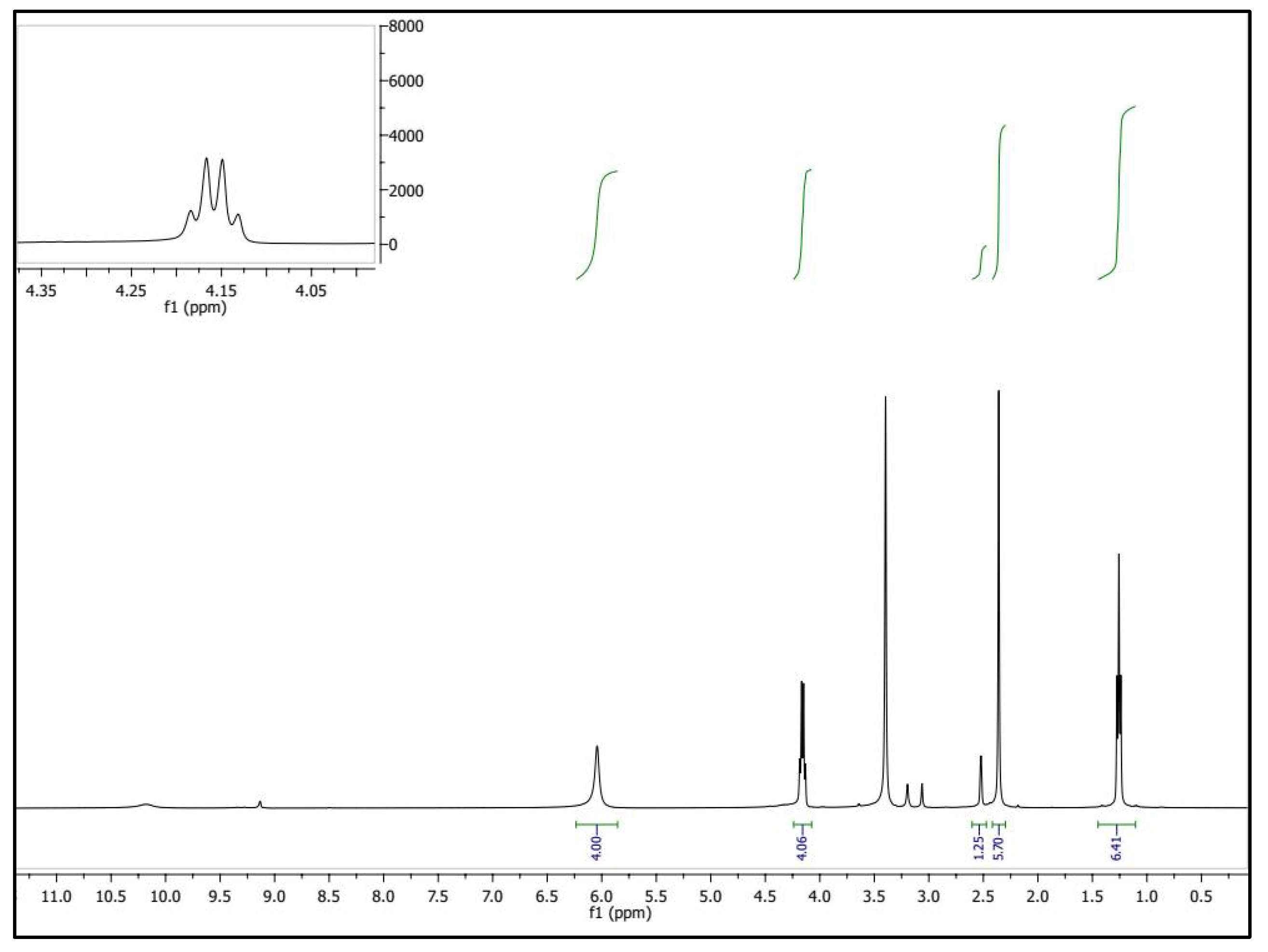
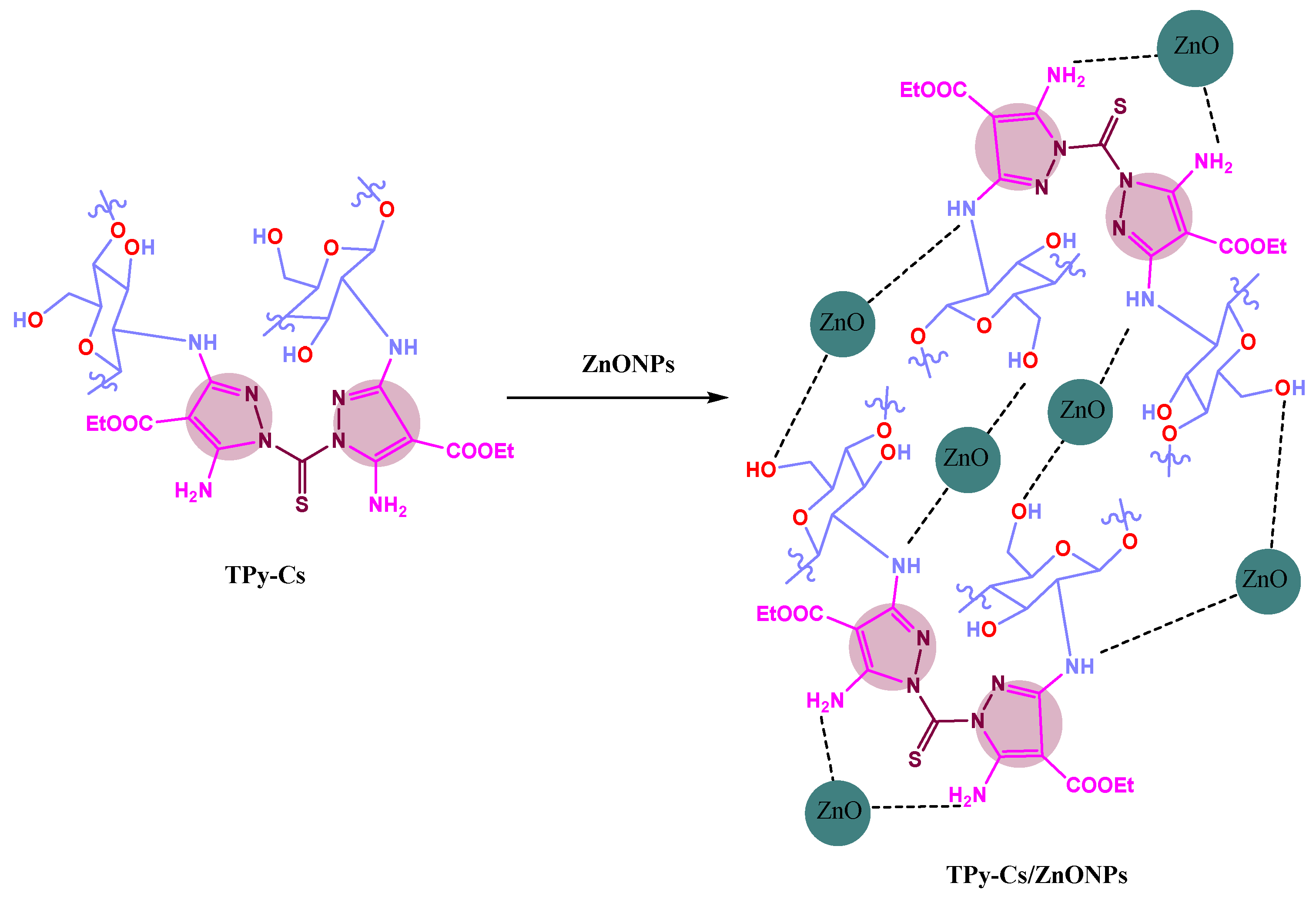
2.2.2. Synthesis of Novel Thiopyrazole Cross-Linked Chitosan (TPy-Cs) Derivatives
2.2.3. Synthesis of TPy-Cs/ZnONPs and MPy-Cs/ZnONP Composites
2.3. Measurements
2.3.1. FTIR Spectroscopy
2.3.2. X-Ray Diffractometry (XRD)
2.3.3. Scanning Electron Microscopy (SEM)
2.3.4. Transmission Electron Microscopy (TEM)
2.3.5. Anticancer and Cytotoxicity Assay
3. Results and Discussion
3.1. Synthesis of the MPy-Cs, TPy-Cs Derivatives and Their ZnONP Composites
3.2. Characterization of MPy-Cs, TPy-Cs Derivatives and Their ZnONP Composites
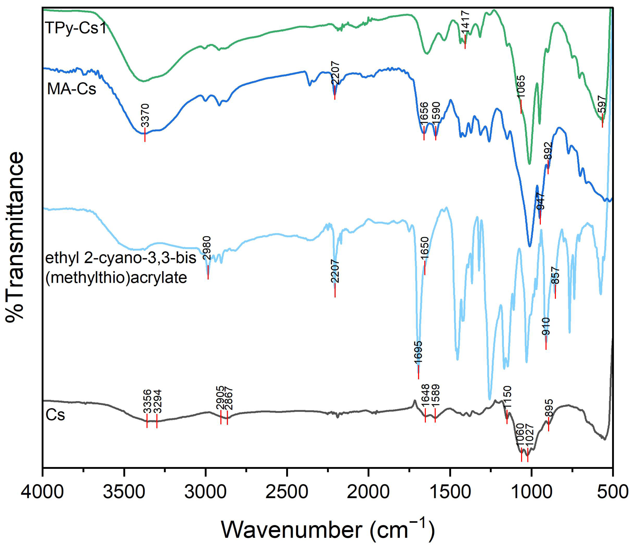
3.2.1. FTIR Analysis
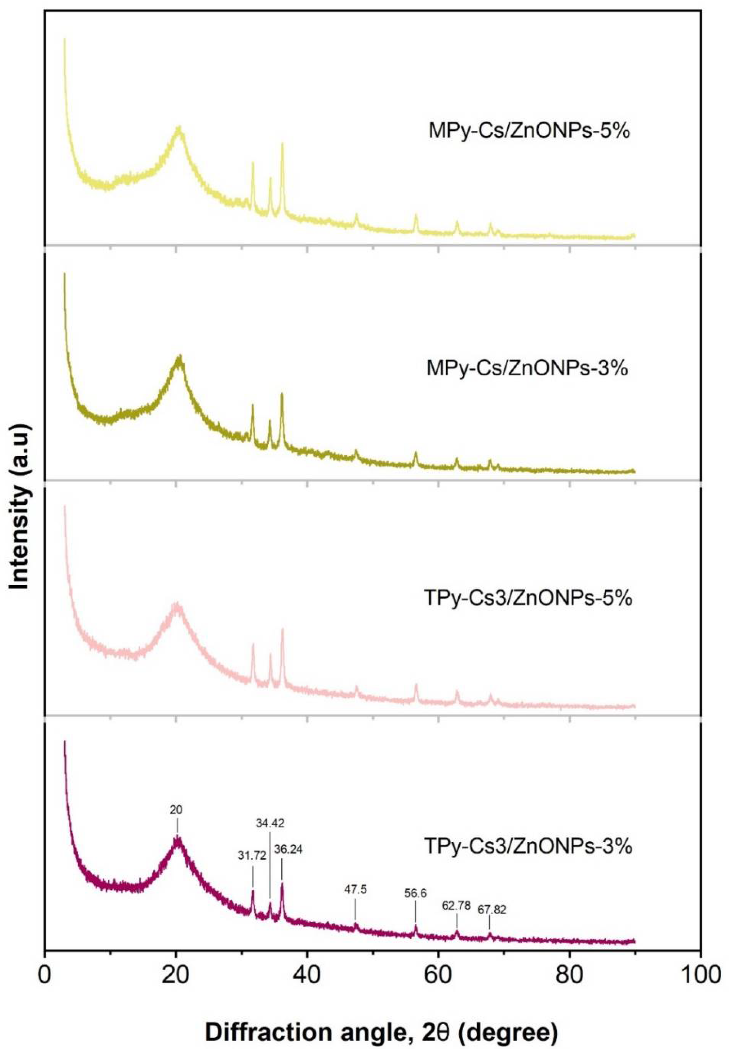
3.2.2. XRD Analysis
3.2.3. SEM Analysis
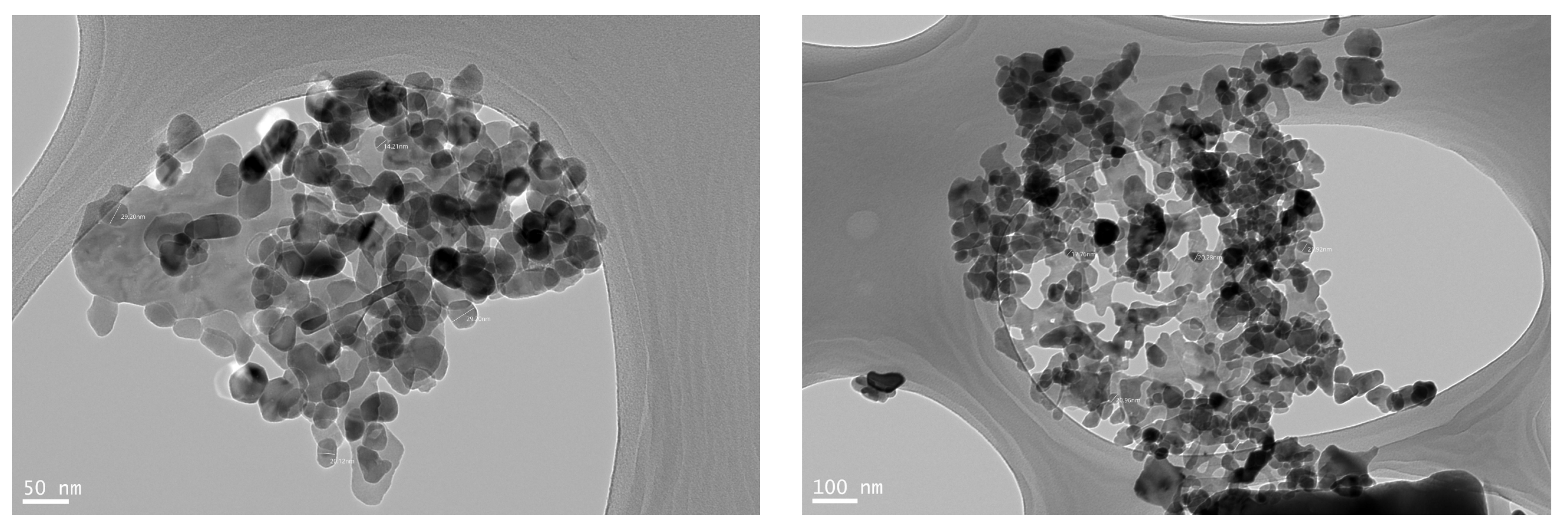
3.2.4. TEM Analysis
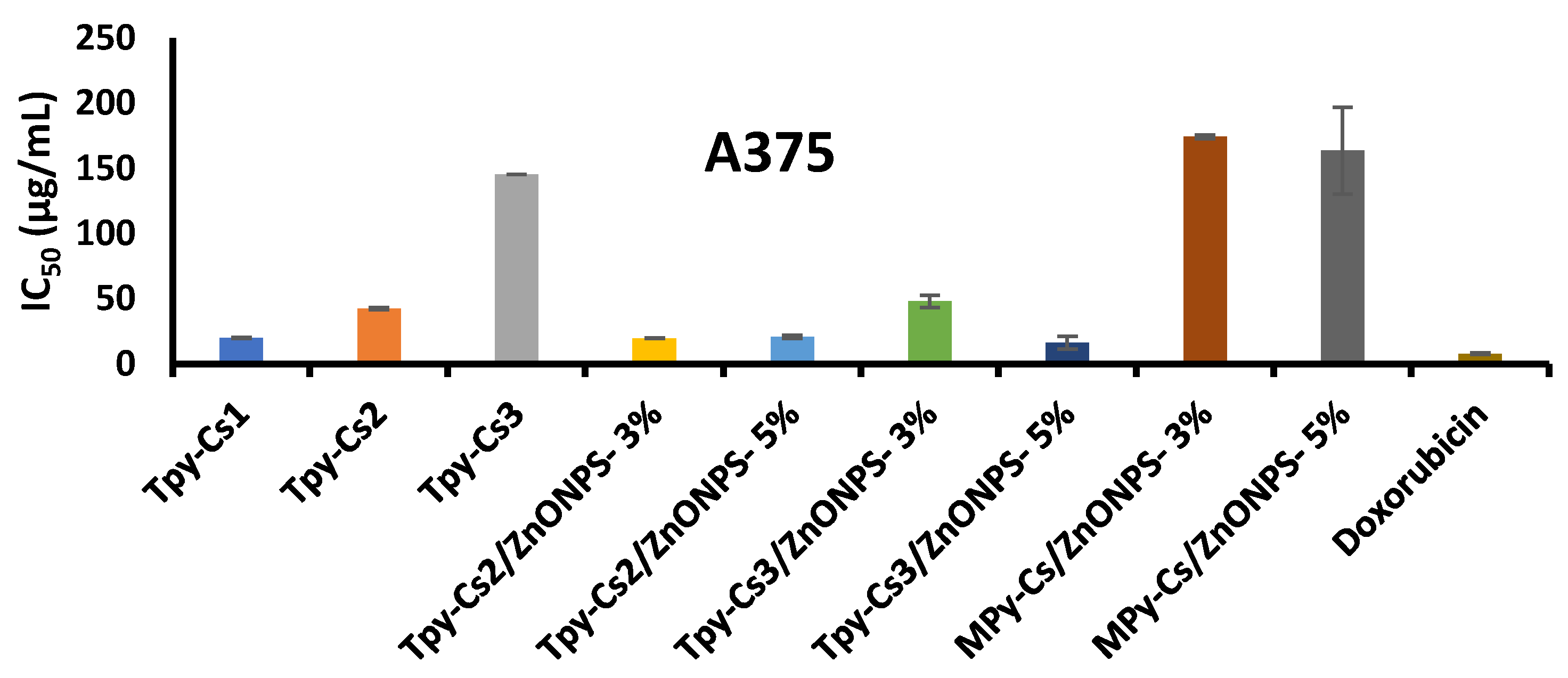
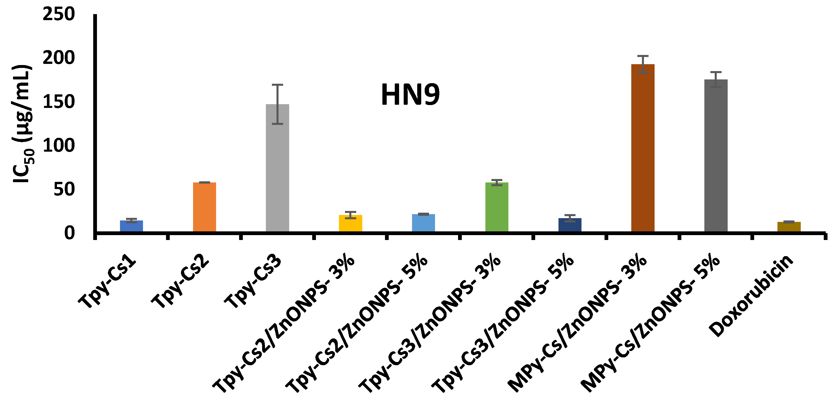
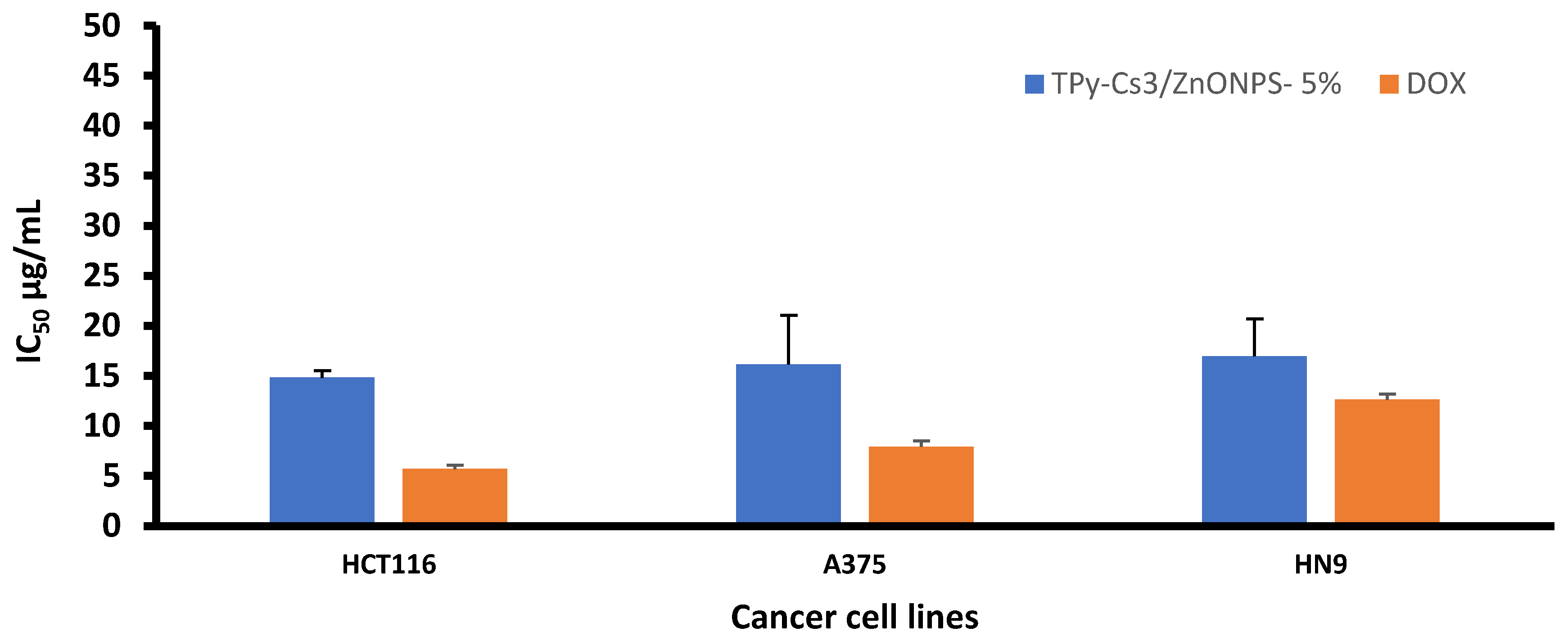
3.3. Anticancer and Cytotoxic Activity
3.3.1. Anticancer Activity
3.3.2. Cytotoxic Activity
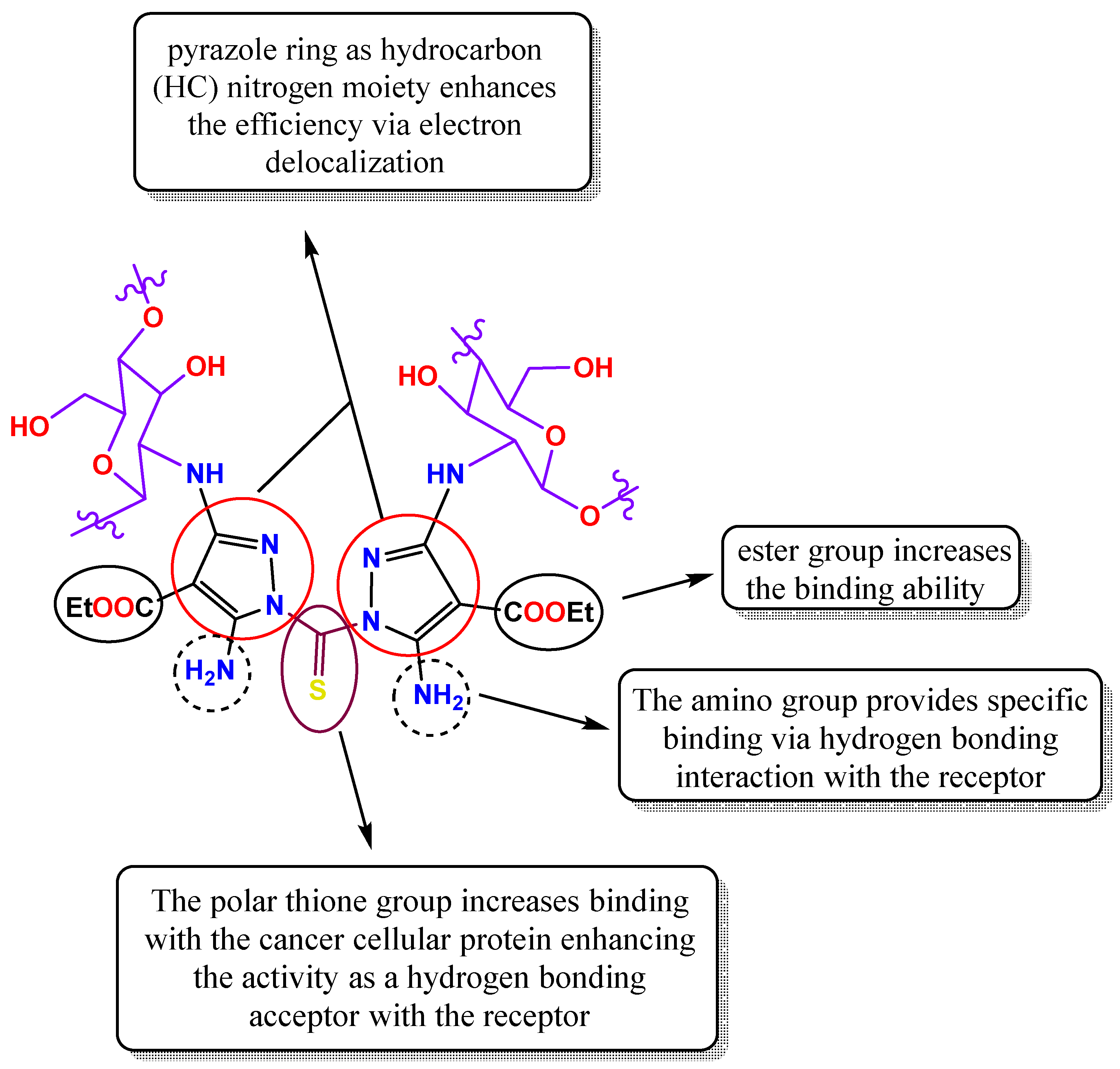
3.3.3. Structure–Activity Relationship (SAR)
4. Conclusions
Author Contributions
Funding
Institutional Review Board Statement
Informed Consent Statement
Data Availability Statement
Conflicts of Interest
References
- Ward, R.A.; Fawell, S.; Floc’h, N.; Flemington, V.; McKerrecher, D.; Smith, P.D. Challenges and Opportunities in Cancer Drug Resistance. Chem. Rev. 2021, 121, 3297–3351. [Google Scholar] [CrossRef]
- Falzone, L.; Salomone, S.; Libra, M. Evolution of Cancer Pharmacological Treatments at the Turn of the Third Millennium. Front. Pharmacol. 2018, 9, 1300. [Google Scholar] [CrossRef]
- Pérez-Herrero, E.; Fernández-Medarde, A. Advanced targeted therapies in cancer: Drug nanocarriers, the future of chemotherapy. Eur. J. Pharm. Biopharm. 2015, 93, 52–79. [Google Scholar] [CrossRef]
- Senapati, S.; Mahanta, A.K.; Kumar, S.; Maiti, P. Controlled drug delivery vehicles for cancer treatment and their performance. Signal Transduct. Target. Ther. 2018, 3, 7. [Google Scholar] [CrossRef] [PubMed]
- Rayhan, M.A.; Hossen, M.S.; Niloy, M.S.; Bhuiyan, M.H.; Paul, S.; Shakil, M.S. Biopolymer and Biomaterial Conjugated Iron Oxide Nanomaterials as Prostate Cancer Theranostic Agents: A Comprehensive Review. Symmetry 2021, 13, 974. [Google Scholar] [CrossRef]
- Parhi, R. Drug delivery applications of chitin and chitosan: A review. Environ. Chem. Lett. 2020, 18, 577–594. [Google Scholar] [CrossRef]
- Elmehbad, N.Y.; Mohamed, N.A.; El-Ghany, N.A.A. Evaluation of the antimicrobial and anti-biofilm activity of novel salicylhydrazido chitosan derivatives impregnated with titanium dioxide nanoparticles. Int. J. Biol. Macromol. 2022, 205, 719–730. [Google Scholar] [CrossRef]
- Mohamed, N.A. Synthesis, characterization and evaluation of in vitro potential antimicrobial efficiency of new chitosan hydrogels and their CuO nanocomposites. Int. J. Biol. Macromol. 2024, 276, 133810. [Google Scholar] [CrossRef]
- Dass, C.R.; Choong, P.F.M. The use of chitosan formulations in cancer therapy. J. Microencapsul. 2008, 25, 275–279. [Google Scholar] [CrossRef]
- Prabaharan, M. Review Paper: Chitosan Derivatives as Promising Materials for Controlled Drug Delivery. J. Biomater. Appl. 2008, 23, 5–36. [Google Scholar] [CrossRef]
- Hasnain, M.S.; Ahmed, S.A.; Alkahtani, S.; Milivojevic, M.; Kandar, C.C.; Dhara, A.K.; Nayak, A.K. Biopolymers for Drug Delivery. In Advanced Biopolymeric Systems for Drug Delivery; Springer: Cham, Switzerland, 2020; pp. 1–29. [Google Scholar] [CrossRef]
- Ahmed, T.; Aljaeid, B. Preparation, characterization, and potential application of chitosan, chitosan derivatives, and chitosan metal nanoparticles in pharmaceutical drug delivery. Drug Des. Dev. Ther. 2016, 10, 483–507. [Google Scholar] [CrossRef]
- Mohamed, N.A.; El-Ghany, N.A.A.; Abdel-Aziz, M.M. Synthesis, characterization, anti-inflammatory and anti-Helicobacter pylori activities of novel benzophenone tetracarboxylimide benzoyl thiourea cross-linked chitosan hydrogels. Int. J. Biol. Macromol. 2021, 181, 956–965. [Google Scholar] [CrossRef] [PubMed]
- Elmehbad, N.Y.; Mohamed, N.A. Terephthalohydrazido cross-linked chitosan hydrogels: Synthesis, characterization and applications. Int. J. Polym. Mater. Polym. Biomater. 2022, 71, 969–982. [Google Scholar] [CrossRef]
- Elieh-Ali-Komi, D.; Hamblin, M.R. Chitin and Chitosan: Production and Application of Versatile Biomedical Nanomaterials. Int. J. Adv. Res. 2016, 4, 411–427. Available online: https://pmc.ncbi.nlm.nih.gov/articles/PMC5094803/ (accessed on 12 April 2025).
- Prateeksha; Sharma, V.K.; Liu, X.; Oyarzún, D.A.; Abdel-Azeem, A.M.; Atanasov, A.G.; Hesham, A.E.-L.; Barik, S.K.; Gupta, V.K.; Singh, B.N. Microbial polysaccharides: An emerging family of natural biomaterials for cancer therapy and diagnostics. Semin. Cancer Biol. 2022, 86, 706–731. [Google Scholar] [CrossRef] [PubMed]
- Alfuraydi, R.T.; Alminderej, F.M.; Mohamed, N.A. Evaluation of antimicrobial and anti-biofilm formation activities of novel poly(vinyl alcohol) hydrogels reinforced with crosslinked chitosan and silver nano-particles. Polymers 2022, 14, 1619. [Google Scholar] [CrossRef]
- Adhikari, H.S.; Yadav, P.N. Anticancer Activity of Chitosan, Chitosan Derivatives, and Their Mechanism of Action. Int. J. Biomater. 2018, 2018, 2952085. [Google Scholar] [CrossRef]
- Park, B.K.; Kim, M.-M. Applications of Chitin and Its Derivatives in Biological Medicine. Int. J. Mol. Sci. 2010, 11, 5152–5164. [Google Scholar] [CrossRef]
- Shanmuganathan, R.; Edison, T.N.J.I.; LewisOscar, F.; Kumar, P.; Shanmugam, S.; Pugazhendhi, A. Chitosan nanopolymers: An overview of drug delivery against cancer. Int. J. Biol. Macromol. 2019, 130, 727–736. [Google Scholar] [CrossRef]
- Elmehbad, N.Y.; Mohamed, N.A.; Abd El-Ghany, N.A.; Abdel-Aziz, M.M. Green synthesis of nano-silver /sodium alginate/carboxymethyl xanthan gum hydrogel and evaluation of its anti-inflammatory and anti-helicobacter pylori activity. Cellul. Chem. Technol. 2022, 56, 983–995. [Google Scholar] [CrossRef]
- Tian, B.; Hua, S.; Liu, J. Multi-functional chitosan-based nanoparticles for drug delivery: Recent advanced insight into cancer therapy. Carbohydr. Polym. 2023, 315, 120972. [Google Scholar] [CrossRef] [PubMed]
- Naskar, S.; Kuotsu, K.; Sharma, S. Chitosan-based nanoparticles as drug delivery systems: A review on two decades of research. J. Drug Target. 2018, 27, 379–393. [Google Scholar] [CrossRef] [PubMed]
- Herdiana, Y.; Wathoni, N.; Shamsuddin, S.; Joni, I.M.; Muchtaridi, M. Chitosan-based nanoparticles of targeted drug delivery system in breast cancer treatment. Polymers 2021, 13, 1717. [Google Scholar] [CrossRef]
- Ding, J.; Guo, Y. Recent advances in chitosan and its derivatives in cancer treatment. Front. Pharmacol. 2022, 13, 888740. [Google Scholar] [CrossRef]
- Fustero, S.; Sánchez-Roselló, M.; Barrio, P.; Simón-Fuentes, A. From 2000 to Mid-2010: A Fruitful Decade for the Synthesis of Pyrazoles. Chem. Rev. 2011, 111, 6984–7034. [Google Scholar] [CrossRef] [PubMed]
- Ansari, A.; Ali, A.; Asif, M.; Shamsuzzaman, S. Review: Biologically active pyrazole derivatives. New J. Chem. 2017, 41, 16–41. [Google Scholar] [CrossRef]
- Uslaner, J.M.; Parmentier-Batteur, S.; Flick, R.B.; Surles, N.O.; Lam, J.S.H.; McNaughton, C.H.; Jacobson, M.A.; Hutson, P.H. Dose-dependent effect of CDPPB, the mGluR5 positive allosteric modulator, on recognition memory is associated with GluR1 and CREB phosphorylation in the prefrontal cortex and hippocampus. Neuropharmacology 2009, 57, 531–538. [Google Scholar] [CrossRef]
- Karrouchi, K.; Radi, S.; Ramli, Y.; Taoufik, J.; Mabkhot, Y.N.; Al-Aizari, F.A.; Ansar, M. Synthesis and Pharmacological Activities of Pyrazole Derivatives: A Review. Molecules 2018, 23, 134. [Google Scholar] [CrossRef]
- Elmehbad, N.Y.; Mohamed, N.A.; Abd El-Ghany, N.A.; Abdel-Aziz, M.M. Evaluation of the in vitro anti-inflammatory and anti-Helicobacter pylori activities of chitosan-based biomaterials modified with copper oxide nanoparticles. Int. J. Biol. Macromol. 2023, 253, 127277. [Google Scholar] [CrossRef]
- Bennani, F.E.; Doudach, L.; Cherrah, Y.; Ramli, Y.; Karrouchi, K.; Ansar, M.; Faouzi, M.E.A. Overview of recent developments of pyrazole derivatives as an anticancer agent in different cell line. Bioorganic Chem. 2020, 97, 103470. [Google Scholar] [CrossRef]
- Alharbi, R.A.; Alminderej, F.M.; Al-Harby, N.F.; Elmehbad, N.Y.; Mohamed, N.A. Design, synthesis, and characterization of novel bis-uracil chitosan hydrogels modified with zinc oxide nanoparticles for boosting their antimicrobial activity. Polymers 2023, 15, 980. [Google Scholar] [CrossRef]
- Mohamed Asik, R.; Gowdhami, B.; Mohamed Jaabir, M.S.; Archunan, G.; Suganthy, N. Anticancer potential of zinc oxide nanoparticles against cervical carcinoma cells synthesized via biogenic route using aqueous extract of Gracilaria edulis. Mater. Sci. Eng. C 2019, 103, 109840. [Google Scholar] [CrossRef]
- Elmehbad, N.Y.; Mohamed, N.A.; El-Ghany, N.A.A.; Abdel-Aziz, M.M. Reinforcement of the antimicrobial activity and biofilm inhibition of novel chitosan-based hydrogels utilizing zinc oxide nanoparticles. Int. J. Biol. Macromol. 2023, 246, 125582. [Google Scholar] [CrossRef]
- Aljohar, A.Y.; Muteeb, G.; Zia, Q.; Siddiqui, S.; Aatif, M.; Farhan, M.; Khan, M.F.; Alsultan, A.; Jamal, A.; Alshoaibi, A.; et al. Anticancer effect of zinc oxide nanoparticles prepared by varying entry time of ion carriers against A431 skin cancer cells in vitro. Front. Chem. 2022, 10, 1069450. [Google Scholar] [CrossRef] [PubMed]
- Xie, J.; Li, H.; Zhang, T.; Song, B.; Wang, X.; Gu, Z. Recent advances in ZnO nanomaterial-mediated biological applications and action mechanisms. Nanomaterials 2023, 13, 1500. [Google Scholar] [CrossRef] [PubMed]
- Zhang, H.; Deng, Y. The synergistic effect and mechanism of doxorubicin-ZnO nanocomplexes as a multimodal agent integrating diverse anticancer therapeutics. Int. J. Nanomed. 2013, 8, 1835–1841. [Google Scholar] [CrossRef]
- Mishra, P.K.; Mishra, H.; Ekielski, A.; Talegaonkar, S.; Vaidya, B. Zinc oxide nanoparticles: A promising nanomaterial for biomedical applications. Drug Discov. Today 2017, 22, 1825. [Google Scholar] [CrossRef]
- Alarfaji, S.S.; Ali, I.H.; Bani-Fwaz, M.Z.; Bedair, M.A. Synthesis and Assessment of Two Malonyl Dihydrazide Derivatives as Corrosion Inhibitors for Carbon Steel in Acidic Media: Experimental and Theoretical Studies. Molecules 2021, 26, 3183. [Google Scholar] [CrossRef]
- Kumar Verma, A.; Singh, A.K.; Manauwarul Islam, M. Synthesis, Characterization and Evaluation of Pyridopyrimidine Carboxylate Derivatives as Potential Antimicrobial and Anticancer Agents. Int. J. Pharm. Pharm. Sci. 2014, 6, 341. [Google Scholar]
- Abdel-Motaal, M.; Aldakhili, D.A.; Farag, A.B.; Elmaaty, A.A.; Sharaky, M.; Mohamed, N.A.; Shaaban, S.; Alzahrani, A.Y.A.; Al-Karmalawy, A.A. Design and synthesis of novel multi-target tetrabromophthalimides as CBS and topo-II inhibitors and DNA intercalators. RSC Med. Chem. 2024, 15, 3800. [Google Scholar] [CrossRef]
- Al-Harby, N.F.; Albahly, E.F.; Mohamed, N.A. Synthesis and characterization of novel uracil-modified chitosan as a promising adsorbent for efficient removal of Congo red dye. Polymers 2022, 14, 271. [Google Scholar] [CrossRef] [PubMed]
- Al-Adiwish, W.M.; Tahir, M.I.M.; Yaacob, W.A. Synthesis of Some Novel α-Cyanoketene-N,S-acetals Derived from Secondary Aliphatic Amines and Their Use in Pyrazole Synthesis. Synth. Commun. 2013, 43, 3203–3216. [Google Scholar] [CrossRef]
- Mohamed, N.A.; Al-Harby, N.F.; Almarshed, M.S. Enhancement of adsorption of Congo Red dye onto novel antimicrobial trimellitic anhydride isothiocyanate-cross linked chitosan hydrogels. Polym. Bull. 2020, 77, 6135–6160. [Google Scholar] [CrossRef]
- Ahmad Yusof, N.A.; Mat Zain, N.; Pauzi, N. Synthesis of Chitosan/Zinc Oxide Nanoparticles Stabilized by Chitosan via Microwave Heating. Bull. Chem. React. Eng. Catal. 2019, 14, 450–458. [Google Scholar] [CrossRef]
- Alharbi, R.A.; Alminderej, F.M.; Al-Harby, N.F.; Elmehbad, N.Y.; Mohamed, N.A. Preparation and characterization of a new bis-uracil chitosan-based hydrogel as efficient adsorbent for removal of anionic Congo red dye. Polymers 2023, 15, 1529. [Google Scholar] [CrossRef] [PubMed]
- Li, J.; Sathasivam, S.; Taylor, A.; Carmalt, C.J.; Parkin, I.P. Single step route to highly transparent, conductive and hazy aluminium doped zinc oxide films. RSC Adv. 2018, 8, 42300–42307. [Google Scholar] [CrossRef]
- Younes, I.; Rinaudo, M. Chitin and chitosan preparation from marine sources. structure, properties and applications. Mar. Drugs 2015, 13, 1133–1174. [Google Scholar] [CrossRef]
- Zhang, Y.; Wu, C.; Zhang, N.; Fan, R.; Ye, Y.; Xu, J. Recent advances in the development of pyrazole derivatives as anticancer agents. Int. J. Mol. Sci. 2023, 24, 12724. [Google Scholar] [CrossRef]
- Chandrasekaran, S.; Anusuya, S.; Anbazhagan, V. Anticancer, anti-diabetic, antimicrobial activity of zinc oxide nanoparticles: A comparative analysis. J. Mol. Struct. 2022, 1263, 133139. [Google Scholar] [CrossRef]
- Singh, T.A.; Das, J.; Sil, P.C. Zinc oxide nanoparticles: A comprehensive review on its synthesis, anticancer and drug delivery applications as well as health risks. Adv. Colloid Interface Sci. 2020, 286, 102317. [Google Scholar] [CrossRef]
- Babayevska, N.; Przysiecka, Ł.; Iatsunskyi, I.; Nowaczyk, G.; Jarek, M.; Janiszewska, E.; Jurga, S. ZnO size and shape effect on antibacterial activity and cytotoxicity profile. Sci. Rep. 2022, 12, 8148. [Google Scholar] [CrossRef] [PubMed]


| Derivative Code | MA-Cs: TCH (Molar Ratio) | MA-Cs (g) | TCH (g) | Elemental Analysis (%) | Crosslinking Degree (%) | ||||
|---|---|---|---|---|---|---|---|---|---|
| C | H | N | O | S | |||||
| Cs | - | - | - | 44.79 | 6.82 | 8.65 | 39.74 | - | - |
| TPy-Cs1 | 1: 0.5 | 2 | 0.32 | 44.80 | 5.09 | 16.60 | 28.79 | 4.72 | 71 |
| TPy-Cs2 | 1: 0.25 | 2 | 0.16 | 44.80 | 5.66 | 13.99 | 32.38 | 3.17 | 48 |
| TPy-Cs3 | 1: 0.125 | 2 | 0.08 | 44.79 | 6.12 | 11.88 | 35.29 | 1.92 | 29 |
| Sample | HCT116 | A375 | HN9 | |||
|---|---|---|---|---|---|---|
| IC50 (μg/mL) | SD (±) | IC50 (μg/mL) | SD (±) | IC50 (μg/mL) | SD (±) | |
| TPy-Cs1 | 32.64 | 1.08 | 20.10 | 0.35 | 14.40 | 1.97 |
| TPy-Cs2 | 90.12 | 13.21 | 42.53 | 0.82 | 57.87 | 0.16 |
| TPy-Cs3 | 98.07 | 3.66 | 145.66 | 0 | 147.19 | 22.34 |
| TPy-Cs2/ZnONPs-3% | 20.98 | 0.33 | 19.67 | 0.28 | 20.57 | 3.52 |
| TPy-Cs2/ZnONPs-5% | 21.68 | 0.28 | 20.63 | 1.21 | 21.60 | 0.61 |
| TPy-Cs3/ZnONPs-3% | 15.21 | 2.29 | 48.06 | 4.59 | 57.69 | 2.89 |
| TPy-Cs3/ZnONPs-5% | 14.82 | 0.69 | 16.14 | 4.91 | 16.95 | 3.74 |
| MPy-Cs | - | - | - | - | - | - |
| MPy-Cs/ZnONPs-3% | 199.35 | 0 | 174.38 | 1.35 | 192.86 | 9.49 |
| MPy-Cs/ZnONPs-5% | 173.89 | 4.69 | 163.79 | 33.28 | 175.33 | 8.63 |
| DOX | 5.68 | 0.37 | 7.89 | 0.59 | 12.60 | 0.60 |
| Sample Conc. (μg/mL) | TPy-Cs2 | TPy-Cs2/ZnONPs- 3% | TPy-Cs2/ZnONPs- 5% | |||
|---|---|---|---|---|---|---|
| Surviving (%) | SD (±) | Surviving (%) | SD (±) | Surviving (%) | SD (±) | |
| 0 | 100 | 0.00 | 100 | 0.00 | 100 | 0.00 |
| 25 | 96.77 | 0.04 | 93.55 | 0.04 | 77.67 | 0.04 |
| 50 | 92.57 | 0.04 | 91.55 | 0.07 | 75.98 | 0.01 |
| 100 | 82.84 | 0.02 | 74.49 | 0.05 | 67.59 | 0.03 |
| 200 | 72.44 | 0.04 | 63.49 | 0.03 | 57.59 | 0.01 |
Disclaimer/Publisher’s Note: The statements, opinions and data contained in all publications are solely those of the individual author(s) and contributor(s) and not of MDPI and/or the editor(s). MDPI and/or the editor(s) disclaim responsibility for any injury to people or property resulting from any ideas, methods, instructions or products referred to in the content. |
© 2025 by the authors. Licensee MDPI, Basel, Switzerland. This article is an open access article distributed under the terms and conditions of the Creative Commons Attribution (CC BY) license (https://creativecommons.org/licenses/by/4.0/).
Share and Cite
Almutairi, H.D.; Abdel-Motaal, M.; Sharaky, M.; Mohamed, N.A. Design, Synthesis, and Characterization of Novel Pyrazole Cross-Linked Chitosan Derivatives Modified with Zinc Oxide Nanoparticles for Boosting Their Anticancer Activity. Polymers 2025, 17, 1061. https://doi.org/10.3390/polym17081061
Almutairi HD, Abdel-Motaal M, Sharaky M, Mohamed NA. Design, Synthesis, and Characterization of Novel Pyrazole Cross-Linked Chitosan Derivatives Modified with Zinc Oxide Nanoparticles for Boosting Their Anticancer Activity. Polymers. 2025; 17(8):1061. https://doi.org/10.3390/polym17081061
Chicago/Turabian StyleAlmutairi, Hanan D., Marwa Abdel-Motaal, Marwa Sharaky, and Nadia A. Mohamed. 2025. "Design, Synthesis, and Characterization of Novel Pyrazole Cross-Linked Chitosan Derivatives Modified with Zinc Oxide Nanoparticles for Boosting Their Anticancer Activity" Polymers 17, no. 8: 1061. https://doi.org/10.3390/polym17081061
APA StyleAlmutairi, H. D., Abdel-Motaal, M., Sharaky, M., & Mohamed, N. A. (2025). Design, Synthesis, and Characterization of Novel Pyrazole Cross-Linked Chitosan Derivatives Modified with Zinc Oxide Nanoparticles for Boosting Their Anticancer Activity. Polymers, 17(8), 1061. https://doi.org/10.3390/polym17081061