Review

Anthocyanin-Loaded Polymers as Promising Nature-Based, Responsive, and Bioactive Materials

by
S.S. Rosales-Murillo
1,
Julia Sánchez-Bodón
2,
S.L. Hernández Olmos
1,
M.F. Ibarra-Vázquez
1,3,
L.G. Guerrero-Ramírez
1,
L. Pérez-Álvarez
2,4,* and
J.L. Vilas-Vilela
2,4
1
Chemistry Department, University Center of Exact Sciences and Engineering, University of Guadalajara, Guadalajara 44430, Mexico
2
Macromolecular Chemistry Group (LQM), Physical Chemistry Department, Faculty of Science and Technology, University of the Basque Country, 48940 Leioa, Spain
3
Technological University of Jalisco, Guadalajara 44970, Mexico
4
BCMaterials, Basque Center for Materials, Applications and Nanostructures, UPV/EHU Science Park, 48940 Leioa, Spain
*
Author to whom correspondence should be addressed.
Polymers 2024, 16(1), 163; https://doi.org/10.3390/polym16010163
Submission received: 30 November 2023 / Revised: 19 December 2023 / Accepted: 30 December 2023 / Published: 4 January 2024
(This article belongs to the Section Polymer Applications)

Abstract

:
Anthocyanins are a specific group of molecules found in nature that have recently received increasing attention due to their interesting biological and colorimetric properties that have been successfully applied in several fields such as food preservation and biomedicine. Consequently, reviews devoted to a general overview of these flavonoids have proliferated in recent years. Meanwhile, the incorporation of anthocyanins into polymeric systems has become an interesting strategy to widen the applicability of these molecules and develop new smart and functional polymers in the above cited areas. However, anthocyanin-based polymers have been scarcely reviewed in the literature. Accordingly, this review aims to be a systematic summary of the most recent approaches for the incorporation of anthocyanins into macro-, micro-, or nanostructured polymers. Moreover, this work describes the fundamentals of the applicability of smart anthocyanin-based polymers and offers an updated review of their most interesting applications as sensors, biological regulators, and active materials.

Graphical Abstract

1. Introduction

Anthocyanins are non-toxic flavonoids pigments that are widely distributed in nature, offering attractive colors to the flower petals and fruits of some plants, such as bright orange, pink, scarlet, red, mauve, violet and blue. The term “anthocyanin” comes from the Greek words anthos (“flower”) and kyanos (“blue”), which was originally used to describe the blue pigment in cornflowers. Anthocyanins are positively charged plant-derived molecules classified as the largest group of water-soluble pigments. They can be found accumulated in the vacuoles of epidermal or subepidermal cells in plant organs such as roots and leaves [1]. They are present in fruits such as blueberries, strawberries, bananas, apples, blackberries, and grapes, to name a few, as well as in vegetables like purple cabbage, red onions, and eggplants [2].
These compounds belong to the flavonoid family. Flavonoids are crucial for plant development and proper functioning due to their role in attracting animals for oviposition and protecting against UV light or infections by phytopathogenic organisms [3]. In addition, flavonoids present remarkable properties related to human health, including antioxidant, anti-inflammatory, antidiabetic, chemopreventive, and antimutagenic properties, among others [4]. Consequently, their ongoing consumption is regarded as a beneficial health promotion practice and a preventive measure against numerous diseases. However, anthocyanins face a challenge in terms of absorption by the human body. The body rapidly metabolizes and excretes anthocyanins, reducing their beneficial activity [5]. Beyond their low bioavailability, anthocyanins also have low stability and are highly sensitive to external factors, making them susceptible to decomposition [6].
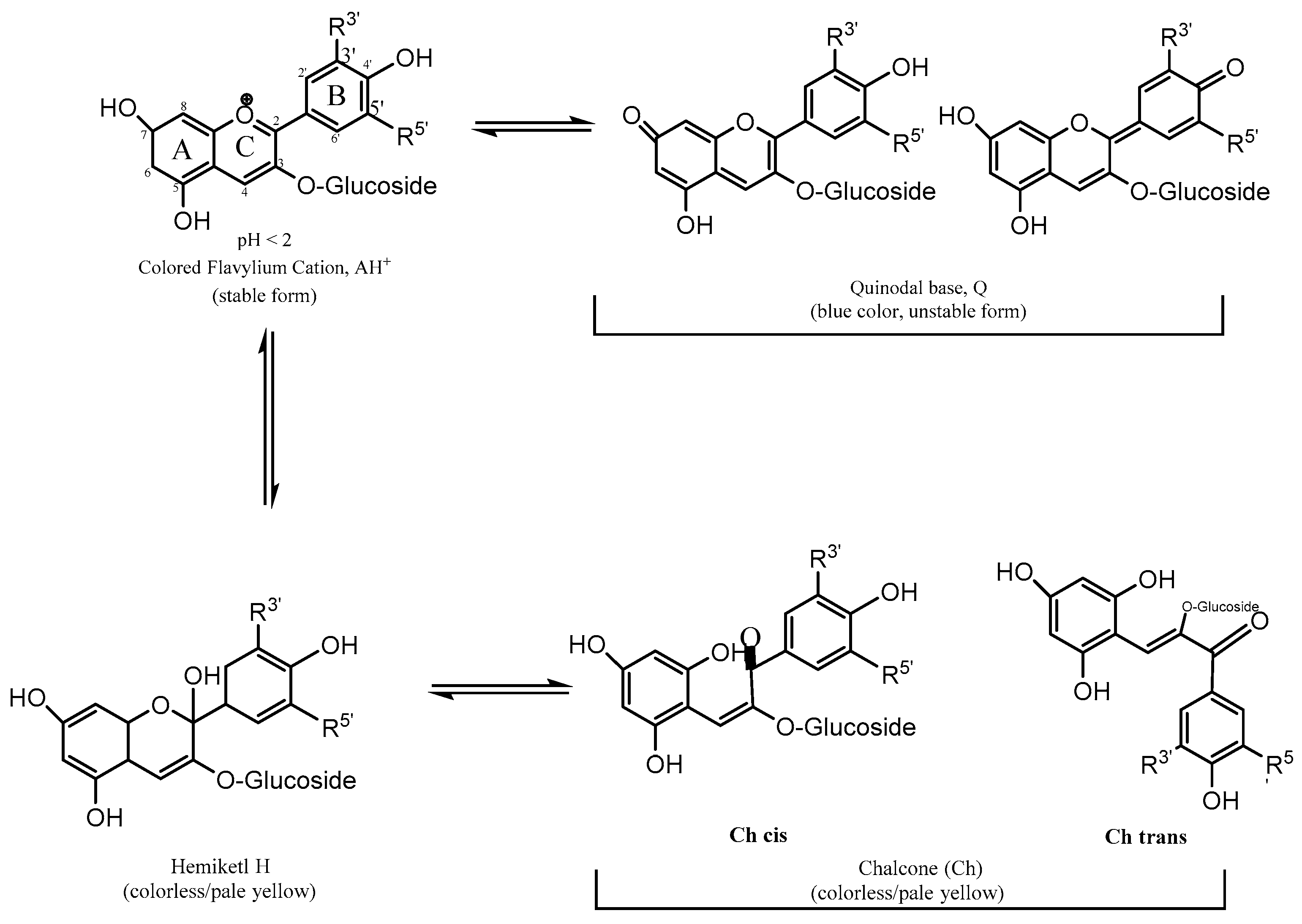
Within anthocyanins’ characteristics, they are known to be water- and alcohol-soluble organic compounds that have a three-ring heteroaromatic polyphenolic skeleton. Anthocyanins are glycosylated analogues, mainly at the C3 position, of anthocyanidins, both based on the 2-phenyl-benzopyrilium chromophore structure (flavylium ion in Figure 1), which shows an extended π conjugation, as well as the presence of a positive charge and several free -OH groups. These characteristics allow anthocyanins to absorb light in the visible region, which generates a great variety of dye colors, making them one of the most important natural pigments besides chlorophyll.
Anthocyanidins possess an almost planar chemical structure characterized by variations in the number and positioning of hydroxyl groups and/or methoxy groups. These structural differences play a crucial role in determining the natural availability of a diverse array of anthocyanins and anthocyanidins [7]. Anthocyanidin glycosides are 3-monoglucosides and 3,5-diglucosides, consisting of the most common sugar, glucose; however, rhamnose, xylose, galactose arabinose, and rutinose can also appear [8].
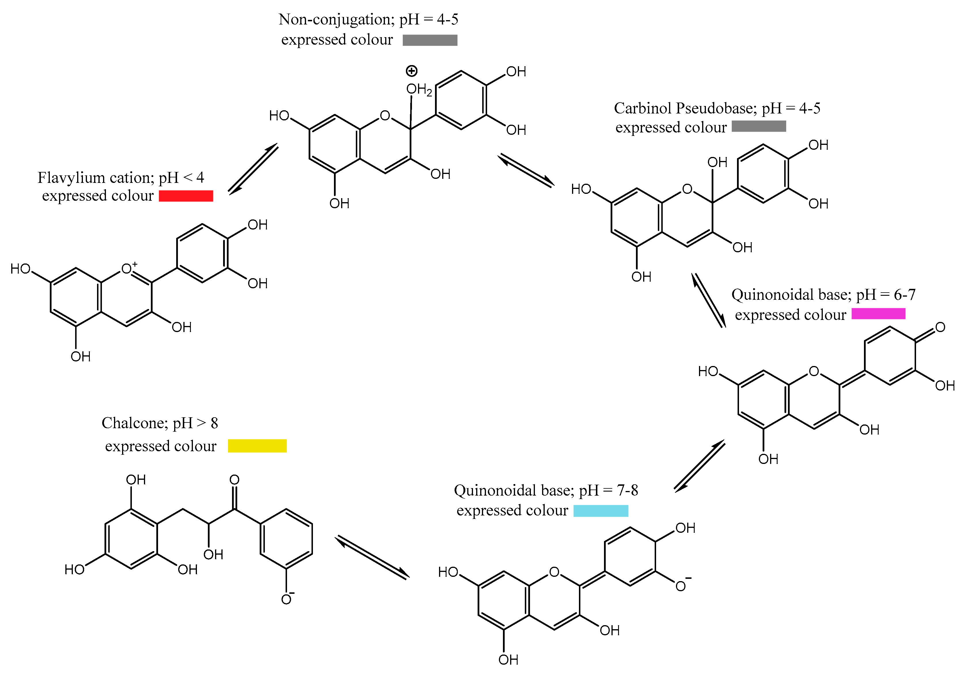
Depending on conditions such as temperature, light, solvent, metal ions, and mainly pH, anthocyanins undergo structural modifications accompanied by photophysical and chemical changes. In fact, different proton concentrations can lead these molecules to hydrated open ring or quinoidal forms [9]. Anthocyanins are very stable at acidic pHs, but as the pH approaches neutral, their stability dramatically decreases, leading to complete degradation at pHs above 7. The kinetics of these changes is one of the main factors that determines the final color of these compounds (Figure 2) [10].
Generally, a molecule with more -OH groups generates a more intense blue color, while in those with -OR groups, the coloration turns red. As for the glycosyl substituents, their presence decreases the coplanarity of the B-ring, so they tend to be less stable, and the coloration is usually not as intense.
If, in addition to sugar, there is an acyl radical in the molecule, they are called acylated anthocyanins. An increase in glucosidic substitution and acylation with cinnamic acids allows them to be more stable and retain their characteristic color at alkaline pHs [11].
Anthocyanins have received increasing attention from researchers in recent years mainly due to their importance in two main application sectors: the food industry and healthcare. Regarding food technology, the utility of anthocyanins extends beyond their role as a natural colorant to enhance the organoleptic properties of food. Their inherently reactive nature has led to their consideration as colorimetric indicators for assessing food quality in advanced smart food packaging technologies. Regarding healthcare and pharmaceutical applications, a large amount of highly interesting health-promoting effects of anthocyanins have been revealed in the last decade. Thus, the nutraceutical effect of the anthocyanin or anthocyanin-rich food intake has attracted research interest. However, their low stability and bioavailability hinder an efficient effect of anthocyanins in the human body. Consequently, research efforts have focused on the development of strategies to improve their stability, bioavailability, and color preservation. Among these strategies, encapsulation has become the most effective and explored one. Encapsulation is based on the development of a protective coverage of anthocyanins, which, in addition to improving their stability and bioavailability, could also provide advanced properties or performance (like selective or controlled release).
Accordingly, there exists a real need for immobilizing anthocyanins within appropriate substrates that fulfill the wide range of physical and chemical requirements specified for the mentioned applications. Among these substrate systems, polymers are versatile materials that show unique properties in a wide range of forms, like films, emulsions, hydrogels, mats, and nanoparticles, among others, that are of great interest in the food-packaging and healthcare sectors. In the light of this combination, the incorporation of anthocyanins within polymeric matrices has proliferated in recent years as a promising approach to utilize the synergistically positive properties of both substrates and active agents.
Until now, several works have reviewed anthocyanin food applications, the physiological effects of anthocyanins as functional food ingredients, as well as anthocyanins applications in health and disease prevention. However, despite there being a considerable number of studies reporting anthocyanin–polymer combinations that warrant highlighting, this specific field lacks a systematic and summarizing review. The objective of this paper is to summarize the most recent advances in the investigation of anthocyanins incorporated into polymers, emphasizing those related to food and healthcare applications. This combination of anthocyanin and polymer matrices is not only a promising alternative to solve the real challenge of the encapsulation of these phytochemicals, but it allows the development of smart biosensors and active materials, which highlights the relevance of the scope of this review.

2. Extraction Procedures for Anthocyanins

Due to their compositional complexity, the extraction from biological matrices and the selective isolation of a specific anthocyanin with chemical features is not a trivial issue. Consequently, it has become the main research target in this field. Moreover, several reviews exist on the methods used to extract and purify anthocyanins [1].
The conventional method for extracting anthocyanins is through maceration according to their polar nature. The use of polar solvents like methanol and ethanol makes the extraction of anthocyanins efficient. For food grade applications, ethanol is preferred over methanol. Often, organic (acetic, citric, or tartaric acids) or mineral acids (hydrochloric acid or phosphoric acid) are added to the extraction solvent to stabilize the flavylium cation. The utilization of hydrochloric acid is limited due to its potential to break down acylated anthocyanins. This restriction stems from the fact that hydrochloric acid may adversely affect the integrity of these particular compounds during the extraction process [12].
The extraction temperature plays a significant role in anthocyanin recovery, as their solubility increases at elevated temperatures. However, it is crucial to note that higher temperatures can also promote decomposition, impacting the overall efficiency of the extraction process [13]. The traditional extraction procedure consists of several extraction rounds, depending on the extractability of the system, as well as the quantities of total anthocyanins present in the matrix. For commercial applications, the extracts are then concentrated into a thick paste under a reduced vacuum in a rotavapor. Finally, the concentrated extract is freeze-dried to obtain a fine anthocyanin powder [14].
Additionally, if the extracts contain lipid materials, the addition of an organic solvent such as hexane to the extract can eliminate some lipid substances contained in said materials. Acid can cause the partial hydrolysis of the acyl moieties in acylated anthocyanins, especially those with dicarboxylic acids such as malonic acid, so the use of weak acids, such as tartaric or citric acid, is desirable to keep the dicarboxylic substituents intact [15,16]. In this sense, pH has also been shown to have a significant influence on the color of anthocyanin extracts and the absorbance readings. At lower pH values (pH < 2), blue and purple wheat extracts exhibited a red to dark red color change [12].
More sophisticated methods include those derived from the combination of sorption methods with membrane (nanofiltration, ultrafiltration, and electrodialysis) technologies [17] that provide a high extraction efficiency and allow for the selective separation of anthocyanins and other polyphenols from multicomponent natural mixtures. In recent years, technologies using ion exchange resins (IERs) or ion exchange membranes (a type of flat resin) for the extraction and purification of phenols and anthocyanins have been increasingly adopted [7].
Recently, Gravier-Rodríguez et al. [18] proposed a factorial experimental design for the ultrasound-assisted extraction of anthocyanins that allowed for a significant improvement in the amount of anthocyanins extracted. This novel procedure for anthocyanin extraction is based on the generation of cavitation bubbles, whose vibrations create fluid currents and disruptive forces in nearby cells and particles.

3. Biological Properties of Anthocyanins

As has been above mentioned, anthocyanins have sparked growing interest due to their bactericidal properties and a large list of unique biological benefits, such as antioxidant, anti-inflammatory, antidiabetic, chemopreventive, antimutagenic, anti-obesity, and neuro- and cardioprotective properties.
Regarding their antibacterial ability, these pigments have been noted for their ability to inhibit the growth of pathogenic bacteria, thus offering significant potential in food preservation and the prevention of foodborne illnesses. Recent research suggests that anthocyanins may act as antimicrobial agents, interfering with the cellular function of harmful bacteria and contributing to the natural preservation of food [19]. Different mechanisms have been proposed for the antimicrobial effect of anthocyanins. In this sense, it is known that anthocyanins can destroy cell wall integrity and destabilize the cytoplasmic membrane. In addition, they have also shown to negatively affect bacteria metabolism by inhibiting extracellular microbial enzymes [20].
Its outstanding antioxidant activity helps neutralize free radicals, unstable molecules that can contribute to cellular aging and various chronic diseases [21]. Indeed, anthocyanins have shown higher antioxidant properties than conventional antioxidant molecules like α-tocopherol. This important antioxidant effect derives from the special chemical structure characterized by a high number of hydroxyl groups, the catechol moiety, and the oxonium ion in the C ring. Indeed, glycosylation, which limits electrons’ delocalization, seems to decrease its free radical scavenger ability. Nevertheless, the antioxidant activity of anthocyanins can be decreased by the antagonistic interactions with other phytochemicals and vitamins also present in ingested fruits and vegetables [20,21].
Anthocyanins have been noted to possess anti-inflammatory properties, which may be especially relevant to cardiovascular health and the prevention of chronic inflammatory conditions. Inflammation is a complex biological response of injured tissue in which different external or internal stimulators, such as cytokines, interleukins, or cyclooxygenase (COX) enzymes, take part. Specifically, anthocyanins have demonstrated an inhibitory effect on COX-1 and COX-2 enzymes that is comparable with synthetic anti-inflammatory drugs such as ibuprofen and naproxen. Anthocyanins have also shown in vitro to inhibit various interleukins and NF-kβ, a nuclear factor that serves as a central inflammatory mediator. In addition, anthocyanins with an ortho-dihydroxyphenyl structure have been shown to suppress the activation of MAPK, which is related to the cellular response to proinflammatory cytokines [20].
These compounds have also been associated with improving cognitive function and a protective function against oxidative stress in the brain [18] due to their special role in inhibiting neuro-inflammation and regulating neural signaling. Different studies relate this action to a cerebral blood flow enhancement and the inhibition of NF-kβ upregulation. In vivo studies have proven that this improvement on the neuronal function results in the delay of Alzheimer’s disease, an enhancement of memory-associated neuronal signaling, and the regulation of synaptic plasticity, which is a key factor in learning and memory.
The ability of anthocyanins to modulate gene expression and regulate various metabolic pathways offers a promising outlook in the research on medical treatments and therapies. Some studies suggest that these substances may have positive effects on blood sugar regulation, which could be beneficial for individuals with diabetes (Type 2) [22].
In addition, it has been observed that anthocyanins can influence the expression of genes related to cell proliferation and apoptosis, which could have implications in the prevention and treatment of cancer [20,23]. These anticancer properties seem to be based on multiple and additive mechanisms that involve cell cycle arrest, apoptosis, anti-angiogenesis, inhibition of DNA oxidative damage, the above-mentioned inhibition of COX-2 enzymes, and anti-mutagenic and anti-carcinogenic effects. Although is widely recognized that anthocyanins play an essential role in the anticancer activity of many fruits and vegetables, several studies have shown that it is the result of the collaborative action with other phytochemicals that are also present.
In summary, anthocyanins, beyond their visual appeal in foods, have bactericidal properties that can contribute to the preservation of foods and have notable biological benefits in the human body. These properties, backed by scientific research, highlight the potential of anthocyanins in promoting health and preventing important diseases.

4. Halochromic Properties of Anthocyanins

Chemically, chromism is a process that induces changes, which are usually reversible, in the colors of compounds. These changes can occur due to a number of external factors, such as temperature (thermochromism), light (photochromism), electricity (electrochromism), ions (ionochromism), etc. The use of these properties in materials categorizes them as smart materials. Within the phenomenon of ionochromism, halochromism corresponds to the color change process affected by pH, being sensitive to the hydrogen ion (H+) (protonation or deprotonation) [24].
Anthocyanins can be found as different chemical structures and colors depending on the pH of the solution as shown in Figure 3 [25]. In general, anthocyanin displays a red color in acidic conditions, pink in neutral conditions, and blue in basic conditions [26]; thus, according to their halochromic properties, anthocyanins are excellent candidates for the development of colorimetric materials.
At pH 1, the flavylium cation (red color) is the predominant species and contributes to purple and red colors (Figure 3). At pH values between 5 and 6, only two colorless species can be observed, which are a carbinol pseudo-base and a chalcone, respectively. At pH values higher than 7, the anthocyanins are degraded depending on their substituent groups (Figure 3, degradation reaction).
At pH values between 4 and 6, four structural forms of the anthocyanins coexist: a flavylium cation, anhydrous quinoidal base, colorless carbinol base, and the pale yellow chalcone. The equilibrium between the quinoidal bases and carbinol occurs via the flavylium cation [27]. Investigations about anthocyanin stability and the color variation with pH concluded that the changes in the color of these compounds are more significant in the alkaline region due to their instability.
The ability of these substances to visually reflect variations in acidity or alkalinity makes them valuable tools in practical applications, such as the evaluation of pH in biological solutions and foods. The versatility of anthocyanins as pH indicators has increased their use in food industry because this specific capacity makes them play a crucial role in the development of non-invasive and environmentally friendly measurement methods.

5. Anthocyanin-Loaded Polymers: Preparation and Characterization Methods

Anthocyanin-loaded polymers have recently been a point of interest due to their potential applications as smart substrates for anthocyanin immobilization, protection, enhanced bioavailability, and controlled release. The most common matrix materials employed are biopolymers, due to their environmentally friendly, biodegradable, non-toxic, and edible nature, which makes them ideal candidates for developing nutraceutical delivery systems. In addition, naturally occurring polymers derived from renewable resources have attained attention as sustainable materials to replace petroleum-based commodity plastics traditionally used in short-term food packaging [28].
Among biopolymers, polysaccharides and proteins are the most commonly employed ones for anthocyanin loading. Both polysaccharides and proteins display excellent gelling properties that facilitate networking in hydrogel-based systems, enabling high loadings as well as remarkable film-forming properties that are essential for forming suitable films for food packaging and wound-healing applications. Polysaccharides are widely investigated and used as substrate and coating materials to encapsulate and deliver bioactive molecules. Moreover, polysaccharides show great affinity to anthocyanins, due to electrostatic, hydrogen bonding, and hydrophobic interactions, which lead to high efficiency entrapment of anthocyanins. Alginate, chitosan, gum arabic, starch, dextran, and their derivatives are the most investigated polysaccharides for anthocyanins immobilization. Proteins are also usually employed as substrate or protective materials for the encapsulation of small molecules; however, they can be easily denaturalized and have low stability, putting them at a disadvantage in comparison with polysaccharides [29]. When it comes to proteins, egg white, soy protein, gelatin, and sodium caseinate are worth highlighting due to their surface- and film-forming characteristics, while hydrophobic proteins like zein and millet protein are known to form nanoparticles by self-assembly in the appropriate conditions [29]. Plant and animal proteins show binding affinity for anthocyanins, driven mainly by Van der Waals forces, hydrophobic interactions, and covalent and H bonding.
Apart from polysaccharides and proteins, biodegradable synthetic polymers have also been explored to combine with anthocyanins. This is the case for poly (lactic acid) and polyvinyl alcohol, which have been widely proposed as biodegradable packaging films.
Since polymers can be processed into a wide variety of forms, polymer-mediated anthocyanins encapsulation has been addressed by different resourceful approaches according to the desired application. The most important encapsulation polymer systems employed in anthocyanin loading and release are described below.

5.1. Films

Anthocyanin-loaded polymer films are obtained mainly by solution casting, a simple and cheap technique that uses aqueous solutions or organic solvents to dissolve anthocyanins as the active agent and polymers as the substrate for immobilization [30]. In brief, polymer/s, plasticizer, and an anthocyanin extract are sequentially dissolved in solution at temperatures lower than 80 °C for a short period of time to minimize the degradation of the anthocyanins. Finally, the obtained solution is incubated onto flat Petri dishes at ~25–40 °C for 24–48 h. Other more sophisticated and expensive techniques have also been reported to fabricate anthocyanin films, such as electrospinning and layer-by-layer and 3D printing [31]. Chitosan, cellulose, starch, zein, gelatin, pectin, agarose, xanthan gum, and combinations of these have been the most commonly employed polymer systems for the development of anthocyanin-loaded films. Glycerol and sorbitol are the most popular plasticizers used in anthocyanin-based films [32].
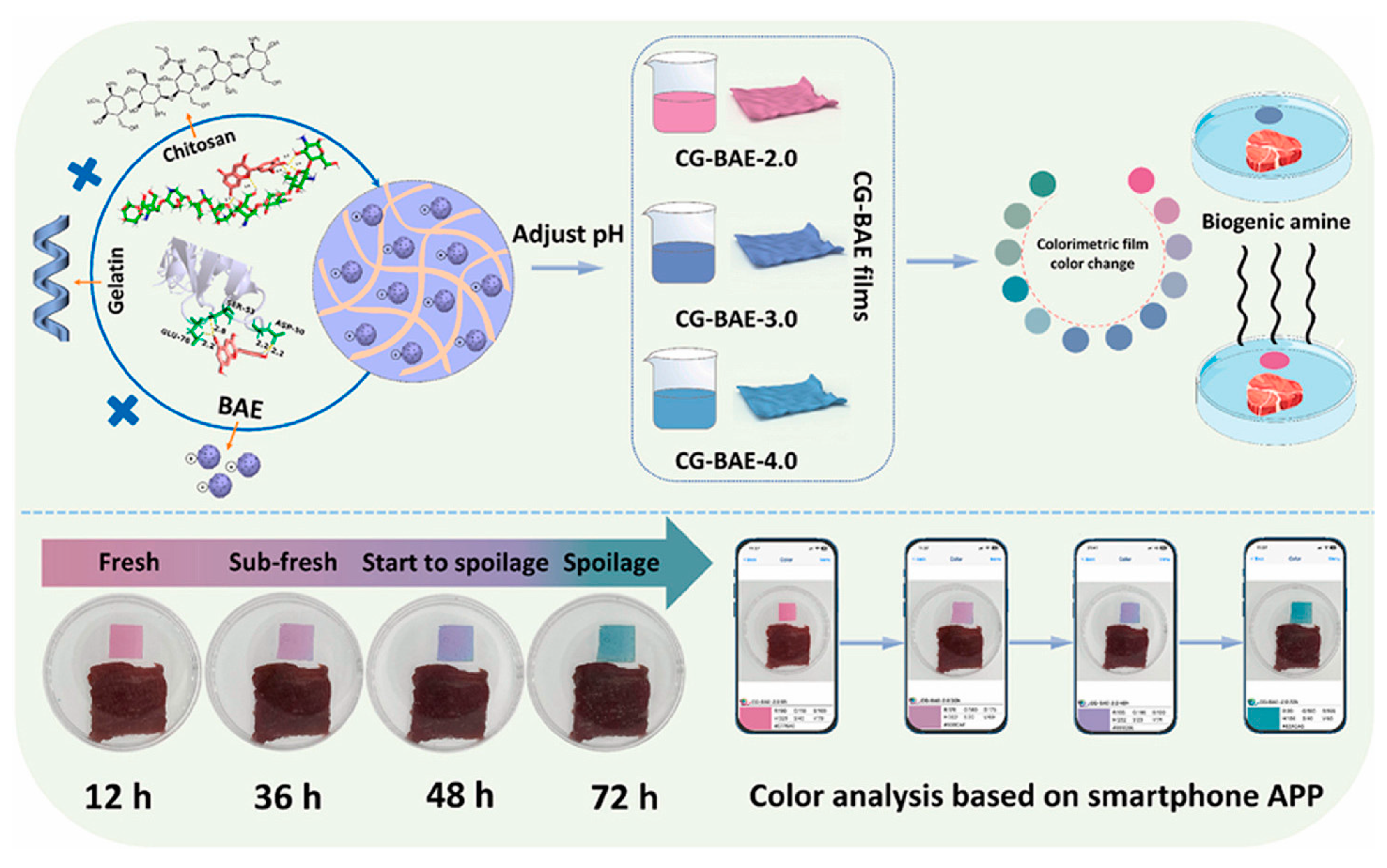
Regarding films for monitoring food spoilage, it has been shown that combining polymers improves the physical properties of the material. For example, Nadi et al. [33] showed that combining basil seed gum with chitosan and adding red cabbage extract as a colorimetric indicator improves properties such as solubility, water vapor permeability, and flexibility in the material. Recently, Li et al. [30] reported the use of a film that combines chitosan and gelatin with a butterfly pea flower anthocyanin extract to evaluate beef freshness. This combination had an outstanding sensitivity to pH changes that are not perceptible to the naked eye, and the color change could be assessed using RGB values obtained from a smartphone app (Figure 4), differentiating four basic stages: freshness, sub-freshness, onset of deterioration, and complete deterioration.
Wu et al. [34] demonstrated that a novel way to promote the stability of anthocyanins in polymer films is the prior chemical modification of the phytochemical.
The incorporation of anthocyanins into films can modify the technological and functional properties of the film. These changes depend on the physical or chemical interactions between the film-forming polymer and the anthocyanin extract, which can lead to structural modifications. The interaction between biopolymers and anthocyanin-rich extracts depends on the nature, chemical characteristics, and concentration of both the polymer and extract, as well as the structural properties of the active components. The addition of anthocyanin-rich extracts can influence the thickness, color, opacity, solubility, water vapor permeability, and mechanical properties of the films [35]. Thickness is typically analyzed because, in most cases, it is modified and, since it hinders the penetration of water, it is a key parameter to take into consideration [31].

5.2. Mats and Fibers

It is known that nanofibrous mats have a high surface-to-volume ratio, small pore size, and high porosity compared to polymer films, which makes electrospun nanofibrous mats very attractive materials for the manufacture of substrates for the immobilization of anthocyanins [36].
The electrospinning process involves the application of an electric field to create fine polymer fibers from a solution containing anthocyanins. Anthocyanins can decrease the conductivity of the polymer solution, and consequently, as Jiang et al. [29] observed by SEM microscopy, heterogeneous fibers are formed.
Anthocyanin nanofiber mats made by electrospinning are used as tools in the field of active/smart packaging where they have been studied to improve their stability, simplify the slow and controlled release of antimicrobial and antioxidant substances [37], and as spoilage sensors [38]. When it comes to healthcare applications, nanofibrous mats of anthocyanins have found their major application as smart wound dressings that can monitor the wound healing process [39].
As an example of this, Khaledian et al. [37] reported a new material based on a potato starch–turnip peel extract and guar gum–cinnamaldehyde to control the growth of spoilage-related microorganisms by extending the shelf life of fresh lamb meat under refrigerated storage conditions for up to 13 days. More complex combinations that including fibers have been also evaluated, for example, in the case of anthocyanin nanoencapsulated in gelatin and immobilized on ethyl cellulose nanofibers reported by Nath et al. [38] that showed pH sensitivity that was stable up to 7 days of storage at room temperature.

5.3. Hydrogels

Hydrogels have been widely used as a method of encapsulating water-soluble active ingredients. The thermo-degradation and stability against sudden pH changes of anthocyanins could be inhibited by encapsulating them within hydrogel systems. This has been demonstrated in the process of simulated digestion and may be very useful in the prevention of intestinal diseases [40,41].
Recent research has demonstrated the versatility of these materials that allow the easy incorporation of multiple active agents. For instance, Lotfinia et al. [42] combined alginate in the form of a hydrogel with honey and red cabbage extract, and could observe improved mechanical properties, the antibacterial activity attributed to the honey, as well as the antioxidant properties and good activity against pH changes from the red cabbage.
Polysaccharides have been traditionally employed in anthocyanin hydrogel preparations due to the simplicity of their corresponding gelation process. This is the case for alginate, which forms cold gels through Ca2+-induced cross-linking [43], or starch [44], another widely used material for these gel-based encapsulation systems.

5.4. Polyelectrolyte Complexes

Polyelectrolytes are macromolecular materials that possess repeating units and dissociate into highly charged polymeric molecules in aqueous solution, forming either positively or negatively charged polymeric chains [45]. There are numerous compounds that can serve as biopolyelectrolytes such as proteins, polysaccharides, and their derivatives. Biopolyelectrolyte complexes can be formed by the titration of a biopolyelectrolyte solution in another biopolyelectrolyte solution with the opposite charge under agitation. Among the advantages of this methodology are the simplicity, quickness, and the fact that it does not require high energy, chemical crosslinkers, specialized equipment, or organic solvents. An advantage of these materials is that polymeric chains can generate dense interconnecting networks, which is useful for inhibiting the penetration of polar reactive compounds and the attack on charged anthocyanins [46].
Tan et al. [47] demonstrated that polyelectrolyte complexes obtained from chitosan and chondroitin sulfate incorporating anthocyanins and several co-pigments (catechol, gallic acid, gallic acid ethyl ester, and (-)-epigallocatechin gallate) present a synergistic effect of co-pigmentation and encapsulation in protecting anthocyanin against the nucleophilic attack of ascorbic acid. This interesting effect was attributed to the dense network created by the hydrogen bonds between the co-pigments and polysaccharides, as well as the formation of hydrophobic environments between the co-pigments and polysaccharides.
The stabilizing effect of polyelectrolyte complexes was comparatively analyzed in polycomplexes of whey protein with four different polysaccharides: chondroitin sulfate, dextran sulfate, arabic gum, and pectin. The results showed that pectin provided the highest protection, followed by arabic gum, chondroitin sulfate, and dextran sulfate. The stabilizing effects are attributed to hydrogen bonding and hydrophobic and electrostatic interactions between the whey protein and the polysaccharide [48].

5.5. Nanoparticles

As in the general case of polymeric nanoparticle preparations, the methods commonly used for the preparation of anthocyanin nanocarriers can be classified as the emulsification cross-linking method, ionic cross-linking method, covalent cross-linking method, and self-assembly methods.
The emulsification crosslinking method is a widely used method. A carrier particle is usually large (tens to hundreds of microns) with a highly homogeneous particle distribution. Anthocyanin nanoparticles are divided into ionically crosslinked and covalently crosslinked depending on the crosslinking method. Covalently cross-linked carriers are more stable. Studies have shown that the type of polymer employed has a relevant effect on the crosslinking efficiency. For example, pectin and carboxymethylcellulose lead to a higher crosslinking density than arabic gum with the 1-ethyl-3-(3-dimethylaminopropyl) carbodiimide/N-hydroxysuccinimide crosslinker, resulting in nanoparticles of anthocyanin with a smaller particle size (160–210 nm), more uniform distribution, high encapsulation rate (up to 80%), slow release, and much higher antioxidant activity [49].
Ionic nanoparticles could be easily formulated through complexation of two different charged biopolymers in diluted solutions. Biopolymers such as alginate, chitosan, whey, and soy protein are commonly extruded as a carrier solution through a needle or nozzle into a gelation solution containing the specified ions. This is a mild process that has shown a high encapsulation efficiency and effective protection of anthocyanins under gastric conditions [50].

5.6. Emulsions

Emulsion-based systems are intended to overcome the low stability and bioavailability of anthocyanins. Among the most used systems are nanoemulsions and microemulsions that have been studied in vivo and in vitro as anthocyanin delivery systems [51,52,53,54].
Nanoemulsions are colloidal dispersions formed by droplets of a liquid dispersed in another liquid that, despite being immiscible, can be stabilized with a surfactant layer. In nanoemulsions, particles showing sizes from 20 to 500 nm can avoid emulsification, flocculation, or precipitation during storage [55,56,57]. Some of the benefits of nanoemulsions are the control of the release rate and the avoidance of decomposition or degradation of the encapsulated anthocyanins [58].
Microemulsions are mainly composed of water, oil, surfactants, and cosurfactants, and have better flowability, more uniform particle dispersion, and stronger stability than emulsions. Microemulsion particles have sizes between 10 and 100 nm, which can provide stable and uniformly dispersed systems [59], which are useful properties for improving the absorption and bioavailability of anthocyanins and enable easy and multiple routes of administration [60,61].
There are several studies that report the development of nanoemulsions and microemulsions with extracts from different sources of anthocyanins, such as mangosteen peel, Brazilian berry, purple sweet potato, cranberry, red cabbage, blueberry, and jaboticaba peel, among others, showing nanoparticles that are stable for long periods of time, ranging from 30 days to 3 months [53,58,59,62,63].
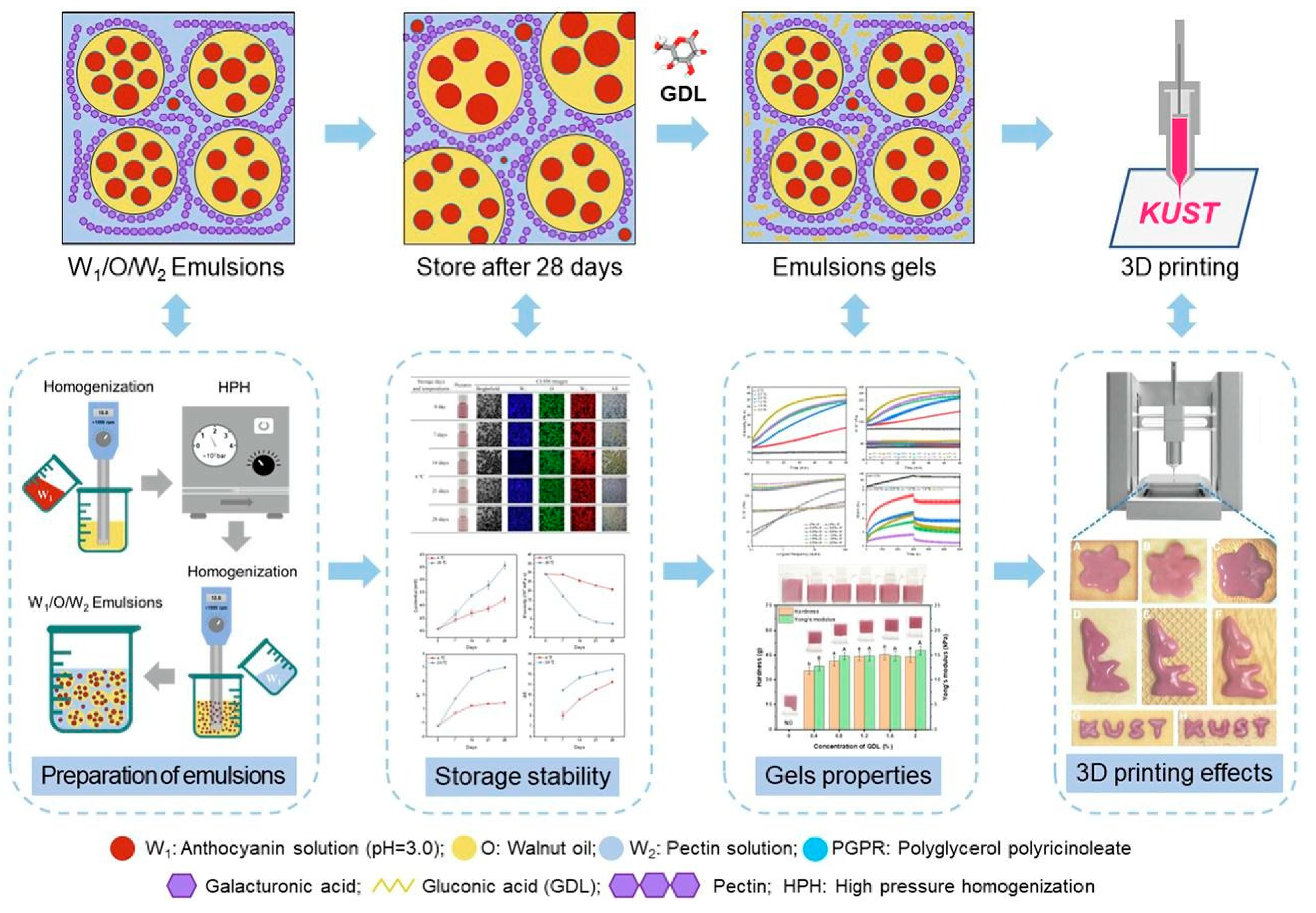
Recently, many researchers have attempted to construct W1/O/W2 double emulsions designed to encapsulate anthocyanins. Among the most recent findings, it is worth highlighting a double emulsion of pectin and glucono-delta-lactone prepared by Li et al. [51] that encapsulated anthocyanins from a mulberry extract that was tested for three-dimensional printing (Figure 5). This manufacturing method has received increasing attention from the food industry due to its shape flexibility and customized design [51,64].

5.7. Self-Assembled Liposomes, Proteins, Peptides, and Phospholipids

Today, liposomes, phospholipids, and proteins are used as anthocyanin nanocarriers and are formed by direct self-assembly. Proteins, as was commented above, have the drawback of being very sensitive to pH changes or temperature. Liposomes also have low stability and their phospholipids are prone to oxidation during long-term storage. Phospholipids have a lot of benefits including the ability to protect sensitive ingredients, increase the bioavailability of nutrients and the efficacy of food additives, and confine undesirable flavors [65].
Amphiphilic peptides with a hydrophobic tail and a hydrophilic head are also used in molecular self-assembly. These amphiphilic peptides have good biocompatibility, biodegradable self-assembly, and chemical variability, which leads to a variety of nanostructures. Indeed, the addition of peptides to foods containing anthocyanins is recognized as the simplest way to improve the color stability of anthocyanins [58,65,66].
Recently, nanoliposomes have been reported as one of the most widely used colloidal delivery systems in food and nutrition research [58]. They are synthesized mainly from surfactants (phospholipids) that form an aqueous core and an amphiphilic lipid bilayer by embedding water-soluble substances in the aqueous phase and lipid-soluble substances in the oil phase [67,68,69]. Anthocyanins can be loaded into the internal aqueous phase of the nanoliposome to avoid degradation by light, pH, and temperature [58]. Their bilayer structure endows them with great biocompatibility, allowing them to easily fuse with bacterial membranes and penetrate microorganisms [70,71].

5.8. Microencapsulates

Microencapsulation consists of the incorporation of the active agent within the encapsulants by noncovalent interactions formed during a mixing process that can include additional coating layers [50]. The final drying step generally takes place by a freeze-drying or spray-drying methodology. Microencapsulation is an alternative to keep the properties of the anthocyanins intact and offer food processors a means of protecting sensitive food components [72,73,74,75,76,77]. However, during the spray drying process, some heat-sensitive anthocyanins may lose their activity or degrade and, as a consequence of the rapid dehydration, they may change their crystal structure. Consequently, freeze-drying is often preferred to mitigate these potential issues and preserve the integrity of the anthocyanins [49].
Microencapsulation allows the encapsulation of anthocyanins within a wide variety of encapsulating agents, such as polysaccharides, starches, inulin, maltodextrin or dextrose, corn syrups, arabic gum, mesquite gum, lipids, and proteins [78].

5.9. Specific Characterization of Anthocyanin-Loaded Polymer

The characterization of anthocyanin-loaded polymers is crucial for understanding their structure and properties. Several techniques are used to obtain significant information on these materials.
UV-Vis spectroscopy is used to analyze the absorption spectra of these materials, providing information about their specific composition, color, stability, and optimum absorption range [79].
FTIR-ATR is also a useful technique because it provides information regarding the interactions between the anthocyanin and the polymer matrix. The main changes are reported in two regions, between 1500 and 1600 cm−1, corresponding to the C=C bonds, and around 3200 cm−1, corresponding to the vibrations of the C-H bonds. In both cases, if the signals are shifted and/or widened, it confirms the immobilization of the anthocyanins in the polymer due the formation of strong interactions with the polymers [30,80,81].
Using Thermo-Gravimetric Analysis (TGA), several studies have shown that the addition of anthocyanins to polymeric matrices or the encapsulation of anthocyanins in a polymer provides greater stability with respect to pure compounds [82,83,84].
In some cases, when the main application is the detection of pH changes through color, color quantification is accomplished using the values of the L*, a*, and b* parameters. The L* value reflects the brightness; the a* value reflects the degree of red and green; and the b* values reflect the degree of yellow and blue. With the standard whiteboard as the color difference reference, the color difference value ΔE was calculated according to the following equation [85]:
E = L L 0 2 + a a 0 2 + b b 0 2
Tassanawatm et al. [86] reported that ΔE values greater than five can be easily indicated and detected with the naked eye [87].
The most recently reported investigations about anthocyanin-loaded polymers are summarized in Table 1.

6. Anthocyanin-Based Polymers for Food Applications

As mentioned above, anthocyanins have been widely studied because they are bioactive substances with many attractive characteristics (antioxidant, anti-inflammatory, antibacterial, and even anticancer properties). Indeed, consuming foods high in flavonoids (of which anthocyanins are a subgroup) has multiple health benefits, such as improvements in the cardiovascular system [105], neuroprotective effects [106], and improvement in skin texture due to their antioxidant and anti-inflammatory properties [107,108]. According to this, the food industry is one of the most important areas for anthocyanin application. For this reason, recent studies have based their research on safety studies of the incorporation of anthocyanins into various polymeric matrices for their use in the food industry.
Research and food industry interest have focused around three specific targets regarding anthocyanin-loaded polymers. Firstly, improving the general health of the human body due to anthocyanins’ beneficial effects against chronic nontransmissible diseases (bioactive substance), which is reflected in the production of food products enriched with anthocyanins. Secondly, providing “intelligence” to those materials into which they are incorporated, e.g., producing so-called “smart materials” that are able to respond to external stimuli. In this sense, anthocyanins act as interactive indicators helping to assess the chemical and microbial quality of food products through pH monitoring. Finally, materials loaded with anthocyanins are “active” since they contain antimicrobial and antioxidant agents that improve food preservation and function as chemical preservatives by reducing lipid oxidation and microbial growth in packaged foods. Figure 6 shows the roles of anthocyanins as a bioactive, active, and smart substance for food applications.

6.1. Anthocyanins as Bioactive Substances

Bioactive substances are chemical compounds present in foods and plants that, when ingested, can benefit human health, as is the case for anthocyanins.
Due to the low bioavailability of anthocyanins, they struggle to reach the desired targets effectively. To address this issue, anthocyanin-containing edible polymers have gained significant attention. These innovative platforms withstand the upper gastrointestinal tract, resisting early molecule release or degradation due to varying pH levels and digestive enzymes. Recent studies have explored the use of different polymers for efficiently transporting anthocyanins through the human body. For example, Deng et al. [109] fabricated anthocyanin-based polysaccharide films with potential antimicrobial and antioxidant effects for food applications. They combined the wine grape pomace anthocyanin with three different plant-based polysaccharides: low methoxyl pectin, sodium alginate, and Ticafilm®. The controlled release of phenolic from the film matrix exhibited its potential antioxidant and antimicrobial functions. Among the synthesized films, pectin-based platforms presented the highest water solubility and a faster phenolic release rate, which make these film strong candidates to be used as an oral fast-dissolving film for pharmaceutical or nutraceutical applications.
Butkevičiūtė et al. [110] used capsules of the natural polymer gelatin to encapsulate freeze-dried apple powder as a bioactive substance to evaluate the release kinetics of phenolic compounds. Mueller et al. [111] encapsulated anthocyanins from bilberries using whey protein or citrus pectin and observed that citrus pectin encapsulation increased intestinal accessibility during passage through the small intestine [112].

6.2. Smart Anthocyanin-Based Polymers

Thanks to their coloring properties, anthocyanins are used as indicators to monitor and evaluate the quality of food products due to their sensitivity of their color to various factors, such as pH changes (Table 2). The detection and quantification of these substances provide valuable information about foods’ freshness, stability, and authenticity. Smart polymers are a technology, primarily used in the food industry, that incorporates materials capable of detecting immediate food spoilage. Anthocyanins have been widely used as quality indicators in packaged foods due to their sensitivity to pH changes [12], creating visual shades varying from red to purple [4].
Milad Tavassoli et al. [113] pioneered the utilization of novel anthocyanins sourced from sumac extracts as doping agents in pectin (PC)/chitosan nanofiber (ChNF) films. These films exhibited promising potential for monitoring shrimp decomposition by showcasing sensitivity to ammonia vapors. Notably, the intelligent film containing sumac anthocyanins showed a rapid response to volatile ammonia within the initial 5 min, as depicted in Figure 7. Furthermore, these films demonstrated remarkable antibacterial activity against S. aureus, E. coli, and P. fluorescence bacteria.
The same author prepared a food quality sensor by combining chitosan nanofibers and gelatin, incorporating anthocyanins from barberry fruit and saffron petals [114]. These polyphenolic compounds served as colorimetric pH-responsive indicators for monitoring fish freshness. The films notably diminished light transmission, particularly in the UV spectrum, owing to the anthocyanins’ ability to absorb UV and visible light. Both types of anthocyanins exhibited color changes corresponding to pH variations. Additionally, their study confirmed that these films effectively inhibited bacterial growth.
Similarly, Milad Bakhshizadeh et al. [115] developed pH-responsive colorimetric films as an intelligent packaging system. These films integrated gelatin along with common poppy anthocyanins (CPs) with aloe vera gel (AVG) and rosemary essential oil in order to provide the material with antioxidant and antimicrobial properties, serving as a pH sensor as well. The intelligent films exhibited color changes under different pH conditions. Another essential characteristic is that the films possessed antioxidant properties as well as antimicrobial properties against both Gram-positive and Gram-negative bacteria.
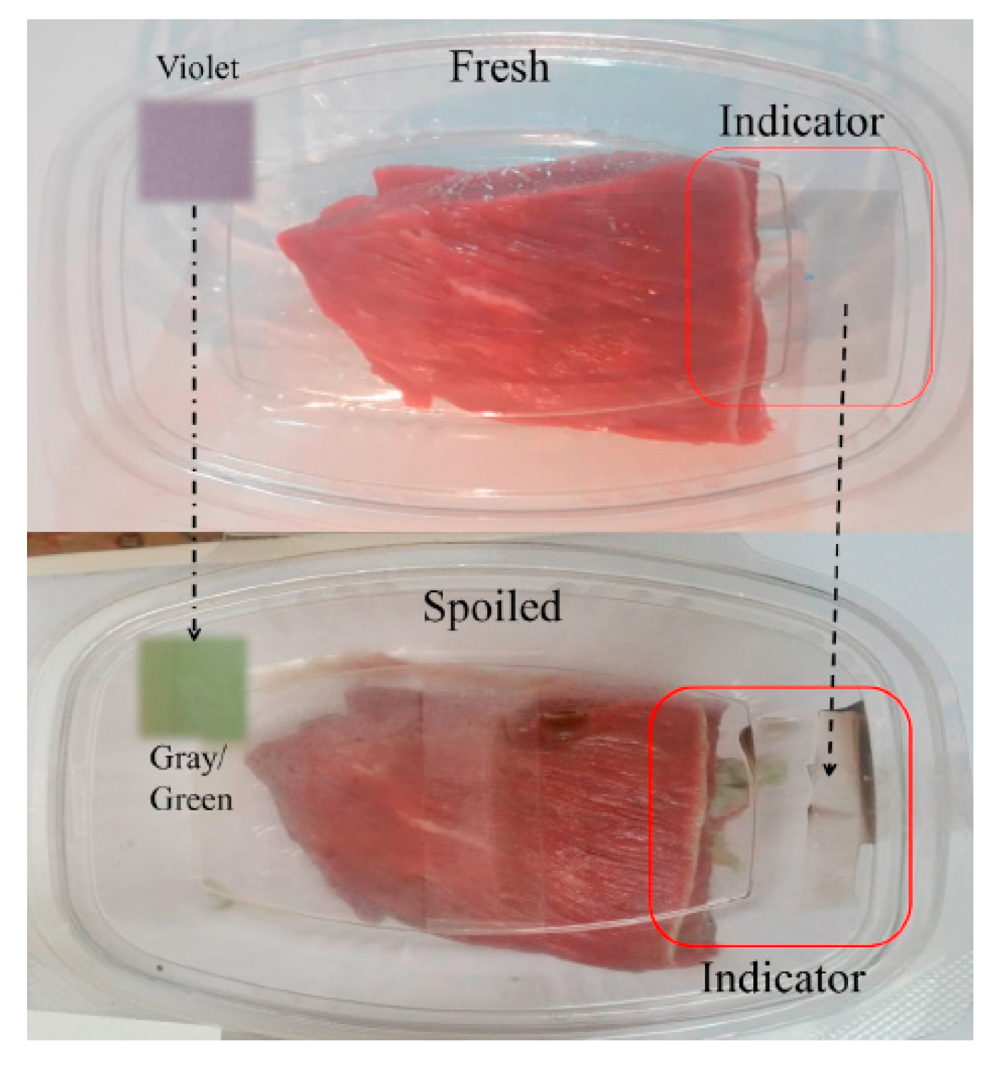
Another use of anthocyanins as food quality sensors was described by Alizadeh-Sani et al. [116]. They prepared halochromic films of methylcellulose/chitosan nanofibers loaded with anthocyanins extracted from saffron petals (Figure 8). These films were mainly characterized as indicators of the quality of meat products (lamb) when used as detectors of ammonium compounds. Another essential characteristic of these materials is that they present antimicrobial properties. The antimicrobial activity of the films was stronger against S. aureus (Gram positive) than against E. coli (Gram negative).
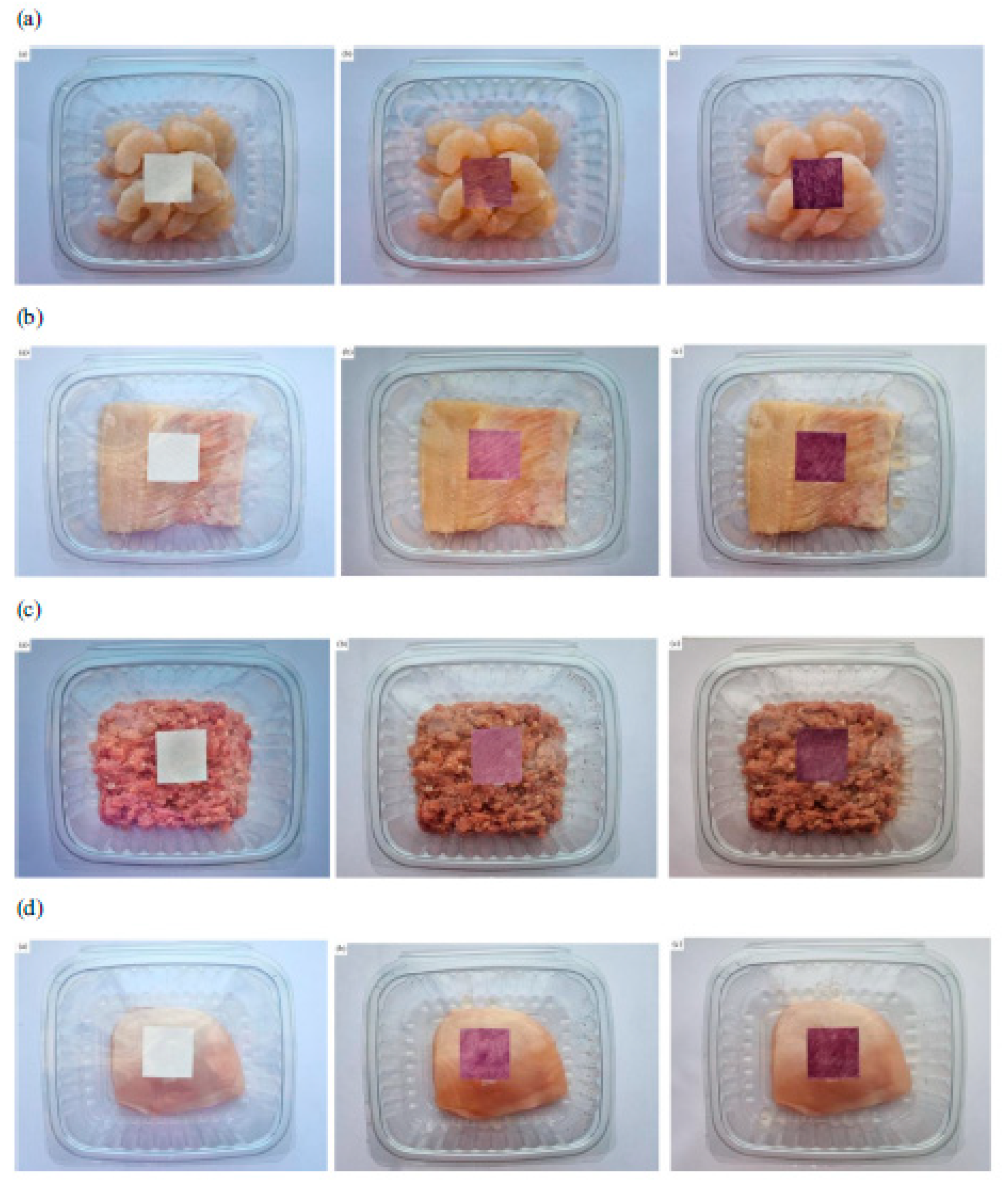
Fatemeh Rezaei et al. [117] also employed anthocyanins for monitoring the freshness of different packaged foods. They synthesized double-layer polymers based on carboxymethyl-cellulose nanocrystals and polylactic acid loaded with anthocyanins extracted from Viola odorata petals (VOE) to control the freshness of Pacific white shrimps, minced lamb meat, chicken fillets, and rainbow trout fillets during refrigerated storage conditions. The color changes associated with VOE anthocyanins were red at pH 1–6, violet at pH 7–8, green at pH 9–10, and brown at pH 11–12; higher pH values were directly correlated with decreased freshness. Hence, these findings showed that the synthesized indicator materials could successfully monitor food freshness in real-time, as shown in Figure 9.
Table 2. Recent research on smart anthocyanin-based polymers in the food industry.
Source of AnthocyaninsType of Studied AnthocyaninAnthocyanin RoleType of Tested FoodPolymer MatrixRef.
Purple corn powderCyanidin-3-glucosidepH indicator for detection of NH3, DMA, and TMA.Muscle food productsAlginate hydrogel beads[43]
Roselle Not indicatedpH indicator for real-time freshness monitoringPenaeus vannamei (white shrimp)PVA/HEMC/RAE/OA films[118]
Sumac powderNot indicatedpH indicator for detection of ammonia vaporsShrimpPectin (PC)/chitosan nanofiber (ChNF) films[113]
Powdered barberry fruit and saffron petalsCyanidin-3-glucosidepH indicator for detection of ammonia vaporsFishGelatin/chitosan nanofibers films[114]
Common poppyNot indicatedpH indicator for detection of ammonia vapors.FishGelatin (G)/rosemary essential oil (REO)[115]
Saffron petalsCyanidin-3-glucosidepH indicator for detection of ammonia vaporsMeat product (lamb)Methyl cellulose/chitosan nanofibers film[116]
Viola odorata petalsDelphinidin-3-(4-p-coumaroyl)-
rutinoside-5-glucoside and cyanidin-3-O-glucoside
pH indicator for detection of ammonia vaporsPacific white shrimps, minced lamb meat, chicken fillets, and rainbow trout fillets Double-layer polymers based on carboxymethyl cellulose/cellulose nanocrystals and poly(lactic acid) [117]
Lycium ruthenicum anthocyaninsNot indicatedpH indicator for detection of volatile acidsMilk Alginate-konjac/glucomannan films[119]
Red cabbage (Brassica oleracea var.
capitata f. rubra)
Not indicatedpH indicator for detection of ammonia vaporsNot studiedChitosan/chitin nanocrystals with curcuma oil[120]
Jacaranda cuspidifolia petalsNot indicatedpH indicator for detection of ammonia vaporsFishChitosan/polyvinyl alcohol [121]

6.3. Active Anthocyanin-Based Polymers

Anthocyanins have sparked interest in food preservation due to their antioxidant and antimicrobial properties [122] and ability to slow the oxidation and deterioration of certain products. Firstly, due to their antioxidant activity, they act as free radical scavengers, helping prevent lipids and protein oxidation in foods [21]. This is particularly relevant to avoid the rancidity of oils and maintain the quality of high-fat products [123]. Secondly, they act as oxidation inhibitors, a process that can cause changes in the flavor, color, and texture of foods [124]. The ability of anthocyanins to inhibit oxidation makes them helpful agents for preserving the freshness and quality of products such as oils, meats, and dairy products [125]. They also have antimicrobial properties, which means they can help inhibit the growth of microorganisms, such as bacteria and fungi, in food [126]. This can contribute to a longer shelf life and microbiological safety of products. Another important factor is color preservation, as products [12] may lose color due to exposure to air, light, or temperature changes. For this reason, various authors have focused their research on incorporating anthocyanins into different polymer matrices for active packaging. For instance, Rashid et al. [127] developed an innovative antibacterial food colorant leveraging anthocyanins through microencapsulation to enhance food preservation. They utilized anthocyanins extracted from Clitoria ternatea and incorporated them onto maltodextrin polysaccharide, an extensively employed food additive known for enhancing flavors. In vitro bactericidal assays concluded that the microencapsulated anthocyanins can inhibit the growth of different foodborne Gram-negative bacteria, showcasing the potential of these complexes as promising antibacterial food colorants for food safety and preservation applications.
Similarly, Wagh et al. [128] fabricated an antibacterial and UV-blocking film tailored for active and smart food packaging applications, which employed anthocyanins and carbon dots (CDs). To produce these films, several anthocyanins from Brassica oleracea were extracted and combined with cellulose nanofibers and CDs. The results confirmed that the addition of anthocyanins significantly enhanced the antioxidant properties compared to those films based only on cellulose. Moreover, in vivo studies demonstrated the real-time monitoring capability of these films and their ability to track the freshness of different foods like pork, fish, or shrimps. These findings underscore the potential of these active films in extending food freshness, while also serving as a suitable tool for monitoring food quality.
Again, anthocyanins and CDs were utilized in food packaging by Khan et al. [122]. They synthesized a carrageenan-based multifunctional film incorporating anthocyanins and Zn-doped CDs to monitor the freshness of shrimps and enhance the utility of the films as a packaging material. According to the antioxidant and antimicrobial tests, both Zn-doped CDs and anthocyanins notably improved the antioxidant properties of the films. Meanwhile, the antibacterial effect was particularly prominent upon the addition of Zn-CDs. Additionally, the incorporation of anthocyanins provided color variations based on pH changes, directly indicating the freshness of the food. These finding suggest that these films possess the capacity to extend shrimp preservation by reducing oxidation and microbial growth while actively monitoring material spoilage. Hence, these films have emerged as an excellent alternative for dual-purpose applications, serving as both smart and active packaging solutions.
More examples of the use of anthocyanins for food preservation and spoilage reduction are described in Table 3.

7. Anthocyanin-Based Polymers for Healthcare Applications

Owing to the vibrant and diverse color spectrum of anthocyanins, they have found multiple applications as natural dyes in various healthcare-related fields. These natural compounds present an intriguing potential as safe and biocompatible alternatives to synthetic dyes. Due to their ability to exhibit color shifts under different conditions, their potential use as biosensors has garnered attention in recent years. In addition, anthocyanins show excellent antioxidant and anti-inflammatory effects as well as potential in the prevention of cancer, neurodegenerative diseases, and diabetes, making them very interesting natural bioactive compounds. However, their limited bioavailability remains as one of the main disadvantages for this family of compounds. Therefore, the development of systems to increase the bioavailability and, consequently, the efficacy of anthocyanins is desirable. This section will focus on two key aspects: the utilization of anthocyanins as biosensors in biomedical applications (Table 4) and the prevalent nanoencapsulation approaches designed to enhance their efficacy through controlled delivery systems (Table 5).

7.1. Biosensors

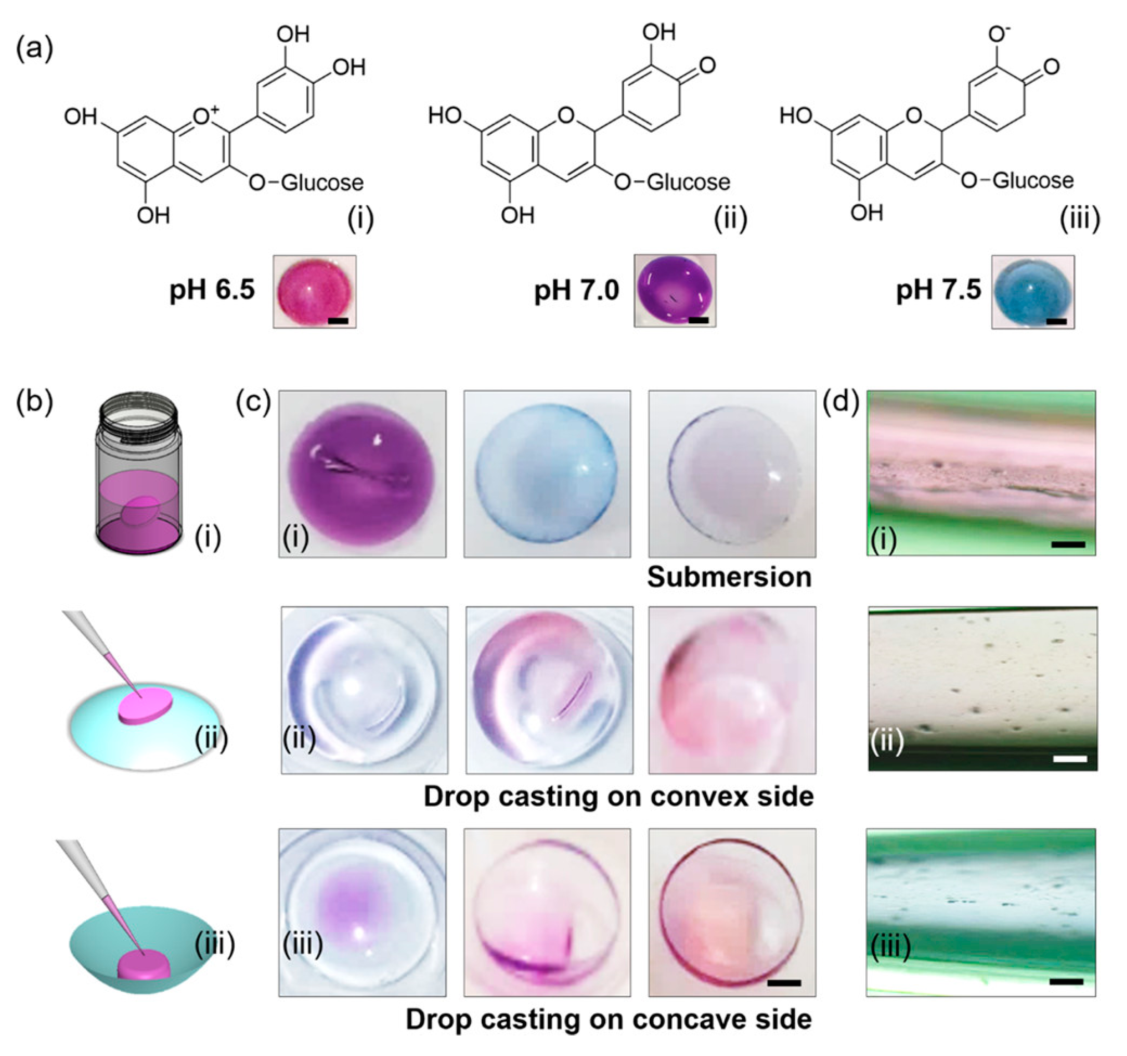
Anthocyanins exhibit changes in their chemical structure in response to varying hydrogen ion concentrations. This characteristic has garnered extensive attention for utilizing anthocyanin pigments as key components in biocompatible pH sensors. Riaz et al. [131] studied the use of anthocyanins on contact lenses to monitor the ocular pH, a crucial parameter in evaluating ocular health post-eye surgery in conditions like keratoconjunctivitis and ocular rosacea. For this purpose, commercially available lacreon and lacreon-free contact lenses were functionalized with the anthocyanins obtained from Brassica oleracea by soaking and drop casting methods. The lacreon contact lenses demonstrated enhanced and uniformly distributed coloration that was noticeable to the naked eye within the physiological pH range of 6.5 and 7.5 (Figure 10).
Similarly, Alsahag et al. [132] developed an economical, reversible, eco-friendly wound dressing based on anthocyanins in order to monitor wound healing progress. This innovative bioassay detected pH changes in a simulated wound solution, which were indicated by shifts in color. They employed anthocyanins sourced from Brassica oleracea and L. var capitate that were integrated onto a carboxymethyl cellulose/polyvinyl alcohol composite. The comfort and durability of these composites were confirmed through favorable colorfastness, air permeability, and bend length. In terms of biological properties, these composites exhibited non-cytotoxic effects and enhanced antimicrobial properties. This colorimetric assay offers an affordable, used-friendly sensor for monitoring wound healing progress. In contrast to prior electric-based sensing tools requiring complex equipment, this chromogenic sensor enables onsite wound pH measurements without intricate procedures.
Another example of anthocyanin-based materials used as biosensors was reported by Al-Qahtani et al. [133]. They encapsulated both natural anthocyanin and urease enzyme in a calcium alginate biopolymer, which were then immobilized within the fabric. The resulting bio-chromic sensor offered quick responses with a detection limit of 300–1000 ppm for urea. This innovative reversible sensor employs urease to convert urea to ammonia, allowing for easy urea detection via the encapsulated anthocyanin pH indicator from red cabbage. This method provides an efficient, eco-friendly, and selective colorimetric approach for measuring urea levels in fluids like blood and urine, demonstrating a potential for practical biomedical applications.
The same authors described the development of a sponge-like colorimetric swab by immobilizing red cabbage extract anthocyanins onto a cellulose aerogel [134]. This biochromic device was synthesized in order to monitor sweat. Sweat is a fluid emitted by the skin and can provide valuable insight into an individual physiology. For instance, in drug testing, sweat might contain measurable amounts of opioids like cocaine, gamma hydroxybutyrates, amphetamines, buprenorphine, and cannabinoids, which can alter pH levels. This makes sweat a significant factor in forensic science and toxicology. When the sponge-like device was exposed to a sweat solution, its color shifted from pink (620 nm) to greenish yellow (529 nm), corresponding to the pH of the solution. The aerogel biosensor displayed remarkable sensitivity and it can provide a real-time sweat analysis. Additionally, they observed that a sponge rich in anthocyanin exhibited no harmful effects on human skin. This eco-friendly biosensor offers a simple, reversible way to detect perspiration conditions using a sponge made from natural cellulose and anthocyanin, making this device great for drug testing applications.

7.2. Nanoencapsulated Delivery Systems

Taking into account the inherent toxicity associated with numerous synthetic drug compounds, the discovery and development of novel and efficient bioactive natural compounds like anthocyanins is highly desirable. However, since these natural phytochemicals are remarkably sensitive to external environmental factors, resulting in a notably brief half-life, as has been described above, the development of innovative delivery systems that can provide stability to anthocyanins without compromising their bioactivity is crucial. Indeed, it is essential to achieve kinetic and thermodynamic stability while simultaneously enhancing solubility and improving bioavailability [135].
Among the diverse array of alternatives explored, encapsulation has garnered substantial attention in recent decades, showcasing immense potential in various sectors, including the pharmaceutical and nutraceutical industries [136]. Furthermore, nanoencapsulation strategies exhibit interactions, such as Van der Waals forces, hydrophobic interactions, and hydrogen bonding, between nanocarriers and natural compounds like anthocyanins, ensuring enhanced material stability and increased bioavailability. Indeed, these advantages endow nanomaterials with the remarkable ability to easily traverse the blood–brain barrier, thereby magnifying the therapeutic potential of the encapsulated molecules [135].
This section highlights the application of three different polymer types, polysaccharides, lipids, and synthetic polymers, for nanoscale coatings of anthocyanin. Polymeric materials used in nanoencapsulation can be classified into two main categories based on their sources: bio-based polymers, from plant or animal sources, and oil (petroleum)-based polymers, from petroleum feedstock. Owing to the defined chemical structure, synthetic polymers offer control over their physical and chemical properties compared to natural polymers. Conversely, bio-based polymeric nanoencapsulations offer key advantages for use in biomedical applications as they present remarkable biocompatibility across a wide range of concentrations and are often cost-effective [137]. In nanomedicine, these polymers predominantly fall into two subcategories: polysaccharides and proteins. It is important to highlight that certain polysaccharides are commonly used alongside anthocyanins due to their inherent charged form. This ionization enables effective interaction with oppositely charged phospholipids, effectively preventing phospholipid hydrolysis under acidic pH conditions or in the presence of enzymes. Chitosan and pectin stand out among these polysaccharides as the most commonly used due to their non-toxic, eco-friendly, biodegradable nature and their high biocompatibility. Apart from these inherent advantages, they demonstrate considerable potential as an efficient drug delivery system, particularly for targeting the colon. For instance, Zhao et al. [138] studied the activity of anthocyanins encapsulated in pectin and chitosan and the controlled release from these nanoparticles, demonstrating enhanced protection for normal rat kidney cells against acrylamide-induced damage. Additionally, they decrease reactive oxygen species as well as matrix metalloproteinases and glutathione levels, providing protection to normal human hepatocyte L02 cells against palmitic acid-induced damage. Nevertheless, it has to be noted that chitosan dissolves only in certain dilute acidic conditions.
Similarly, Sreerekha et al. [139] also employed chitosan to encapsulate anthocyanins and demonstrated excellent antioxidant effects in vitro. They further studied the hypolipidemic effect and the potential dietary supplementation of these nanoparticles in male Wistar rats. The study revealed that these nanoparticles effectively reduced serum total cholesterol and triglyceride levels. Moreover, they mitigated lipid-mediated stress and lowered lipid levels in both the serum and liver. Thus, this highlights the potential of anthocyanin-loaded nanoparticles as functional dietary supplements with definite hypolipidemic effects.
In the same way, Amararathna et al. [140] proposed the use of different polymers to enhance the effectiveness of anthocyanins, by developing a direct delivery method to lung tissues. They employed anthocyanins from ripe haskap berries (HB) and encapsulated them in three types of nanocarriers: polyethylene glycol-poly(lactide-co-glycolide), maltodextrin, and carboxymethyl chitosan (CMC). Among these three nanocarriers, CMC showed the highest encapsulation efficiency at 10%. They tested the cytotoxicity and protective effects of the CMC-encapsulated anthocyanins on lung cells and found no harmful effects. Additionally, HB anthocyanins loaded into CMC helped counteract the oxidative stress caused by carcinogens, restoring the activity of important enzymes. Moreover, in in vivo tests with mice with inhalation exposure to HB-CMC for six days showed the presence of anthocyanins in the lungs within an hour. This suggests that CMC could be a safe encapsulating agent, facilitating the direct delivery to lung tissues and potentially improving therapeutic effectiveness by slowing down metabolic processes.
Another promising polysaccharide explored for the encapsulation of anthocyanins is chondroitin sulfate. Jeong et al. [141] reported the synthesis of black soybean anthocyanins loaded at different concentrations onto chondroitin sulfate polysaccharides in order to improve the structural stability of this natural antioxidant. When compared to anthocyanins alone, these nanoparticle complexes demonstrated superior inhibition in human cervical cancer HeLa cells. Additionally, Liang et al. [142] demonstrated that the combination of chondroitin sulfate and chitosan with loaded black rice anthocyanins induced apoptosis of human HCT-116 colon cancer cells by providing negative charges to the mitochondria. The results show that the addition of anthocyanin exhibited a noteworthy reduction in cell viability, which altered the mitochondrial structure and, consequently, increased apoptosis of cells.
Hyaluronic acid (HA) stands out as another promising natural polysaccharide that has demonstrated enhanced bioavailability and efficacy in encapsulating anthocyanins. In a study conducted by Liu et al. [143], a black rice anthocyanin-loaded HA nanocarrier was developed to reduce xanthine oxidase (XO) activity, a crucial enzyme involved in the generation of reactive oxygen species and the production of uric acid. In a simulated in vitro analysis, the anthocyanin-embedded HA nanocomplex exhibited a rapid release within the first 12 h, which was maintained consistently until reaching 60% release after 60 h, marking a 54 h difference compared to the non-embedded anthocyanin release system. These results indicate that sustained release could reduce the degradation of active compounds, thereby potentially improving the bioavailability of anthocyanins. Furthermore, in vitro assays concluded that the synthesized nanocomposite has potential to inhibit XO activity, suggesting a potential reduction in uric acid levels.
Another polysaccharide example was described by Hanafy et al. [96]. They developed a nanoparticle hydrogel platform based on starch corn, a natural polymer that has garnered attention in recent years. The anthocyanin-loaded starch hydrogel demonstrated promising therapeutic potential in eliminating glycogen from cardiac tissues, overcoming cardiomyopathy, and reducing malondialdehyde levels and collagen fibers. These findings highlights the significance of using biodegradable nanocarriers for encapsulating anthocyanins, showcasing their potential application across various biomedical fields.
On the other hand, much like other drug and active compounds, synthetic polymers have been found to be an excellent approach for the encapsulation of anthocyanins. These polymers offer versatility and enable precise control over the mechanical properties of the material. Among the synthetic polymers, polyesters have been extensively employed in anthocyanin delivery systems. For instance, biodegradable poly(lactic-co-glycolic acid) acid (PLGA) has been used to coat anthocyanins such as pelagonidin, resulting in improved protection against mitochondrial dysfunction [144]. Moreover, it has been demonstrated that the nanoencapsulation of pelargonidin anthocyanin by PLGA can inhibit the production of reactive oxygen species.
Similarly, Amin et al. [145] encapsulated anthocyanins within biodegradable nanoparticles formulated with PLGA and a stabilizing agent, polyethylene glycol (PEG-2000). They concluded that this novel approach bolsters the free radical scavenging abilities of anthocyanins and increases cellular uptake and bioactivity in in vitro assays, even at high doses, without inducing cytotoxicity. They investigated the biological activity and neuroprotective effects of these anthocyanin-loaded nanoparticles (An-NPs) in SH-SY5Y cell lines. The results indicated that anthocyanin-loaded nanoparticle systems effectively mitigated AB-induced neurotoxicity in SH-SY5Y cells, displaying notable antioxidant, anti-apoptotic, and anti-inflammatory properties and prevented Alzheimer’s disease. Overall, these findings suggest that anthocyanin-loaded nanoparticle systems can be a potential approach to treat various neurological disorders, including Alzheimer’s disease.
Regarding the use of other polyesters, in this case polyethylene oxide (PEO), Giaconia et al. [146] developed an anthocyanin-loaded nanofiber composite in order to enhance the antioxidant effect. Anthocyanin compounds obtained from jussara pulp were combined with polyethylene oxide (PEO) and deposited by electrospinning. Subsequently, they were subjected to a simulated digestion process in order to observe the protective effect of these natural compounds. These nanofibers demonstrated superior protection for the bioactive compounds, ensuring increased antioxidant activity compared to lyophilized jussar pulp fibers. Overall, the use of electrospinning to form fibers revealed that the composites could maintain their antioxidant effect even after digestion making these polymeric substrates promising materials to use in food so as to preserve the biological effect of jussara anthocyanins.
On the other hand, Ghiman et al. [147] described the use of a non-biodegradable polyester, polyacrylic acid (PAA), to develop fluorescent nanocarriers loaded with anthocyanins within melanoma cells. For this purpose, chokeberries anthocyanins were encapsulated between PAA layers, generating a polyelectrolyte system between PAA and poly(allylamine hydrochloride)(PAH), where rhodamine B, a fluorophore, was firstly added. According to the in vitro test, the anthocyanin fluorescent system demonstrated that could be used for trafficking over 24 h without compromising the proliferation of melanoma B16-F10 cells. The presented data not only demonstrated the potential of anthocyanin encapsulation, but also offer valuable insight into an innovative formulation for delivering therapeutic molecules or bio-imaging agents directly to melanoma cells.
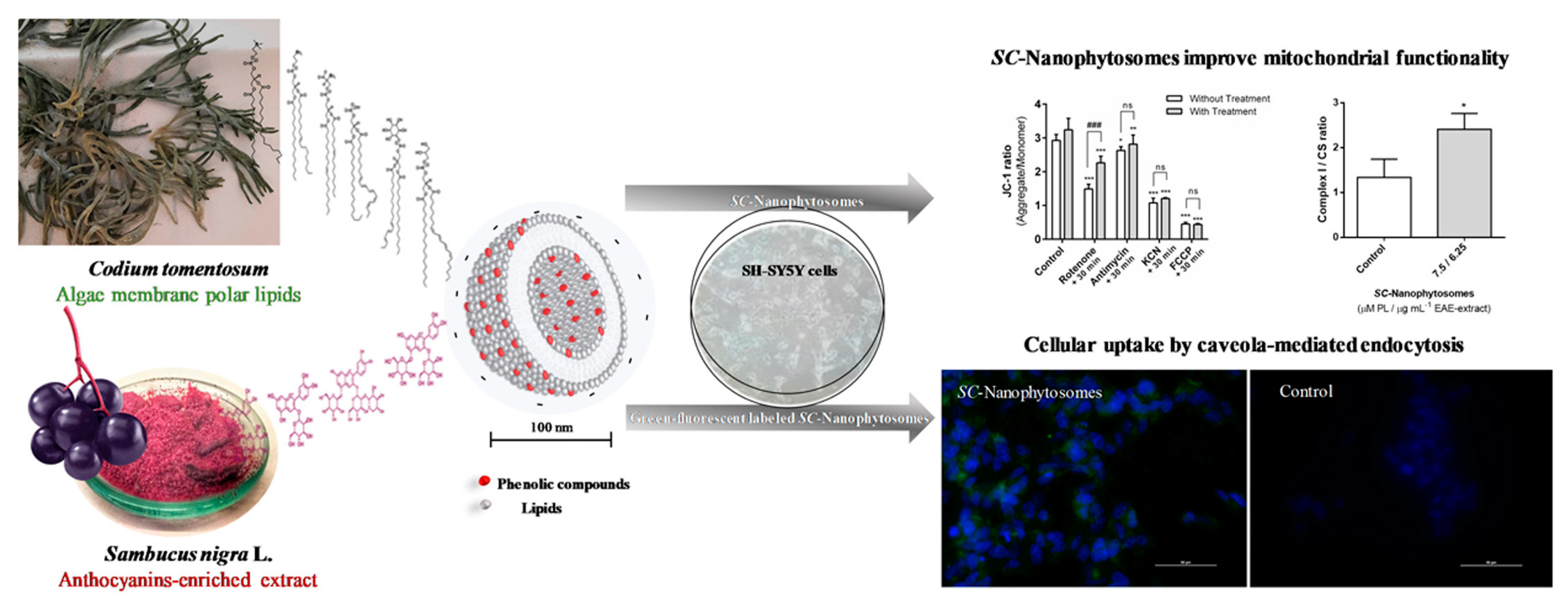
Regarding lipid-based nanoparticles, several studies of anthocyanin nanoencapsulations have been reported. For instance, Mendes et al. [148] studied the nanoencapsulation of anthocyanins derived from elderberries (Sambucus nigra) by combining them with membrane polar lipids through self-assembly. These nanophythosomes presented a remarkable ability to enhance complexes I and II required in the mitochondrial respiratory chain. Additionally, they preserved the mitochondrial membrane in the presence of rotenone, by protecting the cells against rotenone- and glutamate-induced toxicity. Overall, the results suggest that these nanocomplexes are an interesting approach for facilitating targeted mitochondrial therapy in the treatment of neurodegenerative diseases (Figure 11).
Similarly, Fidan et al. [149] reported the attachment of anthocyanins from black carrots onto niosomes, lamellar structures made by non-ionic surfactants from the alkyl polyglycerol ether class, in order to improve the bioavailability of these natural bioactive compounds. The in vitro results suggested that the majority of the encapsulated anthocyanin was released after 10 h, decreasing the viability of Neuro 2A neuroblastoma cells. Overall, these findings indicate that niosomes can serves as promising vesicles for anthocyanin delivery systems. On the other hand, Priprem et al. [150] discovered that anthocyanins extracted from Zea mays and Clitoria ternatea stimulated higher collagen production in human gingival fibroblasts, which can transfer through the esophageal mucosa. In this study, anthocyanins were subjected to nanoclusters, forming crystalline-like aggregates through both intra- and inter-molecular interactions. These nanocomplexes were encapsulated, again, in niosomes and were then integrated into a mucoadhesive gel. According to the in vitro assays, the permeability of the anthocyanins was improved and enhanced due to noisome encapsulation. Moreover, the anthocyanin-loaded noisome gel demonstrated an anti-inflammatory effect and induced oral wound closure in rats. This effect could be attributed to enhanced mucosal permeability and the presence of the anthocyanins released from the noisome gel.

8. Conclusions and Future Outlook

Anthocyanins have significantly impacted human health and the food industry due to their halochromic and valuable biological properties. However, their low bioavailability and unstable nature are large handicaps during the storage, processing, and uptake stages, which restrict their extensive applications and require additional strategies for protecting anthocyanins. Interestingly, the incorporation of anthocyanins into polymer matrices has resulted in an effective method not only to immobilize and protect these molecules, but also to promote their controlled release. Fortunately, there are many and varied general methods of processing polymers to facilitate the encapsulation of anthocyanins. These polymer-based combinations offer many possibilities to create materials with tailored physicochemical properties, which is especially interesting in addressing anthocyanin-related concerns in the food and healthcare sectors.
This review outlines the main polymer-based encapsulation systems designed to enhance the stability and bioavailability of anthocyanins. These systems are based on diverse processing forms of polymers, such as films, emulsions, hydrogels, fibers, microcapsules, nanoparticles, self-assembled structures, and polycomplexes.
Regardless of their processed form, the recent research on anthocyanin-loaded polymer systems has mainly focused on their immobilization and subsequent controlled release, and on their application as colorimetric sensors. The food industry, in particular, has focused on developing anthocyanin polymer films for smart food packaging. These films have shown an effective performance as food quality indicators, antioxidant and antimicrobial packaging to protect against microorganisms, and active films promoting health benefits for consumers.
With respect to the healthcare field, the color-sensing property of anthocyanin-loaded polymers has been exploited to develop chromic biosensors to monitor tissue healing processes, detect the presence of sweat or urea, and prevent chronic diseases. In addition, anthocyanin-loaded nanoparticles have also been intensively investigated as a new, effective, and safe administration route for anthocyanins in preventing chronic diseases.
Despite the extensive literature on anthocyanin-based polymer systems, there are still huge challenges such as improving the processing methods to effectively protect these molecules and increase their bioavailability. These challenges offer plenty of research opportunities that encompass different approaches such as improving the efficiency of the encapsulation, reducing the amount of reagents used, and promoting the sustainable release of anthocyanins, their local delivery at the absorption sites, or their responsive release under specific triggers.
On the other hand, the biodegradability of the employed polymer/s, and the sustainability and economic viability of the processing of anthocyanin-loaded polymers are still challenging issues for future industrial applications. Exploiting current biodegradable alternatives and processing methods has resulted in low yields and is not economically feasible for the food and healthcare industries. Thus, interdisciplinary efforts to explore the opportunities for the conservation of anthocyanin properties, together with the development of environmentally friendly polymer matrices and cost-effective and scalable processes, are required to achieve the successful and widespread application of anthocyanin-loaded polymeric systems in the food and healthcare fields.

Author Contributions

Conceptualization, L.P.-Á. and L.G.G.-R.; writing—original draft preparation, S.S.R.-M., M.F.I.-V., S.L.H.O. and J.S.-B.; writing—review and editing, L.P.-Á. and L.G.G.-R.; supervision, J.L.V.-V.; funding acquisition, J.L.V.-V. and L.G.G.-R. All authors have read and agreed to the published version of the manuscript.

Funding

This research was funded by the Basque Government Grupos Consolidados (grant IT1756-22), The National Council of Sciences, Humanities and Technology (CONAHCYT) of the Mexican government (postdoctoral grant CVU 484866), and PROSNI program of the University of Guadalajara whose resources supported part of this work.

Institutional Review Board Statement

Not applicable.

Data Availability Statement

Not applicable.

Conflicts of Interest

The authors declare no conflicts of interest.

References

  1. Tan, J.; Han, Y.; Han, B.; Qi, X.; Cai, X.; Ge, S.; Xue, H. Extraction and Purification of Anthocyanins: A Review. J. Agric. Food Res. 2022, 8, 100306. [Google Scholar] [CrossRef]
  2. Horbowicz, M.; Grzesiuk, A.; DĘBski, H.; Kosson, R. Anthocyanins of Fruits and Vegetables—Their Occurrence, Analysis and Role in Human. Veg. Crops Res. Bull. 2008, 68, 5–22. [Google Scholar] [CrossRef]
  3. Taylor, L.P.; Grotewold, E. Flavonoids as Developmental Regulators. Curr. Opin. Plant Biol. 2005, 8, 317–323. [Google Scholar] [CrossRef]
  4. Khoo, H.E.; Azlan, A.; Tang, S.T.; Lim, S.M. Anthocyanidins and Anthocyanins: Colored Pigments as Food, Pharmaceutical Ingredients, and the Potential Health Benefits. Food Nutr. Res. 2017, 61, 1361779. [Google Scholar] [CrossRef] [PubMed]
  5. Fang, J. Bioavailability of Anthocyanins. Drug Metab. Rev. 2014, 46, 508–520. [Google Scholar] [CrossRef] [PubMed]
  6. Enaru, B.; Drețcanu, G.; Pop, T.D.; Stǎnilǎ, A.; Diaconeasa, Z. Anthocyanins: Factors Affecting Their Stability and Degradation. Antioxidants 2021, 10, 1967. [Google Scholar] [CrossRef] [PubMed]
  7. Kossyvaki, D.; Contardi, M.; Athanassiou, A.; Fragouli, D. Colorimetric Indicators Based on Anthocyanin Polymer Composites: A Review. Polymers 2022, 14, 4129. [Google Scholar] [CrossRef]
  8. Calogero, G.; Bartolotta, A.; Di Marco, G.; Di Carlo, A.; Bonaccorso, F. Vegetable-based dye-sensitized solar cells. Chem. Soc. Rev. 2015, 44, 3244–3294. [Google Scholar] [CrossRef]
  9. Mirmoeini, S.S.; Moradi, M.; Tajik, H.; Almasi, H.; Gama, F.M. Cellulose/Salep-Based Intelligent Aerogel with Red Grape Anthocyanins: Preparation, Characterization and Application in Beef Packaging. Food Chem. 2023, 425, 136493. [Google Scholar] [CrossRef]
  10. Loypimai, P.; Moongngarm, A.; Chottanom, P. Thermal and PH Degradation Kinetics of Anthocyanins in Natural Food Colorant Prepared from Black Rice Bran. J. Food Sci. Technol. 2016, 53, 461–470. [Google Scholar] [CrossRef]
  11. Fossen, T.; Cabrita, L.; Andersen, M. Colour and Stability of Pure Anthocyanins In¯uenced by PH Including the Alkaline Region. Food Chem. 1998, 4, 435–440. [Google Scholar] [CrossRef]
  12. Alappat, B.; Alappat, J. Anthocyanin Pigments: Beyond Aesthetics. Molecules 2020, 25, 5500. [Google Scholar] [CrossRef] [PubMed]
  13. Tena, N.; Asuero, A.G. Up-To-Date Analysis of the Extraction Methods for Anthocyanins: Principles of the Techniques, Optimization, Technical Progress, and Industrial Application. Antioxidants 2022, 11, 286. [Google Scholar] [CrossRef]
  14. Gomez Mattson, M.; Sozzi, A.; Corfield, R.; Gagneten, M.; Franceschinis, L.; Schebor, C.; Salvatori, D. Colorant and Antioxidant Properties of Freeze-Dried Extracts from Wild Berries: Use of Ultrasound-Assisted Extraction Method and Drivers of Liking of Colored Yogurts. J. Food Sci. Technol. 2022, 59, 944–955. [Google Scholar] [CrossRef] [PubMed]
  15. Castañeda-Ovando, A.; de Lourdes Pacheco-Hernández, M.; Páez-Hernández, M.E.; Rodríguez, J.A.; Galán-Vidal, C.A. Chemical Studies of Anthocyanins: A Review. Food Chem. 2009, 113, 859–871. [Google Scholar] [CrossRef]
  16. Escribano-Bailón, M.T.; Santos-Buelga, C.; Rivas-Gonzalo, J.C. Anthocyanins in Cereals. J. Chromatogr. A 2004, 1054, 129–141. [Google Scholar] [CrossRef]
  17. Van Nguyen, S.; Lee, B.K. PVA/CNC/TiO2 Nanocomposite for Food-Packaging: Improved Mechanical, UV/Water Vapor Barrier, and Antimicrobial Properties. Carbohydr. Polym. 2022, 298, 120064. [Google Scholar] [CrossRef]
  18. Gravier-Rodríguez, G.; Jurado-Basante, S.; Arias-Contreras, A.N.; Gamarra Castillo, O.J.; Porras, A.; Sánchez-Camargo, A.d.P. Ultrasound-Assisted Extraction of Anthocyanins from Andean Blackberry and Their Use as an Indicator in Sustainable Smart Biofilms Developed with Cocoa Bean Shells as Natural Fiber-Filled PLA Composite Materials. Food Packag. Shelf Life 2023, 40, 101165. [Google Scholar] [CrossRef]
  19. Liu, J.; Zhou, H.; Song, L.; Yang, Z.; Qiu, M.; Wang, J.; Shi, S. Anthocyanins: Promising Natural Products with Diverse Pharmacological Activities. Molecules 2021, 26, 3807. [Google Scholar] [CrossRef]
  20. Smeriglio, A.; Barreca, D.; Bellocco, E.; Trombetta, D. Chemistry, Pharmacology and Health Benefits of Anthocyanins. Phytother. Res. 2016, 1286, 1265–1286. [Google Scholar] [CrossRef]
  21. Blando, F.; Calabriso, N.; Berland, H.; Maiorano, G.; Gerardi, C.; Carluccio, M.A.; Andersen, Ø.M. Radical Scavenging and Anti-Inflammatory Activities of Representative Anthocyanin Groupings from Pigment-Rich Fruits and Vegetables. Int. J. Mol. Sci. 2018, 19, 169. [Google Scholar] [CrossRef]
  22. Różańska, D.; Regulska-Ilow, B. The significance of anthocyanins in the prevention and treatment of type 2 diabetes. Adv. Clin. Exp. Med. 2018, 27, 135–142. [Google Scholar] [CrossRef]
  23. Osorio, M.; Posada, L.; Martínez, E.; Estrada, V.; Quintana, G.; Maldonado, M.E.; Peresin, S.; Orozco, J.; Castro, C. Bacterial Nanocellulose Spheres Coated with Meta Acrylic Copolymer: Vaccinium Meridionale Swartz Extract Delivery for Colorectal Cancer Chemoprevention. Food Hydrocoll. 2024, 147, 109310. [Google Scholar] [CrossRef]
  24. Chen, H.; Zhang, M.; Bhandari, B.; Yang, C. Novel PH-Sensitive Films Containing Curcumin and Anthocyanins to Monitor Fish Freshness. Food Hydrocoll. 2020, 100, 109310. [Google Scholar] [CrossRef]
  25. Rakić, Å.V.; Rinnan, T.; Polak, M.; Skrt, M.; Miljković, N.P. Ulrih, pH-induced structural forms of cyanidin and cyanidin 3-O-β-glucopyranoside. Dye. Pigment. 2019, 165, 71–80. [Google Scholar] [CrossRef]
  26. Câmara, J.S.; Locatelli, M.; Pereira, J.A.M.; Oliveira, H.; Arlorio, M.; Fernandes, I.; Perestrelo, R.; Freitas, V.; Bordiga, M. Behind the Scenes of Anthocyanins—From the Health Benefits to Potential Applications in Food, Pharmaceutical and Cosmetic Fields. Nutrients 2022, 14, 5133. [Google Scholar] [CrossRef]
  27. Cruz, L.; Basílio, N.; Mateus, N.; De Freitas, V.; Pina, F. Natural and Synthetic Flavylium-Based Dyes: The Chemistry behind the Color. Chem. Rev. 2022, 122, 1416–1481. [Google Scholar] [CrossRef]
  28. Baranwal, J.; Barse, B.; Fais, A.; Delogu, G.L.; Kumar, A. Biopolymer: A Sustainable Material for Food and Medical Applications. Polymers 2022, 14, 983. [Google Scholar] [CrossRef]
  29. Jiang, M.; Zhang, Y. Biopolymer-Based Encapsulation of Anthocyanins as Reinforced Natural Colorants for Food Applications. J. Agric. Food Res. 2023, 11, 100488. [Google Scholar] [CrossRef]
  30. Li, R.; Wang, S.; Feng, H.; Zhuang, D.; Zhu, J. An Intelligent Chitosan/Gelatin Film via Improving the Anthocyanin-Induced Color Recognition Accuracy for Beef Sub-Freshness Differentiation Monitoring. Food Hydrocoll. 2024, 146, 109219. [Google Scholar] [CrossRef]
  31. Neves, D.; Andrade, P.B.; Videira, R.A.; de Freitas, V.; Cruz, L. Berry Anthocyanin-Based Films in Smart Food Packaging: A Mini-Review. Food Hydrocoll. 2022, 133, 107885. [Google Scholar] [CrossRef]
  32. Yong, H.; Liu, J. Recent Advances in the Preparation, Physical and Functional Properties, and Applications of Anthocyanins-Based Active and Intelligent Packaging Films. Food Packag. Shelf Life 2020, 26, 100550. [Google Scholar] [CrossRef]
  33. Nadi, M.; Razavi, S.M.A.; Shahrampour, D. Fabrication of Green Colorimetric Smart Packaging Based on Basil Seed Gum/Chitosan/Red Cabbage Anthocyanin for Real-Time Monitoring of Fish Freshness. Food Sci. Nutr. 2023, 11, 6360–6375. [Google Scholar] [CrossRef]
  34. Wu, Y.; Li, C. A Double-Layer Smart Film Based on Gellan Gum/Modified Anthocyanin and Sodium Carboxymethyl Cellulose/Starch/Nisin for Application in Chicken Breast. Int. J. Biol. Macromol. 2023, 232, 123464. [Google Scholar] [CrossRef]
  35. de Oliveira Filho, J.G.; Braga, A.R.C.; de Oliveira, B.R.; Gomes, F.P.; Moreira, V.L.; Pereira, V.A.C.; Egea, M.B. The Potential of Anthocyanins in Smart, Active, and Bioactive Eco-Friendly Polymer-Based Films: A Review. Food Res. Int. 2021, 142, 110202. [Google Scholar] [CrossRef]
  36. Aman Mohammadi, M.; Mirza Alizadeh, A.; Mohammadi, M.; Mirzakhani, E.; Sabouri, S.; Pourjafar, H.; Hosseini, S.M. Application and Development of Electrospun Nanofibers as an Efficient Platform for the Delivery of Anthocyanin Compounds in the Food Industry. Food Bioproc Tech. 2023. [Google Scholar] [CrossRef]
  37. Khaledian, Y.; Moshtaghi, H.; Shahbazi, Y. Development and Characterization of Smart Double-Layer Nanofiber Mats Based on Potato Starch-Turnip Peel Anthocyanins and Guar Gum-Cinnamaldehyde. Food Chem. 2024, 434, 137462. [Google Scholar] [CrossRef]
  38. Nath, V.A.; Vijayakumar, R.; Leena, M.M.; Moses, J.A.; Anandharamakrishnan, C. Co-Electrospun-Electrosprayed Ethyl Cellulose-Gelatin Nanocomposite PH-Sensitive Membrane for Food Quality Applications. Food Chem. 2022, 394, 133420. [Google Scholar] [CrossRef]
  39. Pakolpakçıl, A.; Osman, B.; Göktalay, G.; Özer, E.T.; Şahan, Y.; Becerir, B.; Karaca, E. Design and in Vivo Evaluation of Alginate-Based PH-Sensing Electrospun Wound Dressing Containing Anthocyanins. J. Polym. Res. 2021, 28, 50. [Google Scholar] [CrossRef]
  40. Liu, L.; Zhang, D.; Song, X.; Guo, M.; Wang, Z.; Geng, F.; Zhou, X.; Nie, S. Compound Hydrogels Derived from Gelatin and Gellan Gum Regulates the Release of Anthocyanins in Simulated Digestion. Food Hydrocoll. 2022, 127, 107487. [Google Scholar] [CrossRef]
  41. Jin, W.; Xiang, L.; Peng, D.; Liu, G.; He, J.; Cheng, S.; Li, B.; Huang, Q. Study on the Coupling Progress of Thermo-Induced Anthocyanins Degradation and Polysaccharides Gelation. Food Hydrocoll. 2020, 105, 105822. [Google Scholar] [CrossRef]
  42. Lotfinia, F.; Norouzi, M.R.; Ghasemi-Mobarakeh, L.; Naeimirad, M. Anthocyanin/Honey-Incorporated Alginate Hydrogel as a Bio-Based PH-Responsive/Antibacterial/Antioxidant Wound Dressing. J. Funct. Biomater. 2023, 14, 72. [Google Scholar] [CrossRef]
  43. Mohammadalinejhad, S.; Kurek, M.; Jensen, I.J.; Lerfall, J. The Potential of Anthocyanin-Loaded Alginate Hydrogel Beads for Intelligent Packaging Applications: Stability and Sensitivity to Volatile Amines. Curr. Res. Food Sci. 2023, 7, 100560. [Google Scholar] [CrossRef]
  44. Cui, C.; Ji, N.; Wang, Y.; Xiong, L.; Sun, Q. Bioactive and Intelligent Starch-Based Films: A Review. Trends Food Sci. Technol. 2021, 116, 854–869. [Google Scholar] [CrossRef]
  45. Förster, S.; Schmidt, M. Polyelectrolytes in Solution. In Physical Properties of Polymers, Advances in Polymer Science; Springer: Berlin/Heidelberg, Germany, 1995. [Google Scholar]
  46. Tan, C.; Huang, M.; Wang, J.; Sun, B. Biopolyelectrolyte Complex (BioPEC)-Based Carriers for Anthocyanin Delivery. Food Hydrocoll. Health 2021, 1, 100037. [Google Scholar] [CrossRef]
  47. Tan, C.; Celli, G.B.; Abbaspourrad, A. Copigment-Polyelectrolyte Complexes (PECs) Composite Systems for Anthocyanin Stabilization. Food Hydrocoll. 2018, 81, 371–379. [Google Scholar] [CrossRef]
  48. Tan, C.; Sun, Y.; Yao, X.; Zhu, Y.; Jafari, S.M.; Sun, B.; Wang, J. Stabilization of anthocyanins by simultaneous encapsulation-copigmentation via protein-polysaccharide polyelectrolyte complexes. Food Chem. 2023, 416, 135732. [Google Scholar] [CrossRef]
  49. Yuan, Y.; Fan, Q.; Xu, X.; Wang, O.; Zhao, L.; Zhao, L. Nanocarriers Based on Polysaccharides for Improving the Stability and Bioavailability of Anthocyanins: A Review. Carbohydr. Polym. Technol. Appl. 2023, 6, 100346. [Google Scholar] [CrossRef]
  50. Sharif, N.; Khoshnoudi-Nia, S.; Jafari, S.M. Nano/Microencapsulation of Anthocyanins; a Systematic Review and Meta-Analysis. Food Res. Int. 2020, 132, 109077. [Google Scholar] [CrossRef]
  51. Li, J.; Guo, C.; Cai, S.; Yi, J.; Zhou, L. Fabrication of Anthocyanin–Rich W1/O/W2 Emulsion Gels Based on Pectin–GDL Complexes: 3D Printing Performance. Food Res. Int. 2023, 168, 112782. [Google Scholar] [CrossRef]
  52. Baba Shekh, A.O.; Abdul Wahab, R.; Yahya, N.A. Formulation of Roselle Extract Water-in-Oil Nanoemulsion for Controlled Pulmonary Delivery. J. Dispers. Sci. Technol. 2023, 44, 1830–1841. [Google Scholar] [CrossRef]
  53. Kenari, R.E.; Razavi, R. Encapsulation of Bougainvillea (Bougainvillea spectabilis) Flower Extract in Urtica dioica L. Seed Gum: Characterization, Antioxidant/Antimicrobial Properties, and in Vitro Digestion. Food Sci. Nutr. 2022, 10, 3436–3443. [Google Scholar] [CrossRef] [PubMed]
  54. Kanha, N.; Surawang, S.; Pitchakarn, P.; Laokuldilok, T. Microencapsulation of Copigmented Anthocyanins Using Double Emulsion Followed by Complex Coacervation: Preparation, Characterization and Stability. LWT 2020, 133, 110154. [Google Scholar] [CrossRef]
  55. Mushtaq, A.; Mohd Wani, S.; Malik, A.R.; Gull, A.; Ramniwas, S.; Ahmad Nayik, G.; Ercisli, S.; Alina Marc, R.; Ullah, R.; Bari, A. Recent Insights into Nanoemulsions: Their Preparation, Properties and Applications. Food Chem. X 2023, 18, 100684. [Google Scholar] [CrossRef] [PubMed]
  56. Lan, X.; Liu, Y.; Wang, L.; Wang, H.; Hu, Z.; Dong, H.; Yu, Z.; Yuan, Y. A Review of Curcumin in Food Preservation: Delivery System and Photosensitization. Food Chem. 2023, 424, 136464. [Google Scholar] [CrossRef] [PubMed]
  57. Wulansari, A.; Jufri, M.; Budianti, A. Studies on the Formulation, Physical Stability, and in Vitro Antibacterial Activity of Tea Tree Oil (Melaleuca Alternifolia) Nanoemulsion Gel. Int. J. Appl. Pharm. 2017, 9, 135–139. [Google Scholar] [CrossRef] [PubMed]
  58. Cheng, Y.; Liu, J.; Li, L.; Ren, J.; Lu, J.; Luo, F. Advances in Embedding Techniques of Anthocyanins: Improving Stability, Bioactivity and Bioavailability. Food Chem. X 2023, 20, 100983. [Google Scholar] [CrossRef] [PubMed]
  59. Garavand, F.; Jalai-Jivan, M.; Assadpour, E.; Jafari, S.M. Encapsulation of Phenolic Compounds within Nano/Microemulsion Systems: A Review. Food Chem. 2021, 364, 130376. [Google Scholar] [CrossRef]
  60. Nazareth, M.S.; Shreelakshmi, S.V.; Rao, P.J.; Shetty, N.P. Micro and Nanoemulsions of Carissa Spinarum Fruit Polyphenols, Enhances Anthocyanin Stability and Anti-Quorum Sensing Activity: Comparison of Degradation Kinetics. Food Chem. 2021, 359, 129876. [Google Scholar] [CrossRef]
  61. Gorantla, S.; Wadhwa, G.; Jain, S.; Sankar, S.; Nuwal, K.; Mahmood, A.; Dubey, S.K.; Taliyan, R.; Kesharwani, P.; Singhvi, G. Recent Advances in Nanocarriers for Nutrient Delivery. Drug Deliv. Transl. Res. 2022, 12, 2359–2384. [Google Scholar] [CrossRef]
  62. Al-Khayri, J.M.; Asghar, W.; Akhtar, A.; Ayub, H.; Aslam, I.; Khalid, N.; Al-Mssallem, M.Q.; Alessa, F.M.; Ghazzawy, H.S.; Attimarad, M. Anthocyanin Delivery Systems: A Critical Review of Recent Research Findings. Appl. Sci. 2022, 12, 12347. [Google Scholar] [CrossRef]
  63. Chen, B.H.; Inbaraj, B.S. Nanoemulsion and Nanoliposome Based Strategies for Improving Anthocyanin Stability and Bioavailability. Nutrients 2019, 11, 1052. [Google Scholar] [CrossRef] [PubMed]
  64. Feng, T.; Fan, C.; Wang, X.; Wang, X.; Xia, S.; Huang, Q. Food-Grade Pickering Emulsions and High Internal Phase Pickering Emulsions Encapsulating Cinnamaldehyde Based on Pea Protein-Pectin-EGCG Complexes for Extrusion 3D Printing. Food Hydrocoll. 2022, 124, 107265. [Google Scholar] [CrossRef]
  65. Constantin, O.E.; Stănciuc, N.; Yan, Y.; Ghinea, I.O.; Ungureanu, C.; Cîrciumaru, A.; Wang, D.; Poklar Ulrih, N.; Râpeanu, G. Polymers and Protein-Associated Vesicles for the Microencapsulation of Anthocyanins from Grape Skins Used for Food Applications. J. Sci. Food Agric. 2021, 101, 2676–2686. [Google Scholar] [CrossRef] [PubMed]
  66. Qiao, L.; Yang, H.; Gao, S.; Li, L.; Fu, X.; Wei, Q. Research Progress on Self-Assembled Nanodrug Delivery Systems. J. Mater. Chem. B 2022, 10, 1908–1922. [Google Scholar] [CrossRef]
  67. Zhang, R.; Zhang, Q.; Oliveira, H.; Mateus, N.; Ye, S.; Jiang, S.; He, J.; Wu, M. Preparation of Nanoliposomes Loaded with Anthocyanins from Grape Skin Extracts: Stability, Gastric Absorption and Antiproliferative Properties. Food Funct. 2022, 13, 10912–10922. [Google Scholar] [CrossRef] [PubMed]
  68. Chi, J.; Ge, J.; Yue, X.; Liang, J.; Sun, Y.; Gao, X.; Yue, P. Preparation of Nanoliposomal Carriers to Improve the Stability of Anthocyanins. LWT 2019, 109, 101–107. [Google Scholar] [CrossRef]
  69. Surekha, Y.N.; Saravanan, R.; Jyothi, Y.; Swaroop Seetharam, S. Solid Lipid Nanoparticles as Carriers of Natural Phenolic Compounds. Indo Am. J. Pharm. Sci. 2023, 10, 236–255. [Google Scholar]
  70. Yin, Z.; Zheng, T.; Ho, C.T.; Huang, Q.; Wu, Q.; Zhang, M. Improving the Stability and Bioavailability of Tea Polyphenols by Encapsulations: A Review. Food Sci. Hum. Wellness 2022, 11, 537–556. [Google Scholar] [CrossRef]
  71. Cai, D.; Li, X.; Chen, J.; Jiang, X.; Ma, X.; Sun, J.; Tian, L.; Vidyarthi, S.K.; Xu, J.; Pan, Z.; et al. A Comprehensive Review on Innovative and Advanced Stabilization Approaches of Anthocyanin by Modifying Structure and Controlling Environmental Factors. Food Chem. 2022, 366, 130611. [Google Scholar] [CrossRef]
  72. Yavuz-Düzgün, M.; Kareth, S.; Özçelik, B.; Weidner, E. Black Carrot Extract Loaded-Potato Protein Particles by PGSS-Drying: Physico-Chemical Properties and in Vitro Bioaccessability. J. Supercrit. Fluids 2023, 203, 106065. [Google Scholar] [CrossRef]
  73. Rosales-Chimal, S.; Navarro-Cortez, R.O.; Bello-Perez, L.A.; Vargas-Torres, A.; Palma-Rodríguez, H.M. Optimal Conditions for Anthocyanin Extract Microencapsulation in Taro Starch: Physicochemical Characterization and Bioaccessibility in Gastrointestinal Conditions. Int. J. Biol. Macromol. 2023, 227, 83–92. [Google Scholar] [CrossRef] [PubMed]
  74. Kurek, M.A.; Majek, M.; Onopiuk, A.; Szpicer, A.; Napiórkowska, A.; Samborska, K. Encapsulation of Anthocyanins from Chokeberry (Aronia Melanocarpa) with Plazmolyzed Yeast Cells of Different Species. Food Bioprod. Process. 2023, 137, 84–92. [Google Scholar] [CrossRef]
  75. Ligarda-Samanez, C.A.; Choque-Quispe, D.; Moscoso-Moscoso, E.; Palomino-Rincón, H.; Taipe-Pardo, F.; Landa, J.P.A.; Arévalo-Quijano, J.C.; Muñoz-Saenz, J.C.; Quispe-Quezada, U.R.; Huamán-Carrión, M.L.; et al. Nanoencapsulation of Phenolic Extracts from Native Potato Clones (Solanum tuberosum spp. Andigena) by Spray Drying. Molecules 2023, 28, 4961. [Google Scholar] [CrossRef]
  76. Mendes, J.F.; Norcino, L.B.; Manrich, A.; de Oliveira, T.J.P.; Mendes, R.F.; Mattoso, L.H.C. Pectin-Based Color Indicator Films Incorporated with Spray-Dried Hibiscus Extract Microparticles. Food Res. Int. 2022, 162, 111914. [Google Scholar] [CrossRef] [PubMed]
  77. Nadali, N.; Pahlevanlo, A.; Sarabi-Jamab, M.; Balandari, A. Effect of Maltodextrin with Different Dextrose Equivalents on the Physicochemical Properties of Spray-Dried Barberry Juice (Berberis vulgaris L.). J. Food Sci. Technol. 2022, 59, 2855–2866. [Google Scholar] [CrossRef]
  78. Molina, A.K.; Corrêa, R.C.G.; Prieto, M.A.; Pereira, C.; Barros, L. Bioactive Natural Pigments’ Extraction, Isolation, and Stability in Food Applications. Molecules 2023, 28, 1200. [Google Scholar] [CrossRef]
  79. Williams, C.A.; Grayer, R.J. Anthocyanins and Other Flavonoids. Nat. Prod. Rep. 2004, 21, 539–573. [Google Scholar] [CrossRef]
  80. Zhang, K.; Huang, T.S.; Yan, H.; Hu, X.; Ren, T. Novel PH-Sensitive Films Based on Starch/Polyvinyl Alcohol and Food Anthocyanins as a Visual Indicator of Shrimp Deterioration. Int. J. Biol. Macromol. 2020, 145, 768–776. [Google Scholar] [CrossRef]
  81. Pereira, V.A.; de Arruda, I.N.Q.; Stefani, R. Active Chitosan/PVA Films with Anthocyanins from Brassica Oleraceae (Red Cabbage) as Time-Temperature Indicators for Application in Intelligent Food Packaging. Food Hydrocoll. 2015, 43, 180–188. [Google Scholar] [CrossRef]
  82. Li, L.; Zhao, Z.; Wei, S.; Xu, K.; Xia, J.; Wu, Q.; Lü, X.; Wang, L. Development and Application of Multifunctional Films Based on Modified Chitosan/Gelatin Polyelectrolyte Complex for Preservation and Monitoring. Food Hydrocoll. 2024, 147, 109336. [Google Scholar] [CrossRef]
  83. Zhao, R.; Chen, J.; Yu, S.; Niu, R.; Yang, Z.; Wang, H.; Cheng, H.; Ye, X.; Liu, D.; Wang, W. Active Chitosan/Gum Arabic-Based Emulsion Films Reinforced with Thyme Oil Encapsulating Blood Orange Anthocyanins: Improving Multi-Functionality. Food Hydrocoll. 2023, 134, 108094. [Google Scholar] [CrossRef]
  84. Mao, S.; Ren, Y.; Chen, S.; Liu, D.; Ye, X.; Tian, J. Development and Characterization of PH Responsive Sodium Alginate Hydrogel Containing Metal-Phenolic Network for Anthocyanin Delivery. Carbohydr. Polym. 2023, 320, 121234. [Google Scholar] [CrossRef] [PubMed]
  85. Jang, Y.; Koh, E. Effect of Encapsulation on Stability of Anthocyanins and Chlorogenic Acid Isomers in Aronia during in Vitro Digestion and Their Transformation in a Model System. Food Chem. 2024, 434, 137443. [Google Scholar] [CrossRef]
  86. Tassanawat, S.; Phandee, A.; Magaraphan, R.; Nithitanakul, M.; Manuspiya, H. PH-Sensitive PP/Clay Nanocomposites for Beverage Smart Packaging. In Proceedings of the 2nd IEEE International Conference on Nano/Micro Engineered and Molecular Systems, Bangkok, Thailand, 16–19 January 2007. [Google Scholar]
  87. Xue Mei, L.; Mohammadi Nafchi, A.; Ghasemipour, F.; Mat Easa, A.; Jafarzadeh, S.; Al-Hassan, A.A. Characterization of PH Sensitive Sago Starch Films Enriched with Anthocyanin-Rich Torch Ginger Extract. Int. J. Biol. Macromol. 2020, 164, 4603–4612. [Google Scholar] [CrossRef] [PubMed]
  88. Ma, L.; Long, T.; Yuan, S.; Qi, P.; Han, L.; Hao, J. A PH-Indicating Smart Tag Based on Porous Hydrogel as Food Freshness Sensors. J. Colloid Interface Sci. 2023, 647, 32–42. [Google Scholar] [CrossRef]
  89. Goudarzi, J.; Moshtaghi, H.; Shahbazi, Y. Kappa-Carrageenan-Poly(Vinyl Alcohol) Electrospun Fiber Mats Encapsulated with Prunus Domestica Anthocyanins and Epigallocatechin Gallate to Monitor the Freshness and Enhance the Shelf-Life Quality of Minced Beef Meat. Food Packag. Shelf Life 2023, 35, 101017. [Google Scholar] [CrossRef]
  90. He, H.; Song, Y.; Li, M.; Zhang, H.; Li, J.; Huang, H.; Li, Y. A Novel Anthocyanin Electrospun Film by Caffeic Acid Co-Pigmentation for Real-Time Fish Freshness Monitoring. Anal. Methods 2022, 15, 228–239. [Google Scholar] [CrossRef]
  91. Jang, Y.; Koh, E. Characterisation and Storage Stability of Aronia Anthocyanins Encapsulated with Combinations of Maltodextrin with Carboxymethyl Cellulose, Gum Arabic, and Xanthan Gum. Food Chem. 2023, 405, 135002. [Google Scholar] [CrossRef]
  92. Moradi, M.; Tajik, H.; Almasi, H.; Forough, M.; Ezati, P. A Novel PH-Sensing Indicator Based on Bacterial Cellulose Nanofibers and Black Carrot Anthocyanins for Monitoring Fish Freshness. Carbohydr. Polym. 2019, 222, 115030. [Google Scholar] [CrossRef]
  93. Ebrahimi Tirtashi, F.; Moradi, M.; Tajik, H.; Forough, M.; Ezati, P.; Kuswandi, B. Cellulose/Chitosan PH-Responsive Indicator Incorporated with Carrot Anthocyanins for Intelligent Food Packaging. Int. J. Biol. Macromol. 2019, 136, 920–926. [Google Scholar] [CrossRef] [PubMed]
  94. Weston, M.; Phan, M.A.T.; Arcot, J.; Chandrawati, R. Anthocyanin-Based Sensors Derived from Food Waste as an Active Use-by Date Indicator for Milk. Food Chem. 2020, 326, 127017. [Google Scholar] [CrossRef] [PubMed]
  95. Uranga, J.; Etxabide, A.; Guerrero, P.; de la Caba, K. Development of Active Fish Gelatin Films with Anthocyanins by Compression Molding. Food Hydrocoll. 2018, 84, 313–320. [Google Scholar] [CrossRef]
  96. Hanafy, N.A.N. Starch Based Hydrogel NPs Loaded by Anthocyanins Might Treat Glycogen Storage at Cardiomyopathy in Animal Fibrotic Model. Int. J. Biol. Macromol. 2021, 183, 171–181. [Google Scholar] [CrossRef]
  97. Choi, I.; Choi, H.; Lee, J.S.; Han, J. Novel Color Stability and Colorimetry-Enhanced Intelligent CO2 Indicators by Metal Complexation of Anthocyanins for Monitoring Chicken Freshness. Food Chem. 2023, 404, 134534. [Google Scholar] [CrossRef] [PubMed]
  98. Pirayesh, H.; Park, B.D.; Khanjanzadeh, H.; Park, H.J.; Cho, Y.J. Nanocellulose-Based Ammonia Sensitive Smart Colorimetric Hydrogels Integrated with Anthocyanins to Monitor Pork Freshness. Food Control 2023, 147, 109595. [Google Scholar] [CrossRef]
  99. Borges, A.; de Freitas, V.; Mateus, N.; Fernandes, I.; Oliveira, J. Solid Lipid Nanoparticles as Carriers of Natural Phenolic Compounds. Antioxidants 2020, 9, 998. [Google Scholar] [CrossRef] [PubMed]
  100. Yao, L.; Hao, M.; Zhao, F.; Wang, Y.; Zhou, Y.; Liu, Z.; An, X.; Gao, Z.; Wang, J.; Zheng, T.; et al. Fabrication of Silk Sericin-Anthocyanin Nanocoating for Chelating and Saturation-Visualization Detection of Metal Ions. Nanoscale 2022, 14, 17277–17289. [Google Scholar] [CrossRef]
  101. Ge, Y.; Li, Y.; Bai, Y.; Yuan, C.; Wu, C.; Hu, Y. Intelligent Gelatin/Oxidized Chitin Nanocrystals Nanocomposite Films Containing Black Rice Bran Anthocyanins for Fish Freshness Monitorings. Int. J. Biol. Macromol. 2020, 155, 1296–1306. [Google Scholar] [CrossRef]
  102. Lu, X.; Huang, Q.; Xiao, J.; Wang, Y. Milled Miscellaneous Black Rice Particles Stabilized Pickering Emulsions with Enhanced Antioxidation Activity. Food Chem. 2022, 385, 132639. [Google Scholar] [CrossRef]
  103. de Azevedo, E.S.; Noreña, C.P.Z. Anthocyanin-Based Indicators Design by Polyelectrolyte Complexation: A Study on Structural and Thermodynamic Properties, and Application for Milk Freshness Assessment. Food Hydrocoll. 2024, 147, 109389. [Google Scholar] [CrossRef]
  104. Ferron, L.; Milanese, C.; Colombo, R.; Pugliese, R.; Papetti, A. A New Polysaccharide Carrier Isolated from Camelina Cake: Structural Characterization, Rheological Behavior, and Its Influence on Purple Corn Cob Extract’s Bioaccessibility. Foods 2022, 11, 1736. [Google Scholar] [CrossRef] [PubMed]
  105. Wallace, T.C. Anthocyanins in Cardiovascular Disease. Adv. Nutr. 2011, 2, 1–7. [Google Scholar] [CrossRef] [PubMed]
  106. Zaa, C.A.; Marcelo, Á.J.; An, Z.; Medina-Franco, J.L.; Velasco-Velázquez, M.A. Anthocyanins: Molecular Aspects on Their Neuroprotective Activity. Biomolecules 2023, 13, 1598. [Google Scholar] [CrossRef] [PubMed]
  107. Li, S.; Wu, B.; Fu, W.; Reddivari, L. The Anti-Inflammatory Effects of Dietary Anthocyanins against Ulcerative Colitis. Int. J. Mol. Sci. 2019, 20, 2588. [Google Scholar] [CrossRef]
  108. Vendrame, S.; Klimis-Zacas, D. Anti-Inflammatory Effect of Anthocyanins via Modulation of Nuclear Factor-ΚB and Mitogen-Activated Protein Kinase Signaling Cascades. Nutr. Rev. 2015, 73, 348–358. [Google Scholar] [CrossRef]
  109. Deng, Q.; Zhao, Y. Antimicrobial Properties of Wine Grape (Cv. Merlot) Pomace Extract-Based Films. J. Food Sci. 2011, 76, 1–10. [Google Scholar] [CrossRef]
  110. Butkevičiūtė, A.; Liaudanskas, M.; Ramanauskienė, K.; Janulis, V. Biopharmaceutical Evaluation of Capsules with Lyophilized Apple Powder. Molecules 2021, 26, 1095. [Google Scholar] [CrossRef]
  111. Mueller, D.; Jung, K.; Winter, M.; Rogoll, D.; Melcher, R.; Kulozik, U.; Schwarz, K.; Richling, E. Encapsulation of Anthocyanins from Bilberries—Effects on Bioavailability and Intestinal Accessibility in Humans. Food Chem. 2018, 248, 217–224. [Google Scholar] [CrossRef]
  112. Palungwachira, P.; Tancharoen, S.; Phruksaniyom, C.; Klungsaeng, S.; Srichan, R.; Kikuchi, K.; Nararatwanchai, T. Antioxidant and Anti-Inflammatory Properties of Anthocyanins Extracted from Oryza sativa L. In Primary Dermal Fibroblasts. Oxid. Med. Cell Longev. 2019, 2089817, 1–18. [Google Scholar] [CrossRef]
  113. Tavassoli, M.; Khezerlou, A.; Moghaddam, T.N.; Firoozy, S.; Bakhshizadeh, M.; Sani, M.A.; Hashemi, M.; Ehsani, A.; Lorenzo, J.M. Sumac (Rhus Coriaria L.) Anthocyanin Loaded-Pectin and Chitosan Nanofiber Matrices for Real-Time Monitoring of Shrimp Freshness. Int. J. Biol. Macromol. 2023, 242, 125044. [Google Scholar] [CrossRef] [PubMed]
  114. Tavassoli, M.; Sani, M.A.; Khezerlou, A.; Ehsani, A.; Jahed-khaniki, G.; Mcclements, D.J. Smart Biopolymer-Based Nanocomposite Materials Freshness Monitoring. Molecules 2022, 27, 3168. [Google Scholar] [CrossRef] [PubMed]
  115. Bakhshizadeh, M.; Ayaseh, A.; Hamishehkar, H.; Samadi, H.; Niknazar, T.; Baghban, P.; Tavassoli, M.; Amjadi, S.; Manuel, J. Multifunctional Performance of Packaging System Based on Gelatin / Alove Vera Gel Film Containing of Rosemary Essential Oil and Common Poppy Anthocyanins. Food Control 2023, 154, 110017–110028. [Google Scholar] [CrossRef]
  116. Alizadeh-Sani, M.; Tavassoli, M.; McClements, D.J.; Hamishehkar, H. Multifunctional Halochromic Packaging Materials: Saffron Petal Anthocyanin Loaded-Chitosan Nanofiber/Methyl Cellulose Matrices. Food Hydrocoll. 2021, 111, 106237. [Google Scholar] [CrossRef]
  117. Rezaei, F.; Tajik, H.; Shahbazi, Y. Intelligent Double-Layer Polymers Based on Carboxymethyl Cellulose-Cellulose Nanocrystals Film and Poly(Lactic Acid)-Viola Odorata Petal Anthocyanins Nanofibers to Monitor Food Freshness. Int. J. Biol. Macromol. 2023, 252, 126512. [Google Scholar] [CrossRef] [PubMed]
  118. Huang, M.; Sun, Y.; Tan, C. Recent Advances in Emerging Pectin-Derived Nanocarriers for Controlled Delivery of Bioactive Compounds. Food Hydrocoll. 2023, 140, 108682. [Google Scholar] [CrossRef]
  119. Wang, S.; Li, R.; Han, M.; Zhuang, D.; Zhu, J. Intelligent Active Films of Sodium Alginate and Konjac Glucomannan Mixed by Lycium ruthenicum Anthocyanins and Tea Polyphenols for Milk Preservation and Freshness Monitoring. Int. J. Biol. Macromol. 2023, 253, 126674. [Google Scholar] [CrossRef] [PubMed]
  120. Wu, C.; Sun, J.; Zheng, P.; Kang, X.; Chen, M.; Li, Y.; Ge, Y.; Hu, Y.; Pang, J. Preparation of an Intelligent Film Based on Chitosan/Oxidized Chitin Nanocrystals Incorporating Black Rice Bran Anthocyanins for Seafood Spoilage Monitoring. Carbohydr. Polym. 2019, 222, 115006. [Google Scholar] [CrossRef] [PubMed]
  121. Amaregouda, Y.; Kamanna, K.; Gasti, T. Fabrication of Intelligent/Active Films Based on Chitosan/Polyvinyl Alcohol Matrices Containing Jacaranda Cuspidifolia Anthocyanin for Real-Time Monitoring of Fish Freshness. Int. J. Biol. Macromol. 2022, 218, 799–815. [Google Scholar] [CrossRef]
  122. Khan, A.; Riahi, Z.; Kim, J.T.; Rhim, J.W. Carrageenan-Based Multifunctional Packaging Films Containing Zn-Carbon Dots/Anthocyanin Derived from Kohlrabi Peel for Monitoring Quality and Extending the Shelf Life of Shrimps. Food Chem. 2024, 432, 137215. [Google Scholar] [CrossRef]
  123. Stoll, L.; da Silva, A.M.; e.Silva Iahnke, A.O.; Costa, T.M.H.; Flôres, S.H.; de Oliveira Rios, A. Active Biodegradable Film with Encapsulated Anthocyanins: Effect on the Quality Attributes of Extra-Virgin Olive Oil during Storage. J. Food Process Preserv. 2017, 41, e13218. [Google Scholar] [CrossRef]
  124. Estévez, M.; Xiong, Y.L. Protein Oxidation in Foods: Mechanisms, Consequences, and Antioxidant Solutions. Foods 2021, 10, 2346. [Google Scholar] [CrossRef] [PubMed]
  125. Jiao, X.; Xie, J.; Du, H.; Bian, X.; Wang, C.; Zhou, L.; Wen, Y. Antibacterial Smart Absorbent Pad with Janus Structure for Meat Preservation. Food Packag. Shelf Life 2023, 37, 101066. [Google Scholar] [CrossRef]
  126. Ma, Y.; Ding, S.; Fei, Y.; Liu, G.; Jang, H.; Fang, J. Antimicrobial Activity of Anthocyanins and Catechins against Foodborne Pathogens Escherichia Coli and Salmonella. Food Control 2019, 106, 106712. [Google Scholar] [CrossRef]
  127. Rashid, S.A.; Tong, W.Y.; Leong, C.R.; Abdul Ghazali, N.M.; Taher, M.A.; Ahmad, N.; Tan, W.-N.; Teo, S.H. Anthocyanin Microcapsule from Clitoria Ternatea: Potential Bio Preservative and Blue Colorant for Baked Food Products. Arab. J. Sci. Eng. 2021, 46, 65–72. [Google Scholar] [CrossRef]
  128. Wagh, R.V.; Khan, A.; Priyadarshi, R.; Ezati, P.; Rhim, J. Cellulose Nanofiber-Based Multifunctional Films Integrated with Carbon Dots and Anthocyanins from Brassica oleracea for Active and Intelligent Food Packaging Applications. Int. J. Biol. Macromol. 2023, 233, 123567. [Google Scholar] [CrossRef]
  129. Li, Y.; Yang, J.; Sun, L.; Liu, B.; Li, H.; Peng, L. Crosslinked Fish Scale Gelatin/Alginate Dialdehyde Functional Films Incorporated with Carbon Dots Derived from Pomelo Peel Waste for Active Food Packaging. Int. J. Biol. Macromol. 2023, 253, 127290. [Google Scholar] [CrossRef]
  130. Riahi, Z.; Khan, A.; Rhim, J.W.; Shin, G.H.; Kim, J.T. Gelatin/Poly(Vinyl Alcohol)-Based Dual Functional Composite Films Integrated with Metal-Organic Frameworks and Anthocyanin for Active and Intelligent Food Packaging. Int. J. Biol. Macromol. 2023, 249, 126040. [Google Scholar] [CrossRef]
  131. Riaz, R.S.; Elsherif, M.; Moreddu, R.; Rashid, I.; Hassan, M.U.; Yetisen, A.K.; Butt, H. Anthocyanin-Functionalized Contact Lens Sensors for Ocular PH Monitoring. ACS Omega 2019, 4, 21792–21798. [Google Scholar] [CrossRef]
  132. Alsahag, M.; Alisaac, A.; Al-Hazmi, G.A.A.; Pashameah, R.A.; Attar, R.M.S.; Saad, F.A.; El-Metwaly, N.M. Preparation of Carboxymethyl Cellulose/Polyvinyl Alcohol Wound Dressing Composite Immobilized with Anthocyanin Extract for Colorimetric Monitoring of Wound Healing and Prevention of Wound Infection. Int. J. Biol. Macromol. 2023, 224, 233–242. [Google Scholar] [CrossRef]
  133. Al-Qahtani, S.D.; Alzahrani, H.K.; Azher, O.A.; Owidah, Z.O.; Abualnaja, M.; Habeebullah, T.M.; El-Metwaly, N.M. Immobilization of Anthocyanin-Based Red-Cabbage Extract onto Cellulose Fibers toward Environmentally Friendly Biochromic Diagnostic Biosensor for Recognition of Urea. J. Environ. Chem. Eng. 2021, 9, 105493. [Google Scholar] [CrossRef]
  134. Al-Qahtani, S.D.; Azher, O.A.; Felaly, R.; Subaihi, A.; Alkabli, J.; Alaysuy, O.; El-Metwaly, N.M. Development of Sponge-like Cellulose Colorimetric Swab Immobilized with Anthocyanin from Red-Cabbage for Sweat Monitoring. Int. J. Biol. Macromol. 2021, 182, 2037–2047. [Google Scholar] [CrossRef] [PubMed]
  135. Gonçalves, A.C.; Falcão, A.; Alves, G.; Lopes, J.A.; Silva, L.R. Employ of Anthocyanins in Nanocarriers for Nano Delivery: In Vitro and In Vivo Experimental Approaches for Chronic Diseases. Pharmaceutics 2022, 14, 2272. [Google Scholar] [CrossRef] [PubMed]
  136. Ezhilarasi, P.N.; Karthik, P.; Chhanwal, N.; Anandharamakrishnan, C. Nanoencapsulation Techniques for Food Bioactive Components: A Review. Food Bioproc Tech. 2013, 6, 628–647. [Google Scholar] [CrossRef]
  137. Guterres, S.S.; Paese, K.; Pohlmann, A.R. Polymeric Nanoparticles. In Nanoparticles for Biomedical Applications; Chung, E.J., Leon, L., Rinaldi, C., Eds.; Elsevier: Amsterdam, The Netherlands, 2020; pp. 73–94. ISBN 9783030165734. [Google Scholar]
  138. Zhao, X.; Zhang, X.; Tie, S.; Hou, S.; Wang, H.; Song, Y.; Rai, R.; Tan, M. Facile Synthesis of Nano-Nanocarriers from Chitosan and Pectin with Improved Stability and Biocompatibility for Anthocyanins Delivery: An in Vitro and in Vivo Study. Food Hydrocoll. 2020, 109, 106114. [Google Scholar] [CrossRef]
  139. Sreerekha, P.R.; Dara, P.K.; Vijayan, D.K.; Chatterjee, N.S.; Raghavankutty, M.; Mathew, S.; Ravishankar, C.N.; Anandan, R. Dietary Supplementation of Encapsulated Anthocyanin Loaded-Chitosan Nanoparticles Attenuates Hyperlipidemic Aberrations in Male Wistar Rats. Carbohydr. Polym. Technol. Appl. 2021, 2, 100051. [Google Scholar] [CrossRef]
  140. Amararathna, M.; Hoskin, D.W.; Rupasinghe, H.P.V. Anthocyanin Encapsulated Nanoparticles as a Pulmonary Delivery System. Oxid. Med. Cell Longev. 2022, 2022, 1422929. [Google Scholar] [CrossRef]
  141. Jeong, D.; Na, K. Chondroitin Sulfate Based Nanocomplex for Enhancing the Stability and Activity of Anthocyanin. Carbohydr. Polym. 2012, 90, 507–515. [Google Scholar] [CrossRef]
  142. Liang, T.; Zhang, Z.; Jing, P. Black Rice Anthocyanins Embedded in Self-Assembled Chitosan/Chondroitin Sulfate Nanoparticles Enhance Apoptosis in HCT-116 Cells. Food Chem. 2019, 301, 125280–125289. [Google Scholar] [CrossRef]
  143. Liu, Y.; Peng, B. A Novel Hyaluronic Acid-Black Rice Anthocyanins Nanocomposite: Preparation, Characterization, and Its Xanthine Oxidase (XO) -Inhibiting Properties. Front. Nutr. 2022, 9, 879354. [Google Scholar] [CrossRef]
  144. Unosson, E. Antibacterial Strategies for Titanium Biomaterials; Digital Comprehensive Summaries of Uppsala Dissertations from the Faculty of Science and Technology 1250; Acta Universitatis Upsaliensis: Uppsala, Sweden, 2015; p. 72. ISBN 978-91-554-9241-0. [Google Scholar]
  145. Amin, F.U.; Shah, S.A.; Badshah, H.; Khan, M.; Kim, M.O. Anthocyanins Encapsulated by PLGA@PEG Nanoparticles Potentially Improved Its Free Radical Scavenging Capabilities via P38/JNK Pathway against Aβ1-42-Induced Oxidative Stress. J. Nanobiotechnol. 2017, 15, 12. [Google Scholar] [CrossRef] [PubMed]
  146. Giaconia, M.A.; Ramos, S.d.P.; Neves, B.V.; Almeida, L.; Costa-Lotufo, L.; de Rosso, V.V.; Braga, A.R.C. Nanofibers of Jussara Pulp: A Tool to Prevent the Loss of Thermal Stability and the Antioxidant Activity of Anthocyanins after Simulated Digestion. Processes 2022, 10, 2343. [Google Scholar] [CrossRef]
  147. Ghiman, R.; Nistor, M.; Focșan, M.; Pintea, A.; Aștilean, S.; Rugina, D. Fluorescent Polyelectrolyte System to Track Anthocyanins Delivery inside Melanoma Cells. Nanomaterials 2021, 11, 782. [Google Scholar] [CrossRef] [PubMed]
  148. Mendes, D.; Valentão, P.; Oliveira, M.M.; Andrade, P.; Videira, R.A. A Nanophytosomes Formulation Based on Elderberry Anthocyanins and Codium Lipids to Mitigate Mitochondrial Dysfunctions. Biomed. Pharmacother. 2021, 143, 112157–112169. [Google Scholar] [CrossRef] [PubMed]
  149. Fidan-Yardimci, M.; Akay, S.; Sharifi, F.; Sevimli-Gur, C.; Ongen, G.; Yesil-Celiktas, O. A Novel Niosome Formulation for Encapsulation of Anthocyanins and Modelling Intestinal Transport. Food Chem. 2019, 293, 57–65. [Google Scholar] [CrossRef]
  150. Priprem, A.; Damrongrungruang, T.; Limsitthichaikoon, S.; Khampaenjiraroch, B.; Nukulkit, C.; Thapphasaraphong, S.; Limphirat, W. Topical Niosome Gel Containing an Anthocyanin Complex: A Potential Oral Wound Healing in Rats. AAPS PharmSciTech 2018, 19, 1681–1692. [Google Scholar] [CrossRef]
  151. Samadder, A.; Tarafdar, D.; Abraham, S.K.; Ghosh, K.; Khuda-Bukhsh, A.R. Nano-Pelargonidin Protects Hyperglycemic-Induced L6 Cells against Mitochondrial Dysfunction. Planta Med. 2017, 83, 468–475. [Google Scholar] [CrossRef]
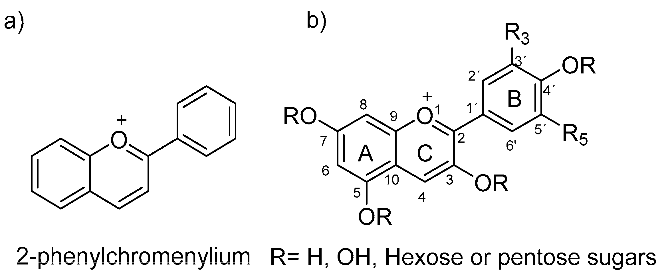
Figure 1. (a) Flavylium ion structure; (b) common numbering of flavylium ion.
Polymers 16 00163 g001
Figure 2. pH-dependent structural changes and color variation of anthocyanins in aqueous solution. Reprinted with permission from Calogero et al. [8]. Copyright (Calogero G) (Chem Soc Rev).
Polymers 16 00163 g002
Figure 3. Chemical structures of anthocyanin at different pH values. Source: [25].
Polymers 16 00163 g003
Figure 4. Illustrative scheme of the synthesis and performance of the smart anthocyanin-loaded chitosan/gelatin films for accurate beef sub-freshness monitoring. Reproduced with permission from Li et al. [30]. Copyright (2024), Elsevier.
Polymers 16 00163 g004
Figure 5. Three-dimensional printing of stable anthocyanin-loaded double emulsion gels formed by pectin and glucono-delta-lactone complexes. Reproduced with permission from Li et al. [51]. Copyright (2023), Elsevier.
Polymers 16 00163 g005
Figure 6. Triple role of anthocyanin-loaded polymers in food applications.
Polymers 16 00163 g006
Figure 7. Changes in shrimp freshness during storage using intelligent halochromic films. (Reproduced with permission from Tavassoli et al. [113]). Copyright (2023), Elsevier.
Polymers 16 00163 g007
Figure 8. Changes in the freshness of lamb meat during storage using halochromic smart films. Reproduced with permission from Alizadeh-Sani et al. [116]. Copyright (2021), Elsevier.
Polymers 16 00163 g008
Figure 9. Changes in the freshness of peeled shrimps (a), rainbow trout fillets (b), minced lamb meat (c), and chicken fillets (d) under cooled conditions using CMC-PLA-VOE 5% after 0 (a), 3 (b), and 7 (c) days. Reproduced with permission from Fatemeh Rezaei et al. [117]. Copyright (2023), Elsevier.
Polymers 16 00163 g009
Figure 10. Preparation of the anthocyanin-loaded pH-sensing soft contact. (a) Anthocyanin chemical formulas at physiological pH levels: (i) pH 6.5, (ii) pH 7.0, and (iii) pH 7.5. Scale bars: 5.0 mm. (b) Illustration of the different preparation methods: (i) soaking and (ii,iii) drop-casting on the convex/concave face of the contact lenses. (c) Color changes in contact lenses according to the preparation method. Scale bars: 5.0 mm. (d) Micrographs of the cross sections of loaded contact lenses at the different pHs: (i) pH 6.5, (ii) pH 7.0, and (iii) pH 7.5. Scale bars: 200 μm. Reproduced with permission from Riaz et al. [131]. Copyright (2019) CC, ACS Publications.
Polymers 16 00163 g010
Figure 11. Nanoencapsulates of anthocyanin-enriched extract formed by lipid assembly generated by Mendes et al. [148] showed successful cellular uptake and improved mitochondrial functionality. Reproduced with the permission of Mendes et al. [148], licensed under CC BY-NC-ND 4.0 DEED. Copyright (2021), Elsevier.
Polymers 16 00163 g011
Table 1. Some recently reported anthocyanin-loaded polymer systems.
Type of MaterialAnthocyanin FunctionExtract UsedRef(s).
AerogelpH indicatorRed grape[9]
CryogelpH indicatorRed radish[88]
Double emulsionsBioactive substanceMulberry[9]
Double-layer nanofiber matspH indicatorTurnip peel extract and potato starch–turnip peel extract[37]
Electrospun fiber matspH indicatorPrunus domestica[89]
Electrospun filmpH indicatorBlueberry[90]
Nanoencapsulation/spray dryingActive ingredientAronia[85,91]
FilmpH indicatorBlack carrot[92,93]
FilmpH indicatorRed cabbage[94,95]
HydrogelpH indicatorRed beets, purple corn[43,96]
HydrogelCO2-sensitive indicatorBlack goji anthocyanin[97]
HydrogelpH indicatorAronia, liriope, and red cabbage[98]
HydrogelBioactive substanceBlueberry[40,83]
FilmpH indicatorRed cabbage[87]
Lipid nanoparticlesBioactive substancePurple sweet potato[99]
Microencapsulation spray dryingBioactive substanceTaro tubers[100]
Microencapsulation spray dryingBioactive substanceChokeberry[73]
NanocoatingColorimetric sensorCyanidin 3-O-glucoside[100]
FilmspH indicatorBlack rice[101]
NanoliposomesBioactive substanceGrape skin[66]
EmulsionBioactive substanceBlack rice[102]
Polyelectrolyte complexpH indicatorNon-pomace residue of grape juice powder[103]
Polysaccharide assemblyBioactive substancePurple corn cob[104]
Table 3. Recent research on active anthocyanin-based polymers in the food industry.
Source of AnthocyaninsPolymer MatrixEffects/ResultsReference
Clitoria ternatea flowersMaltodextrinEnhanced stability.
Improved antibacterial effect.
[127]
Brassica oleracea (BO) extractCellulose nanofiber loaded with carbon dotsReduced spoilage.
Enhanced antioxidant effect.
Improved thermal stability.
UV barrier properties.
[128]
Pomelo peelFish scale gelatin (FSG)/alginate dialdehyde (ADA) loaded with carbon dotsPhysiological qualities post-harvest and extended shelf-life. [129]
Red cabbage (Brassica oleracea)Gelatin/poly(vinyl alcohol)-based matrix integrated with metal–organic frameworksImproved antibacterial and antioxidant effects.
Spoilage detection.
Enhanced shrimp preservation.
[130]
Purple kohlrabi peelCarrageenan-based matrix containing Zn-carbon dotsImproved antioxidant and antimicrobial properties.
Reduced spoilage.
Excellent sensor for food quality.
[122]
Table 4. Recent applications of anthocyanins as biosensors in the biomedical field.
Source of AnthocyaninsType of AnthocyaninAnthocyanin RolePolymer MatrixApplicationsRef. *
Brassica oleraceaNot indicatedpH indicatorACUVUE® contact lenses and poly(hydroxyethyacrylamide)Ocular biosensorRiaz et al. [131]
Brassica oleracea L. Var. capitataNot indicatedpH indicatorCarboxymethyl cellulose (CMC) and polyvinyl alcohol (PVA)Wound dressingAlsahag et al. [132]
Red cabbageNot indicatedpH indicatorCalcium alginate and celluloseUrea detectionAl-Qahtani et al. [133]
Red cabbageNot indicatedpH indicatorCelluloseSweat fluid detectionAl-Qahtani et al. [134]
Ref. *: references.
Table 5. Anthocyanin-loaded nanoparticle activity and applications in different healthcare areas.
Anthocyanin SourceAnthocyanin TypePolymer MatrixAdvantagesApplication(s)Ref.
BilberryNot indicatedChitosan and pectinImproved bioavailability
Gastrointestinal protection
NutraceuticalZhao et al. [138]
Black carrotNot indicatedChitosanInduced hypolipidemic effectNutraceutical as dietary supplementSreerekha et al. [139]
Haskap berry (Lonicera caerulea L.)Cyanidin 3-O-glucoside (C3G)PLGA, maltodextrin, and CMCReduced carcinogen-induced oxidative stressAnticancer therapyAmararathna et al. [140]
Black soybeanNot indicatedChondroitin sulfateInhibited proliferation of cancer cells (HeLa)Anticancer therapyJeong et al. [141]
Black riceNot indicatedChitosan/chondroitin sulfateImproved gastrointestinal bioavailability
Reduced cancer cell viability Induced colon cancer cell apoptosis
Nutraceutical, functional foods, and anticancer therapyLiang et al. [142]
Black riceCyanidin-3-glucoside)HAEnhanced stability
Reduced xanthine oxidase activity
NutraceuticalLiu et al. [143]
Not indicatedNot indicatedStarch from cornReduced glycogen levels
Improved cardiomyopathy
Cardiovascular diseasesHanafy et al. [96]
Not indicatedPelargonidinPLGAIncreased protection and control of
mitochondrial dysfunction
Nutraceutical for diabetes preventionSamadder et al. [151]
Not indicatedNot indicatedPLGA/PEG-2000Protection against free radicals
Excellent antioxidant, antiapoptotic, and anti-inflammatory effects
Protection against Alzheimer’s disease
Prevention and treatment of neurological disordersAmin et al. [145]
Jussara pulpNot indicatedPEOImproved thermal stability and enhanced antioxidant effectNutraceutical and food preservationGiaconia et al. [146]
ChokeberriesNot indicatedPAA and PAHImproved anti-tumoral performance
Monitor and trafficking
Anticancer therapy and biosensorGhiman et al. [147]
Elderberries (Sambucus nigra)Not indicatedLipids (from Codium tomentosun)Improved protection of mitochondrial membranePrevention and treatment of neurodegenerative diseasesMendes et al. [148]
Black carrotsNot indicatedNiosome (cholesterol and non-ionic surfactants)Improved bioavailability
Reduced neuroblastoma cell viability
Pharmaceutical and biotechnological applications
Anticancer therapy
Fidan et al. [149]
Black rice (Zea mays and Clitoria ternatea)Not indicatedNiosome (cholesterol)Enhanced bioavailability
Promoted collagen production
Improved anti-inflammatory effect
Wound healing systemsPriprem et al. [150]
Disclaimer/Publisher’s Note: The statements, opinions and data contained in all publications are solely those of the individual author(s) and contributor(s) and not of MDPI and/or the editor(s). MDPI and/or the editor(s) disclaim responsibility for any injury to people or property resulting from any ideas, methods, instructions or products referred to in the content.