Article

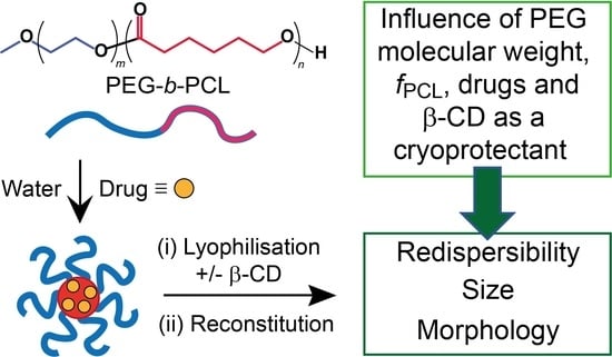
Influence of Lyophilization and Cryoprotection on the Stability and Morphology of Drug-Loaded Poly(ethylene glycol-b-ε-caprolactone) Micelles

by
Md. Saddam Hussain
1,
Khandokar Sadique Faisal
1,
Andrew J. Clulow
2,3,
Hugo Albrecht
4,
Marta Krasowska
5 and
Anton Blencowe
1,*
1
Applied Chemistry and Translational Biomaterials (ACTB) Group, Centre for Pharmaceutical Innovation (CPI), UniSA Clinical and Health Sciences, University of South Australia, Adelaide, SA 5000, Australia
2
Australian Synchrotron, Australian Nuclear Science and Technology Organisation (ANSTO), 800 Blackburn Road, Clayton, Melbourne, VIC 3168, Australia
3
Drug Delivery, Disposition & Dynamics, Monash Institute of Pharmaceutical Sciences, 381 Royal Parade, Parkville, Melbourne, VIC 3052, Australia
4
Drug Discovery and Development Group, UniSA Clinical and Health Sciences, University of South Australia, Adelaide, SA 5000, Australia
5
Future Industries Institute, UniSA STEM, University of South Australia, Mawson Lakes, Adelaide, SA 5095, Australia
*
Author to whom correspondence should be addressed.
Polymers 2023, 15(8), 1974; https://doi.org/10.3390/polym15081974
Submission received: 11 March 2023 / Revised: 17 April 2023 / Accepted: 17 April 2023 / Published: 21 April 2023
(This article belongs to the Section Polymer Applications)

Abstract

:
Polymeric micelles are promising carriers for the delivery of poorly water-soluble drugs, providing enhanced drug solubility, blood circulation times, and bioavailability. Nevertheless, the storage and long-term stability of micelles in solution present challenges requiring the lyophilization and storage of formulations in the solid state, with reconstitution immediately prior to application. Therefore, it is important to understand the effects of lyophilization/reconstitution on micelles, particularly their drug-loaded counterparts. Herein, we investigated the use of β-cyclodextrin (β-CD) as a cryoprotectant for the lyophilization/reconstitution of a library of poly(ethylene glycol-b-ε-caprolactone) (PEG-b-PCL) copolymer micelles and their drug-loaded counterparts, as well as the effect of the physiochemical properties of different drugs (phloretin and gossypol). The critical aggregation concentration (CAC) of the copolymers decreased with increasing weight fraction of the PCL block (fPCL), plateauing at ~1 mg/L when the fPCL was >0.45. The blank (empty) and drug-loaded micelles were lyophilized/reconstituted in the absence and presence of β-CD (9% w/w) and analyzed via dynamic light scattering (DLS) and synchrotron small-angle X-ray scattering (SAXS) to assess for changes in aggregate size (hydrodynamic diameter, Dh) and morphology, respectively. Regardless of the PEG-b-PCL copolymer or the use of β-CD, the blank micelles displayed poor redispersibility (<10% relative to the initial concentration), while the fraction that redispersed displayed similar Dh to the as-prepared micelles, increasing in Dh as the fPCL of the PEG-b-PCL copolymer increased. While most blank micelles displayed discrete morphologies, the addition of β-CD or lyophilization/reconstitution generally resulted in the formation of poorly defined aggregates. Similar results were also obtained for drug-loaded micelles, with the exception of several that retained their primary morphology following lyophilization/reconstitution, although no obvious trends were noted between the microstructure of the copolymers or the physicochemical properties of the drugs and their successful redispersion.

Graphical Abstract

1. Introduction

Polymeric micelles are nanosized aggregates that have been extensively studied for drug delivery [1,2]. In contrast to the static structure of solid nanoparticles, the core-shell dynamic structure of polymer micelles originates from the self-assembly of amphiphilic copolymer constituents. Therefore, solubilization dependence and physical instability (particulate aggregation, fusion, hydrolysis, and therapeutic leakage) during storage in solution remain major challenges for the widespread adoption of polymer micelles in drug delivery applications [3,4]. While lyophilization of polymer micelle solutions has been studied to address some of these issues and extend their shelf-life [5] as well as provide convenient handling, storage, and transport [6], reconstitution without irreversible aggregation remains challenging for many micelle-based systems. The success of lyophilization and reconstitution can be highly dependent on the micelle composition and preparation strategy, polymer composition and encapsulation properties, and lyophilization process parameters [7,8]. In the majority of cases, the addition of a cryoprotectant is necessary to protect micelles from stresses caused during lyophilization that can lead to irreversible secondary aggregation and fusion [9,10].
Typically reported cryoprotectants for the lyophilization of micelles to include saccharides, polymers (e.g., PEG), and surfactants. In particular, cyclodextrin (CD) derivatives including hydroxypropyl-α-CD (HP-α-CD), hydroxypropyl-β-CD (HP-β-CD), sulfobutylether-β-CD (SBE-β-CD), methyl-β-CD (M-β-CD), and hydroxypropyl-γ-CD (HP-γ-CD) have been widely used, as well as saccharides such as maltose, mannitol, sucrose, and trehalose. While the choice of cryoprotectant can depend on many different factors, it is often necessary to screen cryoprotectants for optimal performance with a chosen micelle formulation (i.e., copolymer and payload). Tommaso et al. report that 10% w/v sucrose was the most effective cryoprotectant for the lyophilization of cyclosporin-loaded PEG-b-poly(hexyl-lactide) (PEG-b-hexPLA) micelles [11], while 5% w/v mannitol was found to be more effective for bovine serum albumin (BSA)-loaded PEG-b-poly(lactic acid) (PEG-b-PLA) micelles [12].
PEG-b-PCL copolymer micelles have emerged as efficient and safe drug delivery platforms in preclinical studies due to their cytocompatibility, prolonged blood circulation, accumulation in tumors, low protein adsorption, and immunogenicity [13]. However, during storage in solution, blank and drug-loaded PEG-b-PCL micelles display relatively low physical stability regardless of the overall molecular weight, the weight fraction of the blocks, fPCL, or concentration [6,14]. The physical instability of PEG-b-PCL micelles during long-term storage has been associated with secondary aggregation, phase separation, copolymerization, and drug precipitation [6,10,14]. Therefore, lyophilization with appropriate cryoprotectants (alone or in combination) is a necessary step to maximize the physical stability of PEG-b-PCL micelles during mid- to long-term storage [14,15,16,17,18].
Moretton et al. studied the lyophilization of amphotericin-loaded PCL-b-PEG-b-PCL triblock copolymer micelles and found that lyophilization without a cryoprotectant resulted in an indispersible cake even under strong ultrasonication [6]. In comparison, the application of saccharides (maltose, glucose, or sorbitol), various molecular weight PEGs, and HP-β-CD as cryoprotectants improved redispersibility, with the latter providing a porous cake upon lyophilization that could easily be redisperse with a minimal increase (~10%) in the Dh of the micelles [6]. Miller et al. compared the lyophilization of dexamethasone-loaded PEG-b-poly(4-vinylpyridine) (PEG-PVP) and blank PEG-b-PCL, PEG-b-PLA, and PEG-b-poly(lactide-co-glycolide) (PEG-b-PLGA) micelles with various cryoprotectants, including saccharides, Kolidon (poly(vinylpyrrolidone)), and β-CD derivatives (SBE-β-CD and HP-β-CD), and concluded that 1–2.5% w/v of HP-β-CD was most effective for redispersion of micelles, with similar Dh and particle size distributions (PSDs) as the micelles before lyophilization [10]. The effectiveness of HP-β-CD over saccharides and Kolidon was also demonstrated by Richter et al. for the lyophilization and reconstitution of sagopilone-loaded PEG-b-PCL micelles [19]. Hu et al. further confirmed the ineffectiveness of simple saccharides (i.e., glucose) as cryoprotectants for PCL-b-PEG-b-PCL micelles [14].
The effectiveness of β-CD derivatives for the cryoprotection of PEG-b-PCL micelles has been attributed to host-guest complexation between the β-CD motif and PEG to form PEG-β-CD inclusion complexes [6,10]. The amorphous characteristics and relatively high collapse and glass transition temperatures (Tc and Tg, respectively) of CD derivatives have also been emphasized as important considerations [20], broadening the polymeric transition of the solid to rubbery states and providing the PEG component with more flexibility to endure the stresses imposed during lyophilization [6,10]. These investigations clearly emphasize the importance of cryoprotectants for the lyophilization and redispersion of PEG-b-PCL micelles; however, no studies have investigated a large and systematic library of PEG-b-PCL copolymers and whether a single cryoprotectant is suitable for polymers with different microstructures or micelles loaded with drugs that have different physicochemical properties. While β-CD derivatives have been widely employed for the cryoprotection of PEG-b-PCL micelles, the use of non-functionalized β-CD has not been investigated despite some favorable results with related systems. For example, Lee et al. reported that β-CD was effective as a cryoprotectant for the redispersion of eugenol-loaded Pluronic-based nanocapsules [21].
Thus, the purpose of this study was to investigate if the fPCL and PEG molecular weights influence the redispersion of PEG-b-PCL micelles following lyophilization in the absence and presence of hydrophobic drugs and β-CD as a cryoprotectant. The PEG-b-PCL micelles were analyzed via ultraviolet–visible (UV–vis) spectrophotometry, dynamic light scattering (DLS), and synchrotron small-angle X-ray scattering (SAXS) to determine and compare their ability to redisperse, self-assembly size distributions, and morphologies, respectively, following lyophilization and reconstitution.

2. Experimental

2.1. Materials

α-Methoxy-ω-hydroxy PEG (number-average molecular weight (Mn) = 2 and 5 kDa), ε-caprolactone (ε-CL; ≥97%), deuterated chloroform (CDCl3; ≥99.8% D), deuterium oxide (D2O; ≥99.9% D), anhydrous toluene (≥99.8%), β-cyclodextrin (β-CD, ≥99%), stannous octoate (Sn(Oct)2; 92.5–100%), pyrene (≥98%), and lithium chloride (LiCl; ≥99%) were purchased from Sigma Aldrich (St. Louis, MO, USA). α-Methoxy-ω-hydroxy PEG (Mn = 10 kDa) was purchased from Creative PEG Works (Chapel Hill, NC, USA). Phloretin (PH; 99%) and gossypol (GP; 99%) were purchased from Shaanxi Ciyuan Biotech (Xi’an, China). Analytical reagent grade acetone, diethyl ether (DEE), chloroform, toluene, ethyl acetate, tetrahydrofuran (THF), and N,N-dimethylformamide (DMF) were purchased from ChemSupply (Gilman, Australia). All chemicals were used as received unless otherwise stated. High-purity water was obtained from a Sartorius Arium® Pro Ultrapure Water Systems UV-T-TOC (DEU) and had a resistivity of ≥18.2 MΩ·cm. Ultrahigh purity argon (99.99%) was purchased from BOC and passed through Drierite (W. A Hammond, Xenia, OH, USA) prior to use.

2.2. Characterization

Proton nuclear magnetic resonance (1H NMR) spectroscopy was performed at 25 ± 1 °C on a Bruker NMR AVANCE III HD 500 spectrometer (Bruker BioSpin, DEU) operating at 500 MHz. Polymers were analyzed in CDCl3 using the residual solvent peak (CHCl3; δH 7.26 ppm) as an internal reference.
Gel permeation chromatography (GPC) was performed at 40 °C on a Prominence liquid chromatography system (Shimadzu, Japan) fitted with a Shimadzu RID-10A refractive index detector and two Shimadzu Shim-pack columns in series (GPC-8025D and GPC-805D). THF was used as the mobile phase at a flow rate of 1 mL/min. Samples were prepared at ~10 mg/mL and filtrated through 0.22 μm nylon syringe filters (Millipore, Burlington, MA, USA) prior to analysis. The number-average molecular weight (Mn), weight-average molecular weight (MW), and dispersity (Đ)) of the polymers were determined with reference to a conventional column calibration with narrow Đ PEG standards (Polymer Standards Service GmbH; molar mass at peak maximum (Mp) = 194 to 969 kDa).
Critical aggregation concentrations (CACs) were determined using a Shimadzu RF-6000 fluorescence spectrometer at 25 ± 1 °C and pyrene as a fluorescent probe [22]. UV-vis spectrophotometry was conducted on a NanoDrop OneC (ThermoFischer, Waltham, MA, USA) to determine the extent of lyophilizate redispersion: % redispersibility = (absorbance of the lyophilized sample at peak maxima/absorbance of the non-lyophilized sample at peak maxima) × 100. pH measurements were performed with an Oakton pH 700 bench top pH meter with an Oakton all-in-one pH/ATC electrode, single junction, 12 mm probe (USA).
The PSDs and the polydispersity index (PDI) of micelles were determined from correlograms using the Zetasizer 7.11 software and recorded using a Malvern Zetasizer NANO ZS (Malvern Panalytical, Malvern, UK) fitted with a 4 mW He–Ne laser source operating at a wavelength of 633 nm.
SAXS measurements were performed at 37 °C using the SAXS/WAXS beamline at the Australian Synchrotron (Australia’s Nuclear Science and Technology Organisation (ANSTO), Melbourne, Australia) [23]. Samples were prepared in water, filtrated through 0.22 μM nylon syringe filters, and loaded into thin-walled glass capillaries (Charles Supper, Natick, MA, USA). The capillaries were placed inside a custom-made thermostatted capillary holder. Scattering patterns were recorded using a Dectris Pilatus 2M detector at sample-detector distances of 1350 mm (15.1 keV photon energy, 0.821 Å wavelength) and 7398 mm (11.0 keV photon energy, λ = 1.127 Å wavelength) [24]. 2D scattering patterns were reduced to profiles of scattered X-ray intensity [(I(Q)] versus the scattering vector Q [=(4π/λ)sinθ, 2θ = scattering angle] using ScatterBrain software (Australian Synchrotron). Data from the two detector configurations were combined into a single continuous scattering profile with a Q range from 1.9 × 10⁻³ to 2.0 × 10⁻¹ Å−1 using the IRENA small-angle scattering macros (release 2.68) within the IgorPro software package (version 7.0.8.1) [25]. Multiplication of the background file was required for some samples because of the variable nature of the capillary thicknesses and the potential to take exposures closer to the edge of some capillaries than others in the moving stage. Thus, the sample path length of the X-ray beam was not the same for all samples, which in some cases led to an over-subtraction of the background signal. This was corrected by rescaling the background measurement.

2.3. Procedures

2.3.1. Synthesis of PEGx-b-PCLy Copolymers

PEGxPCLy diblock copolymers (PEG2PCLY, PEG5PCLY, and PEG10PCLY series) were synthesized as previously reported [24], whereby the subscript x and y refer to the Mn (kDa) of the PEG and PCL blocks.

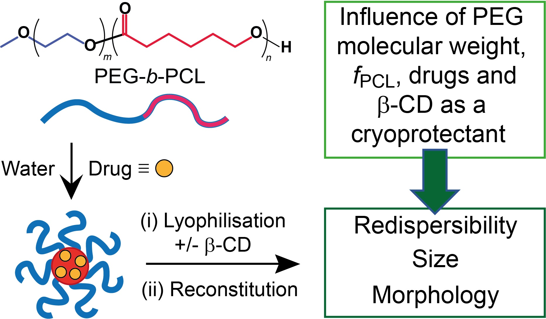
2.3.2. Determination of CAC

Stock solutions of pyrene (6 × 10⁻⁵ M) and copolymers (2 mg/mL) were prepared in acetone. Specific volumes of the pyrene and copolymer solutions were combined in vials, and then high-purity water (2 mL) was added dropwise under gentle agitation to generate a series of solutions with copolymer concentrations ranging from 0.25 to 500 mg/L and a pyrene concentration of 6 × 10⁻⁷ M (Supplementary Information (SI), Table S1). The vials were heated at 60 °C on a heating block (Ratek Instruments Pty Ltd., Boronia, VIC, Australia) for 3 h and then placed on a vacuum line (0.1 mbar) for 1 h. The volume of the solutions was adjusted to 2 mL with high-purity water to account for any water evaporation during the removal of the acetone, and the solutions were filtered through 0.22 μm nylone syringe filters. Pyrene fluorescence emission spectra were recorded between 350 and 470 nm at 2000 nm/s and 3 nm bandwidth following excitation at λex = 334 nm (SI, Figures S1–S3). The intensity ratio of emission peaks at λem = 372 and 384 nm (I384/I372) was plotted against the copolymer concentration, and the CAC was determined from the low concentration plateau.

2.3.3. Preparation of Blank and Drug-Loaded Micelle Solutions

Blank and drug-loaded (GP and PH) micelles were prepared by the solvent evaporation technique (SI, Figure S4). For blank micelles, the copolymers (PEG2PCLY, PEG5PCLY, and PEG10PCLY) were dissolved in acetone (1 mg/mL, 1 mL) in a glass vial, and high-purity water (1 mL) was added dropwise under gentle agitation. The vials were heated at 60 °C for 3 h in a heating block and subsequently placed under a vacuum (0.1 mbar) for 1 h to remove any residual acetone. High-purity water was added as required to adjust the concentration to 1 mg/mL. For drug-loaded micelles, stock solutions (1 mg/mL) of the copolymers (PEG2PCL1.1, PEG2PCL1.8, PEG5PCL1.3, PEG5PCL2.4, PEG5PCL4.2, PEG5PCL9.5, PEG10PCL7.9, and PEG10PCL10.7) and drugs (GP and PH) were prepared in acetone. The copolymer (0.9 mL) and drug (0.1 mL) stock solutions were combined in glass vials, and drug-loaded micelles (1 mg/mL, 10% w/w drug loading) were prepared as described previously for the blank micelles. All micelle solutions were obtained as transparent, homogenous solutions without any observable precipitation or sedimentation. The solutions were filtered through 0.22 µm nylon syringe filters prior to analysis.

2.3.4. Lyophilization and Reconstitution of Micelles

The micelle solutions were frozen at −80 °C for 1 h and then lyophilized (Alpha 1–2 LDplus, Martin Christ GmbH, DEU) at −57 °C (0.350 mbar) for 72 h, as previously reported [24]. The lyophilizates were stored in sealed vials at 23 ± 1 °C in the dark prior to analysis. For micelle lyophilizates containing β-CD as a cryoprotectant, a stock solution of β-CD (1 mg/mL) was prepared in ultrapure water, and 100 µL was added to each micelle solution prior to freezing and lyophilization to give 9% w/w of β-CD relative to the copolymer/drug. The lyophilized blank and drug-loaded (GP and PH) micelles were reconstituted with 1 mL of high-purity water under sonication (Soniclean Pty Ltd., SA, Australia) for 5 min, filtered through 0.22 µm nylon syringe filters, and then analyzed via DLS, SAXS, and UV-vis spectrophotometry.

3. Results and Discussion

Three copolymer series (PEG2PCLY, PEG5PCLY, and PEG10PCLY), with fPCL between 0.1 and 0.7, were studied to determine if the PEG molecular weight or block size ratio influences the redispersibility of their micelles following lyophilization with and without cryoprotection. Phloretin (PH) (logP = 2.2 [26], logS = −3.32 [27]) and gossypol (GP) (logP = 4.5 [28], logS = −5.12 [27]) were selected as model drugs due to differences in hydrophobicity, allowing the influence of this parameter on micelle reconstitution to be studied. Micelles were prepared via the solvent evaporation approach, whereby the copolymers were dissolved in acetone and high-purity water was added, followed by the evaporation of the acetone to afford aqueous micelle solutions (1 mg/mL). For drug-loaded micelles, acetone solutions of the copolymer and drug were combined, and the same procedure was followed to produce micelle solutions (1 mg/mL, 10% w/w drug). All blank and drug-loaded micelle solutions were transparent to the naked eye, and the addition of β-CD (9% w/w relative to the micelles) resulted in no visible change. Following lyophilization and reconstitution in high-purity water with sonication, all micelle solutions were opaque and/or presented with visibly large aggregates. To allow characterization of the dispersed fraction of micelles, all solutions were filtered through 0.22 µm nylon syringe filters prior to analysis.

3.1. Critical Aggregation Concentration of PEG-b-PCL Copolymers

Initially, the CAC of the copolymers was determined using the pyrene probe method in conjunction with fluorescence spectroscopy. The pyrene emission spectra were measured as a function of copolymer concentration from 0.25 to 500 mg/L (SI, Figures S1–S3), and the emission intensity ratio (I384/I372) was plotted against the logarithmic copolymer concentration to estimate the CAC from the crossover of linear fits for the low concentration plateau and slope at intermediate concentrations (excluding the high concentration plateau) (Figure 1a–c).
The emission spectra of most copolymer solutions displayed a narrowing of the first vibronic band (I372) of pyrene and the appearance of the third vibronic band (I384) with increasing copolymer concentration, indicating a decrease in the polarity of the pyrene microenvironment [29,30] associated with aggregation of the PCL block and the formation of micelles (SI, Figures S1–S3). Interestingly, the pyrene emission spectra for PEG2PCL0.4 and PEG5PCL0.6 did not reveal any changes in fine structure despite an apparent increase in the I384/I372 ratio, suggesting that these polymers do form defined aggregates with isolated hydrophobic regions, consistent with previous observations that they form metastable clusters [24]. The CAC was found to decrease with increasing fPCL for all copolymer series, beginning to plateau between 0.9 and 1.1 mg/L when the fPCL > 0.45 (Figure 1d). These results are consistent with previously reported CACs for PEG-b-PCL diblock copolymers [22,31,32,33,34,35,36,37,38,39]. For instance, Choi et al. reported that the CAC of PEG2PCL0.5, PEG2PCL1, and PEG2PCL1.5 copolymers decreased from 5.95 to 0.83 mg/L as the fPCL was increased [22].
The micelles were analyzed under isothermal conditions, so temperature effects could be eliminated. While low-molecular-weight PEG-PCL copolymers (and more specifically PCL-PEG-PCL triblock copolymers) with high fPCL can display lower critical solution temperature (LCST) values in the relevant range for biological applications (ambient to 37 °C) [40,41,42], none of the diblock copolymers studied in this manuscript have similar molecular weights and fPCL. For higher molecular weight PEG-PCL copolymers, it is possible for them to display LCSTs, but these are much higher than physiological temperatures and not relevant for drug-loaded micelles. For these reasons, the thermal properties of the micelles in the solution were not studied.

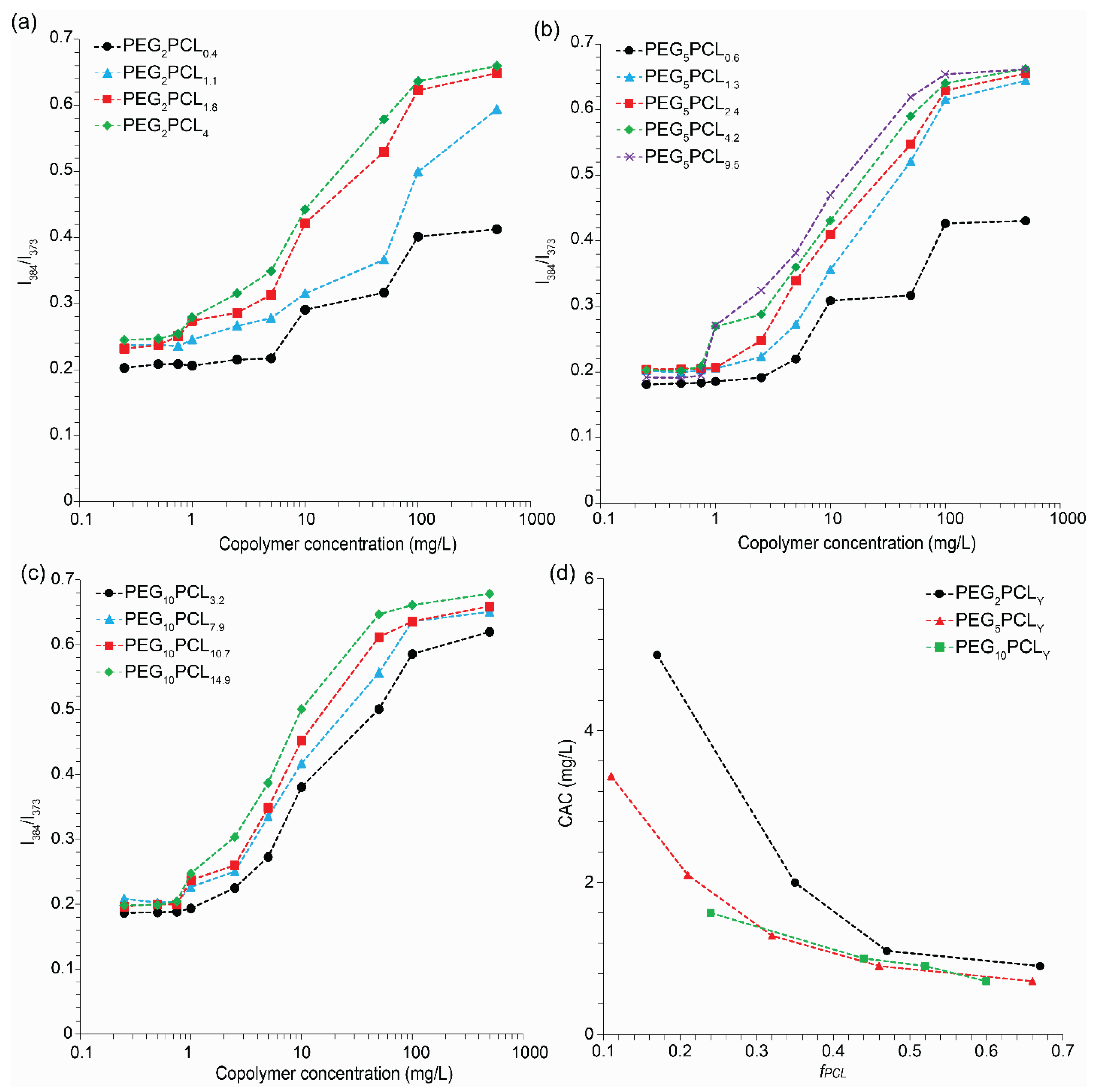
3.2. Effects of Lyophilization and Reconstitution on Blank Micelles

The percentage of blank micelles redispersed was determined by measuring the absorbance at the peak maxima (λmax ≈ 264 nm) of the micelles before and after reconstitution via UV-vis spectrophotometry. In the absence of β-CD, the redispersibility of the blank micelles was found to be very poor (typically < 10%), and there were no obvious trends between the redispersibility and the PEG molecular weight or fPCL. The addition of β-CD did not provide any significant improvement in the redispersibility. Evidently, the majority of the blank micelles are present as larger aggregates after reconstitution and are removed following filtration through 0.22 µm syringe filters.
During this study, we intentionally avoided the use of electron microscopy (e.g., SEM and TEM), as this requires sample preparation (e.g., adsorption of the micelles to substrates, dehydration, staining, and sputter coating) that can distort the micelle morphology, cause disassembly and aggregation, and lead to erroneous results that are not truly representative of the micelles in solution [43]. Furthermore, SEM lacks the resolution to clearly observe small micelles [44,45]. In comparison, non-interfering techniques (such as the light scattering techniques reported herein) allow the characterization of micelles in their native and solvated states at ambient temperatures [24,43].
The Dh and PDI values (SI, Table S2) of the blank micelles were determined from DLS number PSDs before (as prepared (AP)) and after lyophilization/reconstitution (LR) in the absence and presence of β-CD (−/+ CD). While the number of PSDs was generally narrow, volume and intensity PSDs were typically broad and, in many cases, multimodal, indicative of various populations of micelles and secondary aggregates (SI, Figures S5–S7). For the as-prepared micelles, the Dh was found to generally increase as the PEG molecular weight or fPCL was increased (Figure 2), with the exception of the PEG2PCL0.4 and PEG5PCL0.6 copolymers (SI, Figures S5 and S6), which have previously been shown to form metastable clusters rather than well-defined micelles [24]. In most cases, the addition of β-CD before lyophilization had a negligible effect on the micelle Dh, which might indicate that the β-CD is solvated in solution and there is no significant inclusion complexation between the PEG corona of the micelles and the β-CD. While lyophilization and reconstitution in the absence or presence of β-CD did not significantly change the Dh of the redisperse micelles for the PEG2PCLy series when the fPCL was >0.2, a marked increase was noted for many of the PEG5PCLy and PEG10PCLy micelles (Figure 2). The PDI values of the as-prepared micelles ranged between 0.12 and 0.26, with polymers of higher PEG molecular weight and larger fPCL generally having lower values (SI, Table S2). The addition of β-CD before lyophilization resulted in slight changes in the PDI values of the micelles, although no consistent trends were observed. Similarly, following lyophilization/reconstitution in the absence or presence of β-CD, the majority of the PDI values of the redispersed fraction of micelles ranged between 0.12 and 0.25, with no obvious trends resulting from the use of cryoprotectant. In comparison, Richter et al. reported an increase in the PDI of PEG2PCL2.6 and PEG5PCL5 micelles following lyophilization and reconstitution with HP-β-CD as the cryoprotectant, even after filtration (0.22 μm syringe filters) [19].
To assess if the morphology of the redispersed micelles was retained following lyophilization and reconstitution, synchrotron small-angle X-ray scattering (SAXS) was employed. Previously, we observed from SAXS experiments that the PEG-b-PCL assemblies transitioned from loosely associated unimers and metastable aggregates to well-defined micelles with cylindrical or ellipsoidal morphologies as the fPCL increased [24]. For example, PEG5PCLY copolymers with fPCL < 0.2 form metastable aggregates, while fPCL between 0.2 and 0.45 forms cylindrical micelles, and fPCL > 0.45 forms ellipsoidal micelles. These previous results and modeling [22] were used as a point of reference when considering morphological changes in the presence of β-CD or following lyophilization/reconstitution. With the exception of PEG2PCL0.4 solutions (Figure 3a), the SAXS profiles of the as prepared PEG2PCLy micelles prior to the addition of β-CD indicated the presence of primary cylindrical micelles as indicated by the ~Q−1 power law in the intermediate Q region (Figure 3b and SI, Figure S8), with some profiles also displaying an upturn at low Q consistent with scattering from larger secondary aggregates (as indicated on Figure 3c). Following the addition of β-CD or after lyophilization/reconstitution, the form factors generally remained similar, suggesting that the morphology of the primary micelles was conserved, although the upturn at low Q became more pronounced, indicative of an increase in large secondary aggregates not removed by filtration. In contrast, the SAXS profiles of PEG2PCL0.4 revealed a power law increase in scattering with decreasing Q, and an exponent of Q−1.9 corresponds to large fractal aggregates, further confirming the absence of well-defined micelles in this system. The SAXS profile remained unchanged following lyophilization/reconstitution; however, the addition of β-CD led to an increase in background scattering at high Q, likely from β-CD. Similar behavior was observed for the PEG5PCL0.6 copolymer (SI, Figure S9).
For the as prepared PEG5PCLy micelles, when the fPCL > 0.11, the SAXS profiles indicated the presence of discrete primary micelles, accompanied by larger secondary aggregates (SI, Figure S9). While the addition of β-CD or lyophilization/reconstitution did not significantly change the morphology of the PEG5PCL1.3 micelles, the scattering profiles for PEG5PCL2.4, PEG5PCL4.2, and PEG5PCL9.5 revealed the disappearance of discrete form factors and presented solely with power laws at low Q consistent with large poorly defined aggregates (Figure 3b). These results suggest that the addition of β-CD disrupts the micelle morphology when the fPCL > 0.20 for the PEG5PCLy micelles. Lyophilization/reconstitution of these micelles in the absence and presence of β-CD were also found to result in similar morphological changes. Similar trends were also noted for the PEG10PCLy series (SI, Figure S10), indicating that even prior to lyophilization/reconstitution, the addition of β-CD promoted significant aggregation and morphological changes in the micelles when the PEG Mn was >2 kDa. This behavior may be related to the formation of pseudo-rotaxanes between the β-CD and the PEG corona of the micelles, with subsequent hydrogen-bonding between the β-CD leading to aggregation of the micelles [46,47,48,49]. β-CD is also known to form inclusion complexes with PCL, which may also contribute to the disruption of the micelle morphology [50,51,52,53]. An outlier in the PEG10PCLy series was the PEG10PCL10.7 micelle, which following lyophilization/reconstitution in the absence of β-CD presented with similar primary morphology.
Tommaso et al. have also reported changes in the primary morphology of PEG-b-hexPLA micelles after lyophilization/reconstitution in the absence of a cryoprotectant or the presence of certain saccharide cryoprotectants (i.e., glucose, mannitol, and trehalose). Specifically, a morphological change from spherical to worm-like aggregates was observed via transmission electron microscopy, TEM [11]. Miller et al. compared the morphologies of PEG5PLA23, PEG5PLGA28, and PEG5PCL33 micelles before and after lyophilization with or without SBE-β-CD and HP-β-CD cryoprotectants via cryo-TEM [10]. While HP-β-CD was effective in retaining the primary spherical morphology of all micelles, SBE-β-CD was only effective for PEG5PLGA28 micelles [10], highlighting the variability between β-CD derivatives for different micelle compositions.

3.3. Effects of Lyophilization and Reconstitution on Drug-Loaded Micelles

Initially, the incorporation of drugs (10% w/w) into the micelles was investigated using the PEG5PCLy series, with the exclusion of PEG5PCL0.6 as it did not form well-defined micelles. Following lyophilization and reconstitution, the percentage redispersibility of the drug-loaded PEG5PCLy micelles was slightly improved as compared to their blank counterparts (<20%), with the addition of β-CD providing no obvious improvement. The slightly improved redispersibility of the drug-loaded micelles (cf. blank micelles) implies that the drugs either provide some stabilization to the micelles or promote the redispersion of aggregates, even in the absence of a cryoprotectant. While some prior literature supports this observation, in general, the redispersibility appears to be highly dependent on the combination of the micelle, cryoprotectant, and drug [10,19]. Miller et al. reported that the redispersibility of PEG5PCL33 micelles was improved after lyophilization/reconstitution when loaded with dexamethasone (cf. blank micelles) in the absence and presence of certain cryoprotectants [10]. In contrast, Richter et al. reported that sagopilone-loaded PEG2PCL2.6 and PEG5PCL5 micelles were not readily dispersible following lyophilization/reconstitution, even in the presence of HP-β-CD as the cryoprotectant [19]. Tommaso et al. demonstrated that cyclosporin-A (CsA)-loaded PEG-b-hexPLA micelles displayed better redispersibility after lyophilization with sucrose as a cryoprotectant, whereas trehalose and glucose were less effective [11].
For the populations of the drug-loaded micelles that were redispersed after reconstitution, the Dh values were generally similar to those of the micelles prior to lyophilization and followed the same trend of increasing Dh with increasing fPCL observed for the blank micelles. Compared to the blank micelles (Figure 2), the addition of the drugs resulted in a slight increase in the Dh of the micelles by 2–10 nm (Figure 4 and SI, Figures S11–S14), consistent with drug encapsulation. The addition of β-CD to the drug-loaded micelles did not significantly influence the Dh.
The SAXS profiles of the as-prepared drug-loaded PEG5PCLy copolymers were consistent with discrete primary micelles with some larger secondary aggregates, except for PEG5PCL9.5 micelles, which were free of secondary aggregates (Figure 3d and SI, Figures S15–S18). The addition of β-CD had little influence on the scattering profiles as compared to their blank micelle counterparts, implying that drug loading inhibits the β-CD-promoted reorganization of the micelle morphology. Lyophilization/reconstitution of the micelles in the absence of β-CD led to disruption of the morphology and the formation of poorly defined aggregates regardless of the specific drug. Similar results were obtained for the lyophilization/reconstitution in the presence of β-CD, whereby it is conceivable that β-CD may also contribute to the disruption of the morphology through the formation of inclusion complexes with the drugs [54,55]. A notable exception was the GP-loaded PEG5PCL9.5 micelles that retained structural features despite increased secondary aggregates (SI, Figure S18). Thus, it appears that fPCL ≥ 0.66 in the presence of very lipophilic drugs and β-CD provides some benefits for the redispersion of micelles.
While the influence of drug loading on the morphology of polymeric micelles has received limited attention, several studies have reported improved micelle stability following drug loading [56,57]. Akiba et al. investigated the structure of partially benzyl-esterified PEG-b-poly(aspartic acid) using SAXS before and after the loading of LE540 (a hydrophobic retinoid antagonist), whereby the polymer formed a hexagonal arrangement of α-helices that transitioned to spherical micelles upon drug loading [56]. Chang et al. reported that PEG-b-poly(styrene) micelles displayed enhanced stability after curcumin loading with a reduced tendency to aggregate [40]. Similarly, Hu et al. and Moretton et al. have noted that PEG-b-PCL micelles display improved stability upon the loading of hydrophobic drugs [14,58].
To investigate potential differences between copolymers with variable PEG block length and drug-loaded micelles, they were compared with copolymers with a fixed fPCL of ~0.5 (i.e., PEG2PCL1.8, PEG5PCL4.2, and PEG10PCL10.7). DLS analysis revealed that the population of redispersed micelles displayed similar Dh values to the micelles prior to lyophilization (SI, Figures S19–S22). For the PEG2PCL1.8 and PEG5PCL4.2 drug-loaded micelles, the Dh was ~20–30 nm regardless of the identity of the drug, with no effect observed from the addition of β-CD. After lyophilization/reconstitution of the PEG10PCL10.7 drug-loaded micelles, the Dh increased to ~55–65 nm from ~45 nm, regardless of the drug identity, suggesting that the larger molecular weight PEG is prone to forming secondary aggregates that cannot be completely redispersed.
The SAXS profiles of the as-prepared drug-loaded PEG2PCL1.8 micelles (SI, Figure S23) were very similar to their blank counterparts (SI, Figure S8) and indicated the presence of cylindrical/ellipsoidal primary aggregates with some larger secondary aggregates. The addition of β-CD had no effect. In contrast to the drug-loaded PEG5PCL4.2 micelles, the drug-loaded PEG2PCL1.8 micelles retained a discrete morphology after lyophilization/reconstitution, although an increase in secondary aggregates was observed for the PH-loaded PEG2PCL1.8 micelles in the presence of β-CD. The as-prepared drug-loaded PEG10PCL10.7 micelles presented with discrete primary morphologies, although larger secondary aggregates were only noted for the GP-loaded micelles (SI, Figure S24). The addition of β-CD increased the extent of secondary aggregation for the GP-loaded PEG10PCL10.7 micelles but had no effect on the PH-loaded micelles, which is significantly different from the behavior of the blank PEG10PCL10.7 micelles, where the primary morphology was lost (SI, Figure S10). Thus, the addition of lipophilic drugs helps to prevent β-CD mediated disruption of the morphology of the PEG10PCL10.7 micelles (cf. PEG5PCL4.2 micelles). Surprisingly, lyophilization/reconstitution of the GP-loaded PEG10PCL10.7 micelles resulted in discrete morphologies, whereas the PH-loaded PEG10PCL10.7 micelles formed poorly defined aggregates. The addition of β-CD had no influence on this behavior, indicating that the lipophilicity of the drug plays a role in retaining the primary morphology for this particular polymer.

4. Conclusions

Micelles were prepared from a library of PEG-b-PCL copolymers with various PEG molecular weights and fPCL, and the effect of lyophilization and reconstitution on their redispersibility, Dh, and morphology were studied in the absence and presence of β-CD as a cryoprotectant. The Dh of the blank micelles was found to generally increase with increasing PEG molecular weights and fPCL, and when the fPCL > 0.2, the micelles presented with discrete morphologies. While the loading of model drugs (gossypol or phloretin) led to a slight increase in the micelle Dh, the morphology remained unaffected. Following lyophilization and reconstitution, the blank and drug-loaded micelles were poorly dispersible, although drug loading did lead to a slight improvement (<10% vs. <20%). DLS of the redispersible fraction revealed slight changes in the micelle Dh, although there were no obvious trends related to the PEG molecular weight or fPCL. For blank micelles, the primary morphology of the PEG2PCLy series was retained after lyophilization and reconstitution, while the higher molecular weight PEG series predominately formed poorly defined aggregates. The addition of β-CD to the blank micelles provided no improvement in the retention of the morphology and, in several cases, even promoted reorganization of the micelle structure prior to lyophilization and reconstitution. Loading of the micelles with drugs helped to protect against β-CD promoted reorganization, although subsequent lyophilization and reconstitution generally resulted in the formation of poorly defined aggregates, with the exception of gossypol-loaded PEG2PCL1.8 and PEG10PCL10.7 micelles. These results highlight the ineffectiveness of β-CD as a cryoprotectant under the conditions studied, and further studies with a range of cryoprotectants and loadings are required to fully interpret the contribution of any polymer microstructure-behavior relationships to the success of lyophilization and reconstitution.

Supplementary Materials

The following supporting information can be downloaded at: https://www.mdpi.com/article/10.3390/polym15081974/s1, Figure S1: Pyrene fluorescent emission spectra for the PEG2PCLy copolymer series; Figure S2: Pyrene fluorescent emission spectra for the PEG5PCLy copolymer series.; Figure S3: Pyrene fluorescent emission spectra for the PEG10PCLy copolymer series; Figure S4: Illustration of preparation, lyophilization and characterisation of PEGX-b-PCLY micelles; Figure S5: Dynamic light scattering (DLS) particle size distributions (PSDs) for blank PEG2PCLy micelles; Figure S6: DLS PSDs for blank PEG5PCLy micelles; Figure S7: DLS PSDs for blank PEG10PCLy micelles; Figure S8: Small-angle X-ray scattering (SAXS) profiles for blank PEG2PCLy micelles; Figure S9: SAXS profiles for blank PEG5PCLy micelles; Figure S10: SAXS profiles for blank PEG10PCLy micelles; Figure S11: DLS PSDs for drug-loaded PEG5PCL1.3 micelles; Figure S12: DLS PSDs for drug-loaded PEG5PCL2.4 micelles; Figure S13: DLS PSDs for drug-loaded PEG5PCL4.2 micelles; Figure S14: DLS DSDs for drug-loaded PEG5PCL9.5 micelles; Figure S15: SAXS profiles for drug-loaded PEG5PCL1.3 micelles; Figure S16: SAXS profiles for drug-loaded PEG5PCL2.4 micelles; Figure S17: SAXS profiles for drug-loaded PEG5PCL4.2 micelles; Figure S18: SAXS profiles for drug-loaded PEG5PCL9.5 micelles; Figure S19: DLS PSDs for drug-loaded PEG2PCL1.8 micelles; Figure S20: DLS PSDs for drug-loaded PEG10PCL10.7 micelles; Figure S21: Hydrodynamic diameter (Dh) versus weight fraction of PCL (fPCL) as determined from number PSDs for drug-loaded PEG2PCL1.8 micelles; Figure S22: Dh versus fPCL as determined from number PSDs for drug-loaded PEG10PCL10.7 micelles; Figure S23: SAXS profiles for drug-loaded PEG2PCL1.8 micelles; Figure S24: SAXS profiles for drug-loaded PEG10PCL10.7 micelles; Table S1: Determination of the CAC of PEG-b-PCL block copolymer micelles; Table S2: Summary of the PDI values of the blank and drug-loaded PEG-b-PCL micelles.

Author Contributions

Conceptualization, K.S.F. and A.B.; Methodology, M.S.H. and K.S.F.; Validation, A.J.C. and M.K.; Formal analysis, M.S.H., A.J.C., M.K. and A.B.; Investigation, M.S.H., K.S.F. and M.K.; Writing—original draft, M.S.H. and A.B.; Writing—review & editing, M.S.H., A.J.C., M.K. and A.B.; Supervision, A.J.C., H.A. and A.B.; Project administration, A.B.; Funding acquisition, A.B. All authors have read and agreed to the published version of the manuscript.

Funding

This research was funded by Australian Nuclear Science and Technology Organisation (Australian Synchrotron grant number M16667) and the EU research and innovation program “Horizon 2020” (grant number No. 654000).

Institutional Review Board Statement

No applicable.

Data Availability Statement

The data presented in this study are available on request from the corresponding author.

Acknowledgments

MSH thanks the University of South Australia for scholarship support. Part of this research was undertaken on the SAXS/WAXS beamline at the Australian Synchrotron, part of ANSTO. The authors thank the Australian Synchrotron for providing access to facilities and expertise. The authors also acknowledge the SasView application, originally developed under NSF Award DMR-0520547.

Conflicts of Interest

The authors declare no conflict of interest.

References

  1. Gong, J.; Chen, M.; Zheng, Y.; Wang, S.; Wang, Y. Polymeric micelles drug delivery system in oncology. J. Control. Release 2012, 159, 312–323. [Google Scholar] [CrossRef] [PubMed]
  2. Kuperkar, K.; Patel, D.; Atanase, L.I.; Bahadur, P. Amphiphilic Block Copolymers: Their Structures, and Self-Assembly to Polymeric Micelles and Polymersomes as Drug Delivery Vehicles. Polymers 2022, 14, 4702. [Google Scholar] [CrossRef] [PubMed]
  3. Patra, A.; Satpathy, S.; Shenoy, A.K.; Bush, J.A.; Kazi, M.; Hussain, M.D. Formulation and evaluation of mixed polymeric micelles of quercetin for treatment of breast, ovarian, and multidrug resistant cancers. Int. J. Nanomed. 2018, 13, 2869–2881. [Google Scholar] [CrossRef] [PubMed]
  4. Abdelwahed, W.; Degobert, G.; Fessi, H. Investigation of nanocapsules stabilization by amorphous excipients during freeze-drying and storage. Eur. J. Pharm. Biopharm. 2006, 63, 87–94. [Google Scholar] [CrossRef] [PubMed]
  5. Zhang, J.; Zhu, C.; Hu, L.; Liu, H.; Pan, H.-C. Effect of freeze-drying process on the physical stability and properties of Voriconazole complex system. Dry. Technol. 2018, 36, 871–878. [Google Scholar] [CrossRef]
  6. Moretton, M.A.; Chiappetta, D.A.; Sosnik, A. Cryoprotection–lyophilization and physical stabilization of rifampicin-loaded flower-like polymeric micelles. J. R. Soc. Interface 2012, 9, 487–502. [Google Scholar] [CrossRef]
  7. Abdelwahed, W.; Degobert, G.; Stainmesse, S.; Fessi, H. Freeze-drying of nanoparticles: Formulation, process and storage considerations. Adv. Drug Deliv. Rev. 2006, 58, 1688–1713. [Google Scholar] [CrossRef]
  8. Yang, Z.L.; Li, X.R.; Yang, K.W.; Liu, Y. Amphotericin B-loaded poly(ethylene glycol)-poly(lactide) micelles: Preparation, freeze-drying, and in vitro release. J. Biomed. Mater. Res. A 2008, 85, 539–546. [Google Scholar] [CrossRef]
  9. Schwarz, C.; Mehnert, W. Freeze-drying of drug-free and drug-loaded solid lipid nanoparticles (SLN). Int. J. Pharm. 1997, 157, 171–179. [Google Scholar] [CrossRef]
  10. Miller, T.; van Colen, G.; Sander, B.; Golas, M.M.; Uezguen, S.; Weigandt, M.; Goepferich, A. Drug loading of polymeric micelles. Pharm. Res. 2013, 30, 584–595. [Google Scholar] [CrossRef]
  11. Di Tommaso, C.; Como, C.; Gurny, R.; Möller, M. Investigations on the lyophilisation of MPEG–hexPLA micelle based pharmaceutical formulations. Eur. J. Pharm. Sci. 2010, 40, 38–47. [Google Scholar] [CrossRef]
  12. Shi, Y.; Zhu, H.; Ren, Y.; Li, K.; Tian, B.; Han, J.; Feng, D. Preparation of protein-loaded PEG-PLA micelles and the effects of ultrasonication on particle size. Colloid Polym. Sci. 2017, 295, 259–266. [Google Scholar] [CrossRef]
  13. Grossen, P.; Witzigmann, D.; Sieber, S.; Huwyler, J. PEG-PCL-based nanomedicines: A biodegradable drug delivery system and its application. J. Control. Release 2017, 260, 46–60. [Google Scholar] [CrossRef]
  14. Hu, Y.; Ding, Y.; Li, Y.; Jiang, X.; Yang, C.; Yang, Y. Physical stability and lyophilization of poly (ε-caprolactone)-b-poly (ethyleneglycol)-b-Poly (ε-caprolactone) micelles. J. Nanosci. Nanotech. 2006, 6, 3032–3039. [Google Scholar] [CrossRef]
  15. Zhang, Y.; Zhang, H.; Wang, X.; Wang, J.; Zhang, X.; Zhang, Q. The eradication of breast cancer and cancer stem cells using octreotide modified paclitaxel active targeting micelles and salinomycin passive targeting micelles. Biomaterials 2012, 33, 679–691. [Google Scholar] [CrossRef]
  16. Moulahoum, H.; Ghorbanizamani, F.; Bayir, E.; Timur, S.; Zihnioglu, F. A polyplex human saliva peptide histatin 5-grafted methoxy PEG-b-polycaprolactone polymersome for intelligent stimuli-oriented doxorubicin delivery. J. Drug Deliv. Sci. Technol. 2022, 67, 102958. [Google Scholar] [CrossRef]
  17. Tyrrell, Z.; Winoto, W.; Shen, Y.; Radosz, M. Block Copolymer Micelles Formed in Supercritical Fluid Can Become Water-Dispensable Nanoparticles: Poly(ethylene glycol)−block-Poly(ϵ-caprolactone) in Trifluoromethane. Ind. Eng. Chem. Res. 2009, 48, 1928–1932. [Google Scholar] [CrossRef]
  18. Pungkham, H.; Swatdipakdi, N.; Theerasilp, M.; Karnkla, S.; Chittchang, M.; Ploypradith, P.; Nasongkla, N. PEG-b-PCL and PEG-b-PLA polymeric micelles as nanocarrieres for lamellarin N delivery. In Proceedings of the 2011 Annual International Conference of the IEEE Engineering in Medicine and Biology Society, Boston, MA, USA, 30 August–3 September 2011; pp. 3245–3248. [Google Scholar]
  19. Richter, A.; Olbrich, C.; Krause, M.; Hoffmann, J.; Kissel, T. Polymeric micelles for parenteral delivery of Sagopilone: Physicochemical characterization, novel formulation approaches and their toxicity assessment in vitro as well as in vivo. Eur. J. Pharm. Biopharm. 2010, 75, 80–89. [Google Scholar] [CrossRef]
  20. Ressing, M.E.; Jiskoot, W.; Talsma, H.; van Ingen, C.W.; Beuvery, E.C.; Crommelin, D.J. The influence of sucrose, dextran, and hydroxypropyl-β-cyclodextrin as lyoprotectants for a freeze-dried mouse IgG 2a monoclonal antibody (MN12). Pharm. Res. 1992, 9, 266–270. [Google Scholar] [CrossRef]
  21. Lee, M.-Y.; Min, S.-G.; You, S.-K.; Choi, M.-J.; Hong, G.-P.; Chun, J.-Y. Effect of β-cyclodextrin on physical properties of nanocapsules manufactured by emulsion–diffusion method. J. Food Eng. 2013, 119, 588–594. [Google Scholar] [CrossRef]
  22. Choi, C.; Chae, S.Y.; Kim, T.H.; Kweon, J.K.; Cho, C.S.; Jang, M.K.; Nah, J.W. Synthesis and physicochemical characterization of amphiphilic block copolymer self-aggregates formed by poly (ethylene glycol)-block-poly (ϵ-caprolactone). J. Appl. Polym. Sci. 2006, 99, 3520–3527. [Google Scholar] [CrossRef]
  23. Kirby, N.M.; Mudie, S.T.; Hawley, A.M.; Cookson, D.J.; Mertens, H.D.; Cowieson, N.; Samardzic-Boban, V. A low-background-intensity focusing small-angle X-ray scattering undulator beamline. J. Appl. Crystallogr. 2013, 46, 1670–1680. [Google Scholar] [CrossRef]
  24. Faisal, K.S.; Clulow, A.J.; Krasowska, M.; Gillam, T.; Miklavcic, S.J.; Williamson, N.H.; Blencowe, A. Interrogating the relationship between the microstructure of amphiphilic poly(ethylene glycol-b-caprolactone) copolymers and their colloidal assemblies using non-interfering techniques. J. Colloid Interface Sci. 2022, 606, 1140–1152. [Google Scholar] [CrossRef]
  25. Ilavsky, J.; Jemian, P.R. Irena: Tool suite for modeling and analysis of small-angle scattering. J. Appl. Crystallogr. 2009, 42, 347–353. [Google Scholar] [CrossRef]
  26. Wang, Y.; Li, D.; Lin, H.; Jiang, S.; Han, L.; Hou, S.; Lin, S.; Cheng, Z.; Bian, W.; Zhang, X. Enhanced oral bioavailability and bioefficacy of phloretin using mixed polymeric modified self-nanoemulsions. Food Sci. Nutri. 2020, 8, 3545–3558. [Google Scholar] [CrossRef]
  27. Tetko, I.V.; Tanchuk, V.Y. Application of associative neural networks for prediction of lipophilicity in ALOGPS 2.1 program. J. Chem. Inf. Comput. Sci. 2002, 42, 1136–1145. [Google Scholar] [CrossRef]
  28. Adams, R.; Geissman, T.A.; Edwards, J.D. Gossypol, a Pigment of Cottonseed. Chem. Rev. 1960, 60, 555–574. [Google Scholar] [CrossRef]
  29. Glushko, V.; Thaler, M.; Karp, C. Pyrene fluorescence fine structure as a polarity probe of hydrophobic regions: Behavior in model solvents. Arch. Biochem. Biophys. 1981, 210, 33–42. [Google Scholar] [CrossRef]
  30. Dong, D.C.; Winnik, M.A. The Py scale of solvent polarities. Solvent effects on the vibronic fine structure of pyrene fluorescence and empirical correlations with ET and Y values. Photochem. Photobiol. 1982, 35, 17–21. [Google Scholar] [CrossRef]
  31. Satturwar, P.; Eddine, M.N.; Ravenelle, F.; Leroux, J.C. pH-responsive polymeric micelles of poly(ethylene glycol)-b-poly(alkyl(meth)acrylate-co-methacrylic acid): Influence of the copolymer composition on self-assembling properties and release of candesartan cilexetil. Eur. J. Pharm. Biopharm. 2007, 65, 379–387. [Google Scholar] [CrossRef]
  32. Koutroumanis, K.P.; Holdich, R.G.; Georgiadou, S. Synthesis and micellization of a pH-sensitive diblock copolymer for drug delivery. Int. J. Pharm. 2013, 455, 5–13. [Google Scholar] [CrossRef]
  33. Glover, A.L.; Nikles, S.M.; Nikles, J.A.; Brazel, C.S.; Nikles, D.E. Polymer micelles with crystalline cores for thermally triggered release. Langmuir 2012, 28, 10653–10660. [Google Scholar] [CrossRef]
  34. Master, A.M.; Rodriguez, M.E.; Kenney, M.E.; Oleinick, N.L.; Gupta, A.S. Delivery of the photosensitizer Pc 4 in PEG–PCL micelles for in vitro PDT studies. J. Pharm. Sci. 2010, 99, 2386–2398. [Google Scholar] [CrossRef]
  35. El Jundi, A.; Buwalda, S.; Bethry, A.; Hunger, S.; Coudane, J.; Bakkour, Y.; Nottelet, B. Double-hydrophilic block copolymers based on functional poly (ε-caprolactone) s for pH-dependent controlled drug delivery. Biomacromolecules 2019, 21, 397–407. [Google Scholar] [CrossRef]
  36. Gao, X.; Wang, B.; Wei, X.; Rao, W.; Ai, F.; Zhao, F.; Men, K.; Yang, B.; Liu, X.; Huang, M. Preparation, characterization and application of star-shaped PCL/PEG micelles for the delivery of doxorubicin in the treatment of colon cancer. Int. J. Nanomed. 2013, 8, 971. [Google Scholar] [CrossRef] [PubMed]
  37. Luo, S.; Zhang, Y.; Cao, J.; He, B.; Li, S. Arginine modified polymeric micelles as a novel drug delivery system with enhanced endocytosis efficiency. Colloids Surf. B Biointerfaces 2016, 148, 181–192. [Google Scholar] [CrossRef] [PubMed]
  38. Xie, W.; Zhu, W.; Shen, Z. Synthesis, isothermal crystallization and micellization of mPEG–PCL diblock copolymers catalyzed by yttrium complex. Polymer 2007, 48, 6791–6798. [Google Scholar] [CrossRef]
  39. Forrest, M.L.; Won, C.-Y.; Malick, A.W.; Kwon, G.S. In vitro release of the mTOR inhibitor rapamycin from poly(ethylene glycol)-b-poly(ε-caprolactone) micelles. J. Control. Release 2006, 110, 370–377. [Google Scholar] [CrossRef] [PubMed]
  40. Liu, C.B.; Gong, C.Y.; Huang, M.J.; Wang, J.W.; Pan, Y.F.; Zhang, Y.D.; Li, G.Z.; Gou, M.L.; Wang, K.; Tu, M.J.; et al. Thermoreversible gel-sol behavior of biodegradable PCL-PEG-PCL triblock copolymer in aqueous solutions. J. Biomed. Mater. Res. Part B Appl. Biomater. 2008, 84, 165–175. [Google Scholar] [CrossRef]
  41. Hwang, M.J.; Joo, M.K.; Choi, B.G.; Park, M.H.; Hamley, I.W.; Jeong, B. Multiple Sol-Gel Transitions of PEG-PCL-PEG Triblock Copolymer Aqueous Solution. Macromol. Rapid Commun. 2010, 31, 2064–2069. [Google Scholar] [CrossRef]
  42. Bae, S.J.; Joo, M.K.; Jeong, Y.; Kim, S.W.; Lee, W.K.; Sohn, Y.S.; Jeong, B. Gelation behavior of poly (ethylene glycol) and polycaprolactone triblock and multiblock copolymer aqueous solutions. Macromolecules 2006, 39, 4873–4879. [Google Scholar] [CrossRef]
  43. Suys, E.J.; Warren, D.B.; Pham, A.C.; Nowell, C.J.; Clulow, A.J.; Benameur, H.; Porter, C.J.; Pouton, C.W.; Chalmers, D.K. A nonionic polyethylene oxide (PEO) surfactant model: Experimental and molecular dynamics studies of Kolliphor EL. J. Pharm. Sci. 2019, 108, 193–204. [Google Scholar] [CrossRef] [PubMed]
  44. Dalgleish, D.G.; Spagnuolo, P.A.; Goff, H.D. A possible structure of the casein micelle based on high-resolution field-emission scanning electron microscopy. Int. Dairy J. 2004, 14, 1025–1031. [Google Scholar] [CrossRef]
  45. Peacock, H.; Blum, S.A. Single-Micelle and Single-Zinc-Particle Imaging Provides Insights into the Physical Processes Underpinning Organozinc Reactions in Water. J. Am. Chem. Soc. 2022, 144, 3285–3296. [Google Scholar] [CrossRef]
  46. Capacchione, C.; Della Sala, P.; Quaratesi, I.; Bruno, I.; Pauciulo, A.; Bartiromo, A.R.; Iannece, P.; Neri, P.; Talotta, C.; Gliubizzi, R.; et al. Poly(Ethylene Glycol)/β-Cyclodextrin Pseudorotaxane Complexes as Sustainable Dispersing and Retarding Materials in a Cement-Based Mortar. ACS Omega 2021, 6, 12250–12260. [Google Scholar] [CrossRef]
  47. Song, X.; Zhu, J.-l.; Wen, Y.; Zhao, F.; Zhang, Z.-X.; Li, J. Thermoresponsive supramolecular micellar drug delivery system based on star-linear pseudo-block polymer consisting of β-cyclodextrin-poly(N-isopropylacrylamide) and adamantyl-poly(ethylene glycol). J. Colloid Interface Sci. 2017, 490, 372–379. [Google Scholar] [CrossRef]
  48. Guo, C.; Zhang, Y.; Yuan, H.; Zhang, Y.; Yin, T.; He, H.; Gou, J.; Tang, X. Improved Core Viscosity Achieved by PDLLA10kCo-Incorporation Promoted Drug Loading and Stability of mPEG2k-b-PDLLA2.4k Micelles. Pharm. Res. 2022, 39, 369–379. [Google Scholar] [CrossRef]
  49. Zhang, Z.; Wen, Y.; Song, X.; Zhu, J.; Li, J. Nonviral DNA Delivery System with Supramolecular PEGylation Formed by Host–Guest Pseudo-Block Copolymers. ACS Appl. Bio Mater. 2021, 4, 5057–5070. [Google Scholar] [CrossRef]
  50. Yu, G.; Yang, Z.; Fu, X.; Yung, B.C.; Yang, J.; Mao, Z.; Shao, L.; Hua, B.; Liu, Y.; Zhang, F.; et al. Polyrotaxane-based supramolecular theranostics. Nat. Commun. 2018, 9, 766. [Google Scholar] [CrossRef]
  51. Moyers-Montoya, E.; García-Casillas, P.; Vargas-Requena, C.; Escobedo-González, R.; Martel-Estrada, S.A.; Martínez-Pérez, C.A. Polycaprolactone/Amino-β-Cyclodextrin Inclusion Complex Prepared by an Electrospinning Technique. Polymers 2016, 8, 395. [Google Scholar] [CrossRef]
  52. Narayanan, G.; Ormond, B.R.; Gupta, B.S.; Tonelli, A.E. Efficient wound odor removal by β-cyclodextrin functionalized poly (ε-caprolactone) nanofibers. J. Appl. Polym. Sci. 2015, 132. [Google Scholar] [CrossRef]
  53. Varan, C.; Wickström, H.; Sandler, N.; Aktaş, Y.; Bilensoy, E. Inkjet printing of antiviral PCL nanoparticles and anticancer cyclodextrin inclusion complexes on bioadhesive film for cervical administration. Int. J. Pharm. 2017, 531, 701–713. [Google Scholar] [CrossRef] [PubMed]
  54. Wei, Y.; Zhang, J.; Memon, A.H.; Liang, H. Molecular model and in vitro antioxidant activity of a water-soluble and stable phloretin/hydroxypropyl-β-cyclodextrin inclusion complex. J. Mol. Liq. 2017, 236, 68–75. [Google Scholar] [CrossRef]
  55. Shen, Y.-L.; Yang, S.-H.; Wu, L.-M.; Ma, X.-Y. Study on structure and characterization of inclusion complex of gossypol/beta cyclodextrin. Spectrochim. Acta A Mol. Biomol. Spectrosc. 2005, 61, 1025–1028. [Google Scholar] [CrossRef]
  56. Akiba, I.; Terada, N.; Hashida, S.; Sakurai, K.; Sato, T.; Shiraishi, K.; Yokoyama, M.; Masunaga, H.; Ogawa, H.; Ito, K. Encapsulation of a hydrophobic drug into a polymer-micelle core explored with synchrotron SAXS. Langmuir 2010, 26, 7544–7551. [Google Scholar] [CrossRef] [PubMed]
  57. Chang, T.; Trench, D.; Putnam, J.; Stenzel, M.H.; Lord, M.S. Curcumin-loading-dependent stability of PEGMEMA-based micelles affects endocytosis and exocytosis in colon carcinoma cells. Mol. Pharm. 2016, 13, 924–932. [Google Scholar] [CrossRef]
  58. Moretton, M.A.; Glisoni, R.J.; Chiappetta, D.A.; Sosnik, A. Molecular implications in the nanoencapsulation of the anti-tuberculosis drug rifampicin within flower-like polymeric micelles. Colloids Surf. B Biointerfaces 2010, 79, 467–479. [Google Scholar] [CrossRef]
Figure 1. Ratiometric intensity (I384/I372) of the pyrene emission as a function of copolymer concentration in high-purity water for the (a) PEG2PCLy, (b) PEG5PCLy, and (c) PEG10PCLy copolymer series. (d) CAC of the copolymers as a function of fPCL.
Polymers 15 01974 g001
Figure 2. Dh versus fPCL as determined from the number PSDs for the (a) PEG2PCLY, (b) PEG5PCLY, and (c) PEG10PCLY micelles as prepared (AP), after lyophilization/reconstitution (LR), and in the absence and presence of β-CD (−/+CD). All values are reported as the mean + std. dev. (n = 3).
Polymers 15 01974 g002
Figure 3. Representative SAXS scattering profiles complete with power laws to indicate the morphology of the aggregates or micelles: (a) PEG2PCL0.4 (red profile), (b) PEG2PCL1.1, and (c) PEG10PCL3.2 as prepared in the absence of β-CD, (a) PEG5PCL9.5 (green profile) following lyophilization/reconstitution in the absence of β-CD, and (d) gossypol-loaded PEG5PCL9.5 as prepared in the absence of β-CD.
Polymers 15 01974 g003
Figure 4. Dh versus fPCL as determined from number PSDs for (a) gossypol (GP) and (b) phloretin (PH)-loaded PEG5PCL1.3, PEG5PCL2.4, PEG5PCL4.2, and PEG5PCL9.5 micelles as prepared (AP), after lyophilization/reconstitution (LR), and in the absence and presence of β-CD (−/+CD). All values are reported as the mean + std. dev. (n = 3).
Polymers 15 01974 g004
Disclaimer/Publisher’s Note: The statements, opinions and data contained in all publications are solely those of the individual author(s) and contributor(s) and not of MDPI and/or the editor(s). MDPI and/or the editor(s) disclaim responsibility for any injury to people or property resulting from any ideas, methods, instructions or products referred to in the content.

