

Application of Lavender-Oil Microcapsules to Functionalized PET Fibers

 , ,
, ,  , , ,
, , , 

Abstract
:
1. Introduction
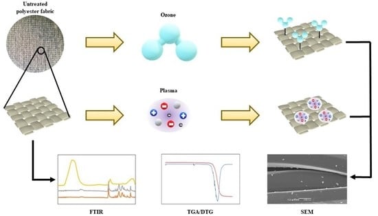
2. Materials and Methods
2.1. Materials
2.2. Chemical Modification of Samples
2.3. Polyester Modification by O3
2.4. Preparation and Application of Lavender-Oil Microcapsules
2.5. Polyester-Fabric Functionalization
2.6. Evaluation of Modified Fabric
2.7. In Vitro Release Behavior
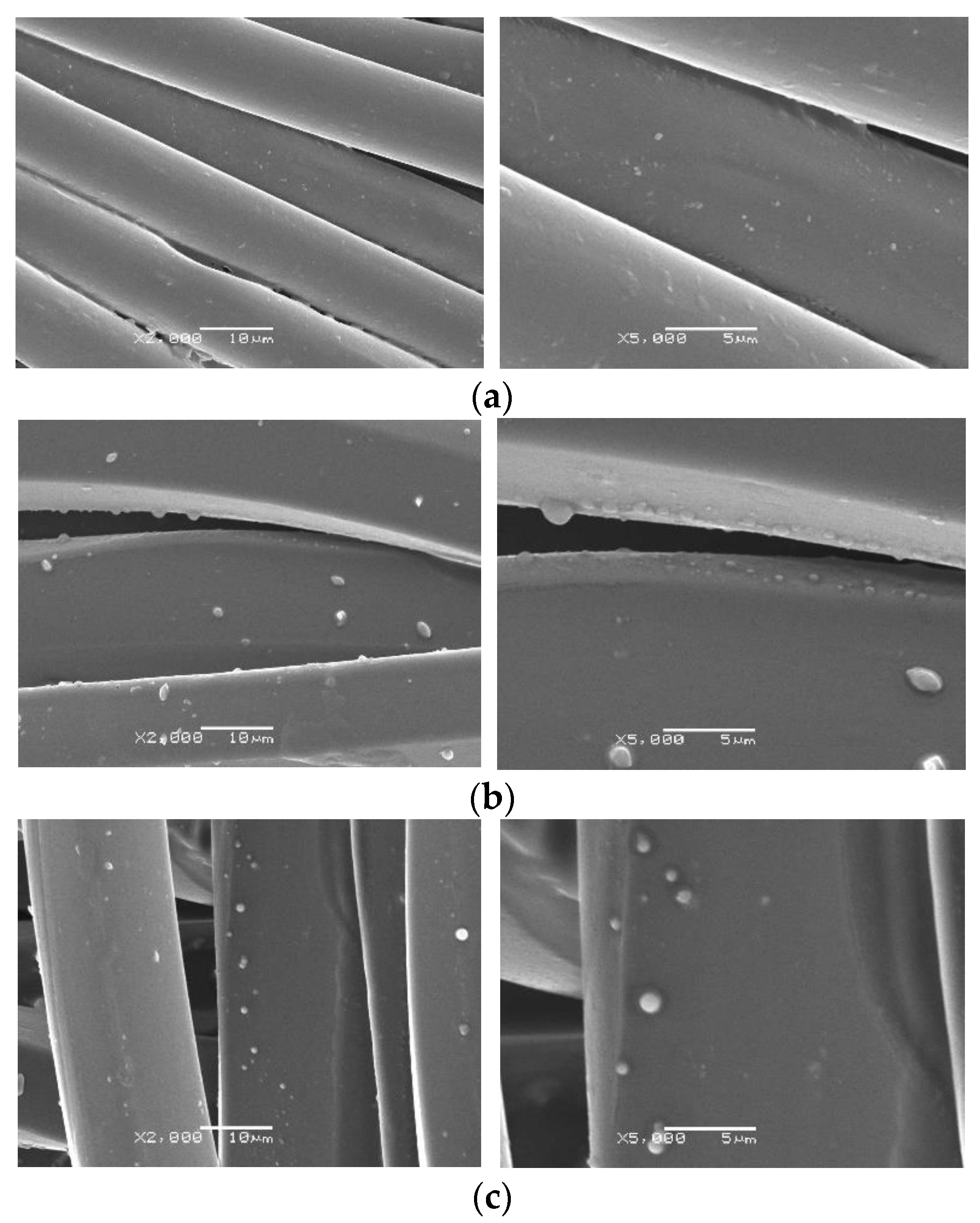
2.8. Scanning Electron and Optical Microscopy
3. Results
3.1. Microcapsules
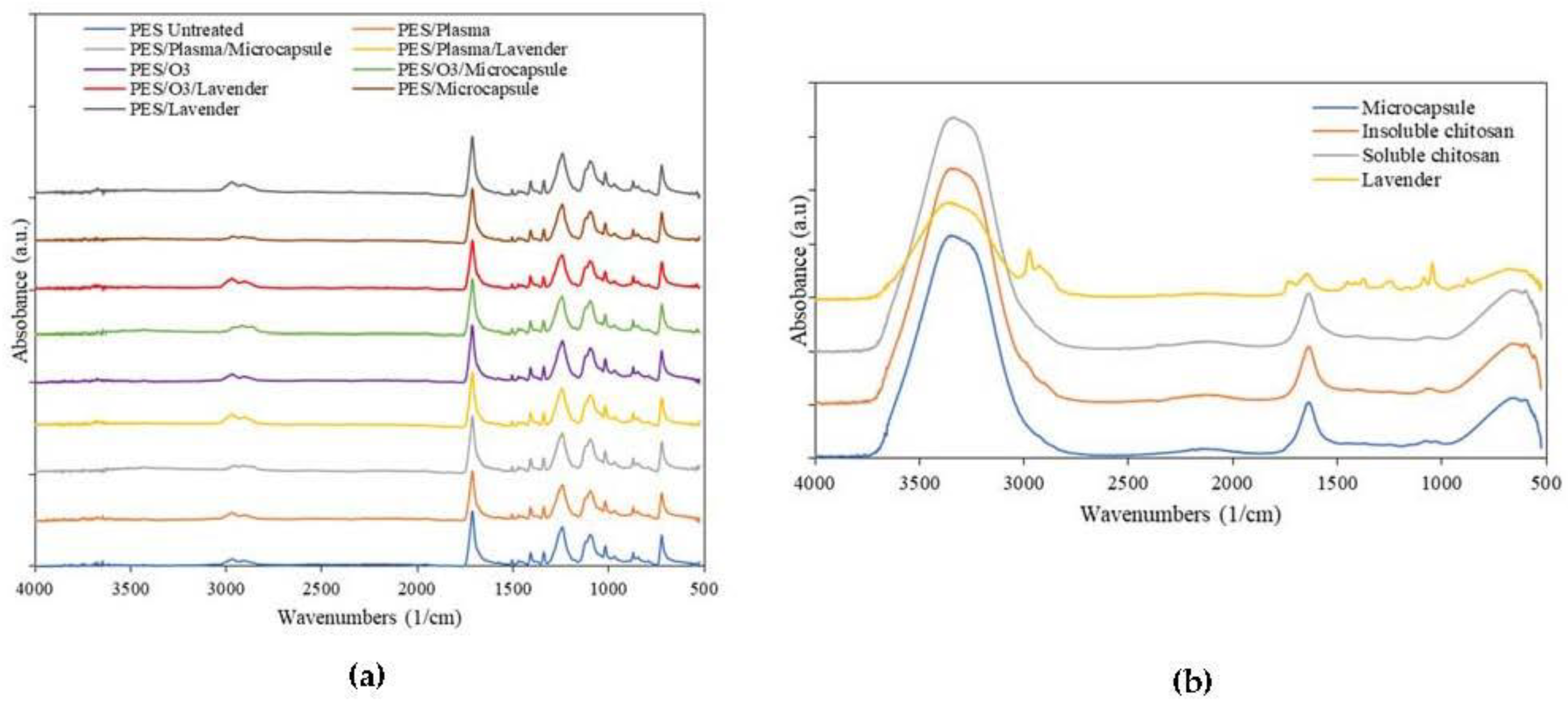
3.2. FTIR
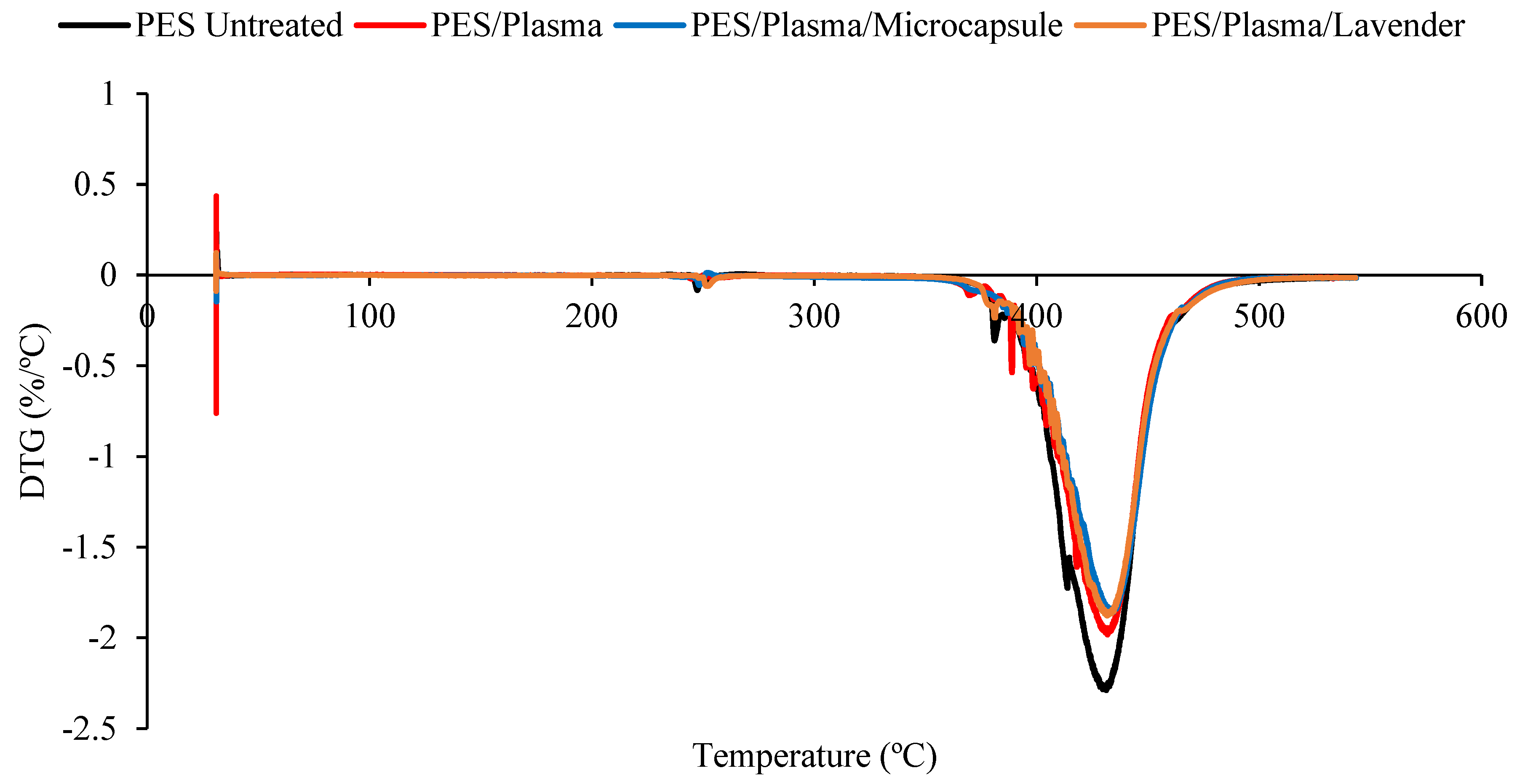
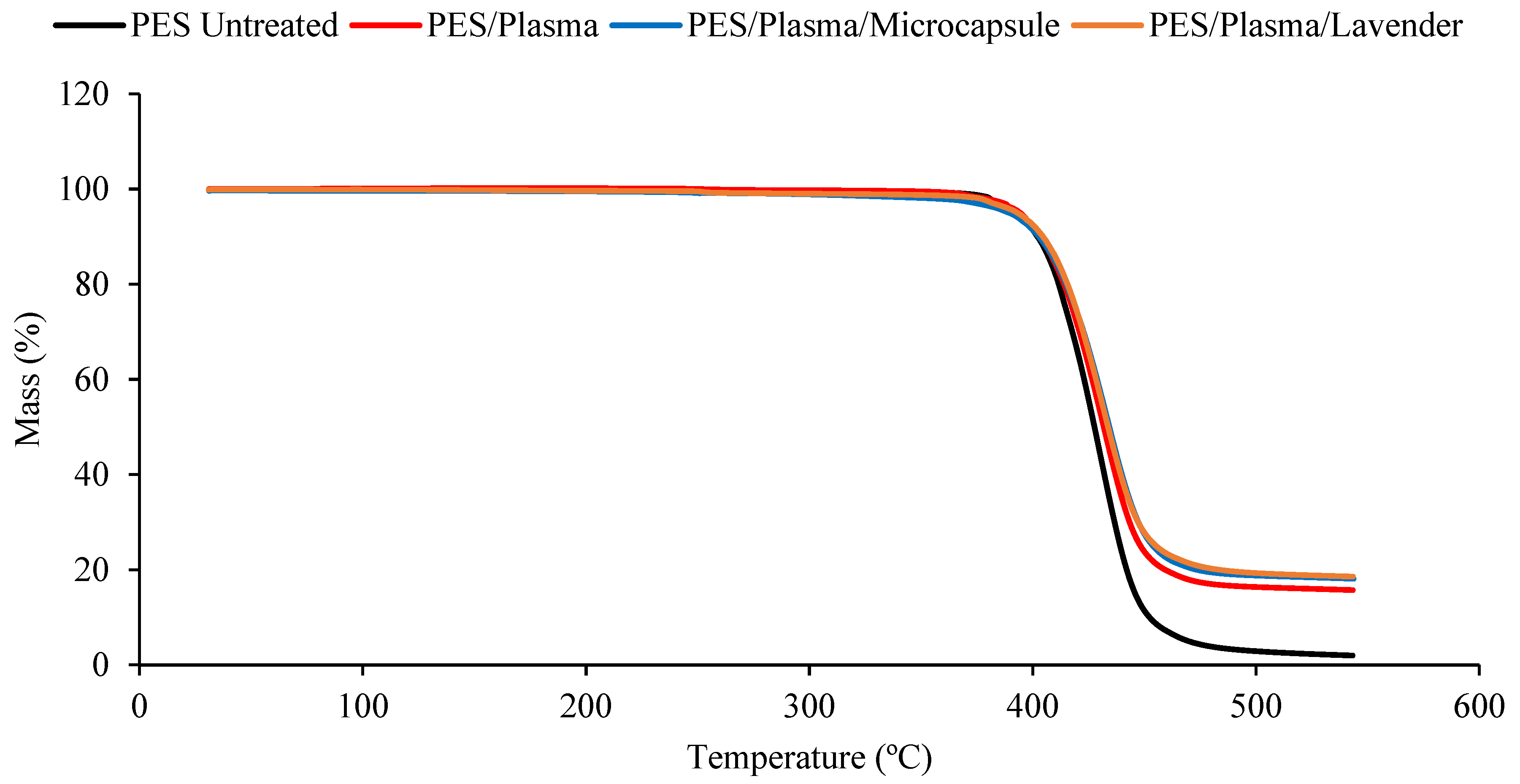
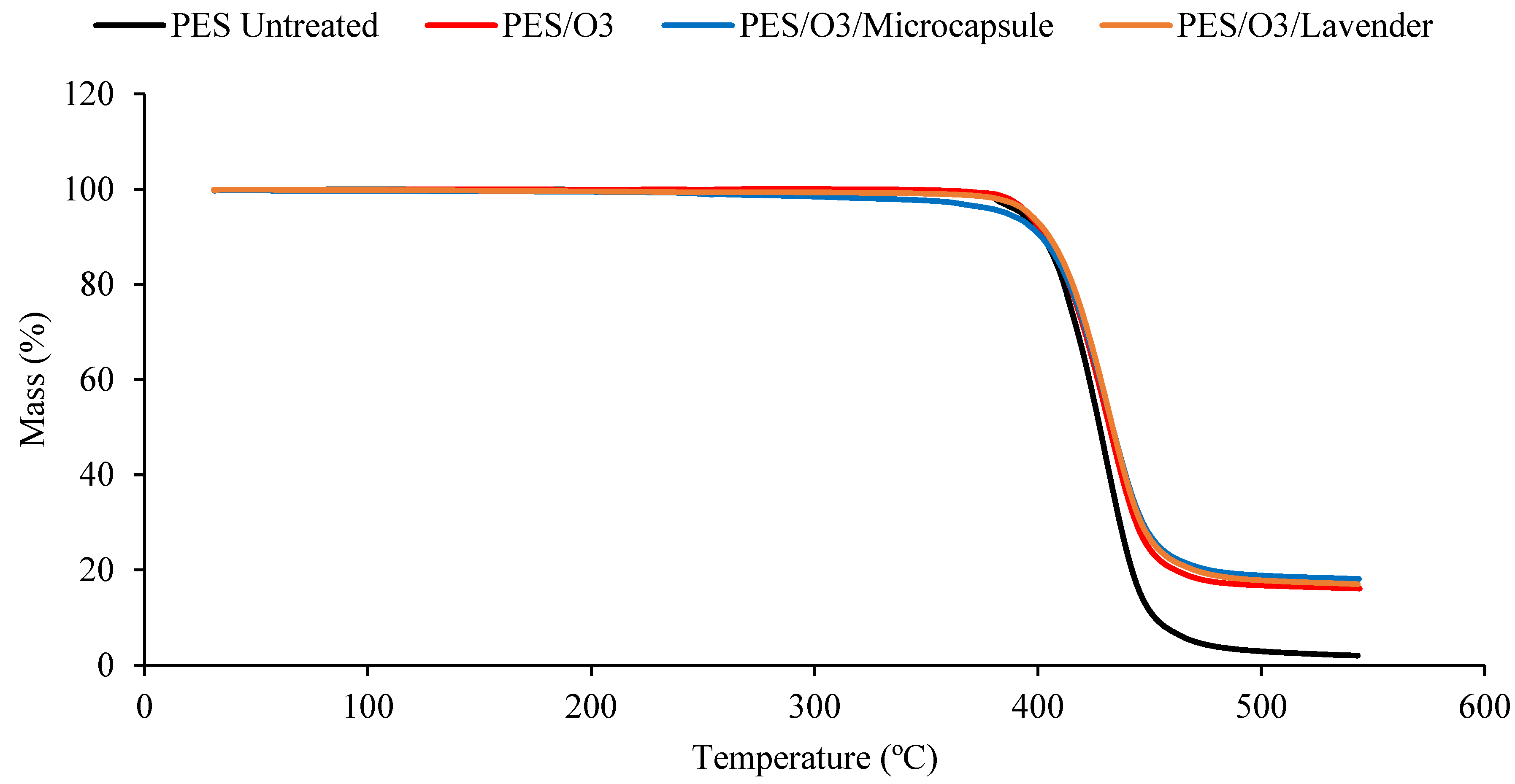
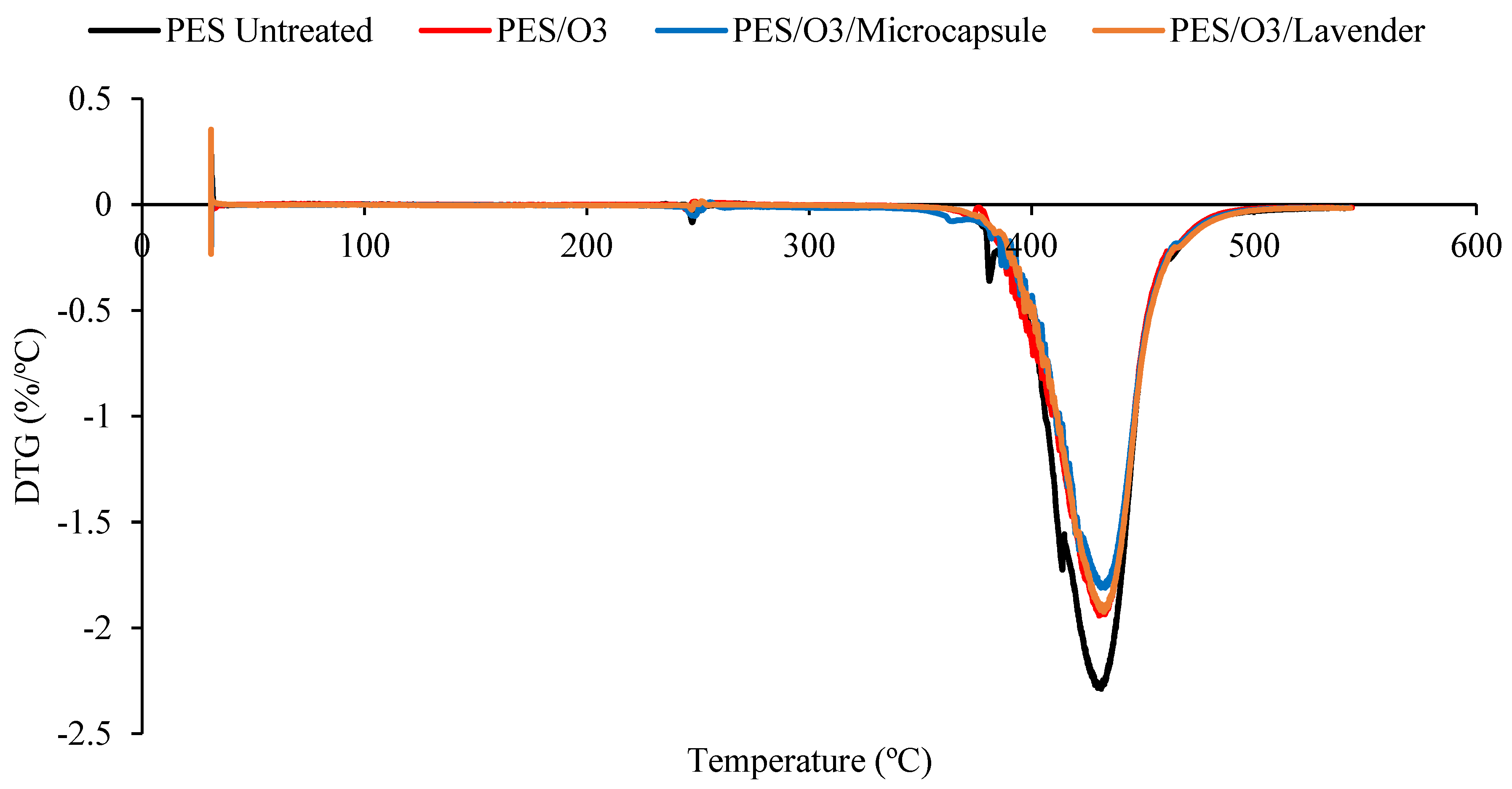
3.3. Thermal Analysis
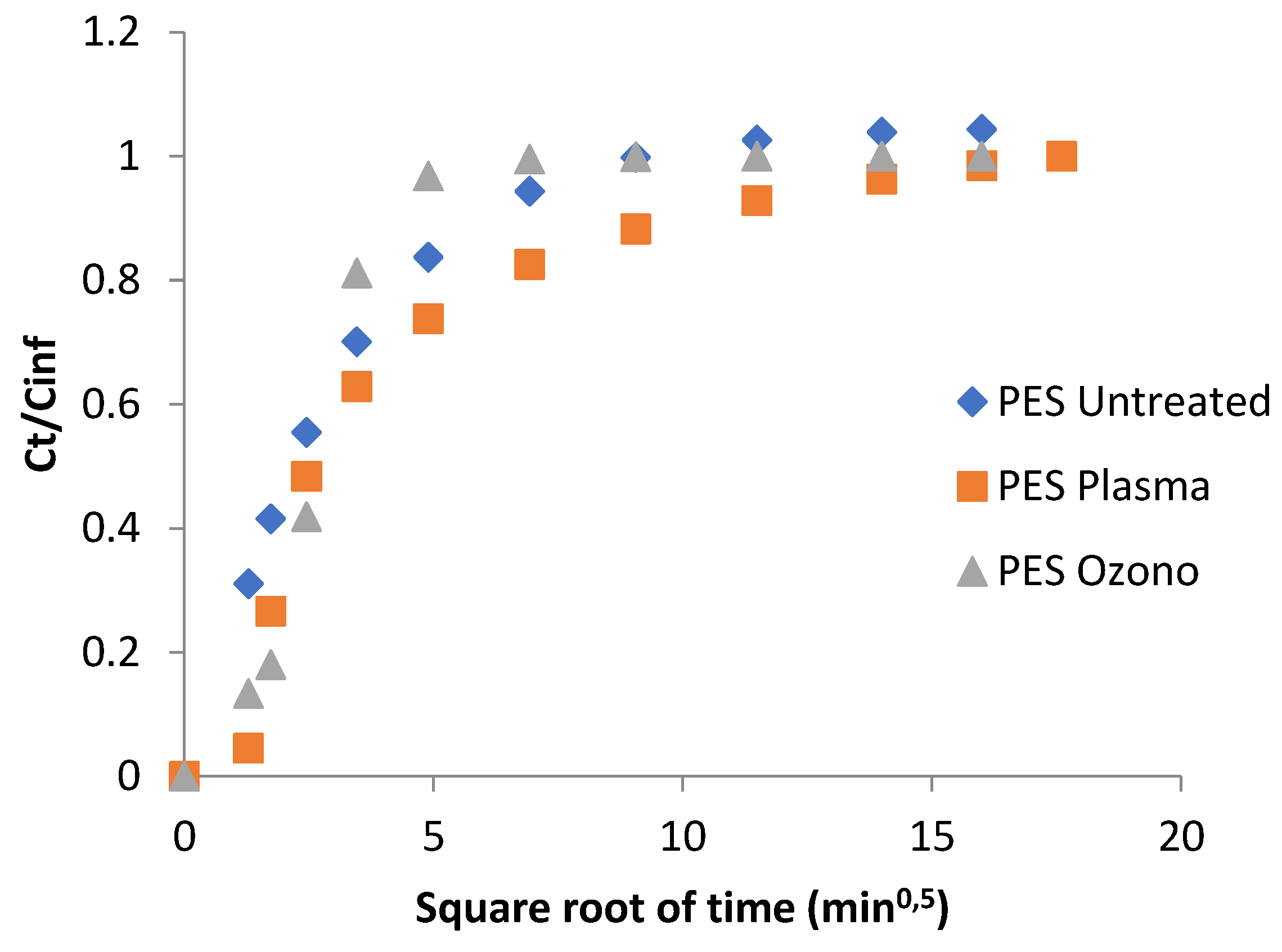
3.4. Lavender Release
4. Conclusions
Author Contributions
Funding
Institutional Review Board Statement
Data Availability Statement
Acknowledgments
Conflicts of Interest
References
- Systematic, B.B.; Podgornik, B.B.; Šandri, S. Microencapsulation for Functional Textile Coatings. Coatings 2021, 11, 1371. [Google Scholar]
- Bezerra, F.M.; Lis, M.; Carmona, Ó.G.; Carmona, C.G.; Moisés, M.P.; Zanin, G.M.; Moraes, F.F. Assessment of the delivery of citronella oil from microcapsules supported on wool fabrics. Powder Technol. 2019, 343, 775–782. [Google Scholar] [CrossRef]
- Singh, N.; Sheikh, J. Multifunctional Linen Fabric Obtained through Finishing with Chitosan-gelatin Microcapsules Loaded with Cinnamon Oil. J. Nat. Fibers. 2021, 19, 1–11. [Google Scholar] [CrossRef]
- Tian, Y.; Huang, X.; Cheng, Y.; Niu, Y.; Meng, Q.; Ma, J.; Zhao, Y.; Kou, X.; Ke, Q. Preparation of self-adhesive microcapsules and their application in functional textiles. J. Appl. Polym. Sci. 2022, 139, e52650. [Google Scholar] [CrossRef]
- Petkova-Slipets, R.; Zlateva, P.; Staneva, D. Influence of the polyester non-wovens production type on their thermal and flammability properties. J. Eng. Appl. Sci. 2022, 69, 51. [Google Scholar] [CrossRef]
- Nimbekar, A.A.; Deshmukh, R.R. Plasma Surface Modification of Flexible Substrates to Improve Grafting for Various Gas Sensing Applications: A Review. IEEE Trans. Plasma Sci. 2022, 50, 1382–1394. [Google Scholar] [CrossRef]
- Sharma, V.; Ali, S.W. A greener approach to impart multiple functionalities on polyester fabric using Schiff base of vanillin and benzyl amine. Sustain. Chem. Pharm. 2022, 27, 100645. [Google Scholar] [CrossRef]
- Luque-Agudo, V.; Hierro-Oliva, M.; Gallardo-Moreno, A.M.; González-Martín, M.L. Effect of plasma treatment on the surface properties of polylactic acid films. Polym. Test. 2021, 96, 107097. [Google Scholar] [CrossRef]
- Moraczewski, K.; Rytlewski, P.; Malinowski, R.; Zenkiewicz, M. Comparison of some effects of modification of a polylactide surface layer by chemical, plasma, and laser methods. Appl. Surf. Sci. 2015, 346, 11–17. [Google Scholar] [CrossRef]
- Deshmukh, R.R.; Bhat, N.V. Pretreatments of Textiles prior to Dyeing: Plasma Processing. In Textile Dying; Hauser, P.J., Ed.; InTech: London, UK, 2011; pp. 33–56. Available online: http://www.intechopen.com/books/textile-dyeing/pretreatments-of-textiles-prior-to-dyeing-plasma-processing (accessed on 14 September 2020).
- Gengenbach, T.R.; Griesser, H.J. Post-deposition ageing reactions differ markedly between plasma polymers deposited from siloxane and silazane monomers. Polymer 1999, 40, 5079–5094. [Google Scholar] [CrossRef]
- Su, L.; Li, J.; Li, J.; Chu, Q.; Li, B.; Liu, Y. Enhancement of the interlayer interaction between polystyrene and polyvinyl alcohol by ozone treatment. Adv. Polym. Technol. 2019, 2019, 7831619. [Google Scholar] [CrossRef]
- Macmanus, L.F.; Walzak, M.J.; Mcintyre, N.S. Study of ultraviolet light and ozone surface modification of polypropylene. J. Polym. Sci. Part A Polym. Chem. 1999, 37, 2489–2501. [Google Scholar] [CrossRef]
- Murakami, T.N.; Fukushima, Y.; Hirano, Y.; Tokuoka, Y.; Takahashi, M.; Kawashima, N. Surface modification of polystyrene and poly(methyl methacrylate) by active oxygen treatment. Colloids Surf. B Biointerfaces 2003, 29, 171–179. [Google Scholar] [CrossRef]
- Golja, B.; Tavčer, P.F. Patterned Printing of Fragrant Microcapsules to Cotton Fabric. Coatings 2022, 12, 593. [Google Scholar] [CrossRef]
- Yingngam, B.; Kacha, W.; Rungseevijitprapa, W.; Sudta, P.; Prasitpuriprecha, C.; Brantner, A. Response surface optimization of spray-dried citronella oil microcapsules with reduced volatility and irritation for cosmetic textile uses. Powder Technol. 2019, 355, 372–385. [Google Scholar] [CrossRef]
- Arias, M.J.L.; Coderch, L.; Martí, M.; Alonso, C.; Carmona, O.G.; Carmona, C.G.; Maesta, F. Vehiculation of active principles as away to create smart and biofunctional textiles. Materials 2018, 11, 2152. [Google Scholar] [CrossRef]
- Özsevinç, A.; Alkan, C. Ethylene glycol based polyurethane shell microcapsules for textile applications releasing medicinal lavender and responding to mechanical stimuli. Colloids Surf. A Physicochem. Eng. Asp. 2022, 652, 1–9. [Google Scholar] [CrossRef]
- Mendes, S.; Catarino, A.; Zille, A.; Fernandes, N.; Bezerra, F.M. Vehiculation of methyl salicylate from microcapsules supported on textile matrix. Materials 2021, 14, 1087. [Google Scholar] [CrossRef]
- Ogilvie-Battersby, J.D.; Nagarajan, R.; Mosurkal, R.; Orbey, N. Microencapsulation and controlled release of insect repellent geraniol in gelatin/gum arabic microcapsules. Colloids Surf. A Physicochem. Eng. Asp. 2022, 640, 128494. [Google Scholar] [CrossRef]
- Valle, J.A.B.; de Valle, R.C.S.C.; Bierhalz, A.C.K.; Bezerra, F.M.; Hernandez, A.K.; Arias, M.L.J. Chitosan microcapsules: Methods of the production and use in the textile finishing. J. Appl. Polym. Sci. 2021, 138, 50482. [Google Scholar] [CrossRef]
- Massella, D.; Giraud, S.; Guan, J.; Ferri, A.; Salaün, F. Textiles for health: A review of textile fabrics treated with chitosan microcapsules. Environ. Chem. Lett. 2019, 17, 1787–1800. [Google Scholar] [CrossRef]
- Tupuna, D.S.; Paese, K.; Guterres, S.S.; Jablonski, A.; Flôres, S.O.; de Rios, A.O. Encapsulation efficiency and thermal stability of norbixin microencapsulated by spray-drying using different combinations of wall materials. Ind. Crop. Prod. 2018, 111, 846–855. [Google Scholar] [CrossRef]
- Raut, J.S.; Karuppayil, S.M. A status review on the medicinal properties of essential oils. Ind. Crop. Prod. 2014, 62, 250–264. [Google Scholar] [CrossRef]
- Joshi, M.; Purwar, R.; Ali, S.W.; Rajendran, S. Antimicrobial Textiles for Health and Hygiene Applications Based on Eco-Friendly Natural Products; Woodhead Publishing Limited: Sawston, UK, 2010. [Google Scholar] [CrossRef]
- Mittal, R.P.; Rana, A.; Jaitak, V. Essential Oils: An Impending Substitute of Synthetic Antimicrobial Agents to Overcome Antimicrobial Resistance. Curr. Drug Targets 2018, 20, 605–624. [Google Scholar] [CrossRef]
- Ghayempour, S.; Montazer, M. Micro/nanoencapsulation of essential oils and fragrances: Focus on perfumed, antimicrobial, mosquito-repellent and medical textiles. J. Microencapsul. 2016, 33, 497–510. [Google Scholar] [CrossRef]
- Lesage-Meessen, L.; Bou, M.; Sigoillot, J.C.; Faulds, C.B.; Lomascolo, A. Essential oils and distilled straws of lavender and lavandin: A review of current use and potential application in white biotechnology. Appl. Microbiol. Biotechnol. 2015, 99, 3375–3385. [Google Scholar] [CrossRef]
- Sousa, V.I.; Parente, J.F.; Marques, J.F.; Forte, M.A.; Tavares, C.J. Microencapsulation of Essential Oils: A Review. Polymers 2022, 14, 1730. [Google Scholar] [CrossRef]
- Prata, A.S.; Grosso, C.R.F. Production of microparticles with gelatin and chitosan. Carbohydr. Polym. 2015, 116, 292–299. [Google Scholar] [CrossRef]
- Wang, S.; Zhang, W.; Chen, Y.; Zhang, S.; Wang, W. The Aromatic Properties of Polyurea-Encapsulated Lavender Oil Microcapsule and Their Application in Cotton Fabrics. J. Nanosci. Nanotechnol. 2019, 19, 4147–4153. [Google Scholar] [CrossRef]
- Kaur, R.; Kukkar, D.; Bhardwaj, S.K.; Kim, K.; Deep, A. Potential use of polymers and their complexes as media for storage and delivery of fragrances. J. Control. Release 2018, 285, 81–95. [Google Scholar] [CrossRef]
- Gabardo, R.S.; De Carvalho Cotre, D.S.; Aria, M.L.J.; Moisés, M.P.; Ferreira, B.M.T.; Samulewski, R.B.; Hinestroza, J.P.; Bezerra, F.M. Surface modification of polyester fabrics by ozone and its effect on coloration using disperse dyes. Materials 2021, 14, 3492. [Google Scholar] [CrossRef] [PubMed]
- Arias, M.J.L.; López, A.; Vilaseca, M.; Vallès, B.; Prieto, R.; Simó, M.; Valle, J.A.B.; Valle, R.D.C.S.C.; Bezerra, F.M.; Bellalta, J.B. Influence of Chitosan Characteristics in the Microencapsulation of Essential Oils. J. Biomed. Sci. Eng. 2021, 14, 119–129. [Google Scholar] [CrossRef]
- Ghaheh, F.S.; Khoddami, A.; Alihosseini, F.; Jing, S.; Ribeiro, A.; Cavaco-Paulo, A.; Silva, C. Antioxidant cosmetotextiles: Cotton coating with nanoparticles containing vitamin E. Process Biochem. 2017, 59, 46–51. [Google Scholar] [CrossRef]
- Carreras, N.; Acuña, V.; Martí, M.; Lis, M.J. Drug release system of ibuprofen in PCL-microspheres. Colloid Polym. Sci. 2013, 291, 157–165. [Google Scholar] [CrossRef]
- Ocak, B. Complex coacervation of collagen hydrolysate extracted from leather solid wastes and chitosan for controlled release of lavender oil. J. Environ. Manage. 2012, 100, 22–28. [Google Scholar] [CrossRef] [PubMed]
- Peran, J.; Ražić, S.E. Application of atmospheric pressure plasma technology for textile surface modification. Text. Res. J. 2020, 90, 1174–1197. [Google Scholar] [CrossRef]
- Ayesh, M.; Horrocks, A.R.; Kandola, B.K. The Impact of Atmospheric Plasma/UV Laser Treatment on the Chemical and Physical Properties of Cotton and Polyester Fabrics. Fibers 2022, 10, 66. [Google Scholar] [CrossRef]
- Lou, J.; Fan, X. Mechanism and application of ozone fading: Oxidative decolorisation of disperse dyes and waste-dyed polyester fabrics. Coloration Technology 2022, 1–12. [Google Scholar] [CrossRef]
- Fechine, G.J.M.; Rabello, M.S.; Souto-Maior, R.M. The effect of ultraviolet stabilizers on the photodegradation of poly(ethylene terephthalate). Polym. Degrad. Stab. 2002, 75, 153–159. [Google Scholar] [CrossRef]
- Iyer, N.; Iyer, P.B.; Patil, K.R.K. An infrared technique for the quick analysis of cotton–polyester blends. J. Appl. Polym. Sci. 1976, 20, 591–595. [Google Scholar] [CrossRef]
- Nowak, N.; Grzebieniarz, W.; Khachatryan, G.; Konieczna-Molenda, A.; Krzan, M.; Khachatryan, K. Preparation of nano/microcapsules of ozonated olive oil in chitosan matrix and analysis of physicochemical and microbiological properties of the obtained films. Innov. Food Sci. Emerg. Technol. 2022, 82, 103181. [Google Scholar] [CrossRef]
- Osman, Z.; Arof, A.K. FTIR studies of chitosan acetate based polymer electrolytes. Electrochim. Acta. 2003, 48, 993–999. [Google Scholar] [CrossRef]
- Ahmad, M.B.; Lim, J.J.; Shameli, K.; Ibrahim, N.A.; Tay, M.Y. Synthesis of silver nanoparticles in chitosan, gelatin and chitosan/gelatin bionanocomposites by a chemical reducing agent and their characterization. Molecules 2011, 16, 7237–7248. [Google Scholar] [CrossRef] [PubMed]
- Predoi, D.; Groza, A.; Iconaru, S.L.; Predoi, G.; Barbuceanu, F.; Guegan, R.; Motelica-Heino, M.S.; Cimpeanu, C. Properties of basil and lavender essential oils adsorbed on the surface of hydroxyapatite. Materials 2018, 11, 652. [Google Scholar] [CrossRef]
- Agatonovic-Kustrin, S.; Ristivojevic, P.; Gegechkori, V.; Litvinova, T.M.; Morton, D.W. Essential oil quality and purity evaluation via ft-ir spectroscopy and pattern recognition techniques. Appl. Sci. 2020, 10, 7294. [Google Scholar] [CrossRef]
- Coates, J. Interpretation of Infrared Spectra, A Practical Approach. Encycl. Anal. Chem. 2000, 10815–10837. [Google Scholar]
- Bezerra, F.M.; Carmona, O.G.; Carmona, C.G.; Lis, M.A.; de Moraes, F.F. Controlled release of microencapsulated citronella essential oil on cotton and polyester matrices. Cellulose 2016, 23, 1459–1470. [Google Scholar] [CrossRef]


| Sample | Tonset | Tmax | Toffset | Mass Loss (%) |
|---|---|---|---|---|
| Untreated Polyester (PES) | 351.22 | 430.74 | 466.39 | 98.01 |
| PES/Microcapsule | 349.18 | 429.95 | 467.09 | 82.17 |
| PES/LAV | 356.91 | 433.50 | 466.30 | 85.09 |
| PES/PLASMA | 367.13 | 431.24 | 466.09 | 84.26 |
| PES/PLASMA/Microcapsule | 367.13 | 433.30 | 461.35 | 81.87 |
| PES/PLASMA/LAVENDER | 364.76 | 433.30 | 466.52 | 81.43 |
| PES/O3 | 363.14 | 432.44 | 467.08 | 83.91 |
| PES/O3/Microcapsule | 363.14 | 436.24 | 467.92 | 81.91 |
| PES/O3/LAVENDER | 360.18 | 432.44 | 466.86 | 82.99 |
Disclaimer/Publisher’s Note: The statements, opinions and data contained in all publications are solely those of the individual author(s) and contributor(s) and not of MDPI and/or the editor(s). MDPI and/or the editor(s) disclaim responsibility for any injury to people or property resulting from any ideas, methods, instructions or products referred to in the content. |
© 2023 by the authors. Licensee MDPI, Basel, Switzerland. This article is an open access article distributed under the terms and conditions of the Creative Commons Attribution (CC BY) license (https://creativecommons.org/licenses/by/4.0/).
Share and Cite
Valle, R.d.C.S.C.; Valle, J.A.B.; Bezerra, F.M.; Correia, J.; da Costa, C.; Martí, M.; Coderch, L.; López, A.; Arias, M.J.L. Application of Lavender-Oil Microcapsules to Functionalized PET Fibers. Polymers 2023, 15, 917. https://doi.org/10.3390/polym15040917
Valle RdCSC, Valle JAB, Bezerra FM, Correia J, da Costa C, Martí M, Coderch L, López A, Arias MJL. Application of Lavender-Oil Microcapsules to Functionalized PET Fibers. Polymers. 2023; 15(4):917. https://doi.org/10.3390/polym15040917
Chicago/Turabian StyleValle, Rita de Cássia Siqueira Curto, José Alexandre Borges Valle, Fabricio Maestá Bezerra, Jeferson Correia, Cristiane da Costa, Meritxell Martí, Luisa Coderch, Arianne López, and Manuel J. Lis Arias. 2023. "Application of Lavender-Oil Microcapsules to Functionalized PET Fibers" Polymers 15, no. 4: 917. https://doi.org/10.3390/polym15040917
APA StyleValle, R. d. C. S. C., Valle, J. A. B., Bezerra, F. M., Correia, J., da Costa, C., Martí, M., Coderch, L., López, A., & Arias, M. J. L. (2023). Application of Lavender-Oil Microcapsules to Functionalized PET Fibers. Polymers, 15(4), 917. https://doi.org/10.3390/polym15040917