Article

Self-Assembly of a Two-Dimensional Coordination Polymer Based on Silver and Lanthanide Tetrakis-Acylpyrazolonates: An Efficient New Strategy for Suppressing Ligand-to-Metal Charge Transfer Quenching of Europium Luminescence

1
Chemistry Department, M.V. Lomonosov Moscow State University, Leninskie Gory Str, Building 1/3, 119991 Moscow, Russia
2
P. N. Lebedev Physical Institute of Russian Academy of Sciences, Leninsky Prospect 53, 119991 Moscow, Russia
3
Kurnakov Institute of General and Inorganic Chemistry of the Russian Academy of Sciences, Leninsky Prospekt 31, 119991 Moscow, Russia
4
School of Science and Tecnology, Chemistry Interdisciplinary Project (ChIP), University of Camerino, Via Madonna delle Carceri, 62032 Camerino, Italy
5
School of Pharmacy, Chemistry Interdisciplinary Project (ChIP), University of Camerino, Via Madonna delle Carceri, 62032 Camerino, Italy
*
Author to whom correspondence should be addressed.
Polymers 2023, 15(4), 867; https://doi.org/10.3390/polym15040867
Submission received: 18 January 2023 / Revised: 2 February 2023 / Accepted: 7 February 2023 / Published: 9 February 2023
(This article belongs to the Special Issue Organoelement Compounds and Polymers)

Abstract

:
A new strategy for the easy polymerization of anionic [Ln(Qcy)4] (HQcy-4-(cyclohexanecarbonyl)-5-methyl-2-phenyl-2,4-dihydro-3H-pyrazol-3-one) into two-dimensional layers of [AgLn(Qcy)4]n (Ln = Sm, Eu, Gd, Tb and Dy) is proposed by binding the single molecular anions [Ln(Qcy)4] to silver cations through the coordination of the pyridinic nitrogen atoms of the pyrazolonate rings. The luminescent properties of [AgLn(Qcy)4]n have been studied in detail, and it was shown that the previously described low photoluminescence quantum yield (PLQY) of [Eu(Qcy)4] is due to Ligand-To-Metal Charge Transfer (LMCT) quenching, which is effectively suppressed in the heterometallic [AgEu(Qcy)4]n polymer. Sensibilization coefficients for H3O[Eu(Qcy)4], [AgEu(Qcy)4]n, and H3O[Sm(Qcy)4] complexes (n ≈ 1) were estimated via theoretical analysis (also by using Judd-Ofelt theory for Sm3+) and PLQY measurements.

Graphical Abstract

1. Introduction

Coordination polymers (CPs) of various dimensions, including porous MOF materials, are attracting unrelenting interest from materials scientists and chemists due to the combination of unique functional properties (magnetic, catalytic, or luminescent) of individual molecular units with the stability of polymeric materials and the presence of pores and channels capable of reversible sorption of guest molecules [1,2]. Undoubtedly, the respectable place in the chemistry of coordination polymers is occupied by lanthanides derivatives [3,4,5]. The directed synthesis of CPs based on the Ln3+ ions is complicated by high and variable values of coordination numbers, as well as the absence of preferred polyhedra for 4f-elements [6,7]. At the same time, the luminescent properties of lanthanide complexes, due to the unique features of the electronic configuration, find application in the creation of various materials for light-emitting devices (LEDs) [8,9,10,11], biological luminescent labels [12,13,14], in thermometry [15,16,17] and in chemical sensors [18,19,20,21,22,23].
The most common approach to solve all these problems involves the use of antenna sensitization of lanthanide luminescence, i.e., inclusion in the complex of organic ligands capable of effectively absorbing exciting radiation and transmitting it to the emission center, the lanthanide cation [6,24,25,26]. Among the anionic ligands, the most effective luminescence sensitizers, β-diketones stand out, which, nevertheless, usually form mononuclear complexes [Ln(β-diketone)3(L)1–2] or molecular anions [Ln(β-diketone)4], which are soluble in typical organic solvents and have moderate chemical stability [27]. The monomeric nature of most studied Ln3+ diketonates can complicate functional applications in a number of problems, for example, in the creation of sensor materials [22,23]. To create oligomeric and polymeric lanthanide coordination compounds, it is necessary to use polydentate ligands, such as carbocyclic [28] and heterocyclic [5] aromatic carboxylic acids, which often have absorption maxima with λ < 300 nm and have low absorption in the near UV region (300–400 nm). Less than 10% of the approximately 4000 structures of lanthanide diketonates deposited in the Cambridge Crystal Data Centre (CCDC) have a polymeric structure. However, some methods are now known for linking diketonate complexes into coordination polymers. This goal can be achieved by introducing additional donor atoms into the ligand structure (as, for example, in pyrazole- [29], imidazole- [30], or pyridine-containing [31,32,33,34,35] β-diketones), or by using additional linker ligands, such as bis(diphenylphosphine oxides) [36,37,38,39,40,41,42], 4,4’-bipyridyl [43] or its N-oxide [44], quinone-based ligands [45], 2,2’-bipyrimidine [46] etc. Tetrakis anions [Ln(β-diketone)4] can be polymerized by forming alkali metal salts under non-aqueous conditions [47,48,49]. In most of the mentioned cases, the dimensionality of the polymer is limited by the coordination number of the lanthanide: if in a typical complex with CN = 8 three diketonate ligands occupy six positions, then the polymer is forced to be linear.
In the chemistry of lanthanides, among β-diketone-related ligands, a special role is played by acylpyrazolones [50,51,52] containing a heterocyclic pyrazolone fragment as a component of the β-diketonate system. This is due to the high energies of the triplet level in comparison with common aromatic β-diketones, such as dibenzolylmethane and thenoyltrifluoroacetone, which make it possible to effectively sensitize the luminescence of Tb3+ [53,54,55,56] and Dy3+ [57,58,59] ions. Acylpyrazolonates of these metals have been repeatedly tested as emitting layers in OLED devices [53,54,56,58] showing high efficiency values. In all cases, the coordination of lanthanides occurs at the acylpyrazolonate Q site with the formation of neutral tris complexes [LnQ3(L)1–2] [37,57,60,61,62,63,64,65,66] or tetrakis anions [LnQ4] [57,67,68,69], or dimers [LnQ3]2 [70,71,72].
Previously, coordination polymers based on rare-earth acylpyrazolonates were obtained only through the use of bridged ligands of the phosphine oxide series [36,37]. In the present paper, we report a fundamentally new approach to the creation of coordination polymers due to the binding of [Ln(Qcy)4] anions by Ag+ cations through nitrogen atoms of pyrazole rings with the formation of 2D-MOF [AgLn(Qcy)4]n, where Ln = Sm-Dy, and HQcy = 4-(cyclohexanecarbonyl)-5-methyl-2-phenyl-2,4-dihydro-3H-pyrazol-3-one. Such a reaction is possible due to the fact that silver cations, being soft Pearson acids, form complexes with diketonate ligands [73] only with difficulty, preferring coordination with softer donor atoms, for example, the nitrogen atom [73,74]. Moreover, the H3O[Ln(Qcy)4] (Ln = Sm, Tb, Dy) coordination compounds have pronounced ion-centered luminescence with the quantum yields (~2, 56 and 3%, respectively) close to the highest ones to date [75,76]. However, a curiously low value of overall PLQY was obtained for the H3O[Eu(Qcy)4] complex, below the measurement limit. From our investigation we could state that this uncommon result is due to the luminescence quenching via LMCT. Effective suppression of the LMCT processes was accomplished by the new method, provided in our research, of a change-over to polymer complexes with an Ag+ ion instead of H3O+ in the outer coordination sphere. Specifically, we were able to totally prevent the LMCT participation in the luminescent process in the [AgEu(Qcy)4]n complex with significantly high PLQY increase of up to two orders compared to the H3O[Eu(Qcy)4] one.

2. Materials and Methods

2.1. Synthesis and Spectroscopic Characterization of the Complexes

The hydroxonium complexes H3O[Ln(Qcy)4] were prepared according to the procedure previously reported [57] for a H3O[Dy(Qcy)4] complex using LnCl3 hexahydrates as starting reagents. The isostructurality of the complexes 1–5 was confirmed by powder diffraction (see SI, Figure S1).
H3O[Sm(Qcy)4], 1. Pale yellow plates, 78%. Anal. Calc. for C68H79N8O9Sm: C, 62.69; H, 6.11; N, 8.60; Sm, 11.54%. Found: C, 62.0; H, 6.7; N, 8.5; Sm, 11.9. IR (cm−1): 3392 m (ν O–HH3O+), 2929 m, 2853 m (ν C-HCyclohexyl), 1662 w (δ O–HH3O+), 1612 m (ν C=ODiketone), 1593 m, 1584 m, 1499 m (ν C–C), 1400 m, 1235 m, 1145 w, 1078 vs, 1027 s, 902 m, 812 m, 691 w.
H3O[Eu(Qcy)4], 2. Colorless plates, 73%. Anal. Calc. for C68H79N8O9Eu: C, 62.62; H, 6.10; N, 8.59; Eu, 11.65%. Found: C, 62.5; H, 6.7; N, 8.6; Eu, 12.0. IR (cm−1): 3392 m (ν O–HH3O+), 2929 m, 2852 m (ν C-HCyclohexyl), 1662 w (δ O–HH3O+), 1614 m (ν C=ODiketone), 1593 m, 1584 m, 1499 m (ν C–C), 1400 m, 1232 m, 1145 w, 1078 vs, 1028 s, 902 m, 812 m, 692 w.
H3O[Gd(Qcy)4], 3. Colorless plates, 74%. Anal. Calc. for C68H79N8O9Gd: C, 62.36; H, 6.08; N, 8.56; Gd, 12.01%. Found: C, 62.1; H, 6.4; N, 8.4; Gd, 12.2. IR (cm−1): 3391 m (ν O–HH3O+), 2930 m, 2852 m (ν C-HCyclohexyl), 1661 w (δ O–HH3O+), 1616 m (ν C=ODiketone), 1592 m, 1584 m, 1500 m (ν C–C), 1400 m, 1232 m, 1145 w, 1078 vs, 1028 s, 902 m, 812 m, 692 w.
H3O[Tb(Qcy)4], 4. Colorless plates, 73%. Anal. Calc. for: C68H79N8O9Tb: C, 62.28; H, 6.07; N, 8.55; Tb, 12.12%. Found: C, 62.5; H, 6.2; N, 8.6; Tb, 11.7. IR (cm−1): 3391 m (ν O–HH3O+), 2929 m, 2852 m (ν C-HCyclohexyl), 1661 w (δ O–HH3O+), 1616 m (ν C=ODiketone), 1593 m, 1584 m, 1500 m (ν C–C), 1400 m, 1230 m, 1145 w, 1078 vs, 1028 s, 902 m, 812 m, 692 w.
H3O[Dy(Qcy)4], 5. Pale yellow plates, 70%. Anal. Calc. for C68H79N8O9Dy: C, 62.11; H, 6.06; N, 8.52; Dy, 12.36%. Found: C, 61.9; H, 6.1; N, 7.9%. IR (cm−1): 3390 m (ν O–HH3O+), 2929 m, 2852 m (ν C-HCyclohexyl), 1660 w (δ O–HH3O+), 1616 m (ν C=ODiketone), 1594 m, 1584 m, 1500 m (ν C–C), 1400 m, 1230 m, 1153 w, 1078 vs, 1028 s, 902 m, 812 m, 692w.
The silver complexes [AgLn(Qcy)4]n 6–10 were obtained according to the following general procedure given in detail for [AgSm(Qcy)4]n.
[AgSm(Qcy)4]n, 6. 0.069 mg (0.222 mmol) of a SmCl3.6H2O and 1.5 mL of H2O were placed in a centrifugation tube. Then, 0.100 mL of a NH3 solution ~12 M were added, the volume was adjusted to 2 mL with water and centrifuged (8000 rpm, 5 min). The precipitate was carefully washed with water (4 × 2 mL), 96% ethanol (2 × 2 mL) and added to a solution of 0.284 g (1.00 mmol) of HQcy in 20 mL of 96% ethanol at room temperature. The mixture was stirred without heating until the precipitation of the formed lanthanide hydroxide. Then, 0.224 mL of a 10% solution of triethylamine (0.222 mmol) in 96% ethanol was added dropwise and the solution was filtered through a 0.25 μm PTFE syringe membrane filter. Subsequent operations were performed in the dark.
A solution of 0.0377 g (0.222 mmol) silver nitrate in 5 mL of acetonitrile was added dropwise at a rate of 1 mL per minute. An abundant precipitate begins to form already when the first drops are added. The suspension was stirred at room temperature for another 15 min, after which it was filtered on a glass porous filter, washed with ethanol (2 × 5 mL), acetonitrile (2 × 5 mL) and diethyl ether (2 × 5 mL). The precipitate was dried in a darkened desiccator over P4O10 for 48 h.
[AgSm(Qcy)4]n, 6. Pale yellow powder, 82%. Anal. Calc. for C68H76N8O8AgSm: C, 58.69; H, 5.50; N, 8.05; Ag, 7.75; Sm, 10.80%. Found: C, 58.2; H, 5.6; N, 8.0; Ag, 7.5; Sm, 11.3%. IR (cm−1): 3058 m, 2929 m, 2852 m(ν C-HCyclohexyl), 1627 vs (ν C=ODiketone), 1600 m, 1500 s, 1434 w, 1395 w, 1370 w, 1080 m, 1035 vw, 1010 vw, 979 m, 812 vw, 758 w, 693 w, 622 w, 452 w.
[AgEu(Qcy)4]n, 7. White powder, 83%. Anal. Calc. for C68H76N8O8AgEu: C, 58.62; H, 5.50; N, 8.04; Ag, 7.74; Eu, 10.91%. Found: C, 58.9; H, 5.5; N, 8.2; Ag, 7.6; Eu, 10.8%. IR (cm−1): 3060 m, 2929 m, 2852 m(ν C-HCyclohexyl), 1628 vs (ν C=ODiketone), 1599 m, 1500 s, 1435 w, 1392 w, 1377 vw, 1080 m, 1010 w, 979 m, 756 w, 691 w, 623 w, 450 w.
[AgGd(Qcy)4]n, 8. White powder, 74%. Anal. Calc. for C68H76N8O8AgGd: C, 58.40; H, 5.48; N, 8.01; Ag, 7.71; Gd, 11.24%. Found: C, 58.2; H, 5.8; N, 8.0; Ag, 7.7; Gd, 11.4%. IR (cm−1): 3061 m, 2929 m, 2852 m (ν C-HCyclohexyl), 1628 vs(ν C=ODiketone), 1598 m, 1500 s, 1436 w, 1392 w, 1372 vw, 1080 m, 1010 w, 978 m, 756 w, 690 w, 623 w, 448 vw.
[AgTb(Qcy)4]n, 9. White powder, 82%. Anal. Calc. for C68H76N8O8AgTb: C, 58.33; H, 5.47; N, 8.00; Ag, 7.70; Tb, 11.35%. Found: C, 58.4; H, 5.6; N, 8.2; Ag, 7.7; Tb, 11.4%. IR (cm−1): 3070 m, 2928 m, 2852 m, (ν C-HCyclohexyl) 1631 vs(ν C=ODiketone), 1596 m, 1501 s, 1543, 1435 w, 1371 w, 1080 m, 979 m, 902 vw, 756 w, 691 w, 622 w, 449 w.
[AgDy(Qcy)4]n, 10. White powder, 77%. Anal. Calc. for C68H76N8O8AgDy: C, 58.18; H, 5.46; N, 7.98; Ag, 7.68; Dy, 11.58%. Found: C, 58.6; H, 5.2; N, 7.9; Ag, 7.8; Dy, 11.2%. IR (cm−1): 3071 m, 2929 m, 2852 m (ν C-HCyclohexyl), 1631 vs(ν C=ODiketone), 1595 m, 1502 s, 1542, 1435 w, 1371 w, 1081 m, 979 m, 692 w, 622 w, 449 w.
Single crystals of [AgGd(Qcy)4]n (8) were obtained by suspending 0.05 g of the powder in 10 mL of acetonitrile at room temperature in the dark. The precipitate was then separated by centrifugation (8000 rpm), and the solution was filtered through a 0.25 μm PTFE syringe membrane filter. Slow evaporation of the solution at room temperature allowed the obtaining of small single crystals of 8 suitable for investigations on the synchrotron equipment. The isostructurality of other compounds 6, 7, 9 and 10 was confirmed by powder diffraction methods (see SI, Figure S2).

2.2. Apparatus

Elemental analyses were performed with an Elemental Vario MicroCube CHNO(S) (Elementar Americas Inc., Ronkonkoma, NY, USA) analyser. The Ln3+ content was determined by complexometric titration with a Trilon B solution in the presence of Xylenol Orange as an indicator [77]. The silver content was determined by titration with standard KSCN solution using Fe3+ to indicate the end point [78]. Before the analysis, the complexes were decomposed by heating with concentrated HNO3.
Absorption spectra for all the complexes were obtained on a JASCO V-770 (Jasco, Tokyo, Japan) spectrophotometer operating within 200–3200 nm. Concentrations of the solutions were approximately 10−5 M. For solutions, the measurements were performed using quartz cells with a 1 cm pathlength.
IR spectra were registered in the range 4000–400 cm−1 in KBr pellets using a Perkin-Elmer system Spectrum One 100 FTIR spectrometer (PerkinElmer, Inc., Waltham, MA, USA).
The X-ray diffraction data sets for crystals of H3O[Ln(Qcy)4] (1: Ln = Sm; 2: Ln = Eu; 3: Ln = Gd; 4: Ln = Tb) were collected on a Bruker SMART APEX II (Bruker, Billerica, MA, USA) diffractometer equipped with a CCD detector (Mo-Kα, λ = 0.71073 Å, graphite monochromator) [79]. X-ray diffraction data for [AgGd(Qcy)4]n 8 were collected on the ‘Belok’ beamline of the Kurchatov Synchrotron Radiation Source (National Research Center ‘Kurchatov Institute’, Moscow, Russia) in φ-scan mode with 1° step (λ = 0.79312 Å) using a Rayonix SX165 (Rayonix, L.L.C., Evanston, IL, USA) CCD detector [80]. The structures were solved by direct methods and refined by the full-matrix least squares in the anisotropic approximation for non-hydrogen atoms. The calculations were carried out by the SHELX-2014/2018 program package [81] using Olex2 1.2 [82]. The hydrogen atoms of the ligands were positioned geometrically and refined using the riding model. Solvent molecules in [AgGd(Qcy)4]n 8 which could not be localized were removed by the SQUEEZE procedure [83]. The crystallographic parameters for investigated crystals and the structure refinement details are given in Table 1. Crystallographic data for structures reported in this paper have been deposited with the Cambridge Crystallographic Data Center (H3O[Eu(Qcy)4]—2233167, H3O[Gd(Qcy)4]—2233168, H3O[Tb(Qcy)4]—2233169, [AgGd(Qcy)4]n—2233170).
Photoluminescence and excitation spectra in the visible region for all the complexes were measured in solid state using a Horiba Jobin-Yvon Fluorolog QM-75-22-C spectrofluorimeter with an installed 75 W xenon arc lamp (PowerArc, HORIBA, Ltd., Kyoto, Japan). A Hamamatsu R13456 cooled photomultiplier tube sensitive in the UV-Vis-NIR region (200–950 nm) was used as the detector. For the NIR spectral region measurements the same setup was used, except for the detector, which was replaced by a Hamamatsu H10330 (Hamamatsu Photonics, Hamamatsu, Japan) cooled photomultiplier tube sensitive in the NIR region (950–1700 nm). Photoluminescent decays in the visible region were recorded for all the complexes in solid state on the same device; however, the excitation source was changed to a pulsed xenon lamp with a 50 μ s pulse duration and 100 Hz repetition rate.
Photoluminescence quantum yields were obtained for solid samples by the absolute method with the use of the same experimental setup, equipped by integration sphere G8 (GMP, Renens, Switzerland).
For all optical measurements, the corresponding instrument response functions were taken into account. The experiments were performed in air at atmospheric pressure. Degradation of the optical properties was not observed during the experiments. Commercially available reagents and solvents (Sigma-Aldrich, Darmstadt, Germany) were used as received. Ln3+ chlorides were obtained by dissolution of the corresponding oxides (99.999%, LANHIT, Moscow, Russia) in concentrated hydrochloric acid (reagent grade, XPC). Ligand HQcy was prepared according to the published method [37].

3. Results

3.1. Synthesis

The synthesis of complex tetrakis acids H3O[Ln(Qcy)4] was carried out according to the general procedure proposed by us for a dysprosium derivative [57]. Temperature control is important to avoid formation of tris complexes [Ln(Qcy)3(H2O)] [57,84]. Polymer complexes can be obtained by the reaction of silver (I) nitrate with a solution of H3O[Ln(Qcy)4] acids, but the yield of the reaction somewhat increases after its preliminary neutralization with the use of triethylamine. The complexes gradually darken in the light, so all operations must be performed in subdued light. The polymer structure makes [AgLn(Qcy)4]n complexes extremely poorly soluble in most common solvents. The use of DMF for recrystallization is impossible, since it is accompanied by the decomposition of the complex with the formation of a silver mirror on the walls of the vessel. DMSO also causes decomposition of the complexes with the formation of an unspecified black precipitate (possibly silver sulfide). Among other tested solvents (methanol, ethanol, acetone, chloroform, ethyl acetate), only acetonitrile can be a possible medium for recrystallization. Attempts to grow large crystals under solvothermal conditions were unsuccessful, and only small crystals of the gadolinium complex were studied at the synchrotron facility. However, IR spectroscopy, elemental analysis, and powder diffraction data confirm that all [AgLn(Qcy)4]n complexes are isomorphic to the gadolinium polymeric complex [AgGd(Qcy)4]n.

3.2. Single Crystal Structures

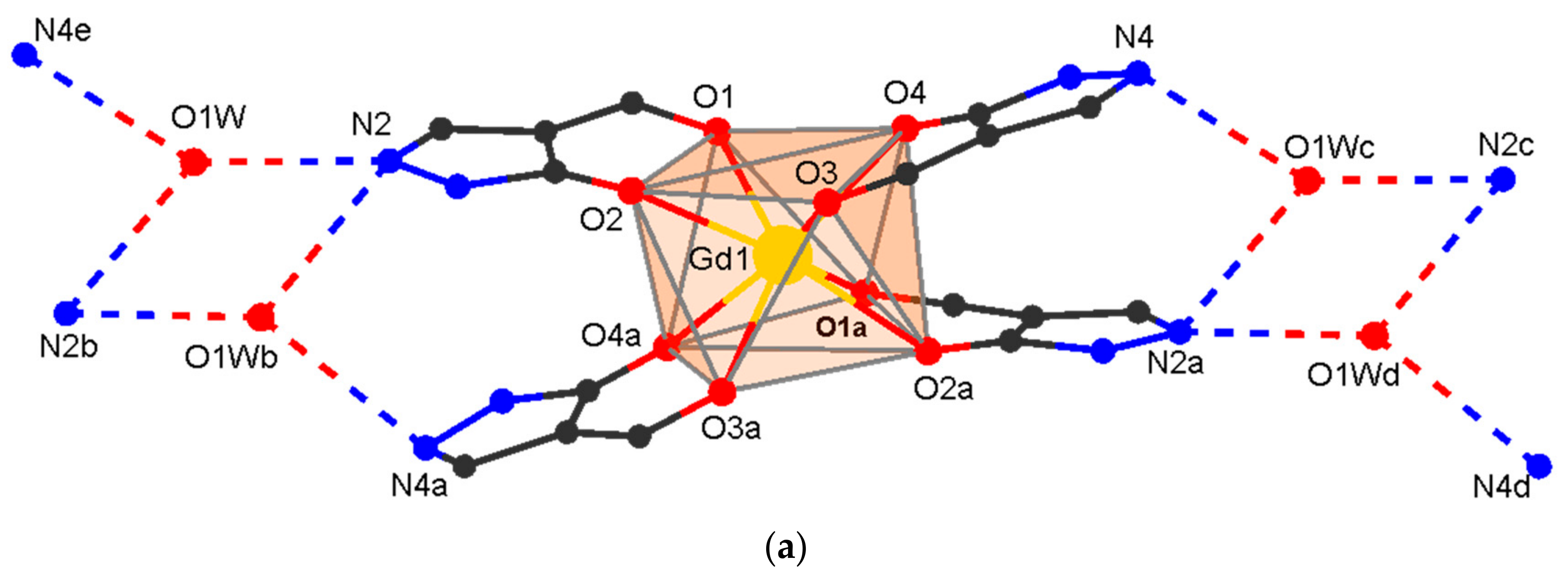
Complexes [H3O][Ln(Qcy)4] 1–4 (1: Ln = Sm; 2: Ln = Eu; 3: Ln = Gd; 4: Ln = Tb) crystallize in the monoclinic space group C2/c (for crystals with Ln = Sm, only cell parameters were determined; Table 1) and are isostructural to the previously described dysprosium complex [H3O][Dy(Qcy)4] 5 [57]. These compounds consist of the [Ln(Qcy)4] anion and of the outer sphere H3O+ (Figure 1a). The Ln atom is coordinated by four chelating Qcy ligands to form a LnO8 polyhedron in a square antiprismatic geometry (main distances and angles are given in Table 2). The oxygen atom of H3O+ is disordered in two positions (a two-fold axis passes between them) and form H-bonds with two N atoms of one complex molecule and one N atom of the neighboring molecule (Figure 2a; Table S1). Additionally, the molecule is stabilized by intramolecular C-H…O/N as well as C-H…π interactions (Tables S1 and S2). In the crystal, π-π intermolecular interactions occur between the pyrazole rings of neighboring molecules (Table S3). As in the other described H(H2O)n[LnQ4] tetrakis-acids, stabilization is achieved by strong intermolecular hydrogen bonds [57,67,68,69]. H-bonding between the anionic fragments [Ln(Qcy)4] leads to the formation of 1D supramolecular chains.
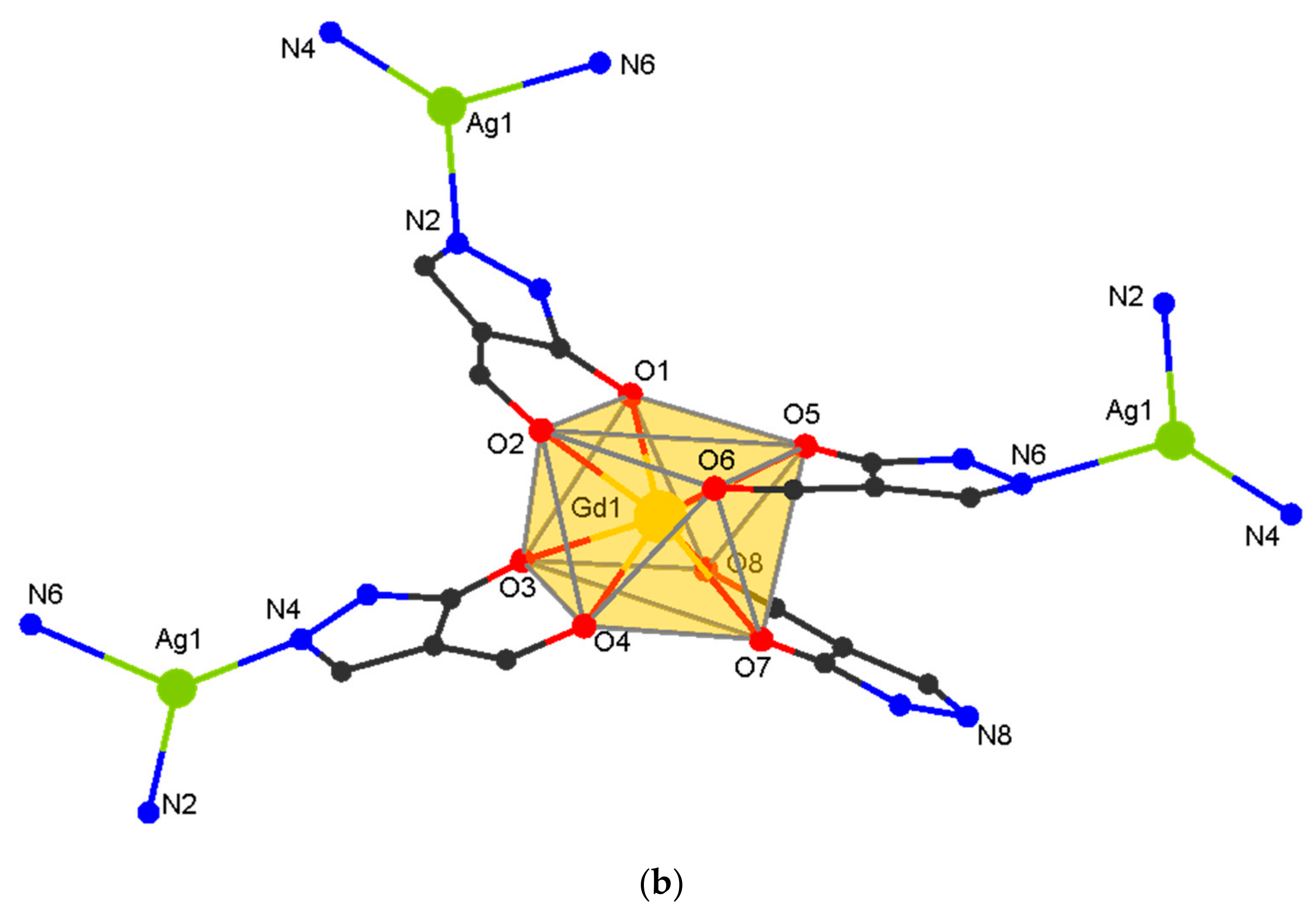
[AgGd(Qcy)4]n 8 crystallizes in the monoclinic space group P21/n. It contains the fragment [Gd(Qcy)4] and the Ag center. The structure of the [Gd(Qcy)4] fragment in 8 is similar to that in 3 (Figure 1b), the geometry of the metal atoms environment (GdO8) being slightly distorted (Figure 2, Table 2). The molecule is stabilized by intramolecular C-H…O/N interactions (Table S1). Each Ag atom is coordinated by three pyridinic N atoms of the pyrazole rings from three [Gd(Qcy)4] fragments. Each [Gd(Qcy)4] fragment is bonded to three Ag atoms; thus, three of the four pyrazole rings are involved in formation of the polymeric structure (Figure 2b). Binding of [Gd(Qcy)4] to Ag+ leads to the formation of a layered structure with a well-known topological type fes [85], where both structural building units are 3-coordinated nodes (Figure 3). In the crystal, π-π intermolecular interactions occur between the pyrazole and the phenyl rings of neighboring anionic framents (Table S3). The minimum interatomic distance between successive Gd atoms in the layer is 8.788 Å, and that of neighboring layers is 13.040 Å. The interlayer space is filled by cyclohexyl substituents.

3.3. Optical Properties

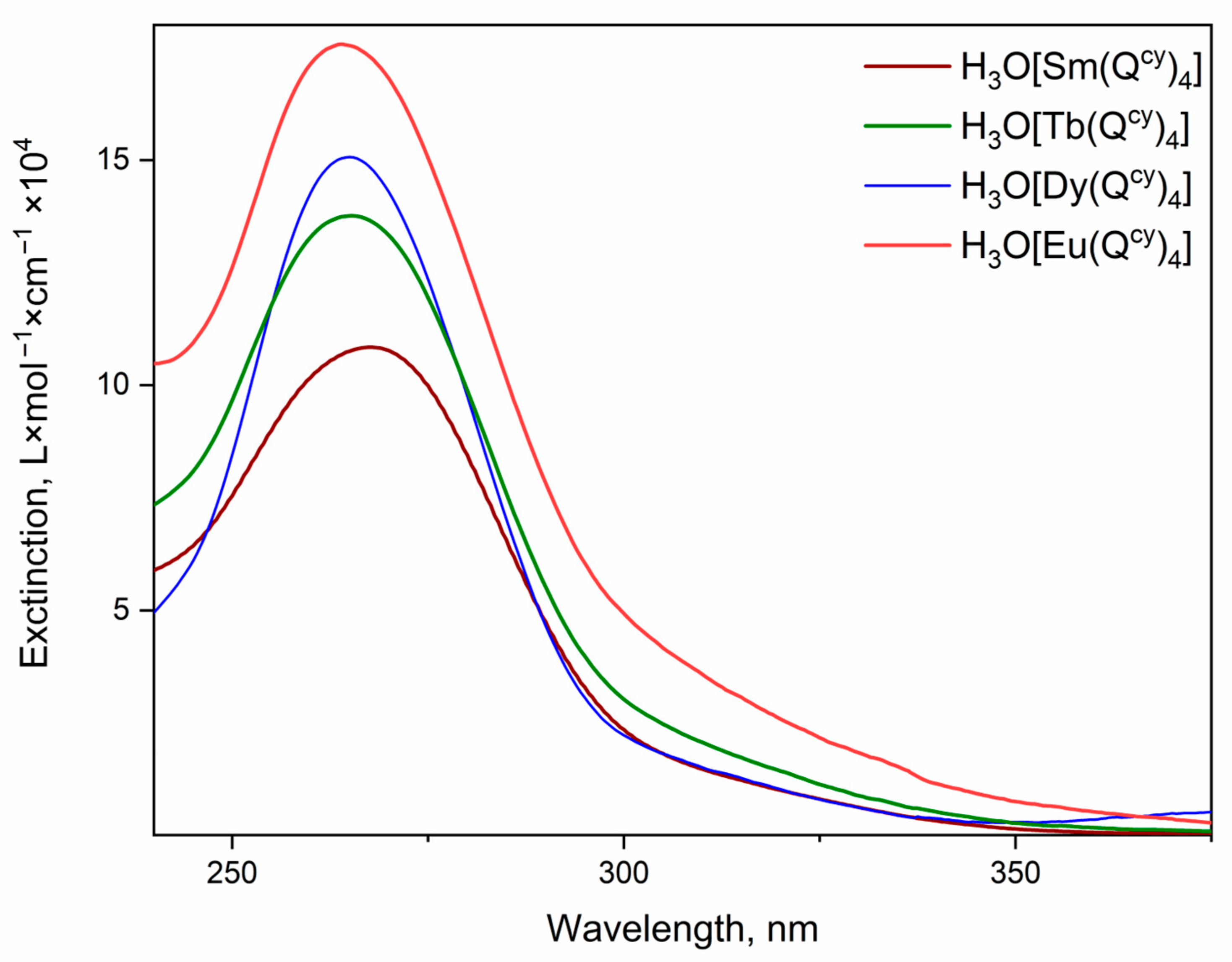
The absorption spectra of H3O[Ln(Qcy)4] 1–4 dissolved in MeCN are shown in Figure 4. All complexes 1–4 reveal intensive absorption in the range 250–375 nm. The observed absorption maximum at 265 nm is originated from the π-π* transition in the β-diketone moiety [57]. The spectra qualitatively resemble each other; therefore, despite the variation of the central ion, the ligand environment energy structure remains the same. Moreover, the coordination of the central ion by the ligands allows an increase in the molar extinction of the complexes up to 105 L × mol−1 × cm−1, with respect to the low values obtained for the free ions (~10 L × mol−1 × cm−1). Notably, we did not observe any absorption bands related to ion transitions due to a high ligand absorption rate.
The energy of the first excited singlet state (S1) was estimated using the tangent method [26] for the absorption spectra. For all the complexes, the S1 energies are nearly equal and are 27,300 cm−1. Due to the low solubility of [AgLn(Qcy)4]n 6–10 in most of the solvents generally employed (acetonitrile, methanol, dichloromethane), it is impossible to record their absorption spectra. For this reason, diffuse reflectance spectra were studied (see below).
PL excitation spectra, recorded for all the complexes in the solid state, are shown in Figure 5. Apart from H3O[Eu(Qcy)4], all the other complexes reveal the strong ion-centered luminescence under the optical excitation in a wide spectral range from 285 to 425 nm. The wide excitation band with the maximum at 340 nm is specific of the sensitization of the luminescence through the π*-π transition in the β-diketone moiety. However, the spectrum of the H3O[Eu(Qcy)4] shows a maximum at 380 nm with the FWHM (full width at half maximum) of 75 nm. We suggest that this band is related to the excitation due to ligand-to-metal charge-transfer (LMCT) [86]. Actually, the long wavelength shoulder observed for 2 in the diffuse reflection spectrum (see Figure 6) can result from different charge transfer processes. No LMCT with such energies has been observed for 4 due to the high redox potential of terbium ion. As the spectrum of 4 reveals no absorption within 360–450 nm, the shoulder in 2 can be associated to LMCT.
Interestingly, the spectra of complexes containing Ag+ are qualitatively similar, while the diffuse reflection spectra (see Figure 6) of [AgSm(Qcy)4]n 6 and [AgEu(Qcy)4]n 7 also reveal charge transfer processes.
The weak narrow bands observed in the excitation spectra of 1 and 2 and of 6 and 7 are related to the H 5 / 2 4 P 5 / 2 6 and H 5 / 2 4 P 3 / 2 6 transitions of the Sm3+ ion and the F 0 7 D 2 5 transition of the Eu3+ ion. Such behavior implicitly proves effective energy transfer of electronic excitation from donor-ligand to acceptors-ions.
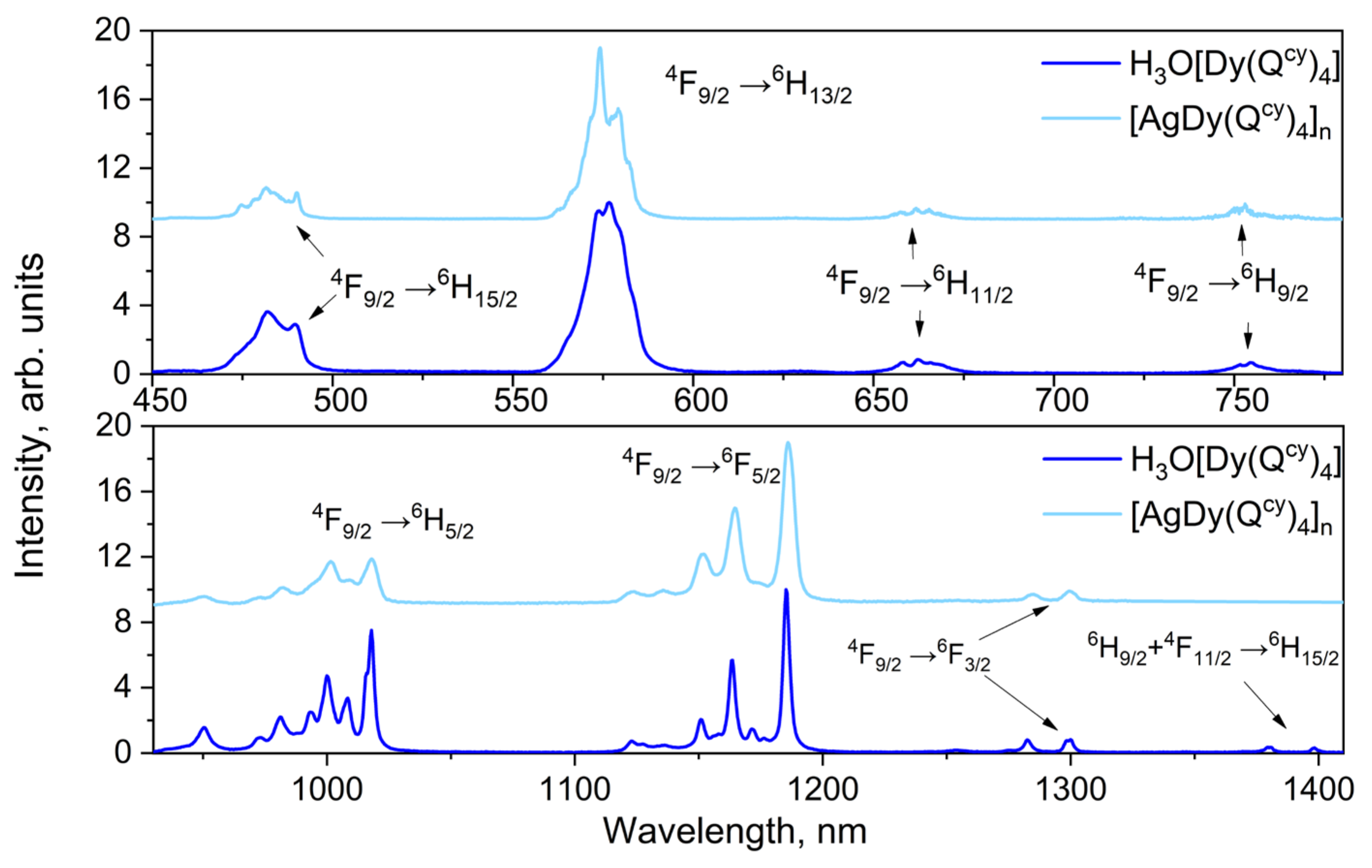
Intensive luminescence of all the investigated complexes is observed under optical excitation via absorption bands associated with ligand environment (see Figure 7, Figure 8 and Figure 9). In all the ionic species, we observed the narrow spectral bands typical of f*-f transitions (see Table S5). The correlation of the emission bands with the f*-f transitions was performed according to [57,87,88]. Notably, we observed no ligand emission in the emission spectra, which also indicates the relatively effective energy transfer of electronic excitation from ligand to ions [26].
The luminescence spectra of the complexes containing H3O+ and Ag+ have no significant differences pairwise in Stark splitting and emission band positions. Thereby, the replacement of H3O+ with the Ag+ produces no change in the symmetry of coordination polyhedral [26,89,90].
PL decays were recorded under the excitation wavelengths corresponding to maxima of excitation spectra with the registration at the photoluminescence maxima (see Figures S3 and S4). However, all the decay curves were estimated by a biexponential function. The obtained characteristic lifetimes are presented in Table 3. The replacement of H3O+ with Ag+ in the ligand environment leads to an increase of observed lifetimes for all the complexes except for Tb3+ ion complexes. Photoluminescence overall quantum yield (PLQY, Ф) values recorded under UV excitation are presented in Table 3.
To determine the triplet level energy of the ligand in lanthanide complexes, measurements of the low-temperature phosphorescence spectra of Gd3+ derivatives are usually used [26,91]. The combination of the high energy of the natural resonance level (>30,000 cm−1) of Gd3+ ions, the paramagnetic nature and the heavy atom effect contribute to the fact that the low-temperature emission spectra of gadolinium complexes mainly contain a phosphorescence transition whose energy corresponds to the triplet level value [92].
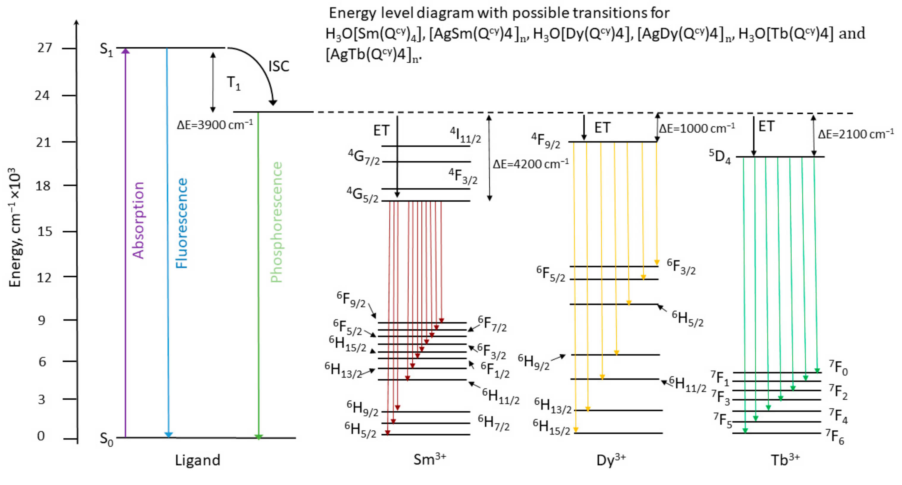
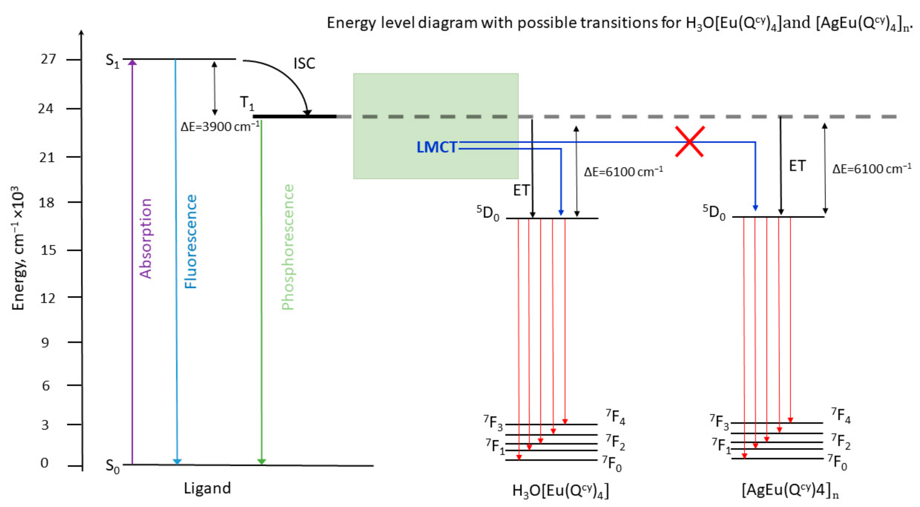
The phosphorescence spectra measured at 77 K for the solid state Gd3+ complexes are shown in Figure S5. The energy of the first excited triplet state was derived from the maximum of the fitting component corresponding to the zero-phonon line in the spectrum, according to the well-known procedure [93]. Thus, based on the spectral and kinetic measurements, we were able to construct the Jablonsky–Crossby energy diagrams for complexes reported here (Figure 10 and Figure 11). The luminescence characteristics of the europium complexes are caused by the influence of the LMCT state, which quenches the luminescence in the H3O[Eu(Qcy)4] complex and is associated with the ease ( E E u 3 + / E u 2 + 0 = 0.35   V ) [86] reduction of the europium ion. The high oxidation potential ( E A g + / A g 0 = 0.799   V ) of silver blocks this mechanism, which makes it possible to improve the luminescence characteristics of europium complexes. We believe that the low luminescence efficiency of other previously described europium acylpyrazolonates can also be explained by LMCT quenching [37,65].
The replacement of H3O+ with Ag+ in the ligand environment results in a significant decrease of PLQY, except for the Eu3+ complexes. The distance between the ions in the crystal package decreases practically two times after the replacement of H3O+ with Ag+, which results in cross-relaxation processes between the ions, which suppresses the radiative relaxation. Surprisingly, the Eu3+ complexes have low values of quantum yield; in particular, the Ф of [AgEu(Qcy)4]n is 0.3% and H3O[Eu(Qcy)4] PLQY is lower than 0.005%.
All the complexes have rather low extinction coefficients for the absorption bands characteristic of the lanthanide ions (see Figure S6). However, we successfully performed the Judd-Ofelt analysis [94,95] for the H3O[Sm(Qcy)4] complex to estimate the τrad radiative lifetime value for the 4G5/2 emission state of the Sm3+ ion. The general procedure was the same as reported in [87]. It should be noted that for minimizing the root-mean-square deviation (RMS) in our calculations we include only transitions within the energy range 8000–22,000 cm−1; see Table 4. The oscillator strengths of the electric dipole transitions determined from the optical absorption spectrum and calculated ones, Ωt (t = 2,4,6) Judd–Ofelt intensity parameters and radiative rate of the emission state of Sm3+ ion in our complex H3O[Sm(Qcy)4] 1 are presented in Table 4. The refractive index value 1.47 was used in our calculations according to [96]. Theoretical branching ratios for luminescent transitions are in good agreement with the experimental data and the low RMS value allow us to consider our results to be accurate; see Table 5.
The evaluated τrad value of ca. 3.2 ms is of the same order of magnitude of those reported for other Sm3+ complexes with similar chemical environments [87,97]. Measured luminescence decay times, obtained for the resonant excitation of the Sm3+ ion in the complex at 405 nm, are slightly higher than those presented in Table 5, and could also be fitted by a biexponential function with τ1 = 49.1 ± 0.8 μ s and τ2 = 80.1 ± 1.4 μ s . The estimated internal quantum yield of the complex is near 2% (1.5–2.4%, with respect to the biexponential luminescence decay behavior and accuracy of the calculations); the estimated and measured values are very close, and the sensibilization coefficient is close to 1.
The complexes reported in this work brightly emit in red, yellow and green spectral regions. For each compound, color diagrams were made (see Figure 12). The color coordinates for all the complexes are presented in Table S6.
The internal quantum yield can be estimated by the following formula
P L Q Y i n = k r a d k r a d + k n r a d .
Since the rate constant of the magnetic-dipole transition 5D0 7F1 in Eu3+, k M D = 14.65 s−1, does not depend on the electric field induced by the ligand, the value of k r a d can be determined by the relationship:
k r a d = k M D n 3 I t o t I M D ,
where n is the refractive index and I t o t I M D is the ratio between the total integral luminescence intensity and the integral intensity of the magnetic-dipole transition. The k n r a d constant was estimated using a simple dependence:
k n r a d = 1 τ o b s k r a d  
comprising the calculated k r a d and the observed attenuation time τ o b s measured with resonant excitation of Eu3+ at a wavelength of 464 nm. The calculated internal quantum yields P L Q Y i n , the total quantum yields PLQY measured by the absolute method, and the sensitization coefficients η are presented in Table 6.

4. Conclusions

The synthesis of coordination polymers based on lanthanides acylpyrazolonates due to the formation of a heterometallic 4d-4f complexes is shown for the first time. The resulting coordination polymers exhibit bright luminescence caused by f-f transitions of the central ions. The change from a monomeric to a polymeric structure leads to various changes in the luminescence characteristics: while for samarium, terbium, and dysprosium derivatives the luminescence efficiency somewhat decreases, in the case of europium, on the contrary, it increases significantly. This phenomenon is due to the special role of LMCT states in europium acylpyrazolonates.

Supplementary Materials

The following supporting information can be downloaded at: https://www.mdpi.com/article/10.3390/polym15040867/s1, Figure S1: PXRD patterns of 1–5 and simulated from single crystal data of 2, 3 and 4.; Figure S2: PXRD patterns of 6–10 and simulated from single crystal data of 8; Figure S3: PL decays for the complexes containing H3O+; Figure S4: PL decays for the complexes containing Ag+; Figure S5: Phosphorescence spectrum of Gd3+ complexes at 77K; Figure S6: Absorption spectra for DMSO solution of complex H3O[Sm(Qcy)4] (1) with concentration 3·10−3 M; Table S1. D-H…A (D = O, C, A = O, F) interactions in crystals H3O[Ln(Qcy)4] (Ln = Eu, Gd, Tb) and [AgGd(Qcy)4]n; Table S2. C-H…π interactions in the crystal [AgGd(Qcy)4]n; Table S3. Selected parameters of π-π intermolecular interactions in H3O[Ln(Qcy)4] (Ln = Nd, Sm, Eu, Gd, Tb) and [AgGd(Qcy)4]n; Table S4. Continuous Shape Measures (CShM) values [98] for the potential coordination polyhedron of Ln in H3O[Ln(Qcy)4] (Ln = Eu, Gd, Tb) and [AgGd(Qcy)4]n; Table S5. Electronic transitions for all the complexes; Table S6. Color coordinates for all the complexes.

Author Contributions

Conceptualization, Y.A.B., M.T.M. and C.P.; Data curation, M.T.M., D.A.M., M.A.K., I.A.Y. and T.A.P.; formal analysis, Y.A.B., M.T.M., M.A.K., D.A.M., C.P., A.A.D., I.V.T. and F.M.; funding acquisition, I.V.T. and C.P.; resources, I.V.T., Y.A.B., C.P. and A.A.D.; investigation, Y.A.B., M.T.M., D.A.M., T.A.P., M.A.K., I.A.Y. and I.V.T.; data curation, M.T.M., D.A.M., T.A.P., M.A.K. and I.A.Y.; writing—original draft preparation, Y.A.B., M.T.M., D.A.M. and A.A.D.; writing—review and editing, I.V.T., C.P. and F.M.; visualization, M.T.M., D.A.M., T.A.P. and M.A.K.; supervision, Y.A.B., A.A.D. and C.P.; project administration, I.V.T., C.P., F.M. and Y.A.B. All authors have read and agreed to the published version of the manuscript.

Funding

The photophysic study was supported by the Russian Science Foundation (project No. 19-13-00272).

Institutional Review Board Statement

Not applicable.

Informed Consent Statement

Not applicable.

Data Availability Statement

Data available upon request.

Acknowledgments

This photophysic study was supported by the Russian Science Foundation (project No. 19-13-00272). X-ray diffraction analysis were performed using the equipment at the Center for Collective Use of the Kurnakov Institute RAS and Kurchatov Synchrotron Radiation Source. University of Camerino is gratefully acknowledged.

Conflicts of Interest

The authors declare no conflict of interest.

References

  1. Cui, Y.; Yue, Y.; Qian, G.; Chen, B. Luminescent Functional Metal–Organic Frameworks. Chem. Rev. 2012, 112, 1126–1162. [Google Scholar] [CrossRef] [PubMed]
  2. Lustig, W.P.; Li, J. Luminescent Metal–Organic Frameworks and Coordination Polymers as Alternative Phosphors for Energy Efficient Lighting Devices. Coord. Chem. Rev. 2018, 373, 116–147. [Google Scholar] [CrossRef]
  3. Hasegawa, Y.; Nakanishi, T. Luminescent Lanthanide Coordination Polymers for Photonic Applications. RSC Adv. 2015, 5, 338–353. [Google Scholar] [CrossRef]
  4. Li, B.; Wen, H.-M.; Cui, Y.; Qian, G.; Chen, B. Multifunctional Lanthanide Coordination Polymers. Prog. Polym. Sci. 2015, 48, 40–84. [Google Scholar] [CrossRef]
  5. Belousov, Y.A.; Drozdov, A.A.; Taydakov, I.V.; Marchetti, F.; Pettinari, R.; Pettinari, C. Lanthanide Azolecarboxylate Compounds: Structure, Luminescent Properties and Applications. Coord. Chem. Rev. 2021, 445, 214084. [Google Scholar] [CrossRef]
  6. Bünzli, J.C.G.; Piguet, C. Lanthanide-Containing Molecular and Supramolecular Polymetallic Functional Assemblies. Chem. Rev. 2002, 102, 1897–1928. [Google Scholar] [CrossRef]
  7. Cotton, S. Lanthanide and Actinide Chemistry; John Wiley & Sons, Ltd: Chichester, UK, 2006; ISBN 9780470010082. [Google Scholar]
  8. Xu, H.; Sun, Q.; An, Z.; Wei, Y.; Liu, X. Electroluminescence from Europium(III) Complexes. Coord. Chem. Rev. 2015, 293–294, 228–249. [Google Scholar] [CrossRef]
  9. Álvarez, Á.L.; Coya, C. OLEDs Based on Ln(III) Complexes for near-Infrared Emission. In Lanthanide-Based Multifunctional Materials; Elsevier: Amsterdam, The Netherlands, 2018; pp. 133–170. ISBN 9780128138403. [Google Scholar]
  10. Wang, L.; Zhao, Z.; Wei, C.; Wei, H.; Liu, Z.; Bian, Z.; Huang, C. Review on the Electroluminescence Study of Lanthanide Complexes. Adv. Opt. Mater. 2019, 7, 1801256. [Google Scholar] [CrossRef]
  11. Zinna, F.; Pasini, M.; Galeotti, F.; Botta, C.; Di Bari, L.; Giovanella, U. Design of Lanthanide-Based OLEDs with Remarkable Circularly Polarized Electroluminescence. Adv. Funct. Mater. 2017, 27, 1603719. [Google Scholar] [CrossRef]
  12. Dreyer, D.R.; Park, S.; Bielawski, C.W.; Ruoff, R.S. Lanthanide Luminescence for Functional Materials and Bio-Sciences. Chem. Soc. Rev. 2010, 1, 189–227. [Google Scholar] [CrossRef]
  13. Heffern, M.C.; Matosziuk, L.M.; Meade, T.J. Lanthanide Probes for Bioresponsive Imaging. Chem. Rev. 2014, 114, 4496–4539. [Google Scholar] [CrossRef]
  14. Yang, Y.; Zhao, Q.; Feng, W.; Li, F. Luminescent Chemodosimeters for Bioimaging. Chem. Rev. 2013, 113, 192–270. [Google Scholar] [CrossRef]
  15. Grebenyuk, D.; Zobel, M.; Tsymbarenko, D. Partially Ordered Lanthanide Carboxylates with a Highly Adaptable 1D Polymeric Structure. Polymers 2022, 14, 3328. [Google Scholar] [CrossRef]
  16. Brites, C.D.S.; Millán, A.; Carlos, L.D. Lanthanides in Luminescent Thermometry. In Handbook on the Physics and Chemistry of Rare Earths; Elsevier B.V.: Amsterdam, The Netherlands, 2016; Volume 49, pp. 339–427. [Google Scholar]
  17. Rocha, J.; Brites, C.D.S.; Carlos, L.D. Lanthanide Organic Framework Luminescent Thermometers. Chem. A Eur. J. 2016, 22, 14782–14795. [Google Scholar] [CrossRef]
  18. Lee, S.; Lin, M.; Lee, A.; Park, Y. Lanthanide-Doped Nanoparticles for Diagnostic Sensing. Nanomaterials 2017, 7, 411. [Google Scholar] [CrossRef]
  19. Aulsebrook, M.L.; Graham, B.; Grace, M.R.; Tuck, K.L. Lanthanide Complexes for Luminescence-Based Sensing of Low Molecular Weight Analytes. Coord. Chem. Rev. 2018, 375, 191–220. [Google Scholar] [CrossRef]
  20. Aletti, A.B.; Gillen, D.M.; Gunnlaugsson, T. Luminescent/Colorimetric Probes and (Chemo-) Sensors for Detecting Anions Based on Transition and Lanthanide Ion Receptor/Binding Complexes. Coord. Chem. Rev. 2018, 354, 98–120. [Google Scholar] [CrossRef]
  21. Xu, H.; Cao, C.S.; Kang, X.M.; Zhao, B. Lanthanide-Based Metal-Organic Frameworks as Luminescent Probes. Dalt. Trans. 2016, 45, 18003–18017. [Google Scholar] [CrossRef]
  22. Zhao, S.-N.; Wang, G.; Poelman, D.; Voort, P. Luminescent Lanthanide MOFs: A Unique Platform for Chemical Sensing. Materials 2018, 11, 572. [Google Scholar] [CrossRef]
  23. Lunev, A.M.; Belousov, Y.A. Luminescent Sensor Materials Based on Rare-Earth Element Complexes for Detecting Cations, Anions, and Small Molecules. Russ. Chem. Bull. 2022, 71, 825–857. [Google Scholar] [CrossRef]
  24. Crosby, G.A.; Whan, R.E.; Freeman, J.J. Spectroscopic Studies of Rare Earth Chelates. J. Phys. Chem. 1962, 66, 2493–2499. [Google Scholar] [CrossRef]
  25. Bünzli, J.-C.G.; Piguet, C. Taking Advantage of Luminescent Lanthanide Ions. Chem. Soc. Rev. 2005, 34, 1048. [Google Scholar] [CrossRef]
  26. Bünzli, J.; Eliseeva, S.V. Basics of Lanthanides Photophysics. In Lanthanide Luminescence; Hänninen, P., Härmä, H., Eds.; Springer Series on Fluorescence; Springer: Berlin/Heidelberg, Germany, 2010; Volume 7, pp. 1–45. [Google Scholar] [CrossRef]
  27. Nehra, K.; Dalal, A.; Hooda, A.; Bhagwan, S.; Saini, R.K.; Mari, B.; Kumar, S.; Singh, D. Lanthanides β-Diketonate Complexes as Energy-Efficient Emissive Materials: A Review. J. Mol. Struct. 2022, 1249, 131531. [Google Scholar] [CrossRef]
  28. Janicki, R.; Mondry, A.; Starynowicz, P. Carboxylates of Rare Earth Elements. Coord. Chem. Rev. 2017, 340, 98–133. [Google Scholar] [CrossRef]
  29. Taidakov, I.V.; Lobanov, A.N.; Vitukhnovskii, A.G.; Starikova, Z.A. Synthesis and Unusual Crystal Structure of the Eu(III) Complex with 1-(1,5-Dimethyl-1H-Pyrazol-4-Yl)-4,4,4-Trifluorobutane-1,3-Dione. Russ. J. Coord. Chem. 2013, 39, 437–441. [Google Scholar] [CrossRef]
  30. Yang, P. Synthesis, Structure, and Luminescence Properties of Rare Earth Complexes with β-Diketone Containing Imidazole Group. Z. Anorg. Allg. Chem. 2018, 644, 838–843. [Google Scholar] [CrossRef]
  31. Burrows, A.D.; Mahon, M.F.; Renouf, C.L.; Richardson, C.; Warren, A.J.; Warren, J.E. Dipyridyl β-Diketonate Complexes and Their Use as Metalloligands in the Formation of Mixed-Metal Coordination Networks. Dalt. Trans. 2012, 41, 4153. [Google Scholar] [CrossRef]
  32. Hui, Y.-C.; Meng, Y.-S.; Li, Z.; Chen, Q.; Sun, H.-L.; Zhang, Y.-Q.; Gao, S. Construction and Theoretical Study of a New Dy-β-Diketone Chain Featuring Slow Magnetic Relaxation. CrystEngComm 2015, 17, 5620–5624. [Google Scholar] [CrossRef]
  33. Liu, F.; Zhou, Y. Polymeric [Eu(L)3(H2O)] (HL = 1-(Pyridin-4-Yl)Butane- 1,3-Dione and Dimeric [Eu2(L)6(H2O)2] (HL = 1,3-Di(Pyridin-3-Yl)Propane-1,3-Dione) as Selective and Sensitive Chemosensors for Hg(II) Ion. Inorg. Chem. Commun. 2010, 13, 1410–1413. [Google Scholar] [CrossRef]
  34. Guettas, D.; Montigaud, V.; Garcia, G.F.; Larini, P.; Cador, O.; Le Guennic, B.; Pilet, G. Fine Control of the Metal Environment within Dysprosium-Based Mononuclear Single-Molecule Magnets. Eur. J. Inorg. Chem. 2018, 2018, 333–339. [Google Scholar] [CrossRef]
  35. Andrews, P.C.; Deacon, G.B.; Frank, R.; Fraser, B.H.; Junk, P.C.; MacLellan, J.G.; Massi, M.; Moubaraki, B.; Murray, K.S.; Silberstein, M. Formation of Ho III Trinuclear Clusters and Gd III Monodimensional Polymers Induced by Ortho and Para Regioisomers of Pyridyl-Functionalised Β-Diketones: Synthesis, Structure, and Magnetic Properties. Eur. J. Inorg. Chem. 2009, 2009, 744–751. [Google Scholar] [CrossRef]
  36. Semenov, S.N.; Rogachev, A.Y.; Eliseeva, S.V.; Pettinari, C.; Marchetti, F.; Drozdov, A.A.; Troyanov, S.I. First Direct Assembly of Molecular Helical Complexes into a Coordination Polymer. Chem. Commun. 2008, 17, 1992–1994. [Google Scholar] [CrossRef]
  37. Marchetti, F.; Pettinari, C.; Pizzabiocca, A.; Drozdov, A.A.; Troyanov, S.I.; Zhuravlev, C.O.; Semenov, S.N.; Belousov, Y.A.; Timokhin, I.G. Syntheses, Structures, and Spectroscopy of Mono- and Polynuclear Lanthanide Complexes Containing 4-Acyl-Pyrazolones and Diphosphineoxide. Inorg. Chim. Acta 2010, 363, 4038–4047. [Google Scholar] [CrossRef]
  38. Miyata, K.; Ohba, T.; Kobayashi, A.; Kato, M.; Nakanishi, T.; Fushimi, K.; Hasegawa, Y. Thermostable Organo-Phosphor: Low-Vibrational Coordination Polymers That Exhibit Different Intermolecular Interactions. Chempluschem 2012, 77, 277–280. [Google Scholar] [CrossRef]
  39. Hasegawa, Y.; Miura, Y.; Kitagawa, Y.; Wada, S.; Nakanishi, T.; Fushimi, K.; Seki, T.; Ito, H.; Iwasa, T.; Taketsugu, T.; et al. Spiral Eu(iii) Coordination Polymers with Circularly Polarized Luminescence. Chem. Commun. 2018, 54, 10695–10697. [Google Scholar] [CrossRef]
  40. Kitagawa, Y.; Naito, A.; Fushimi, K.; Hasegawa, Y. Bright Sky-Blue Fluorescence with High Color Purity: Assembly of Luminescent Diphenyl-Anthracene Lutetium-Based Coordination Polymer. RSC Adv. 2021, 11, 6604–6606. [Google Scholar] [CrossRef]
  41. Zhang, L.; Chen, P.; Li, H.-F.; Tian, Y.-M.; Yan, P.-F.; Sun, W.-B. From Zero-Dimensional to One-Dimensional Chain N -Oxide Bridged Compounds with Enhanced Single-Molecule Magnetic Performance. Dalt. Trans. 2019, 48, 4324–4332. [Google Scholar] [CrossRef]
  42. Hirai, Y.; Ferreira Da Rosa, P.P.; Nakanishi, T.; Kitagawa, Y.; Fushimi, K.; Seki, T.; Ito, H.; Hasegawa, Y. Structural Manipulation of Triboluminescent Lanthanide Coordination Polymers by Side-Group Alteration. Inorg. Chem. 2018, 57, 14653–14659. [Google Scholar] [CrossRef]
  43. Armelao, L.; Belli Dell’Amico, D.; Bellucci, L.; Bottaro, G.; Labella, L.; Marchetti, F.; Samaritani, S. A Convenient Synthesis of Highly Luminescent Lanthanide 1D-Zigzag Coordination Chains Based Only on 4,4′-Bipyridine as Connector. Polyhedron 2016, 119, 371–376. [Google Scholar] [CrossRef]
  44. Wang, J.; Yang, M.; Sun, J.; Li, H.; Liu, J.; Wang, Q.; Li, L.; Ma, Y.; Zhao, B.; Cheng, P. Enhancing the Energy Barrier of Dysprosium(iii) Single-Molecule Magnets by Tuning the Magnetic Interactions through Different N -Oxide Bridging Ligands. CrystEngComm 2019, 21, 6219–6225. [Google Scholar] [CrossRef]
  45. Flores Gonzalez, J.; Cador, O.; Ouahab, L.; Norkov, S.; Kuropatov, V.; Pointillart, F. Field-Induced Dysprosium Single-Molecule Magnet Involving a Fused o-Semiquinone-Extended-Tetrathiafulvalene-o-Semiquinone Bridging Triad. Inorganics 2018, 6, 45. [Google Scholar] [CrossRef]
  46. Fratini, A.; Richards, G.; Larder, E.; Swavey, S. Neodymium, Gadolinium, and Terbium Complexes Containing Hexafluoroacetylacetonate and 2,2‘-Bipyrimidine: Structural and Spectroscopic Characterization. Inorg. Chem. 2008, 47, 1030–1036. [Google Scholar] [CrossRef] [PubMed]
  47. Tan, R.H.C.; Motevalli, M.; Abrahams, I.; Wyatt, P.B.; Gillin, W.P. Quenching of IR Luminescence of Erbium, Neodymium, and Ytterbium β-Diketonate Complexes by Ligand C−H and C−D Bonds. J. Phys. Chem. B 2006, 110, 24476–24479. [Google Scholar] [CrossRef] [PubMed]
  48. Mara, D.; Artizzu, F.; Laforce, B.; Vincze, L.; Van Hecke, K.; Van Deun, R.; Kaczmarek, A.M. Novel Tetrakis Lanthanide β-Diketonate Complexes: Structural Study, Luminescence Properties and Temperature Sensing. J. Lumin. 2019, 213, 343–355. [Google Scholar] [CrossRef]
  49. Abad Galán, L.; Sobolev, A.N.; Zysman-Colman, E.; Ogden, M.I.; Massi, M. Lanthanoid Complexes Supported by Retro-Claisen Condensation Products of β-Triketonates. Dalt. Trans. 2018, 47, 17469–17478. [Google Scholar] [CrossRef]
  50. Marchetti, F.; Pettinari, C.; Pettinari, R. Acylpyrazolone Ligands: Synthesis, Structures, Metal Coordination Chemistry and Applications. Coord. Chem. Rev. 2005, 249, 2909–2945. [Google Scholar] [CrossRef]
  51. Marchetti, F.; Pettinari, R.; Pettinari, C. Recent Advances in Acylpyrazolone Metal Complexes and Their Potential Applications. Coord. Chem. Rev. 2015, 303, 1–31. [Google Scholar] [CrossRef]
  52. Belousov, Y.A.; Drozdov, A.A. Lanthanide Acylpyrazolonates: Synthesis, Properties and Structural Features. Russ. Chem. Rev. 2012, 81, 1159–1169. [Google Scholar] [CrossRef]
  53. Capecchi, S.; Renault, O.; Moon, D.G.; Halim, M.; Etchells, M.; Dobson, P.J.; Salata, O.V.; Christou, V. High-Efficiency Organic Electroluminescent Devices Using an Organoterbium Emitter. Adv. Mater. 2000, 12, 1591–1594. [Google Scholar] [CrossRef]
  54. Thorne, J.R.G.; Rey, J.M.; Denning, R.G.; Watkins, S.E.; Etchells, M.; Green, M.; Christou, V. Excited State Dynamics of Organo-Lanthanide Electroluminescent Phosphors: The Properties of Tb(Tb-Pmp)3 and Gd(Tb-Pmp)3. J. Phys. Chem. A 2002, 106, 4014–4021. [Google Scholar] [CrossRef]
  55. Chen, Z.; Ding, F.; Hao, F.; Bian, Z.; Ding, B.; Zhu, Y.; Chen, F.; Huang, C. A Highly Efficient OLED Based on Terbium Complexes. Org. Electron. 2009, 10, 939–947. [Google Scholar] [CrossRef]
  56. Girotto, E.; Pereira, A.; Arantes, C.; Cremona, M.; Bortoluzzi, A.J.; Salla, C.A.M.; Bechtold, I.H.; Gallardo, H. Efficient Terbium Complex Based on a Novel Pyrazolone Derivative Ligand Used in Solution-Processed OLEDs. J. Lumin. 2019, 208, 57–62. [Google Scholar] [CrossRef]
  57. Belousov, Y.A.; Korshunov, V.M.; Metlin, M.T.; Metlina, D.A.; Kiskin, M.A.; Aminev, D.F.; Datskevich, N.P.; Drozdov, A.A.; Pettinari, C.; Marchetti, F.; et al. Towards Bright Dysprosium Emitters: Single and Combined Effects of Environmental Symmetry, Deuteration, and Gadolinium Dilution. Dye. Pigment. 2022, 199, 110078. [Google Scholar] [CrossRef]
  58. Li, Z.F.; Zhou, L.; Yu, J.B.; Zhang, H.J.; Deng, R.P.; Peng, Z.P.; Guo, Z.Y. Synthesis, Structure, Photoluminescence, and Electroluminescence Properties of a New Dysprosium Complex. J. Phys. Chem. C 2007, 111, 2295–2300. [Google Scholar] [CrossRef]
  59. Li, X.L.; Li, J.; Zhu, C.; Han, B.; Liu, Y.; Yin, Z.; Li, F.; Liu, C.M. An Intense Luminescent Dy(iii) Single-Ion Magnet with the Acylpyrazolonate Ligand Showing Two Slow Magnetic Relaxation Processes. New J. Chem. 2018, 42, 16992–16998. [Google Scholar] [CrossRef]
  60. Pettinari, C.; Marchetti, F.; Pettinari, R.; Drozdov, A.; Troyanov, S.; Voloshin, A.I.; Shavaleev, N.M. Synthesis, Structure and Luminescence Properties of New Rare Earth Metal Complexes with 1-Phenyl-3-Methyl-4-Acylpyrazol-5-Ones. J. Chem. Soc. Dalt. Trans. 2002, 7, 1409–1415. [Google Scholar] [CrossRef]
  61. Zhang, D.; Shi, M.; Liu, Z.; Li, F.; Yi, T.; Huang, C. Luminescence Modulation of a Terbium Complex with Anions and Its Application as a Reagent. Eur. J. Inorg. Chem. 2006, 3, 2277–2284. [Google Scholar] [CrossRef]
  62. Shen, L.; Shi, M.; Li, F.; Zhang, D.; Li, X.; Shi, E.; Yi, T.; Du, Y.; Huang, C. Polyaryl Ether Dendrimer with a 4-Phenylacetyl-5-Pyrazolone-Based Terbium(III) Complex as Core: Synthesis and Photopysical Properties. Inorg. Chem. 2006, 45, 6188–6197. [Google Scholar] [CrossRef]
  63. Liu, J.; Shi, Q.; He, Y.; Fu, G.; Li, W.; Miao, T.; Lü, X. Single-Molecule White-Light of Tris-Pyrazolonate-Dy3+ Complexes. Inorg. Chem. Commun. 2019, 109, 107573. [Google Scholar] [CrossRef]
  64. Xin, H.; Shi, M.; Zhang, X.M.; Li, F.Y.; Bian, Z.Q.; Ibrahim, K.; Liu, F.Q.; Huang, C.H. Carrier-Transport, Photoluminescence, and Electroluminescence Properties Comparison of a Series of Terbium Complexes with Different Structures. Chem. Mater. 2003, 15, 3728–3733. [Google Scholar] [CrossRef]
  65. Taydakov, I.V.; Belousov, Y.A.; Lyssenko, K.A.; Varaksina, E.; Drozdov, A.A.; Marchetti, F.; Pettinari, R.; Pettinari, C. Synthesis, Phosphorescence and Luminescence Properties of Novel Europium and Gadolinium Tris-Acylpyrazolonate Complexes. Inorg. Chim. Acta 2020, 502, 119279. [Google Scholar] [CrossRef]
  66. Li, J.; Zhang, L.; Liu, L.; Liu, G.; Jia, D.; Xu, G. A Series of Pyrazolone Lanthanide (III) Complexes: Synthesis, Crystal Structures and Fluorescence. Inorg. Chim. Acta 2007, 360, 1995–2001. [Google Scholar] [CrossRef]
  67. Pettinari, C.; Marchetti, F.; Pettinari, R.; Drozdov, A.; Semenov, S.; Troyanov, S.I.; Zolin, V. A New Rare-Earth Metal Acylpyrazolonate Containing the Zundel Ion H5O2+ Stabilized by Strong Hydrogen Bonding. Inorg. Chem. Commun. 2006, 9, 634–637. [Google Scholar] [CrossRef]
  68. Pettinari, C.; Marchetti, F.; Pettinari, R.; Natanti, P.; Drozdov, A.; Semenov, S.; Troyanov, S.I.; Zolin, V. Syntheses, spectroscopic characterization and X-ray structural studies of lanthanide complexes with adamantyl substituted 4-acylpyrazol-5-one. Inorg. Chim. Acta 2006, 359, 4063–4070. [Google Scholar] [CrossRef]
  69. Pettinari, C.; Marchetti, F.; Cingolani, A.; Drozdov, A.; Troyanov, S.; Timokhin, I.; Vertlib, V. Lanthanide Metal Complexes Containing the First Structurally Characterized β-Diketonate Acid Stabilized by Hydrogen Bonding. Inorg. Chem. Commun. 2003, 6, 48–51. [Google Scholar] [CrossRef]
  70. Sukhikh, T.S.; Kolybalov, D.S.; Pylova, E.K.; Bashirov, D.A.; Komarov, V.Y.; Kuratieva, N.V.; Smolentsev, A.I.; Fitch, A.N.; Konchenko, S.N. A Fresh Look at the Structural Diversity of Dibenzoylmethanide Complexes of Lanthanides. New J. Chem. 2019, 43, 9934–9942. [Google Scholar] [CrossRef]
  71. Safronova, A.V.; Bochkarev, L.N.; Baranov, E.V. Synthesis of Lanthanide Pyrazolonate Complexes by the Reactions of 1-Phenyl-3-Methyl-4-(2,2-Dimethylpropan-1-Oyl)Pyrazol-5-One with Metallic Lanthanides. Crystal Structures of [Ln(Bu t -PMP)3] 2 (Ln = Gd, Tb, and Tm). Russ. J. Coord. Chem. Khimiya 2013, 39, 537–543. [Google Scholar] [CrossRef]
  72. Safronova, A.V.; Bochkarev, L.N.; Malysheva, I.P.; Baranov, E. V Inorganica Chimica Acta Facile Synthesis of Rare-Earth Pyrazolonates by the Reaction of Rare-Earth Metals with 1-Phenyl-3-Methyl-4-Isobutyryl-5-Pyrazolone. Crystal Structures of [Ln(PMIP)3]2 (Ln = Y, Gd, Tb, Er, Tm). Inorg. Chim. Acta 2012, 392, 454–458. [Google Scholar] [CrossRef]
  73. Cingolani, A.; Effendy; Marchetti, F.; Pettinari, C.; Pettinari, R.; Skelton, B.W.; White, A.H. First Structurally Characterized Silver(I) Derivatives with Nonfluorinated β-Diketones. Inorg. Chem. 2002, 41, 1151–1161. [Google Scholar] [CrossRef]
  74. Marchetti, F.; Palmucci, J.; Pettinari, C.; Pettinari, R.; Condello, F.; Ferraro, S.; Marangoni, M.; Crispini, A.; Scuri, S.; Grappasonni, I.; et al. Novel Composite Plastics Containing Silver(I) Acylpyrazolonato Additives Display Potent Antimicrobial Activity by Contact. Chem. A Eur. J. 2015, 21, 836–850. [Google Scholar] [CrossRef]
  75. Bui, A.T.; Roux, A.; Grichine, A.; Duperray, A.; Andraud, C.; Maury, O. Twisted Charge-Transfer Antennae for Ultra-Bright Terbium(III) and Dysprosium(III) Bioprobes. Chem. A Eur. J. 2018, 24, 3408–3412. [Google Scholar] [CrossRef]
  76. Lo, W.-S.; Zhang, J.; Wong, W.-T.; Law, G.-L. Highly Luminescent Sm III Complexes with Intraligand Charge-Transfer Sensitization and the Effect of Solvent Polarity on Their Luminescent Properties. Inorg. Chem. 2015, 54, 3725–3727. [Google Scholar] [CrossRef]
  77. Barge, A.; Cravotto, G.; Gianolio, E.; Fedeli, F. How to Determine Free Gd and Free Ligand in Solution of Gd Chelates. A Technical Note. Contrast Media Mol. Imaging 2006, 1, 184–188. [Google Scholar] [CrossRef]
  78. Hillebrand, W.F.; Lundell, G.E.F. Applied Inorganic Analysis; John Wiley & Sons, Ltd: New York City, NY, USA, 1929. [Google Scholar]
  79. SMART (Control) and SAINT (Integration) Software, Version 5.0; Bruker AXS Inc.: Madison, WI, USA, 1997.
  80. Svetogorov, R.D.; Dorovatovskii, P.V.; Lazarenko, V.A. Belok/XSA Diffraction Beamline for Studying Crystalline Samples at Kurchatov Synchrotron Radiation Source. Cryst. Res. Technol. 2020, 55, 1900184. [Google Scholar] [CrossRef]
  81. Sheldrick, G.M. A Short History of SHELX. Acta Crystallogr. Sect. A Found. Adv. 2008, 64, 112–122. [Google Scholar] [CrossRef]
  82. Dolomanov, O.V.; Bourhis, L.J.; Gildea, R.J.; Howard, J.A.K.; Puschmann, H. OLEX2: A Complete Structure Solution, Refinement and Analysis Program. J. Appl. Crystallogr. 2009, 42, 339–341. [Google Scholar] [CrossRef]
  83. van der Sluis, P.; Spek, A.L. BYPASS: An Effective Method for the Refinement of Crystal Structures Containing Disordered Solvent Regions. Acta Crystallogr. Sect. A Found. Crystallogr. Adv. 1990, 46, 194–201. [Google Scholar] [CrossRef]
  84. Kordeyro Magrino, D.A.; Korshunov, V.M.; Lyssenko, K.A.; Gontcharenko, V.E.; Belousov, Y.A.; Pettinari, C.; Taydakov, I. V Luminescent Complexes of Eu3+,Tb3+ and Gd3+ Nitrates with Polytopic Ligand 2,4,6-Tris(1H-Pyrazol-1-Yl)-1,3,5-Triazine. Inorg. Chim. Acta 2020, 510, 119764. [Google Scholar] [CrossRef]
  85. Alexandrov, E.V.; Blatov, V.A.; Proserpio, D.M. How 2-Periodic Coordination Networks Are Interweaved: Entanglement Isomerism and Polymorphism. CrystEngComm 2017, 19, 1993–2006. [Google Scholar] [CrossRef] [Green Version]
  86. Kovacs, D.; Borbas, K.E. The Role of Photoinduced Electron Transfer in the Quenching of Sensitized Europium Emission. Coord. Chem. Rev. 2018, 364, 1–9. [Google Scholar] [CrossRef]
  87. Sizov, V.S.; Komissar, D.A.; Metlina, D.A.; Aminev, D.F.; Ambrozevich, S.A.; Nefedov, S.E.; Varaksina, E.A.; Metlin, M.T.; Mislavskií, V.V.; Taydakov, I.V. Effect of Ancillary Ligands on Visible and NIR Luminescence of Sm3+ β-Diketonate Complexes. Spectrochim. Acta Part A Mol. Biomol. Spectrosc. 2020, 225, 117503. [Google Scholar] [CrossRef] [PubMed]
  88. Gontcharenko, V.E.; Kiskin, M.A.; Dolzhenko, V.D.; Korshunov, V.M.; Taydakov, I.V.; Belousov, Y.A. Mono- and Mixed Metal Complexes of Eu3+, Gd3+, and Tb3+ with a Diketone, Bearing Pyrazole Moiety and Chf2-Group: Structure, Color Tuning, and Kinetics of Energy Transfer between Lanthanide Ions. Molecules 2021, 26, 2655. [Google Scholar] [CrossRef] [PubMed]
  89. Binnemans, K. Interpretation of Europium (III) Spectra. Coord. Chem. Rev. 2015, 295, 1–45. [Google Scholar] [CrossRef]
  90. Vicentini, G.; Zinner, L.B.; Zukerman-Schpector, J.; Zinner, K. Luminescence and Structure of Europium Compounds. Coord. Chem. Rev. 2000, 196, 353–382. [Google Scholar] [CrossRef]
  91. Sato, S.; Wada, M. Relations between Intramolecular Energy Transfer Efficiencies and Triplet State Energies in Rare Earth β-Diketone Chelates. Bull. Chem. Soc. Jpn. 1970, 43, 1955–1962. [Google Scholar] [CrossRef]
  92. Latva, M.; Takalob, H.; Mukkala, V.M.; Matachescu, C.; Rodríguez-Ubis, J.C.; Kankare, J. Correlation between the Lowest Triplet State Energy Level of the Ligand and Lanthanide(III) Luminescence Quantum Yield. J. Lumin. 1997, 75, 149–169. [Google Scholar] [CrossRef]
  93. Lewis, G.N.; Kasha, M. Phosphorescence and the Triplet State. J. Am. Chem. Soc. 1944, 66, 2100–2116. [Google Scholar] [CrossRef]
  94. Judd, B.R. Optical Absorption Intensities of Rare-Earth Ions. Phys. Rev. 1962, 127, 750–761. [Google Scholar] [CrossRef]
  95. Opelt, G.S. Intensities of Crystal Spectra of Rare-Earth Ions. J. Chem. Phys. 1962, 37, 511–520. [Google Scholar] [CrossRef]
  96. Kozma, I.Z.; Krok, P.; Riedle, E. Direct Measurement of the Group-Velocity Mismatch and Derivation of the Refractive-Index Dispersion for a Variety of Solvents in the Ultraviolet. J. Opt. Soc. Am. B 2005, 22, 1479. [Google Scholar] [CrossRef]
  97. Liang, H.; Xie, F. Optical Investigation of Sm(III)-β-Diketonate Complexes with Different Neutral Ligands. Spectrochim. Acta Part A: Mol. Biomol. Spectrosc. 2009, 73, 309–312. [Google Scholar] [CrossRef]
  98. Casanova, D.; Llunell, M.; Alemany, P.; Alvarez, S. The Rich Stereochemistry of Eight-Vertex Polyhedra: A Continuous Shape Measures Study. Chem. A Eur. J. 2005, 11, 1479–1494. [Google Scholar] [CrossRef]
Figure 1. Structure of the anionic moiety of [Gd(Qcy)4] in H3O[Gd(Qcy)4] 3 (a) and in [AgGd(Qcy)4]n 8 (b) (H atoms are omitted).
Polymers 15 00867 g001
Figure 2. Fragment of the crystal packing in H3O[Gd(Qcy)4] 3 (a) and [AgGd(Qcy)4]n 8 (b) (phenyl, cyclohexyl and methyl groups are omitted for clarity).
Polymers 15 00867 g002aPolymers 15 00867 g002b
Figure 3. Crystal packing of [AgGd(Qcy)4]n 8.
Polymers 15 00867 g003
Figure 4. UV-VIS spectra of [H3O][Ln(Qcy)4] complexes.
Polymers 15 00867 g004
Figure 5. PL excitation spectra for all the complexes pairwise.
Polymers 15 00867 g005
Figure 6. Diffuse reflection spectra for all the complexes pairwise.
Polymers 15 00867 g006
Figure 7. Photoluminescence spectra of Sm3+ complexes 1 and 6.
Polymers 15 00867 g007
Figure 8. Photoluminescence spectra for Dy3+ complexes 5 and 10.
Polymers 15 00867 g008
Figure 9. Photoluminescence spectra for Eu3+ (2 and 7) and Tb3+ (4 and 9) complexes.
Polymers 15 00867 g009
Figure 10. Energy diagram with possible transitions for 1 and 6 (Sm3+), 5 and 10 (Dy3+), 4 and 9 (Tb3+).
Polymers 15 00867 g010
Figure 11. Energy diagram with possible transitions for [H3O][Eu(Qcy)4] 2 and [AgEu(Qcy)4]n 7.
Polymers 15 00867 g011
Figure 12. CIE diagram for complexes 1, 6, 2, 7, 3, 8, 4 and 9.
Polymers 15 00867 g012
Table 1. The main crystallography data and refinement details for structures H3O[Ln(Qcy)4] (1–4) and [AgGd(Qcy)4]n 8.
Complex/Parameters1 a2348
Empirical formula C68H79EuN8O9C68H79GdN8O9C68H79N8O9TbC68H76AgGdN8O8
Formula weight 1304.351309.641311.311398.48
Crystal systemMonoclinic
Space groupCC2/cP21/n
T, K296296150150100
a, Å14.907 (8)14.8703 (6)14.8096 (5)14.7960 (5)15.0083 (11)
b, Å25.244 (16)25.2111 (14)25.1474 (9)25.1641 (11)17.7224 (16)
c, Å16.773 (10)16.9977 (11)16.7390 (9)16.7208 (9)25.1710 (19)
β, deg.93.87 (2)94.7700 (10)94.1050 (10)94.3260 (10)92.623 (3)
V, Å36297 (2)6350.3 (6)6218.0 (5)6207.9 (5)6688.0 (9)
Z 4444
Dcalc, g cm3 1.3641.3991.4031.389
μ, mm −1 1.0511.1311.2041.782
θmax, deg. 27.1030.5226.6129.716
Index ranges −18 ≤ h ≤ 19
−32 ≤ k ≤ 32
−19 ≤ l ≤ 21
−20 ≤ h ≤ 21
−35 ≤ k ≤ 35
−23 ≤ l ≤ 23
−18 ≤ h ≤ 18
−31 ≤ k ≤ 31
−19 ≤ l ≤ 21
−18 ≤ h ≤ 18
−22 ≤ k ≤ 22
−31 ≤ l ≤ 31
F (000) 2712271627202860
Rint 0.16120.13190.06970.2473
Number of collected/total reflections 34,077/700237,516/947628,718/641460,971/13,654
Number of reflections (I > 2σ(I)) 5265692055704961
Number of parameters 393395395779
GooF 1.0601.0771.0950.881
R1, wR2 (I > 2σ(I)) 0.0699, 0.15420.0875, 0.15350.0467, 0.09800.0911/0.1939
Δρmax, ρmin(e/Å3) 0.895, −1.7745.325, −2.8151.467, −1.2380.783/−2.114
a Only cell parameters were determined.
Table 2. Selected bond lengths, shortest interatomic distances (Å), angles (deg.) and symmetry of polyhedron LnO8 in H3O[Ln(Qcy)4] 2–4 (Ln = Eu, Gd, Tb) and [AgGd(Qcy)4]n 8.
Complex/Parameter2348
Ln-O2.354 (4)–2.436 (4)2.349 (4)–2.414 (4)2.328 (3)–2.412 (3)2.287 (8)–2.448 (9)
O-Ln-O (1,3-diketone)71.14 (13), 72.13 (14)71.75 (13), 72.28 (14)72.22 (9), 72.66 (9)68.1 (3)–70.8 (3)
Ag-N---2.229 (11)–2.403 (9)
N-Ag-N---99.4 (4), 129.3 (4), 131.2 (4)
Symmetry of the LnO8 polyhedron with SQ (p) value aSquare antiprism, D4d, 0.322Square antiprism, D4d, 0.254Square antiprism, D4d, 0.246Square antiprism, D4d, 0.322
a The geometry of the polyhedron corresponds to the symmetry of the analyzed environment; the details of the analysis of the polyhedron are given in Table S4.
Table 3. PL quantum yields and PL decays characteristic lifetimes for Sm, Tb, Dy and Eu complexes.
Complexλexc, nmPLQY, %λreg, nm τ 1 ,   μ s τ 2 ,   μ s
13502.065044.1 ± 0.573.6 ± 1.2
63500.865066.7 ± 3.188.2 ± 7.6
435055.6550593.9 ± 4.81403.9 ± 1.9
935014.8550292.9 ± 2.9805.7 ± 1.6
53502.8 [57]57013.0 ± 0.142.7 ± 0.1
103501.057025.3 ± 1.349.8 ± 0.1
2370n.a.61597.9 ± 0.5486.9 ± 2.0
73500.3615179.8 ± 2.2592.5 ± 1.4
Table 4. Experimental fexp and calculated fcalc oscillator strengths, Judd–Ofelt parameters Ωt (t = 2,4,6), and root-mean-squared deviation RMS for complex H3O[Sm(Qcy)4] 1.
6H5/22S+1LJWavelength, nmfexp × 108fcalc × 108
6F7/2123799.499.4
6F9/2108649.049.2
6F11/29488.57.4
4F5/24541.61.8
Ω2, cm2 8.1 × 10⁻²⁰
Ω4, cm2 1.8 × 10⁻²⁰
Ω6, cm2 0.5 × 10⁻²⁰
RMS = 1.3 × 10⁻⁸
Table 5. Calculated electric-dipole transition probabilities Arad, branching ratios βcalc, radiative lifetime τrad for complex H3O[Sm(Qcy)4].
4G5/22S+1LJWavelength, nmArad, s−1βcalc, %
6H5/25658.652.7
6H7/261046.2114.9
6H9/2650178.6757.6
6H11/271514.274.6
6H13/28000.850.27
6F3/29366.141.98
6F5/294934.9911.2
6F7/210361.470.47
6F9/2118018.676.02
τrad = 3.2 ms; RMS = 1.3 × 10⁻⁸
Table 6. Photophysical parameters for complexes 2 and 7 in the solid state. Internal quantum yields Φin and sensitization coefficients η.
ComplexnItot/IMDkrad, s−1knrad, s−1τobs−1, s−1PLQYin, %PLQY, %η
21.58.874382000243917.90
719.03941748168955.700.30.05385
Disclaimer/Publisher’s Note: The statements, opinions and data contained in all publications are solely those of the individual author(s) and contributor(s) and not of MDPI and/or the editor(s). MDPI and/or the editor(s) disclaim responsibility for any injury to people or property resulting from any ideas, methods, instructions or products referred to in the content.