
Development and Characterization of PEGylated Fatty Acid-Block-Poly(ε-caprolactone) Novel Block Copolymers and Their Self-Assembled Nanostructures for Ocular Delivery of Cyclosporine A

 , ,
, ,  and
and 
Abstract
:
1. Introduction
2. Materials and Methods
2.1. Materials
2.2. Methods
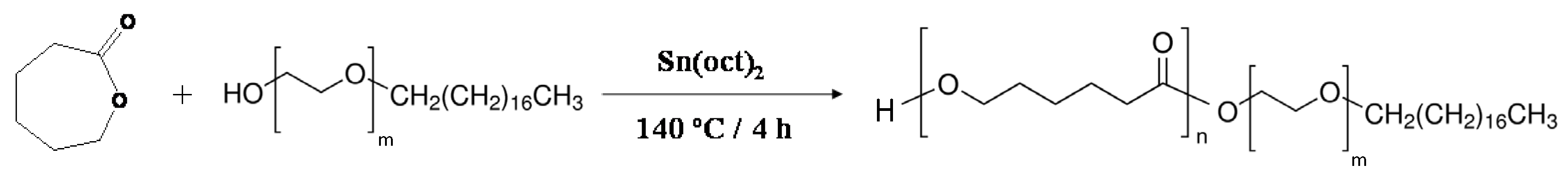
2.2.1. Synthesis of Myrj-b-PCL Copolymers
2.2.2. Characterization of the Myrj-b-PCL Copolymers
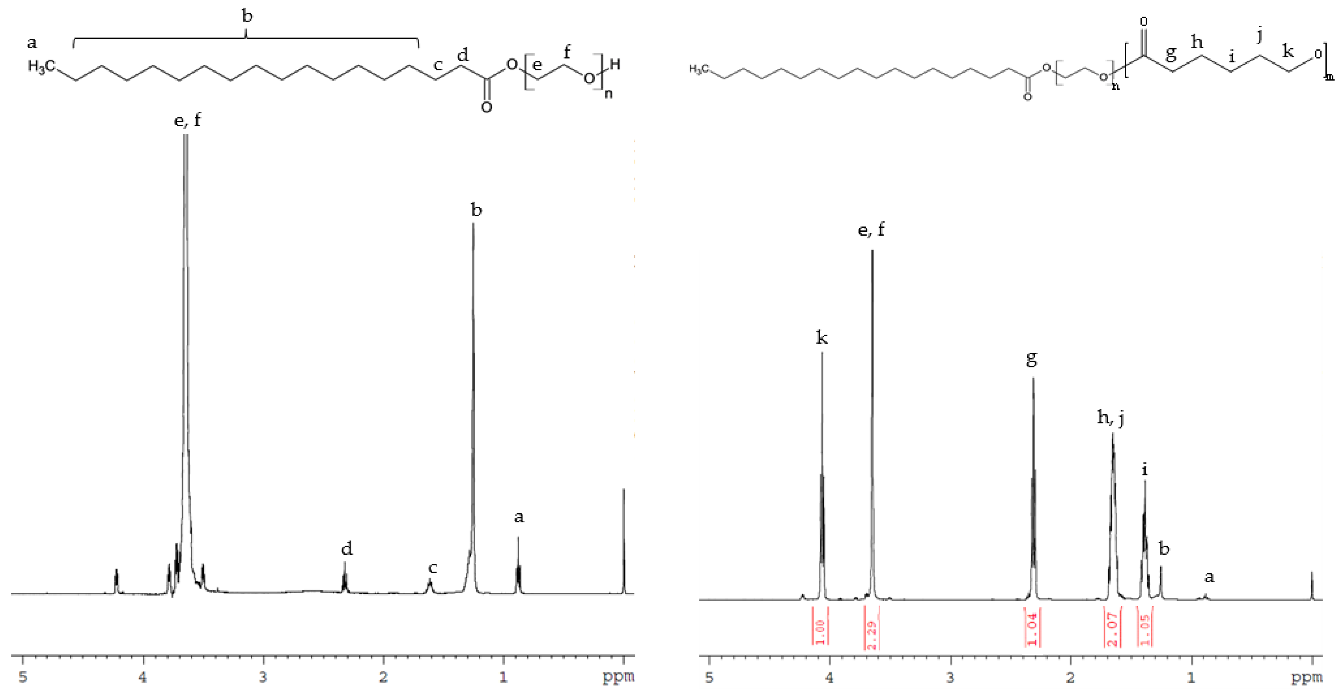
1H Nuclear Magnetic Resonance (1H NMR) Spectroscopy
GPC Chromatography
Fourier-Transform Infrared (FTIR) Spectroscopy
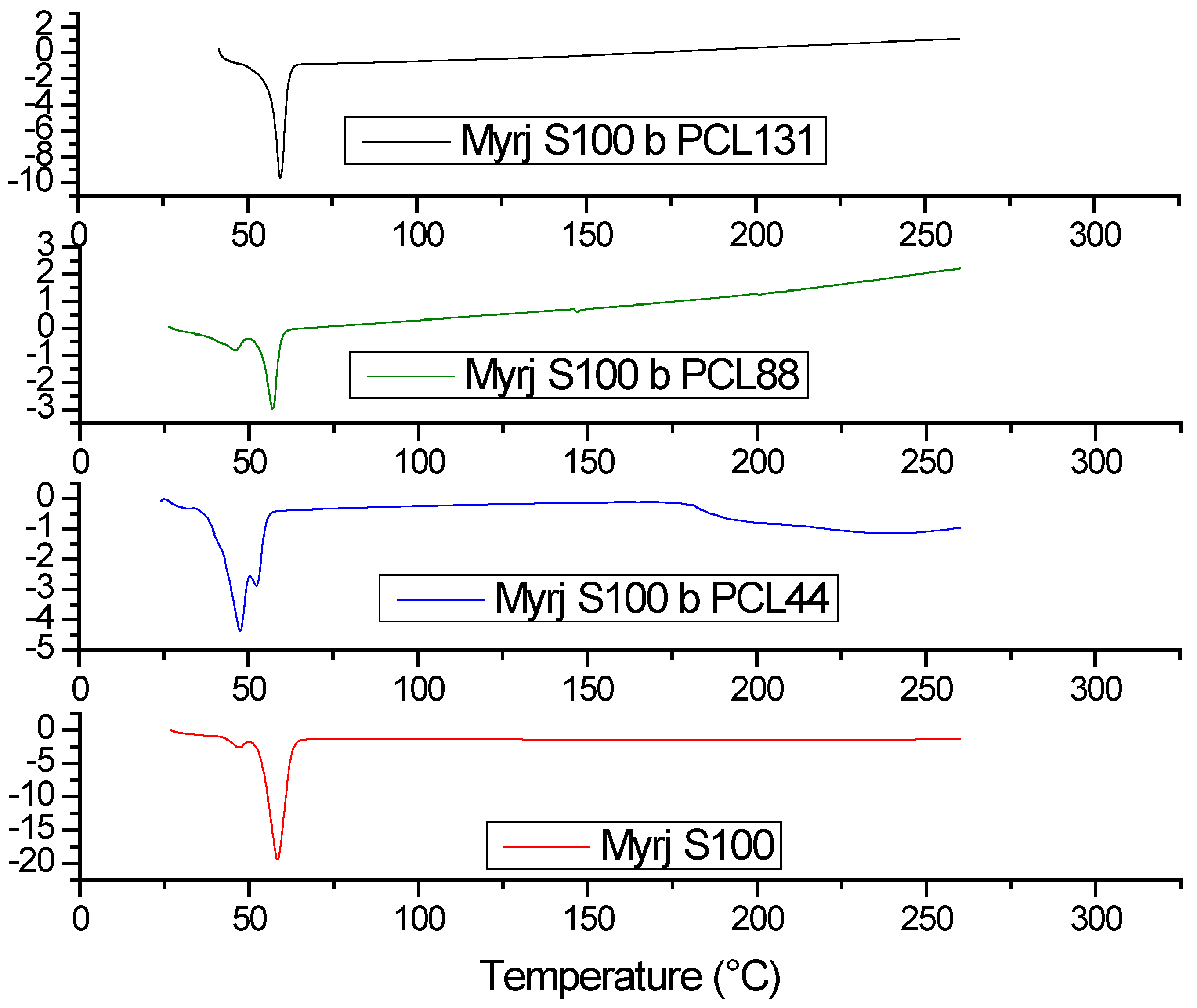
Differential Scanning Calorimetry (DSC)
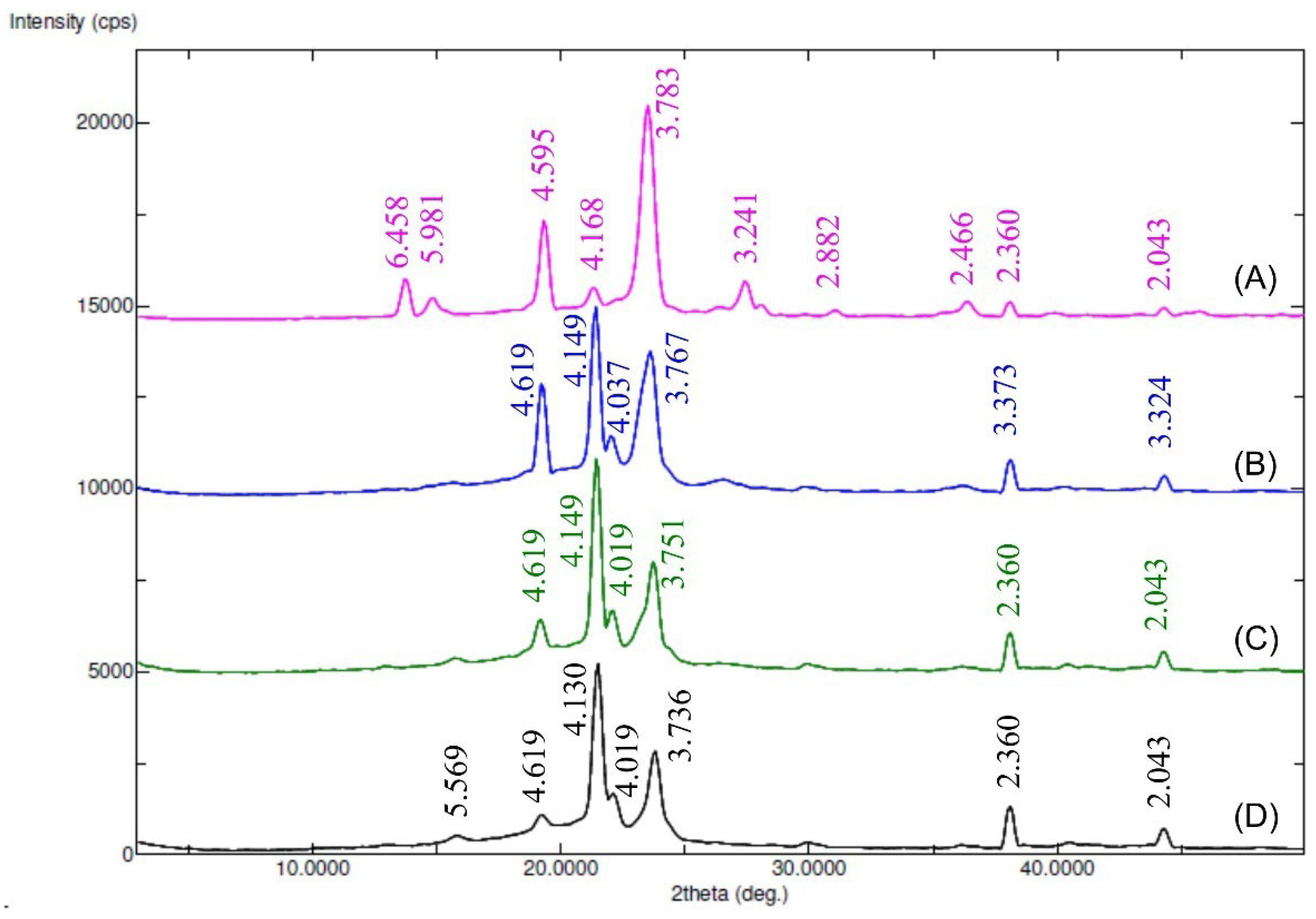
X-ray Diffraction (XRD)
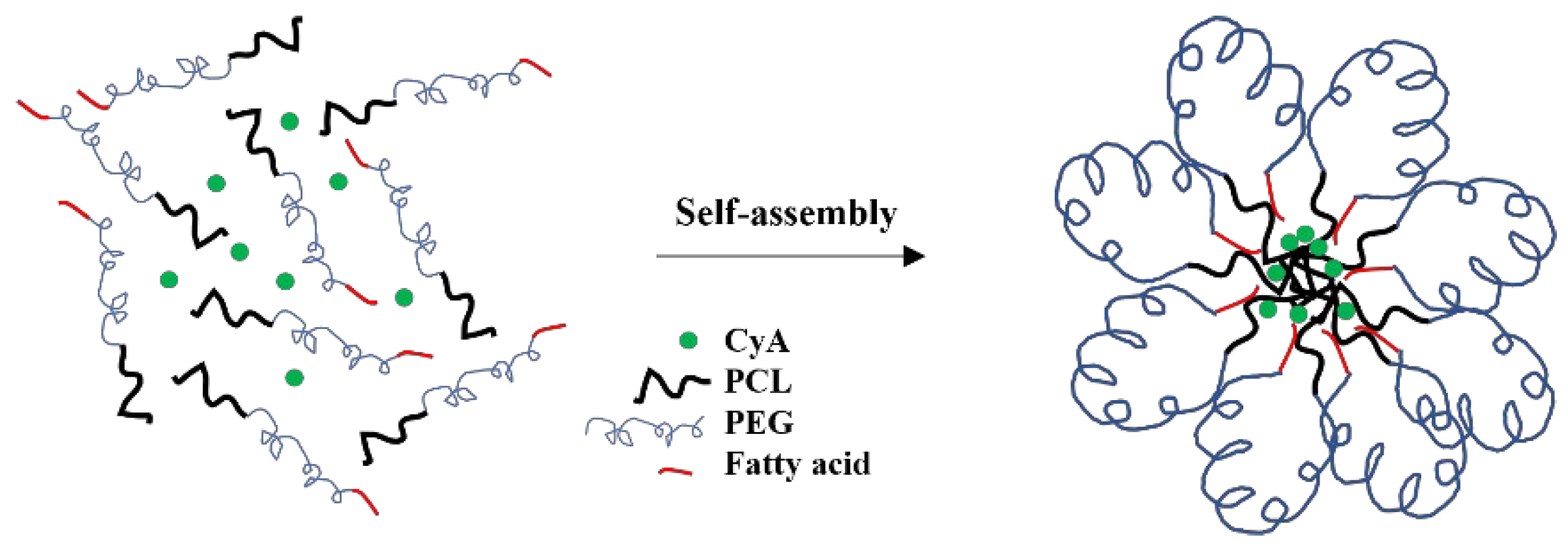
Preparation of Drug-Free and CyA-Loaded Myrj-b-PCL Micelles
2.2.3. Characterization of Myrj-b-PCL Micelles
Size, Polydispersity, and ζ-Potential
Critical Micelle Concentration (CMC)
Drug Encapsulation Efficiency % and Drug Loading %
Morphology
Ex Vivo Corneal Permeation
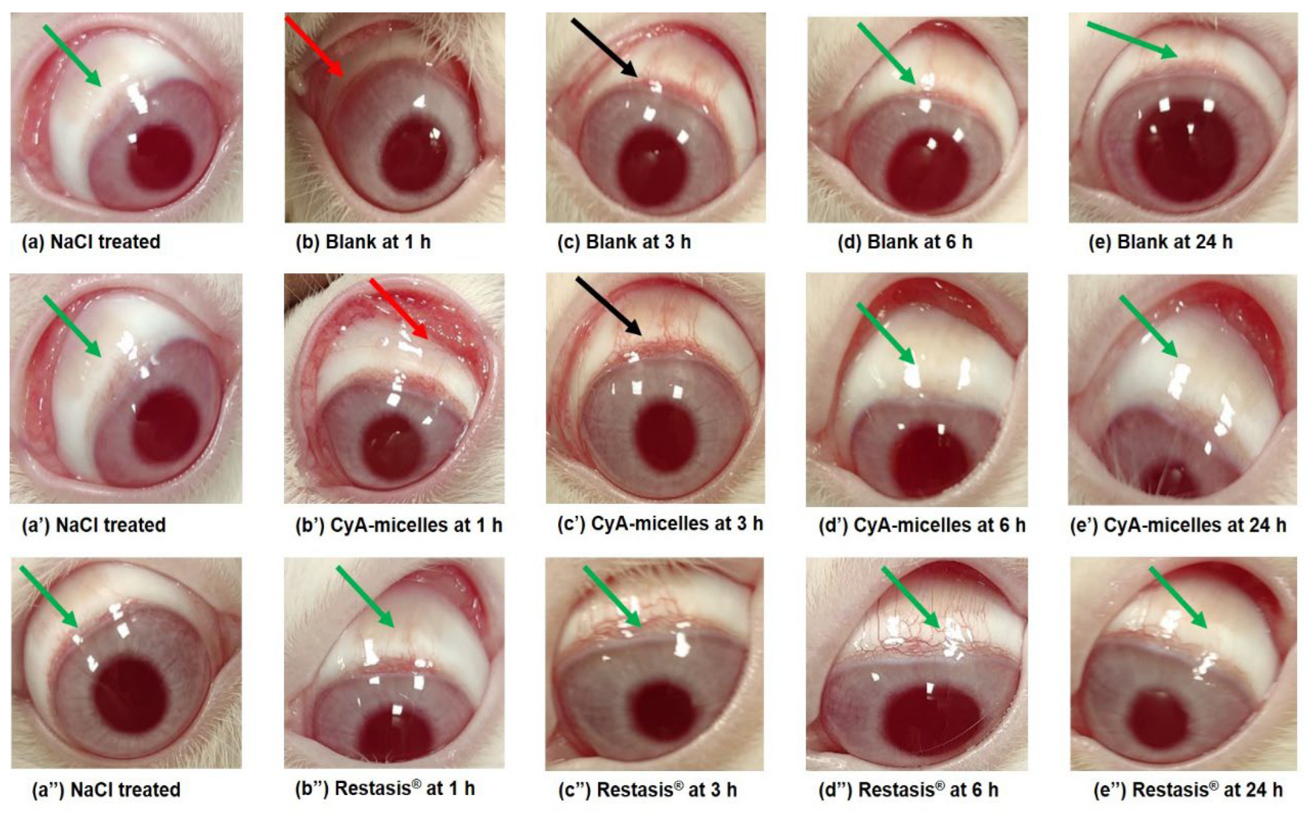
2.2.4. In Vivo Ocular Irritation Test
2.2.5. Data Analysis
3. Results and Discussion
3.1. Synthesis and Characterization of Myrj-b-PCL Copolymers
3.1.1. 1H NMR
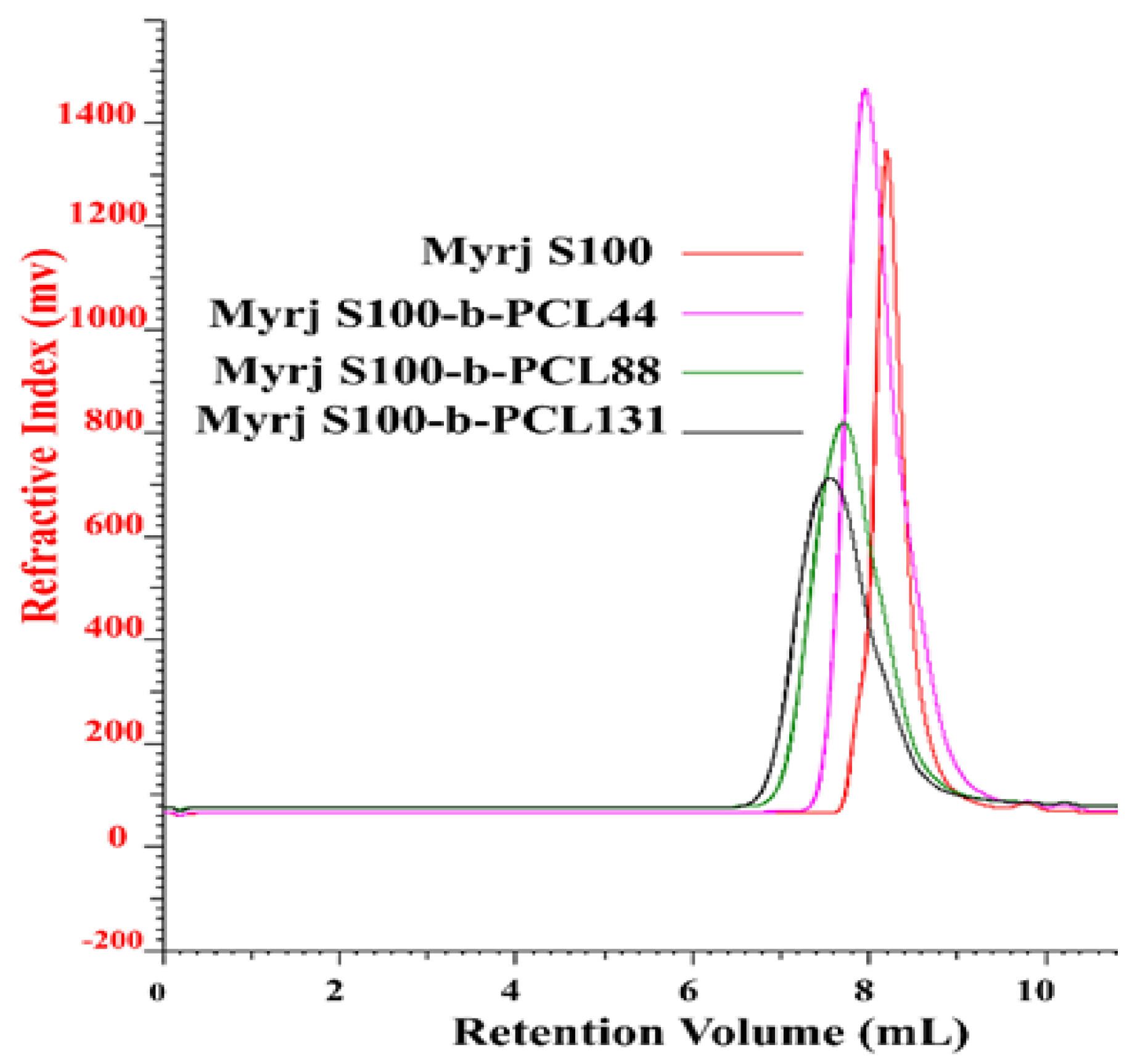
3.1.2. GPC
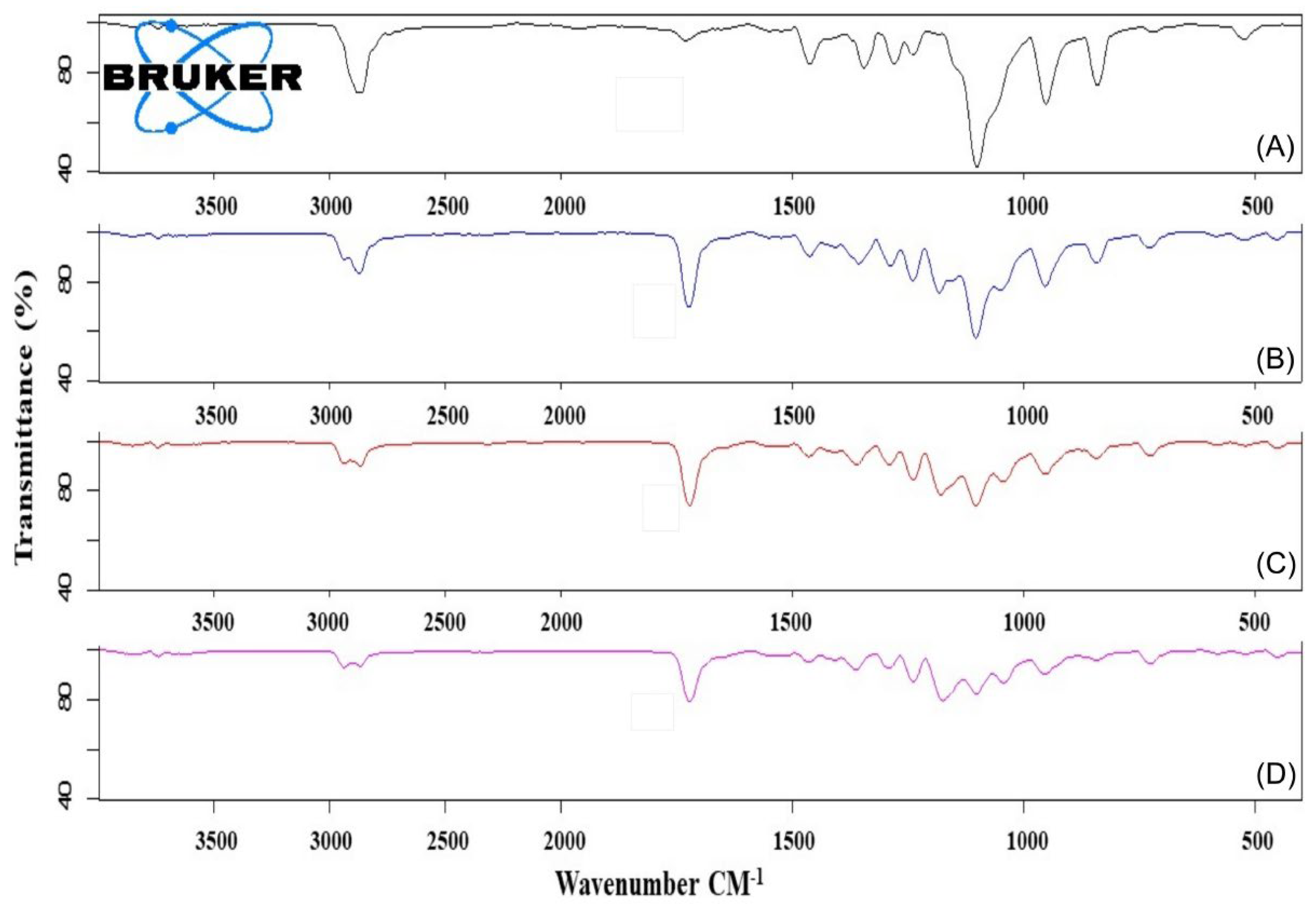
3.1.3. FTIR
3.1.4. XRD
3.1.5. DSC
3.2. Preparation and Characterization of Drug-Free and CyA-Loaded Myrj-b-PCL Micelles
3.2.1. Size, Polydispersity Index, and ζ-Potential
3.2.2. CMC
3.2.3. Drug Encapsulation Efficiency (EE%) and Drug Loading (DL%)
3.2.4. Morphology
3.3. Transcorneal Permeation of CyA-Loaded Myrj-b-PCL Micelles
3.4. In Vivo Ocular Irritation Test
4. Conclusions
Supplementary Materials
Author Contributions
Funding
Institutional Review Board Statement
Data Availability Statement
Conflicts of Interest
References
- Rowe, R.C.; Sheskey, P.J.; Owen, S.C.; Association, A.P. Handbook of Pharmaceutical Excipients; Pharmaceutical Press: London, UK, 2006. [Google Scholar]
- Liu, C.; Wu, J.; Shi, B.; Zhang, Y.; Gao, T.; Pei, Y. Enhancing the Bioavailability of Cyclosporine A Using Solid Dispersion Containing Polyoxyethylene (40) Stearate. Drug Dev. Ind. Pharm. 2006, 32, 115–123. [Google Scholar] [CrossRef] [PubMed]
- Lo, Y.L. Relationships between the hydrophilic-lipophilic balance values of pharmaceutical excipients and their multidrug resistance modulating effect in Caco-2 cells and rat intestines. J. Control. Release 2003, 90, 37–48. [Google Scholar] [CrossRef]
- Wang, S.-W.; Monagle, J.; McNulty, C.; Putnam, D.; Chen, H. Determination of P-glycoprotein inhibition by excipients and their combinations using an integrated high-throughput process. J. Pharm. Sci. 2004, 93, 2755–2767. [Google Scholar] [CrossRef]
- Dash, T.K.; Konkimalla, V.B. Poly-epsilon-caprolactone based formulations for drug delivery and tissue engineering: A review. J Control. Release 2012, 158, 15–33. [Google Scholar] [CrossRef] [PubMed]
- Yusuf, O.; Ali, R.; Alomrani, A.H.; Alshamsan, A.; Alshememry, A.K.; Almalik, A.M.; Lavasanifar, A.; Binkhathlan, Z. Design and Development of D–α–Tocopheryl Polyethylene Glycol Succinate–block–Poly(ε-Caprolactone) (TPGS-b-PCL) Nanocarriers for Solubilization and Controlled Release of Paclitaxel. Molecules 2021, 26, 2690. [Google Scholar] [CrossRef]
- Sinha, V.R.; Bansal, K.; Kaushik, R.; Kumria, R.; Trehan, A. Poly-epsilon-caprolactone microspheres and nanospheres: An overview. Int. J. Pharm. 2004, 278, 1–23. [Google Scholar] [CrossRef]
- Woodruff, M.A.; Hutmacher, D.W. The return of a forgotten polymer—Polycaprolactone in the 21st century. Prog. Polym. Sci. 2010, 35, 1217–1256. [Google Scholar] [CrossRef] [Green Version]
- Aliabadi, H.M.; Brocks, D.R.; Lavasanifar, A. Polymeric micelles for the solubilization and delivery of cyclosporine A: Pharmacokinetics and biodistribution. Biomaterials 2005, 26, 7251–7259. [Google Scholar] [CrossRef]
- Binkhathlan, Z.; Hamdy, D.A.; Brocks, D.R.; Lavasanifar, A. Development of a polymeric micellar formulation for valspodar and assessment of its pharmacokinetics in rat. Eur. J. Pharm. Biopharm. 2010, 75, 90–95. [Google Scholar] [CrossRef]
- Shuai, X.; Merdan, T.; Schaper, A.K.; Xi, F.; Kissel, T. Core-cross-linked polymeric micelles as paclitaxel carriers. Bioconjugate Chem. 2004, 15, 441–448. [Google Scholar] [CrossRef]
- Amber, T.; Tabassum, S. Cyclosporin in dermatology: A practical compendium. Dermatol. Ther. 2020, 33, e13934. [Google Scholar] [CrossRef] [PubMed]
- Airody, A.; Heath, G.; Lightman, S.; Gale, R. Non-Infectious Uveitis: Optimising the Therapeutic Response. Drugs 2016, 76, 27–39. [Google Scholar] [CrossRef] [PubMed]
- Schultz, C. Safety and Efficacy of Cyclosporine in the Treatment of Chronic Dry Eye. Ophthalmol. Eye Dis. 2014, 6, 37–42. [Google Scholar] [CrossRef] [PubMed]
- Peng, C.C.; Bengani, L.C.; Jung, H.J.; Leclerc, J.; Gupta, C.; Chauhan, A. Emulsions and microemulsions for ocular drug delivery. J. Drug Deliv. Sci. Technol. 2011, 21, 111–121. [Google Scholar] [CrossRef]
- Binkhathlan, Z.; Alomrani, A.H.; Alshamsan, A.; Aljuffali, I.I.; Ali, R. Poly e-Caprolactone-Ethoxylated Fatty Acid Copolymers. U.S. Patent 9,622,973, 18 April 2017. [Google Scholar]
- Topel, Ö.; Çakır, B.A.; Budama, L.; Hoda, N. Determination of critical micelle concentration of polybutadiene-block-poly(ethyleneoxide) diblock copolymer by fluorescence spectroscopy and dynamic light scattering. J. Mol. Liq. 2013, 177, 40–43. [Google Scholar] [CrossRef]
- Soleymani Abyaneh, H.; Vakili, M.R.; Zhang, F.; Choi, P.; Lavasanifar, A. Rational design of block copolymer micelles to control burst drug release at a nanoscale dimension. Acta Biomater. 2015, 24, 127–139. [Google Scholar] [CrossRef] [PubMed]
- Binkhathlan, Z.; Ali, R.; Qamar, W.; Lavasanifar, A. Pharmacokinetics of Orally Administered Poly(Ethylene Oxide)-block-Poly(ε-Caprolactone) Micelles of Cyclosporine A in Rats: Comparison with Neoral®. J. Pharm. Pharm. Sci. 2018, 21, 192s–199s. [Google Scholar] [CrossRef]
- Al-Jenoobi, F.I.; Alam, M.A.; Alkharfy, K.M.; Al-Suwayeh, S.A.; Korashy, H.M.; Al-Mohizea, A.M.; Iqbal, M.; Ahad, A.; Raish, M. Pharmacokinetic interaction studies of fenugreek with CYP3A substrates cyclosporine and carbamazepine. Eur. J. Drug Metab. Pharmacokinet. 2014, 39, 147–153. [Google Scholar] [CrossRef]
- Alshamsan, A.; Abul Kalam, M.; Vakili, M.R.; Binkhathlan, Z.; Raish, M.; Ali, R.; Alturki, T.A.; Safaei Nikouei, N.; Lavasanifar, A. Treatment of endotoxin-induced uveitis by topical application of cyclosporine a-loaded PolyGel in rabbit eyes. Int. J. Pharm. 2019, 569, 118573. [Google Scholar] [CrossRef]
- Kalam, M.A.; Alshamsan, A. Poly (d, l-lactide-co-glycolide) nanoparticles for sustained release of tacrolimus in rabbit eyes. Biomed. Pharmacother. 2017, 94, 402–411. [Google Scholar] [CrossRef]
- Draize, J.H.; Woodard, G.; Calvery, H.O. Methods for the study of irritation and toxicity of substances applied topically to the skin and mucous membranes. J. Pharmacol. Exp. Ther. 1944, 82, 377–390. [Google Scholar]
- Lee, M.; Hwang, J.H.; Lim, K.M. Alternatives to In Vivo Draize Rabbit Eye and Skin Irritation Tests with a Focus on 3D Reconstructed Human Cornea-Like Epithelium and Epidermis Models. Toxicol. Res. 2017, 33, 191–203. [Google Scholar] [CrossRef] [PubMed] [Green Version]
- Gyun Shin, I.L.; Yeon Kim, S.; Moo Lee, Y.; Soo Cho, C.; Yong Kiel, S. Methoxy poly(ethylene glycol)/ϵ-caprolactone amphiphilic block copolymeric micelle containing indomethacin.: I. Preparation and characterization. J. Control. Release 1998, 51, 1–11. [Google Scholar] [CrossRef]
- Sun, J.; He, C.; Zhuang, X.; Jing, X.; Chen, X. The crystallization behavior of poly(ethylene glycol)-poly(ε-caprolactone) diblock copolymers with asymmetric block compositions. J. Polym. Res. 2011, 18, 2161–2168. [Google Scholar] [CrossRef]
- Sharma, R.; Murali, R.; Murthy, C.N. Clouding and Aggregation Behavior of PPO-PEO-PPO Triblock Copolymer (Pluronic®25R4) in Surfactant Additives Environment. Tenside Surfactants Deterg. 2012, 49, 136–144. [Google Scholar] [CrossRef]
- Mahmud, A.; Xiong, X.-B.; Lavasanifar, A. Novel Self-Associating Poly(ethylene oxide)-block-poly(ε-caprolactone) Block Copolymers with Functional Side Groups on the Polyester Block for Drug Delivery. Macromolecules 2006, 39, 9419–9428. [Google Scholar] [CrossRef]
- Atanase, L.I.; Winninger, J.; Delaite, C.; Riess, G. Micellization and demicellization of amphiphilic poly(vinyl acetate)-graft-poly(N-vinyl-pyrrolidone) graft copolymers in the presence of sodium dodecyl sulfate. Colloids Surf. A Physicochem. Eng. Asp. 2014, 461, 287–294. [Google Scholar] [CrossRef]
- Fairley, N.; Hoang, B.; Allen, C. Morphological Control of Poly(ethylene glycol)-block-poly(ε-caprolactone) Copolymer Aggregates in Aqueous Solution. Biomacromolecules 2008, 9, 2283–2291. [Google Scholar] [CrossRef]
- Qi, W.; Ghoroghchian, P.P.; Li, G.; Hammer, D.A.; Therien, M.J. Aqueous self-assembly of poly(ethylene oxide)-block-poly(ε-caprolactone) (PEO-b-PCL) copolymers: Disparate diblock copolymer compositions give rise to nano- and meso-scale bilayered vesicles. Nanoscale 2013, 5, 10908–10915. [Google Scholar] [CrossRef]
- Danaei, M.; Dehghankhold, M.; Ataei, S.; Hasanzadeh Davarani, F.; Javanmard, R.; Dokhani, A.; Khorasani, S.; Mozafari, M.R. Impact of Particle Size and Polydispersity Index on the Clinical Applications of Lipidic Nanocarrier Systems. Pharmaceutics 2018, 57. [Google Scholar] [CrossRef] [Green Version]
- Lallemand, F.; Schmitt, M.; Bourges, J.L.; Gurny, R.; Benita, S.; Garrigue, J.S. Cyclosporine A delivery to the eye: A comprehensive review of academic and industrial efforts. Eur. J. Pharm. Biopharm. 2017, 117, 14–28. [Google Scholar] [CrossRef] [PubMed]
- Kennah, H.E., 2nd; Hignet, S.; Laux, P.E.; Dorko, J.D.; Barrow, C.S. An objective procedure for quantitating eye irritation based upon changes of corneal thickness. Fundam. Appl. Toxicol. 1989, 12, 258–268. [Google Scholar] [CrossRef]
- Falahee, K.J.; Rose, C.S.; Olin, S.S. Eye Irritation Testing: An Assessment of Methods and Guidelines for Testing Materials for Eye Irritancy; Office of Pesticides and Toxic Substances, US Environmental Protection Agency: Washington, DC, USA, 1981. [Google Scholar]
- Kay, J.H.; Calandra, J.C. Interpretation of eye irritation tests. J. Soc. Cosmet. Chem. 1962, 13, 281–289. [Google Scholar]
- Di Tommaso, C.; Torriglia, A.; Furrer, P.; Behar-Cohen, F.; Gurny, R.; Moller, M. Ocular biocompatibility of novel Cyclosporin A formulations based on methoxy poly(ethylene glycol)-hexylsubstituted poly(lactide) micelle carriers. Int. J. Pharm. 2011, 416, 515–524. [Google Scholar] [CrossRef] [PubMed]
- Civiale, C.; Licciardi, M.; Cavallaro, G.; Giammona, G.; Mazzone, M.G. Polyhydroxyethylaspartamide-based micelles for ocular drug delivery. Int. J. Pharm. 2009, 378, 177–186. [Google Scholar] [CrossRef] [PubMed]


| Block Copolymer a | Theoretical MW (g/mol) | Mn b (g/mol) | Mn c (g/mol) | Mw d (g/mol) | Dispersity e (Ð) |
|---|---|---|---|---|---|
| Myrj S40-b-PCL18 | 4100 | 3500 | 3007 | 4037 | 1.34 |
| Myrj S40-b-PCL35 | 6040 | 5700 | 4116 | 5745 | 1.40 |
| Myrj S100-b-PCL44 | 9700 | 9400 | 6192 | 8628 | 1.39 |
| Myrj S100-b-PCL88 | 14,650 | 14,600 | 10,150 | 15,724 | 1.55 |
| Myrj S100-b-PCL131 | 19,700 | 19,600 | 12,019 | 28,880 | 1.67 |
| Polymeric Micelles | Particle Size (nm) | Polydispersity Index | ζ-Potential (mV) | EE (%) | DL (%) |
|---|---|---|---|---|---|
| Micelles before CyA loading | |||||
| Myrj S100-b-PCL44 | 49.8 ± 14.9 | 0.38 ± 0.14 | −7.0 ± 1.8 | – | – |
| Myrj S100-b-PCL88 | 70.3 ± 2.4 | 0.20 ± 0.04 | −4.7 ± 0.3 | – | – |
| Myrj S100-b-PCL131 | 72.6 ± 1.3 | 0.21 ± 0.02 | −9.4 ± 0.7 | – | – |
| Micelles after CyA loading | |||||
| Myrj S100-b-PCL44 | 41.1 ± 5.6 | 0.44 ± 0.06 | −4.9 ± 3.2 | 15.23 ± 0.38 | 1.38 ± 0.03 |
| Myrj S100-b-PCL88 | 81.9 ± 2.9 | 0.18 ± 0.01 | −6.8 ± 2.1 | 54.15 ± 3.03 | 5.62 ± 0.27 |
| Myrj S100-b-PCL131 | 63.5 ± 13.1 | 0.28 ± 0.05 | −1.6 ± 1.7 | 23.04 ± 1.35 | 2.25 ± 0.13 |
| Parameters | CyA-Micelles | Restasis® |
|---|---|---|
| Cumulative amount of CyA permeated (µg cm−2) at 4th h | 51.14 ± 5.23 | 59.49 ± 8.67 |
| Steady-state flux, J (µg cm−2 h−1) | 18.89 ± 2.99 | 17.14 ± 2.79 |
| Permeability coefficient, Papp (cm h−1) | (3.78 ± 0.59) × 10−2 | (3.43 ± 0.56) × 10−2 |
| Lesions in the Treated Eyes | Individual Scores for Eye Irritation | ||||||||
|---|---|---|---|---|---|---|---|---|---|
| Restasis® | Blank Micelles | CyA-Micelles | |||||||
| Animal # | Animal # | Animal # | |||||||
| 1st | 2nd | 3rd | 1st | 2nd | 3rd | 1st | 2nd | 3rd | |
| Cornea | |||||||||
| I. Opacity (Degree of density) | 0 | 1 | 0 | 0 | 1 | 1 | 0 | 1 | 0 |
| II. Area of cornea | 4 | 4 | 4 | 4 | 4 | 4 | 4 | 4 | 4 |
| Total scores = (I × II × 5) = | 0 | 20 | 0 | 0 | 20 | 20 | 0 | 20 | 0 |
| Iris | |||||||||
| I. Lesion values | 0 | 1 | 0 | 0 | 1 | 1 | 0 | 1 | 0 |
| Total scores = (I × 5) = | 0 | 5 | 0 | 0 | 5 | 5 | 0 | 5 | 0 |
| Conjunctiva | |||||||||
| I. Redness | 0 | 0 | 1 | 1 | 1 | 1 | 0 | 1 | 0 |
| II. Chemosis | 0 | 0 | 0 | 0 | 0 | 0 | 0 | 0 | 0 |
| III. Mucoidal discharge | 0 | 0 | 0 | 0 | 1 | 0 | 0 | 0 | 0 |
| Total scores = (I + II + III) × 2 = | 0 | 0 | 2 | 2 | 4 | 2 | 0 | 2 | 0 |
| Restasis® | |||||
| Animal #→ | 1st | 2nd | 3rd | SUM | Average (SUM/3) |
| Cornea | 0 | 20 | 0 | 20 | 6.67 |
| Iris | 0 | 5 | 0 | 5 | 1.67 |
| Conjunctiva | 0 | 0 | 2 | 2 | 0.66 |
| SUM total = | 0 | 25 | 2 | 27 | 9.00 |
| Blank micelles | |||||
| Animal #→ | 1st | 2nd | 3rd | SUM | Average (SUM/3) |
| Cornea | 0 | 20 | 20 | 40 | 13.33 |
| Iris | 0 | 5 | 5 | 10 | 3.33 |
| Conjunctiva | 2 | 4 | 2 | 8 | 2.67 |
| SUM total = | 2 | 29 | 27 | 58 | 19.33 |
| CyA-Micelles | |||||
| Animal #→ | 1st | 2nd | 3rd | SUM | Average (SUM/3) |
| Cornea | 0 | 20 | 0 | 20 | 6.67 |
| Iris | 0 | 5 | 0 | 5 | 1.67 |
| Conjunctiva | 0 | 2 | 0 | 2 | 0.66 |
| Total = | 0 | 27 | 0 | 27 | 9.00 |
Publisher’s Note: MDPI stays neutral with regard to jurisdictional claims in published maps and institutional affiliations. |
© 2022 by the authors. Licensee MDPI, Basel, Switzerland. This article is an open access article distributed under the terms and conditions of the Creative Commons Attribution (CC BY) license (https://creativecommons.org/licenses/by/4.0/).
Share and Cite
Binkhathlan, Z.; Alomrani, A.H.; Hoxha, O.; Ali, R.; Kalam, M.A.; Alshamsan, A. Development and Characterization of PEGylated Fatty Acid-Block-Poly(ε-caprolactone) Novel Block Copolymers and Their Self-Assembled Nanostructures for Ocular Delivery of Cyclosporine A. Polymers 2022, 14, 1635. https://doi.org/10.3390/polym14091635
Binkhathlan Z, Alomrani AH, Hoxha O, Ali R, Kalam MA, Alshamsan A. Development and Characterization of PEGylated Fatty Acid-Block-Poly(ε-caprolactone) Novel Block Copolymers and Their Self-Assembled Nanostructures for Ocular Delivery of Cyclosporine A. Polymers. 2022; 14(9):1635. https://doi.org/10.3390/polym14091635
Chicago/Turabian StyleBinkhathlan, Ziyad, Abdullah H. Alomrani, Olsi Hoxha, Raisuddin Ali, Mohd Abul Kalam, and Aws Alshamsan. 2022. "Development and Characterization of PEGylated Fatty Acid-Block-Poly(ε-caprolactone) Novel Block Copolymers and Their Self-Assembled Nanostructures for Ocular Delivery of Cyclosporine A" Polymers 14, no. 9: 1635. https://doi.org/10.3390/polym14091635
APA StyleBinkhathlan, Z., Alomrani, A. H., Hoxha, O., Ali, R., Kalam, M. A., & Alshamsan, A. (2022). Development and Characterization of PEGylated Fatty Acid-Block-Poly(ε-caprolactone) Novel Block Copolymers and Their Self-Assembled Nanostructures for Ocular Delivery of Cyclosporine A. Polymers, 14(9), 1635. https://doi.org/10.3390/polym14091635