
Iodo Silanes as Superior Substrates for the Solid Phase Synthesis of Molecularly Imprinted Polymer Nanoparticles

 , ,
, ,  , and
, and
Abstract
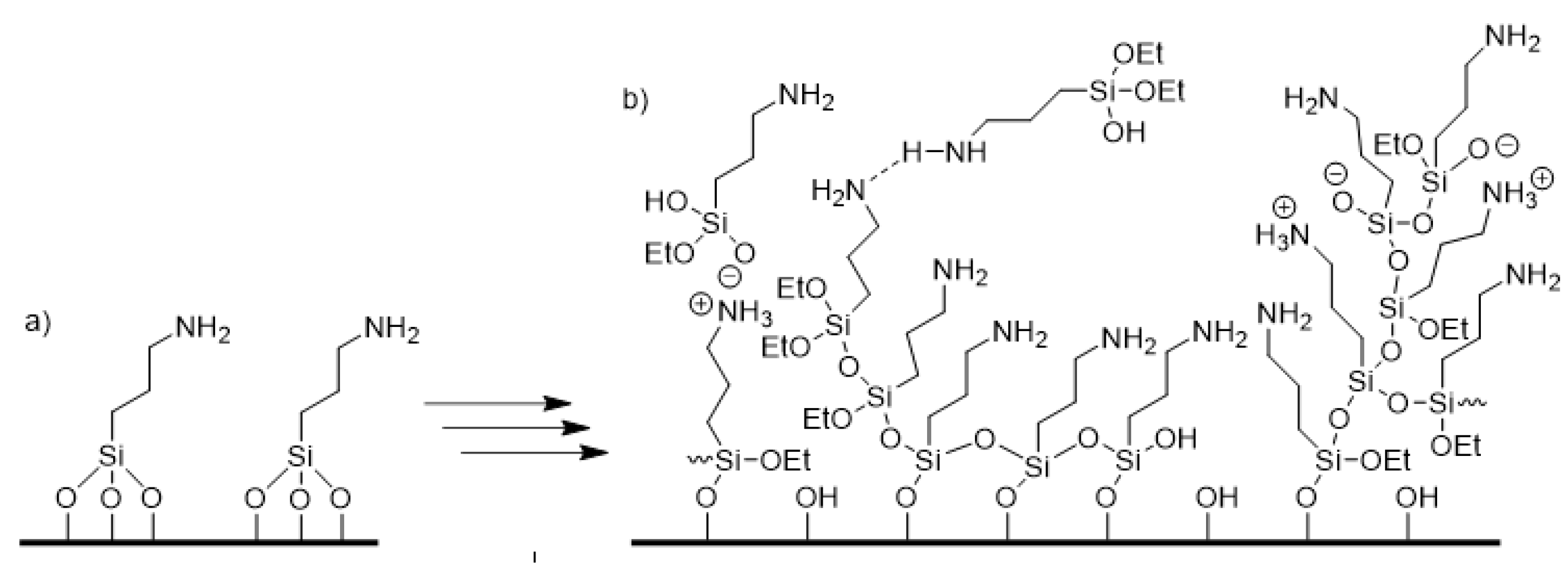
:1. Introduction
2. Materials and Methods
2.1. Materials
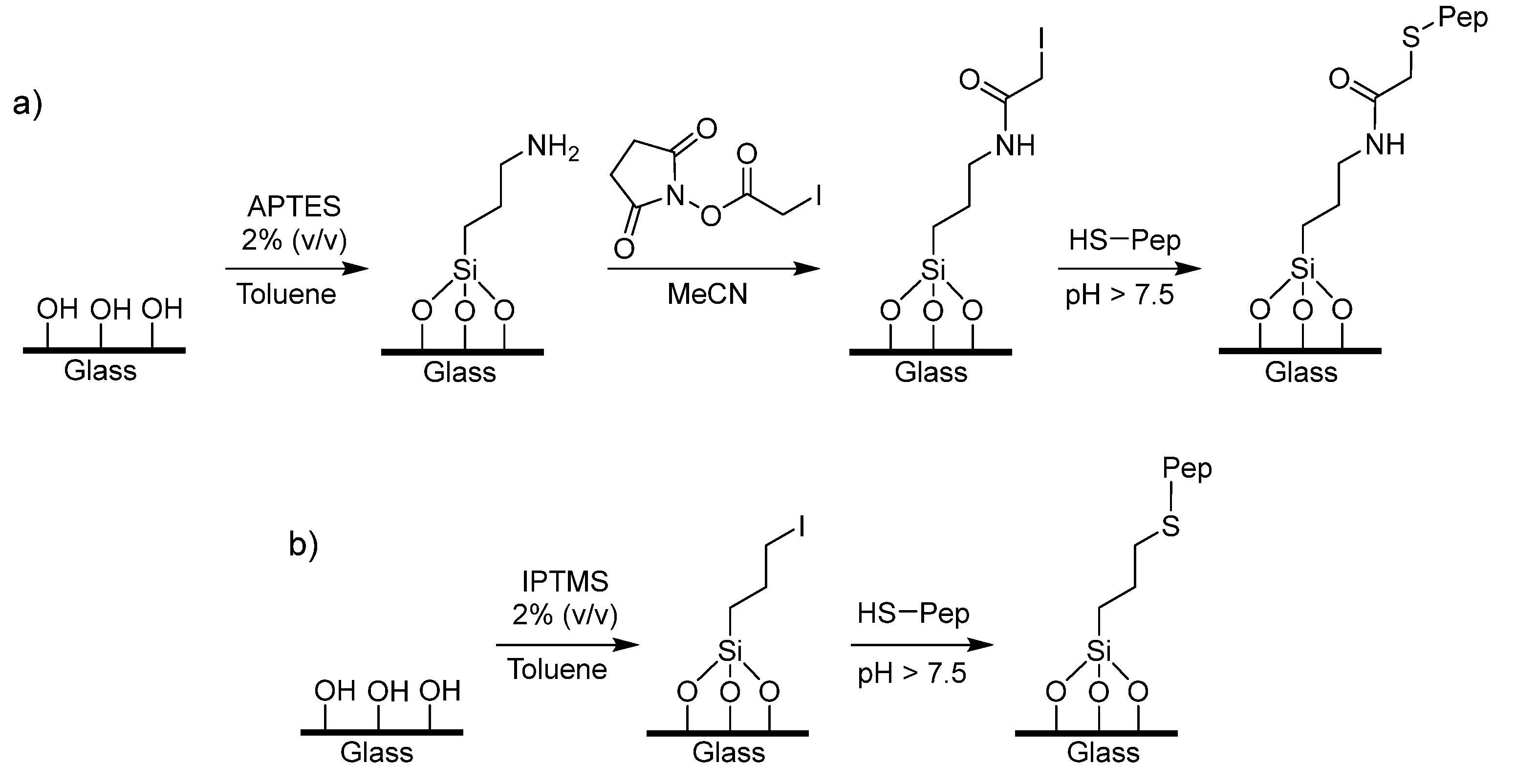
2.2. Preparation of APTES, AHAMTES and IPTMS Solid Phase
2.3. Immobilisation of Peptide on AHAMTES Glass
2.4. Immobilisation of Peptide on IPTMS Glass
2.5. Peptide Density Measurement
2.6. MIP Synthesis
2.7. Surface Plasmon Resonance (SPR) Measurement
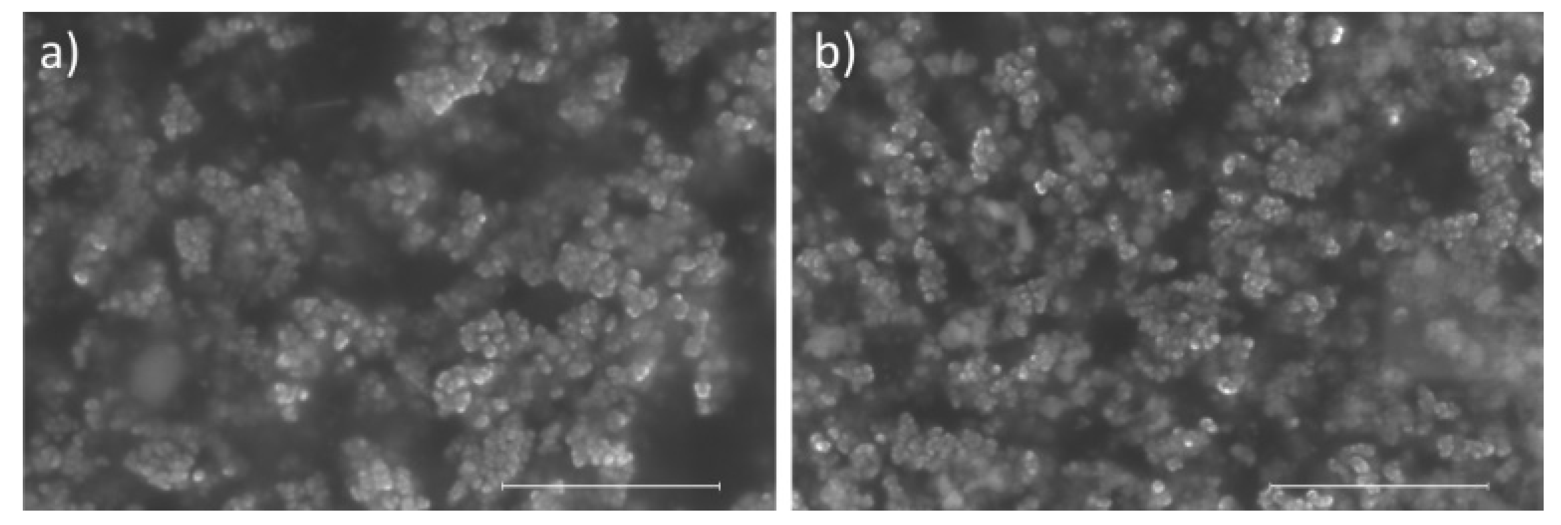
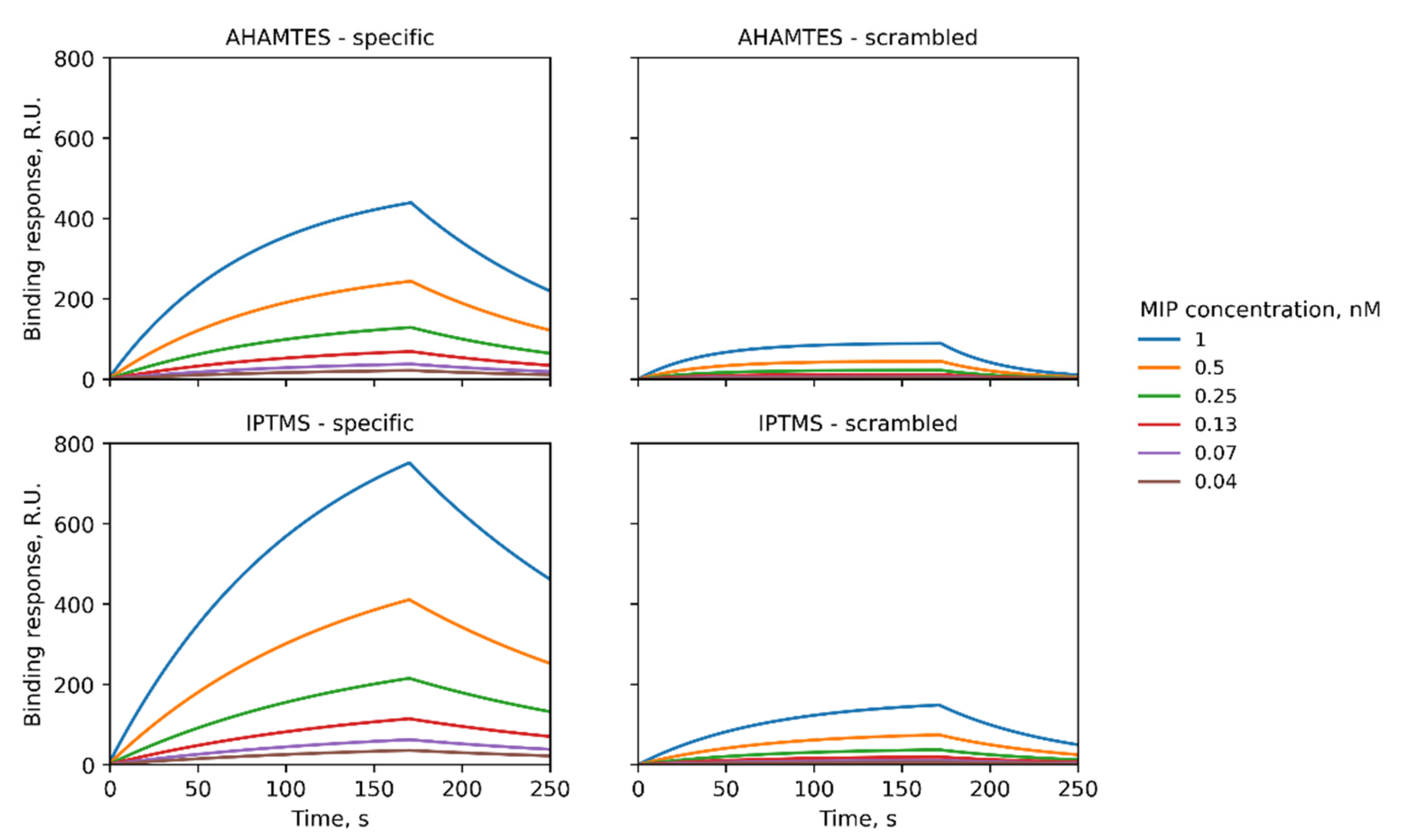
3. Results and Discussion
4. Conclusions
Supplementary Materials
Author Contributions
Funding
Institutional Review Board Statement
Informed Consent Statement
Data Availability Statement
Conflicts of Interest
References
- Olsen, J.; Martin, P.; Wilson, I.D. Molecular Imprints as Sorbents for Solid Phase Extraction: Potential and Applications. Anal. Commun. 1998, 35, 13H–14H. [Google Scholar] [CrossRef]
- Andersson, L.I. Molecular Imprinting for Drug Bioanalysis: A Review on the Application of Imprinted Polymers to Solid-Phase Extraction and Binding Assay. J. Chromatogr. B: Biomed. Sci. Appl. 2000, 739, 163–173. [Google Scholar] [CrossRef]
- Jodlbauer, J.; Maier, N.M.; Lindner, W. Towards Ochratoxin A Selective Molecularly Imprinted Polymers for Solid-Phase Extraction. J. Chromatogr. A 2002, 945, 45–63. [Google Scholar] [CrossRef]
- Piletsky, S.S.; Cass, A.E.G.; Piletska, E.V.; Czulak, J.; Piletsky, S.A. A Novel Assay Format as an Alternative to ELISA: MINA Test for Biotin. ChemNanoMat 2018, 4, 1214–1222. [Google Scholar] [CrossRef]
- Piletsky, S.S.; Rabinowicz, S.; Yang, Z.; Zagar, C.; Piletska, E.V.; Guerreiro, A.; Piletsky, S.A. Development of Molecularly Imprinted Polymers Specific for Blood Antigens for Application in Antibody-Free Blood Typing. Chem. Commun. 2017, 53, 1793–1796. [Google Scholar] [CrossRef] [Green Version]
- Saylan, Y.; Akgönüllü, S.; Yavuz, H.; Ünal, S.; Denizli, A. Molecularly Imprinted Polymer Based Sensors for Medical Applications. Sensors 2019, 19, 1279. [Google Scholar] [CrossRef] [Green Version]
- Canfarotta, F.; Lezina, L.; Guerreiro, A.; Czulak, J.; Petukhov, A.; Daks, A.; Smolinska-Kempisty, K.; Poma, A.; Piletsky, S.; Barlev, N.A. Specific Drug Delivery to Cancer Cells with Double-Imprinted Nanoparticles against Epidermal Growth Factor Receptor. Nano Lett. 2018, 18, 4641–4646. [Google Scholar] [CrossRef]
- Piletsky, S.S.; Piletska, E.; Poblocka, M.; Macip, S.; Jones, D.J.L.; Braga, M.; Cao, T.H.; Singh, R.; Spivey, A.C.; Aboagye, E.O.; et al. Snapshot Imprinting: Rapid Identification of Cancer Cell Surface Proteins and Epitopes Using Molecularly Imprinted Polymers. Nano Today 2021, 41, 101304. [Google Scholar] [CrossRef]
- Xing, R.; Wen, Y.; He, H.; Guo, Z.; Liu, Z. Recent Progress in the Combination of Molecularly Imprinted Polymer-Based Affinity Extraction and Mass Spectrometry for Targeted Proteomic Analysis. TrAC Trends Anal. Chem. 2019, 110, 417–428. [Google Scholar] [CrossRef]
- Poma, A.; Guerreiro, A.; Whitcombe, M.J.; Piletska, E.; Turner, A.P.F.; Piletsky, S.A. Solid-Phase Synthesis of Molecularly Imprinted Polymer Nanoparticles with a Reusable Template–“Plastic Antibodies”. Adv. Funct. Mater. 2013, 23, 2821–2827. [Google Scholar] [CrossRef] [Green Version]
- Medina Rangel, P.X.; Laclef, S.; Xu, J.; Panagiotopoulou, M.; Kovensky, J.; Tse Sum Bui, B.; Haupt, K. Solid-Phase Synthesis of Molecularly Imprinted Polymer Nanolabels: Affinity Tools for Cellular Bioimaging of Glycans. Sci. Rep. 2019, 9, 3923. [Google Scholar] [CrossRef] [PubMed] [Green Version]
- Ambrosini, S.; Beyazit, S.; Haupt, K.; Tse Sum Bui, B. Solid-Phase Synthesis of Molecularly Imprinted Nanoparticles for Protein Recognition. Chem. Commun. 2013, 49, 6746–6748. [Google Scholar] [CrossRef] [PubMed]
- Canfarotta, F.; Poma, A.; Guerreiro, A.; Piletsky, S. Solid-Phase Synthesis of Molecularly Imprinted Nanoparticles. Nat. Protoc. 2016, 11, 443–455. [Google Scholar] [CrossRef] [PubMed]
- Ekpenyong-Akiba, A.E.; Canfarotta, F.; Abd H., B.; Poblocka, M.; Casulleras, M.; Castilla-Vallmanya, L.; Kocsis-Fodor, G.; Kelly, M.E.; Janus, J.; Althubiti, M.; et al. Detecting and Targeting Senescent Cells Using Molecularly Imprinted Nanoparticles. Nanoscale Horiz. 2019, 4, 757–768. [Google Scholar] [CrossRef]
- Zhu, M.; Lerum, M.Z.; Chen, W. How to Prepare Reproducible, Homogeneous, and Hydrolytically Stable Aminosilane-Derived Layers on Silica. Langmuir 2012, 28, 416–423. [Google Scholar] [CrossRef] [PubMed] [Green Version]
- Stayton, P.S.; Olinger, J.M.; Jiang, M.; Bohn, P.W.; Sligar, S.G. Genetic Engineering of Surface Attachment Sites Yields Oriented Protein Monolayers. J. Am. Chem Soc. 1992, 114, 9298–9299. [Google Scholar] [CrossRef]
- Zhang, S.; Shi, Z.; Xu, H.; Ma, X.; Yin, J.; Tian, M. Revisiting the Mechanism of Redox-Polymerization to Build the Hydrogel with Excellent Properties Using a Novel Initiator. Soft Matter 2016, 12, 2575–2582. [Google Scholar] [CrossRef]
- Poma, A.; Guerreiro, A.; Caygill, S.; Moczko, E.; Piletsky, S. Automatic Reactor for Solid-Phase Synthesis of Molecularly Imprinted Polymeric Nanoparticles (MIP NPs) in Water. RSC Adv. 2014, 4, 4203–4206. [Google Scholar] [CrossRef] [Green Version]
- García, Y.; Czulak, J.; Pereira, E.D.; Piletsky, S.A.; Piletska, E. A Magnetic Molecularly Imprinted Nanoparticle Assay (MINA) for Detection of Pepsin. React. Funct. Polym. 2022, 170, 105133. [Google Scholar] [CrossRef]
- Cuoq, F.; Masion, A.; Labille, J.; Rose, J.; Ziarelli, F.; Prelot, B.; Bottero, J.Y. Preparation of Amino-Functionalized Silica in Aqueous Conditions. Appl. Surf. Sci. 2013, 266, 155–160. [Google Scholar] [CrossRef]
- Gómez-Caballero, A.; Elejaga-Jimeno, A.; García del Caño, G.; Unceta, N.; Guerreiro, A.; Saumell-Esnaola, M.; Sallés, J.; Goicolea, M.A.; Barrio, R.J. Solid-Phase Synthesis of Imprinted Nanoparticles as Artificial Antibodies against the C-Terminus of the Cannabinoid CB1 Receptor: Exploring a Viable Alternative for Bioanalysis. Mikrochim. Acta 2021, 188, 3. [Google Scholar] [CrossRef] [PubMed]

| AHAMTES | IPTMS | |
|---|---|---|
| Silanisation | Incubate glass beads (60 g) in 2% (v/v) AHAMTES overnight in dry toluene (60 mL). Wash with acetone (4 × 100 mL). Dry at 120 °C for 30 min. | Incubate glass beads (60 g) in 2% (v/v) IPTMS overnight in dry toluene (60 mL), protected from light. Wash with acetone (4 × 100 mL). Dry at 120 °C for 30 min. |
| Removal of multilayers | Soak glass beads overnight in a large excess of water. Wash with acetone (4 × 100 mL). | - |
| Surface activation | Incubate silanised glass beads (60 g) in a solution of succinimidyl iodoacetate (SIA) (5 mg) in anhydrous acetonitrile (25 ml) for 2 h, protected from light. Wash with acetonitrile (5 × 50 mL). | - |
| Peptide conjugation | Prepare ethylenediaminetetraacetic acid (EDTA) (74 mg, 500 µmol, 5mM) in phosphate buffered saline (PBS, 10 mM, 50 mL), adjust to pH 8.2 with sodium hydroxide. Add SIA-functionalised glass beads (60 g) and EGFR peptide (5 mg), incubate overnight protected from light. | Prepare EGFR peptide (5 mg) in borate buffer (pH 9.2, 30 mM sodium tetraborate, 25 mL). Add IPTMS-functionalised glass beads (60 g) and incubate overnight, protected from light. |
| Surface quenching | Add mercaptoethanol (20 µL) to a mixture of glass beads and peptide, and incubate for 2 h protected from light. Wash with water (2 × 500 mL) and acetone (100 mL), and allow to dry. | Add mercaptoethanol (20 µL) to a mixture of glass beads and peptide, and incubate for 2 h protected from light. Wash with water (2 × 500 mL) and acetone (100 mL), and allow to dry. |
| Silane | Peptide | Kd [nM] (χ2) |
|---|---|---|
| AHAMTES | Specific | 2.5 (7.14 × 10⁻⁵) |
| AHAMTES | Scrambled | 676 (1.58 × 10⁻⁵) |
| IPTMS | Specific | 2.3 (7.14 × 10⁻⁵) |
| IPTMS | Scrambled | 766 (2.06 × 10⁻⁵) |
Publisher’s Note: MDPI stays neutral with regard to jurisdictional claims in published maps and institutional affiliations. |
© 2022 by the authors. Licensee MDPI, Basel, Switzerland. This article is an open access article distributed under the terms and conditions of the Creative Commons Attribution (CC BY) license (https://creativecommons.org/licenses/by/4.0/).
Share and Cite
Piletsky, S.S.; Garcia Cruz, A.; Piletska, E.; Piletsky, S.A.; Aboagye, E.O.; Spivey, A.C. Iodo Silanes as Superior Substrates for the Solid Phase Synthesis of Molecularly Imprinted Polymer Nanoparticles. Polymers 2022, 14, 1595. https://doi.org/10.3390/polym14081595
Piletsky SS, Garcia Cruz A, Piletska E, Piletsky SA, Aboagye EO, Spivey AC. Iodo Silanes as Superior Substrates for the Solid Phase Synthesis of Molecularly Imprinted Polymer Nanoparticles. Polymers. 2022; 14(8):1595. https://doi.org/10.3390/polym14081595
Chicago/Turabian StylePiletsky, Stanislav S., Alvaro Garcia Cruz, Elena Piletska, Sergey A. Piletsky, Eric O. Aboagye, and Alan C. Spivey. 2022. "Iodo Silanes as Superior Substrates for the Solid Phase Synthesis of Molecularly Imprinted Polymer Nanoparticles" Polymers 14, no. 8: 1595. https://doi.org/10.3390/polym14081595
APA StylePiletsky, S. S., Garcia Cruz, A., Piletska, E., Piletsky, S. A., Aboagye, E. O., & Spivey, A. C. (2022). Iodo Silanes as Superior Substrates for the Solid Phase Synthesis of Molecularly Imprinted Polymer Nanoparticles. Polymers, 14(8), 1595. https://doi.org/10.3390/polym14081595