
Sensing beyond Senses: An Overview of Outstanding Strides in Architecting Nanopolymer-Enabled Sensors for Biomedical Applications

 , , , , ,
  and
and
Abstract
1. Introduction
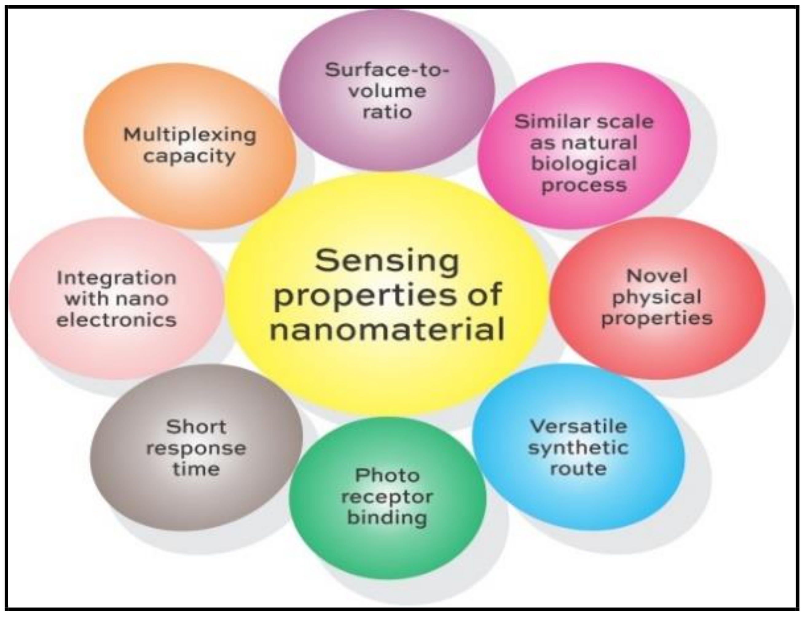
2. Properties of Nanomaterial in Sensors
3. Transformational Applications of Nanomaterial for Sensing Phenomena
3.1. Agriculture
3.2. Biological Detection
3.3. Food Industry
3.4. Environmental Monitoring
Plasmonic Structures
3.5. Nano-Based Sensors in Pharmaceuticals
3.6. Nano-Based Sensors in Diagnostics
4. Sensing Mechanism
4.1. Physisorption-Chemisorption
4.2. Surface Modification
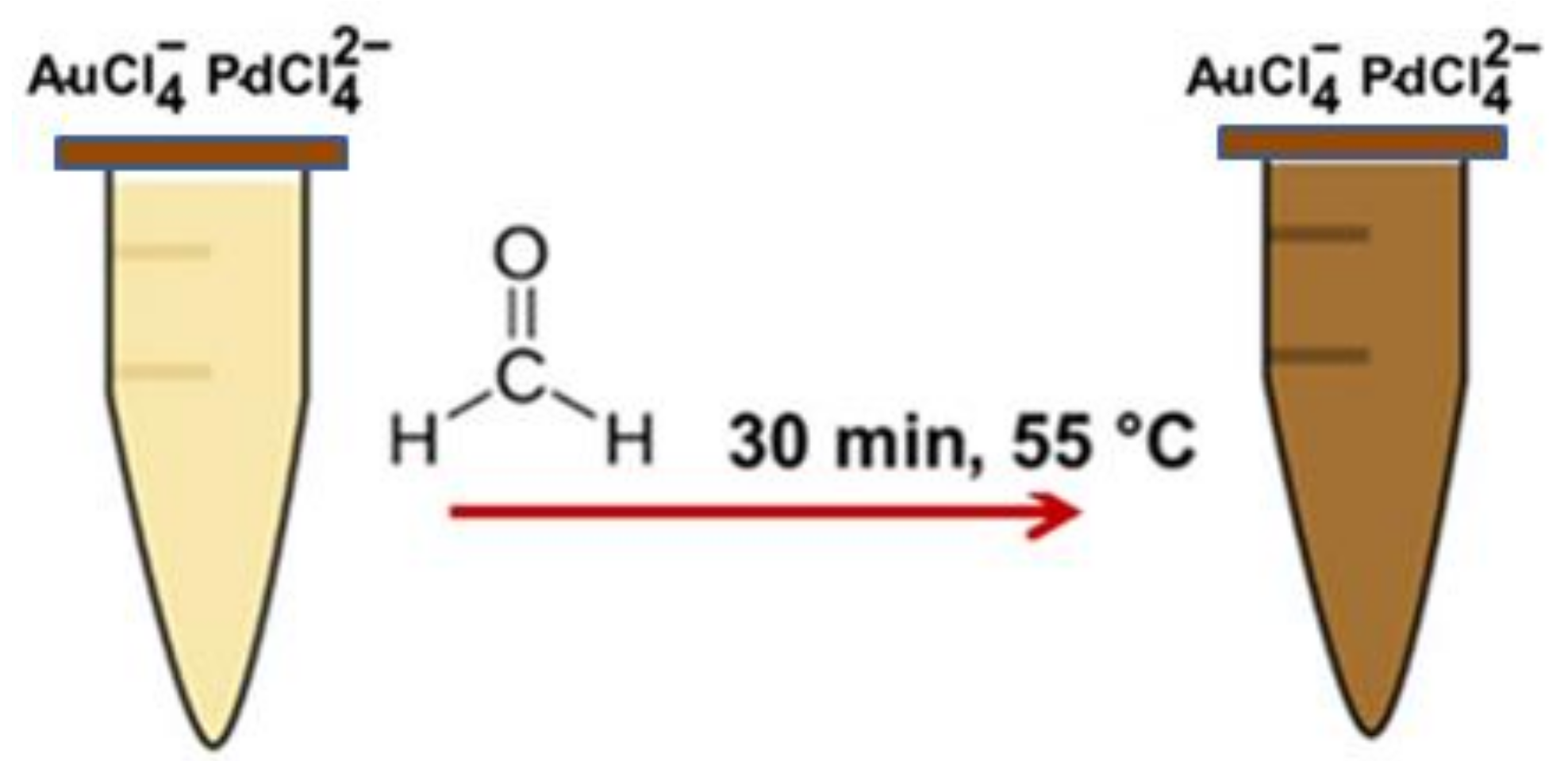
4.3. Colorimetric Detection
4.4. Electrochemical Reduction
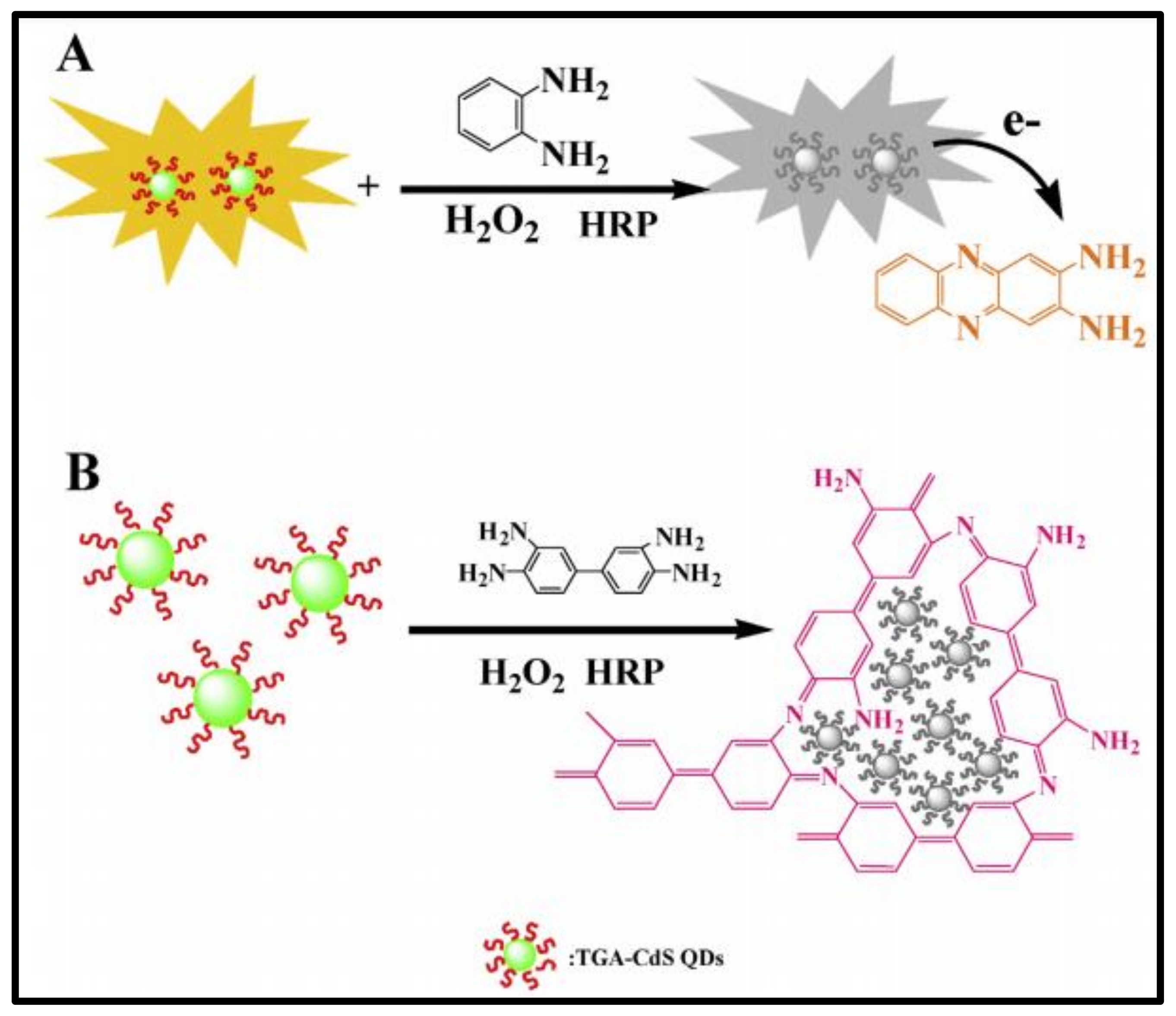
4.5. Fluorescence Quenching
4.6. Nonlocal Theories
5. Regulatory Facet of Nano-Enabled Sensors
6. Safety Concerns in Nano-Enabled Sensors
7. Unresolved Problems and Future Outlook
- Sensors are much more than just a molecule, and, hence, the molecular recognition through mere analytical protocols or “sensing schemes” would not suffice.
- Sensors must be applicable as such to monitor a chemical substance under varying environmental conditions.
- Sensors must be able to operate over different durations, such as few hours through a surgery and a few years as a component in automobile, or a month to monitor glucose in blood of diabetic.
- Sensors must support reversible reactions and respond reversibly towards surrounding oxygen, temperature etc.
- Sensors must not be equated to probes, as probes cannot undergo irreversible reactions while responding to unidirectional signals.
8. Our Contribution to This Field
9. Conclusions
Author Contributions
Funding
Data Availability Statement
Acknowledgments
Conflicts of Interest
References
- Yu, X.; Kilani, M.; Siddiqui, A.; Mao, G. One-Step Synthesis of Charge-Transfer Salt Nanosensors on Microelectrode Patterns. Adv. Mater. Technol. 2020, 5, 2000554. [Google Scholar] [CrossRef]
- Di Lecce, V.; Calabrese, M. From Smart to Intelligent Sensors: A Case Study, Sensors and Transducers, Toronto. Sens. Transducers 2012, 14, 1–17. Available online: https://www.sensorsportal.com/HTML/DIGEST/P_SI_178.htm (accessed on 31 December 2021).
- Bogue, R. Towards the trillion sensors market. Sens. Rev. 2014, 34, 137–142. [Google Scholar] [CrossRef]
- Rasmussen, M.K.; Pedersen, J.N.; Marie, R. A fit to the size and surface charge characterization of nanoparticles with a salt gradient. Nat. Commun. 2020, 11, 2337. Available online: https://www.nature.com/articles/s41467-020-15889-3 (accessed on 31 December 2021). [CrossRef]
- Xie, L.; Wang, P.; Qian, Y.; Rao, L.; Yin, H.; Wang, X.; Chen, H.; Zhou, G.; Nötzel, R. Spatial Surface Charge Engineering for Electrochemical Electrodes. Sci. Rep. 2019, 9, 14489. [Google Scholar] [CrossRef] [PubMed]
- Zhou, H.; Liu, J.; Xu, J.J.; Zhang, S.S.; Chen, H.Y. Optical nano-biosensing interface via nucleic acid amplification strategy: Construction and application. Chem. Soc. Rev. 2018, 47, 1996. [Google Scholar] [CrossRef]
- Dong, B.; Shi, Q.; He, T.; Zhu, S.; Zhang, Z.; Sun, Z.; Ma, Y.; Kwong, D.; Lee, C. Wearable Triboelectric/Aluminum Nitride Nano-Energy-Nano-System with Self-Sustainable Photonic Modulation and Continuous Force Sensing. Adv. Sci. 2020, 7, 1903636. [Google Scholar] [CrossRef]
- Arachchige, H.M.M.; Zappa, D.; Poli, N.; Gunawardhana, N.; Comini, E. Gold functionalized MoO3 nano flakes for gas sensing applications. Sens. Actuators B Chem. 2018, 269, 331–339. [Google Scholar] [CrossRef]
- Ma, Y.; Bao, J.; Zhang, Y.; Li, Z.; Zhou, X.; Wan, C.; Huang, L.; Zhao, Y.; Han, G.; Xue, T. Mammalian Near-Infrared Image Vision through Injectable and Self-Powered Retinal Nanoantennae. Cell 2019, 177, 243–255. [Google Scholar] [CrossRef]
- Dogan, E.; Ozkazanc, E.; Ozkazanc, H. Multifunctional polyindole/nanometal-oxide composites: Optoelectronic and charge transport properties. Synth. Met. 2019, 256, 116154. [Google Scholar] [CrossRef]
- Dheyab, A.B.; Mohammed, S.I.; Mustafa, M.K.; Fayadh, R.S.; Hussein, N.L. Fabrication and characterization of PSi/nanometal hybrid structures by laser for CO gas sensor. J. Theor. Appl. Phys. 2020, 14, 107–112. [Google Scholar] [CrossRef]
- Xue, B.; Yang, Y.; Tang, R.; Sun, Y.; Sun, S.; Cao, X.; Li, P.; Zhang, Z.; Li, X. One-step hydrothermal synthesis of a flexible nanopaper-based Fe3+ sensor using carbon quantum dot grafted cellulose nanofibrils. Cellulose 2019, 27, 729–742. [Google Scholar] [CrossRef]
- Zhang, L.; Zhang, H.; Chu, X.; Han, X. One-dimensional mesoporous CO3O4 tubules for enhanced performance supercapacitor and enzymeless glucose sensing. Ionics 2019, 25, 5445–5458. [Google Scholar] [CrossRef]
- Tian, Y.; Lu, Q.; Guo, X.; Wang, S.; Gao, Y.; Wang, L.-H. Au nanoparticles deposited on ultrathin two-dimensional covalent organic framework nanosheets for in vitro and intracellular sensing. Nanoscale 2020, 12, 7776–7781. [Google Scholar] [CrossRef] [PubMed]
- Ricciardella, F.; Lee, K.; Stelz, T.; Hartwig, O.; Prechtl, M.; McCrystall, M.; McEvoy, N.; Duesberg, G.S. Calibration of Nonstationary Gas Sensors Based on Two-Dimensional Materials. ACS Omega 2020, 5, 5959–5963. [Google Scholar] [CrossRef]
- Litti, L.; Ramundo, A.; Biscaglia, F.; Toffoli, G.; Gobbo, M.; Meneghetti, M. A surface enhanced Raman scattering based colloid nanosensor for developing therapeutic drug monitoring. J. Colloid Interface Sci. 2018, 533, 621–626. [Google Scholar] [CrossRef]
- Yang, F.; Guo, J.; Zhao, L.; Shang, W.; Gao, Y.; Zhang, S.; Gu, G.; Zhang, B.; Cui, P.; Cheng, G.; et al. Tuning oxygen vacancies and improving UV sensing of ZnO nanowire by micro-plasma powered by a triboelectric nanogenerator. Nano Energy 2019, 67, 104210. [Google Scholar] [CrossRef]
- Gao, H.; Kam, C.; Chou, T.Y.; Wu, M.Y.; Zhao, X.; Chen, S. A simple yet effective AIE-based fluorescent nano-thermometer for temperature mapping in living cells using fluorescence lifetime imaging microscopy. Nanoscale Horiz. 2020, 5, 488–494. [Google Scholar] [CrossRef]
- Chen, J.; Qiu, H.; Zhao, S. Fabrication of chemiluminescence resonance energy transfer platform based on nanomaterial and its application in optical sensing, biological imaging and photodynamic therapy. TrAC Trends Anal. Chem. 2019, 122, 115747. [Google Scholar] [CrossRef]
- Zhang, X.; Cao, H.; Zhao, J.; Wang, H.; Xing, B.; Chen, Z.; Li, X.; Zhang, J. Graphene oxide exhibited positive effects on the growth of Aloe vera L. Physiol. Mol. Biol. Plants 2021, 27, 815–824. [Google Scholar] [CrossRef]
- Mahmoud, N.E.; Abdelhameed, R.M. Superiority of modified graphene oxide for enhancing the growth, yield, and antioxidant potential of pearl millet (Pennisetum glaucum L.) under salt stress. Plant Stress 2021, 2, 100025. [Google Scholar] [CrossRef]
- Siddiqui, M.S.; Palaparthy, V.S.; Kalita, H.; Baghini, M.S.; Aslam, M. Graphene Oxide Array for In-Depth Soil Moisture Sensing toward Optimized Irrigation. ACS Appl. Electron. Mater. 2020, 2, 4111–4121. [Google Scholar] [CrossRef]
- Lakshmiprasanna, H.R.; Manjunatha, K.; Husain, J. Effect of cerium on structural, microstructural, magnetic and humidity sensing properties of Mn–Bi ferrites. Nano-Struct. Nano-Objects 2020, 24, 100608. [Google Scholar] [CrossRef]
- Surya, S.G.; Yuvaraja, S.; Varrla, E.; Baghini, M.S.; Palaparthy, V.S.; Salama, K.N. An in-field integrated capacitive sensor for rapid detection and quantification of soil moisture. Sens. Actuators B Chem. 2020, 321, 128542. [Google Scholar] [CrossRef]
- Serban, B.C.; Buiu, O.; Dumbravescu, N.; Cobianu, C.; Avramescu, V.; Brezeanu, M.; Bumbac, M.; Pachiu, C.; Nicolescu, C.M. Oxidized Carbon Nanohorn-Hydrophilic Polymer Nanocomposite as the Resistive Sensing Layer for Relative Humidity. Anal. Lett. 2020, 54, 527–540. [Google Scholar] [CrossRef]
- Atar, N.; Yola, M.L. Core-Shell Nanoparticles/Two-Dimensional (2D) Hexagonal Boron Nitride Nanosheets with Molecularly Imprinted Polymer for Electrochemical Sensing of Cypermethrin. J. Electrochem. Soc. 2018, 165, H255–H262. [Google Scholar] [CrossRef]
- Ebrahimiasl, S.; Seifi, R.; Nahli, R.E.; Zakaria, A. Ppy/Nanographene Modified Pencil Graphite Electrode Nanosensor for Detection and Determination of Herbicides in Agricultural Water. Sci. Adv. Mater. 2017, 9, 2045–2053. [Google Scholar] [CrossRef]
- Tafreshi, F.A.; Fatahi, Z.; Ghasemi, S.F.; Taherian, A.; Esfandiari, N. Ultrasensitive fluorescent detection of pesticides in real sample by using green carbon dots. PLoS ONE 2020, 15, e0230646. [Google Scholar] [CrossRef]
- Yang, L.; Zhang, X.; Jiang, L. Determination of Organophosphorus Pesticides in Fortified Tomatoes by Fluorescence Quenching of Cadmium Selenium–Zinc Sulfide Quantum Dots. Anal. Lett. 2018, 52, 729–744. [Google Scholar] [CrossRef]
- Park, M.; Kim, H.S.; Kim, T.; Kim, J.; Seo, S.; Lee, B.Y. Real-time monitoring of microbial activity using hydrogel-hybridized carbon nanotube transistors. Sens. Actuators B Chem. 2018, 263, 486–492. [Google Scholar] [CrossRef]
- Cheraghi, S.; Taher, M.A.; Karimi-Maleh, H.; Karimi, F.; Shabani-Nooshabadi, M.; Alizadeh, M.; Al-Othman, A.; Erk, N.; Raman, P.K.Y.; Karaman, C. Novel enzymatic graphene oxide based biosensor for the detection of glutathione in biological body fluids. Chemosphere 2021, 287, 132187. [Google Scholar] [CrossRef] [PubMed]
- Wang, T.; Tao, Z.; Qu, C.; Wang, S.; Liu, Y. A cerium-based fluorescent nanosensor for highly specific detection of glutathione over cysteine and homocysteine. Analyst 2020, 146, 283–288. [Google Scholar] [CrossRef] [PubMed]
- Park, K.; Kuo, Y.; Shvadchak, V.; Ingargiola, A.; Dai, X.; Hsiung, L.; Kim, W.; Zhou, Z.H.; Zou, P.; Levine, A.J.; et al. Membrane insertion of—And membrane potential sensing by—Semiconductor voltage nanosensors: Feasibility demonstration. Sci. Adv. 2018, 4, e1601453. [Google Scholar] [CrossRef] [PubMed]
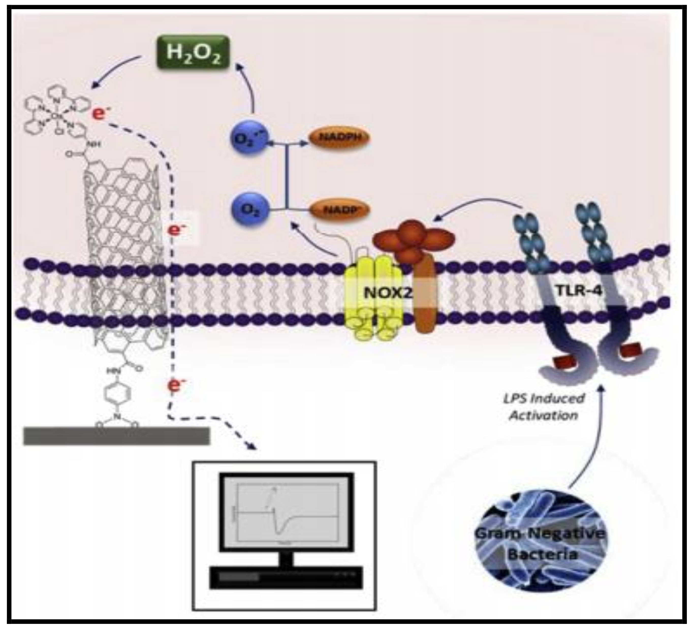
- Hicks, J.; Halkerston, R.; Silman, N.; Jackson, S.; Aylott, J.; Rawson, F. Real-time bacterial detection with an intracellular ROS sensing platform. Biosens. Bioelectron. 2019, 141, 111430. [Google Scholar] [CrossRef]
- Narang, J.; Mishra, A.; Pilloton, R.; Vv, A.; Wadhwa, S.; Pundir, C.S.; Khanuja, M. Development of MoSe2 Nano-Urchins as a Sensing Platform for a Selective Bio-Capturing of Escherichia. coli Shiga Toxin DNA. Biosensors 2018, 8, 77. [Google Scholar] [CrossRef]
- Avsievich, T.; Tarakanchikova, Y.; Zhu, R.; Popov, A.; Bykov, A.; Skovorodkin, I.; Vainio, S.; Meglinski, I. Impact of Nanocapsules on Red Blood Cells Interplay Jointly Assessed by Optical Tweezers and Microscopy. Micromachines 2019, 11, 19. [Google Scholar] [CrossRef]
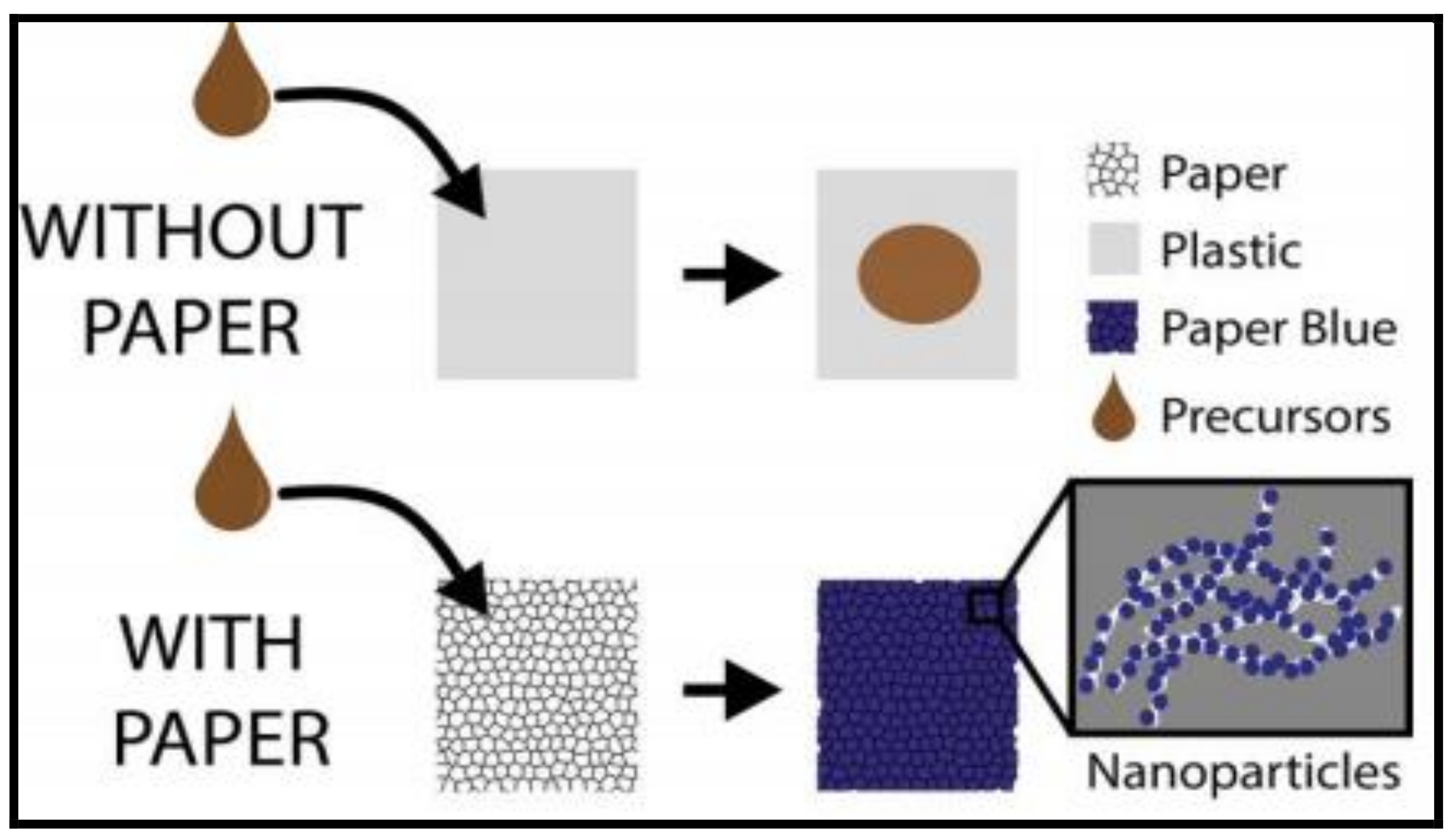
- Cinti, S.; Cusenza, R.; Moscone, D.; Arduini, F. Paper-based synthesis of Prussian Blue Nanoparticles for the development of whole blood glucose electrochemical biosensor. Talanta 2018, 187, 59–64. [Google Scholar] [CrossRef]
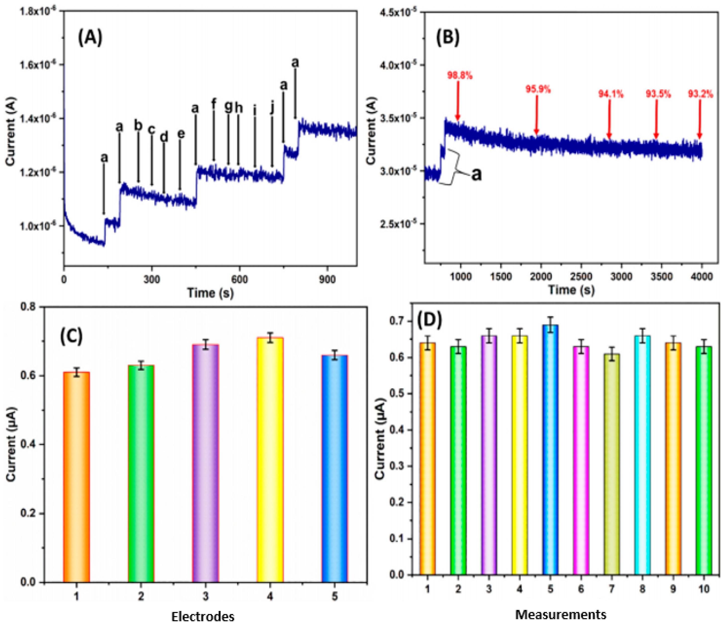
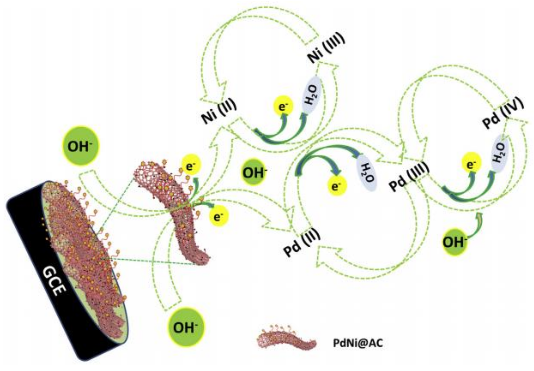
- Koskun, Y.; Şavk, A.; Şen, B.; Şen, F. Highly sensitive glucose sensor based on monodisperse palladium nickel/activated carbon nanocomposites. Anal. Chim. Acta 2018, 1010, 37–43. [Google Scholar] [CrossRef]
- Boussema, F.; Gross, A.; Hmida, F.; Ayed, B.; Majdoub, H.; Cosnier, S.; Maaref, A.; Holzinger, M. Dawson-type polyoxometalate nanoclusters confined in a carbon nanotube matrix as efficient redox mediators for enzymatic glucose biofuel cell anodes and glucose biosensors. Biosens. Bioelectron. 2018, 109, 20–26. [Google Scholar] [CrossRef]
- Muthusankar, E.; Ragupathy, D. Graphene/Poly(aniline-co-diphenylamine) nanohybrid for ultrasensitive electrochemical glucose sensor. Nano-Struct. Nano-Objects 2019, 20, 100390. [Google Scholar] [CrossRef]
- Samie, H.A.; Arvand, M. RuO2 nanowires on electrospun CeO2-Au nanofibers/functionalized carbon nanotubes/graphite oxide nanocomposite modified screen-printed carbon electrode for simultaneous determination of serotonin, dopamine and ascorbic acid. J. Alloys Compd. 2018, 782, 824–836. [Google Scholar] [CrossRef]
- Yao, G.; Lei, T.; Zhong, J.; Jiang, P.; Jia, W. Comparative evaluation of background subtraction algorithms in remote scene videos captured by MWIR sensors. Sensors 2017, 17, 1945. [Google Scholar] [CrossRef]
- Du, F.; Cheng, Z.; Wang, G.; Li, M.; Lu, W.; Shuang, S.; Dong, C. Carbon Nanodots as a Multifunctional Fluorescent Sensing Platform for Ratiometric Determination of Vitamin B2 and “Turn-Off” Detection of pH. J. Agric. Food Chem. 2021, 69, 2836–2844. [Google Scholar] [CrossRef]
- Liu, L.; Mi, Z.; Huo, X.; Yuan, L.; Bao, Y.; Liu, Z.; Feng, F. A label-free fluorescence nanosensor based on nitrogen and phosphorus co-doped carbon quantum dots for ultra-sensitive detection of new coccine in food samples. Food Chem. 2021, 368, 130829. [Google Scholar] [CrossRef]
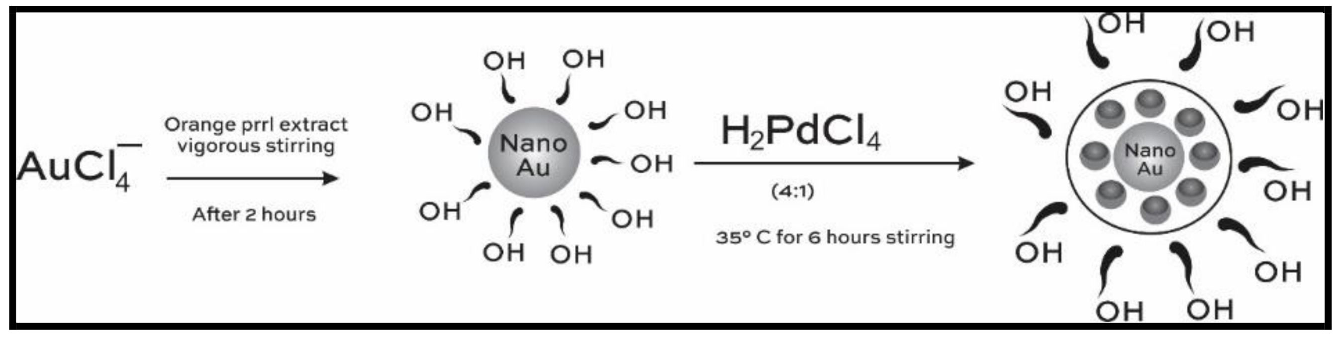
- Wicaksono, W.P.; Kadjabcd, T.M.G.; Amaliaa, D.; Uyuna, L.; Rini, W.P.; Hidayata, A.; Fahmi, R.L.; Nasriyantia, D.; Leun, S.G.V.; Ariyanta, H.A.; et al. A green synthesis of gold–palladium core–shell nanoparticles using orange peel extract through two-step reduction method and its formaldehyde colorimetric sensing performance. Nano-Struct. Nano-Objects 2020, 24, 100535. [Google Scholar] [CrossRef]
- Li, H.; Ahmad, W.; Rong, Y.; Chen, Q.; Zuo, M.; Ouyang, Q.; Guo, Z. Designing an aptamer based magnetic and upconversion nanoparticles conjugated fluorescence sensor for screening Escherichia coli in food. Food Control 2019, 107, 106761. [Google Scholar] [CrossRef]
- Xu, Y.; Dai, Y.; Li, C.; Zhang, H.; Guo, M.; Yang, Y. PC software-based portable cyclic voltammetry system with PB-MCNT-GNPs-modified electrodes for E. coli detection. Rev. Sci. Instrum. 2020, 91, 014103. [Google Scholar] [CrossRef]
- Zhong, M.; Yang, L.; Yang, H.; Cheng, C.; Deng, W.; Tan, Y.; Xie, Q.; Yao, S. An electrochemical immunobiosensor for ultrasensitive detection of Escherichia coli O157:H7 using CdS quantum dots-encapsulated metal-organic frameworks as signal-amplifying tags. Biosens. Bioelectron. 2018, 126, 493–500. [Google Scholar] [CrossRef] [PubMed]
- Shaibani, P.M.; Etayash, H.R.; Jiang, K.; Sohrabi, A.; Hassanpourfard, M.; Naicker, S.; Sadrzadeh, M.; Thundat, T. Portable Nanofiber-Light Addressable Potentiometric Sensor for Rapid Escherichia coli Detection in Orange Juice. ACS Sens. 2018, 3, 815–822. [Google Scholar] [CrossRef]
- Chen, Z.-G.; Zhong, H.-X.; Luo, H.; Zhang, R.-Y.; Huang, J.-R. Recombinase Polymerase Amplification Combined with Unmodified Gold Nanoparticles for Salmonella Detection in Milk. Food Anal. Methods 2018, 12, 190–197. [Google Scholar] [CrossRef]
- Du, J.; Wu, S.; Niu, L.; Li, J.; Zhao, D.; Bai, Y. A gold nanoparticles-assisted multiplex PCR assay for simultaneous detection of Salmonella typhimurium, Listeria monocytogenes and Escherichia coli O157:H7. Anal. Methods 2019, 12, 212–217. [Google Scholar] [CrossRef]
- Yousefi, A.; Babaei, A.; Delavar, M. Application of modified screen-printed carbon electrode with MWCNTs-Pt-doped CdS nanocomposite as a sensitive sensor for determination of natamycin in yoghurt drink and cheese. J. Electroanal. Chem. 2018, 822, 1–9. [Google Scholar] [CrossRef]
- Kampeera, J.; Pasakon, P.; Karuwan, C.; Arunrut, N.; Sappat, A.; Sirithammajak, S.; Dechokiattawan, N.; Sumranwanich, T.; Chaivisuthangkura, P.; Ounjai, P.; et al. Point-of-care rapid detection of Vibrio parahaemolyticus in seafood using loop-mediated isothermal amplification and graphene-based screen-printed electrochemical sensor. Biosens. Bioelectron. 2019, 132, 271–278. [Google Scholar] [CrossRef]
- Tian, Y.; Chen, Y.; Chen, M.; Song, Z.-L.; Xiong, B.; Zhang, X.-B. Peroxidase-like Au@Pt nanozyme as an integrated nanosensor for Ag+ detection by LSPR spectroscopy. Talanta 2020, 221, 121627. [Google Scholar] [CrossRef]
- Liu, J.; Ye, L.Y.; Mo, Y.Y.; Yang, H. Highly sensitive fluorescent quantification of acid phosphatase activity and its inhibitor pesticide Dufulin by a functional metal–organic framework nanosensor for environment assessment and food safety. Food Chem. 2021, 370, 131034. [Google Scholar] [CrossRef]
- Hashemi, S.A.; Mousavi, S.M.; Bahrani, S.; Ramakrishna, S. Integrated polyaniline with graphene oxide-iron tungsten nitride nanoflakes as ultrasensitive electrochemical sensor for precise detection of 4-nitrophenol within aquatic media. J. Electroanal. Chem. 2020, 873, 114406. [Google Scholar] [CrossRef]
- Meena, S.; Anantharaju, K.S.; Vidya, Y.S.; Renuka, L.; Malini, S.; Sharma, S.C.; Nagabhushana, H. MnFe2O4/ZrO2 nanocomposite as an efficient magnetically separable photocatalyst with good response to sunlight: Preparation, characterization and catalytic mechanism. SN Appl. Sci. 2020, 2, 328. [Google Scholar] [CrossRef]
- Murthy, H.C.A.; Ghotekar, S.; Kumar, B.V.; Roy, A. Graphene: A Multifunctional Nanomaterial with Versatile Applications. Adv. Mater. Sci. Eng. 2021, 2021, 1–8. [Google Scholar] [CrossRef]
- Roy, A.; Elzaki, A.; Tirth, V.; Kajoak, S.; Osman, H.; Algahtani, A.; Islam, S.; Faizo, N.L.; Khandaker, M.U.; Islam, M.N.; et al. Biological Synthesis of Nanocatalysts and Their Applications. Catalysts 2021, 11, 1494. [Google Scholar] [CrossRef]
- Karimi-Maleh, H.; Fakude, C.; Mabuba, N.; Peleyeju, G.M.; Arotiba, O.A. The determination of 2-phenylphenol in the presence of 4-chlorophenol using nano-Fe3O4/ionic liquid paste electrode as an electrochemical sensor. J. Colloid Interface Sci. 2019, 554, 603–610. [Google Scholar] [CrossRef]
- Myung, Y.; Jung, S.; Tung, T.T.; Tripathi, K.M.; Kim, T. Graphene-Based Aerogels Derived from Biomass for Energy Storage and Environmental Remediation. ACS Sustain. Chem. Eng. 2019, 7, 3772–3782. [Google Scholar] [CrossRef]
- Hou, X.; Pan, Y.; Xiao, H.; Liu, J. Controlled Release of Agrochemicals Using pH and Redox Dual-Responsive Cellulose Nanogels. J. Agric. Food Chem. 2019, 67, 6700–6707. [Google Scholar] [CrossRef] [PubMed]
- Chauhan, P.; Saini, J.; Chaudhary, S. Agarose waste derived toxicologically screened carbon dots as dual sensor: A mechanistic insight into luminescence and solvatochromic behaviour. Nano-Struct. Nano-Objects 2020, 24, 100585. [Google Scholar] [CrossRef]
- Anju, M.; Akhila, A.; Renuka, N. Non-covalently functionalised rGO–fluorescein unit for selective detection of fluoride ions. Nano-Struct. Nano-Objects 2020, 24, 100606. [Google Scholar] [CrossRef]
- Maruthapandi, M.; Das, P.; Saravanan, A.; Natan, M.; Banin, E.; Kannan, S.; Michaeli, S.; Luong, J.H.; Gedanken, A. Biocompatible N-doped carbon dots for the eradication of methicillin-resistant S. aureus (MRSA) and sensitive analysis for europium (III). Nano-Struct. Nano-Objects 2021, 26, 100724. [Google Scholar] [CrossRef]
- Oluwafemi, O.S.; Anyik, J.L.; Zikalala, N.E.; Sakho, E.H.M. Biosynthesis of silver nanoparticles from water hyacinth plant leaves extract for colourimetric sensing of heavy metals. Nano-Struct. Nano-Objects 2019, 20, 100387. [Google Scholar] [CrossRef]
- Xiao, X.; Liu, L.; Ma, J.; Ren, Y.; Cheng, X.; Zhu, Y.; Zhao, D.; Elzatahry, A.A.; Alghamdi, A.; Deng, Y. Ordered Mesoporous Tin Oxide Semiconductors with Large Pores and Crystallized Walls for High-Performance Gas Sensing. ACS Appl. Mater. Interfaces 2018, 10, 1871–1880. [Google Scholar] [CrossRef]
- Tit, N.; Othman, W.; Shaheen, A.; Ali, M. High selectivity of N-doped ZnO nano-ribbons in detecting H2, O2 and CO2 molecules: Effect of negative-differential resistance on gas-sensing. Sens. Actuators B Chem. 2018, 270, 167–178. [Google Scholar] [CrossRef]
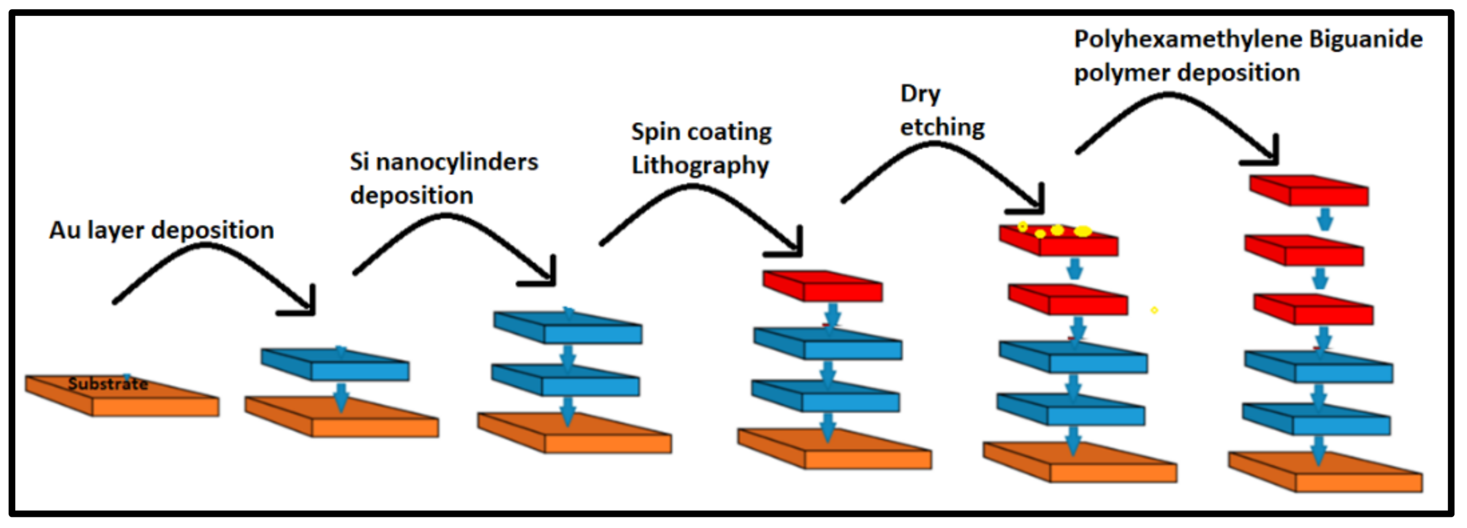
- Kazanskiy, N.L.; Butt, M.A.; Khonina, S.N. Carbon Dioxide Gas Sensor Based on Polyhexamethylene Biguanide Polymer Deposited on Silicon Nano-Cylinders Metasurface. Sensors 2021, 21, 378. [Google Scholar] [CrossRef]
- Bilge, S.; Dogan-Topal, B.; Atici, E.B.; Sınağ, A.; Ozkan, S.A. Rod-like CuO nanoparticles/waste masks carbon modified glassy carbon electrode as a voltammetric nanosensor for the sensitive determination of anti-cancer drug pazopanib in biological and pharmaceutical samples. Sens. Actuators B Chem. 2021, 343, 130109. [Google Scholar] [CrossRef]
- Bilici, A.; Denizhan, N.; Emre, D.; Soylukan, C.; Algi, F.; Yilmaz, S. Fabrication of PAMP/Au and GO/PAMP/Au nanosensors for electrochemical detection of paracetamol in pharmaceutical preparations. Mon. Für Chem.–Chem. 2021, 152, 1539–1552. [Google Scholar] [CrossRef]
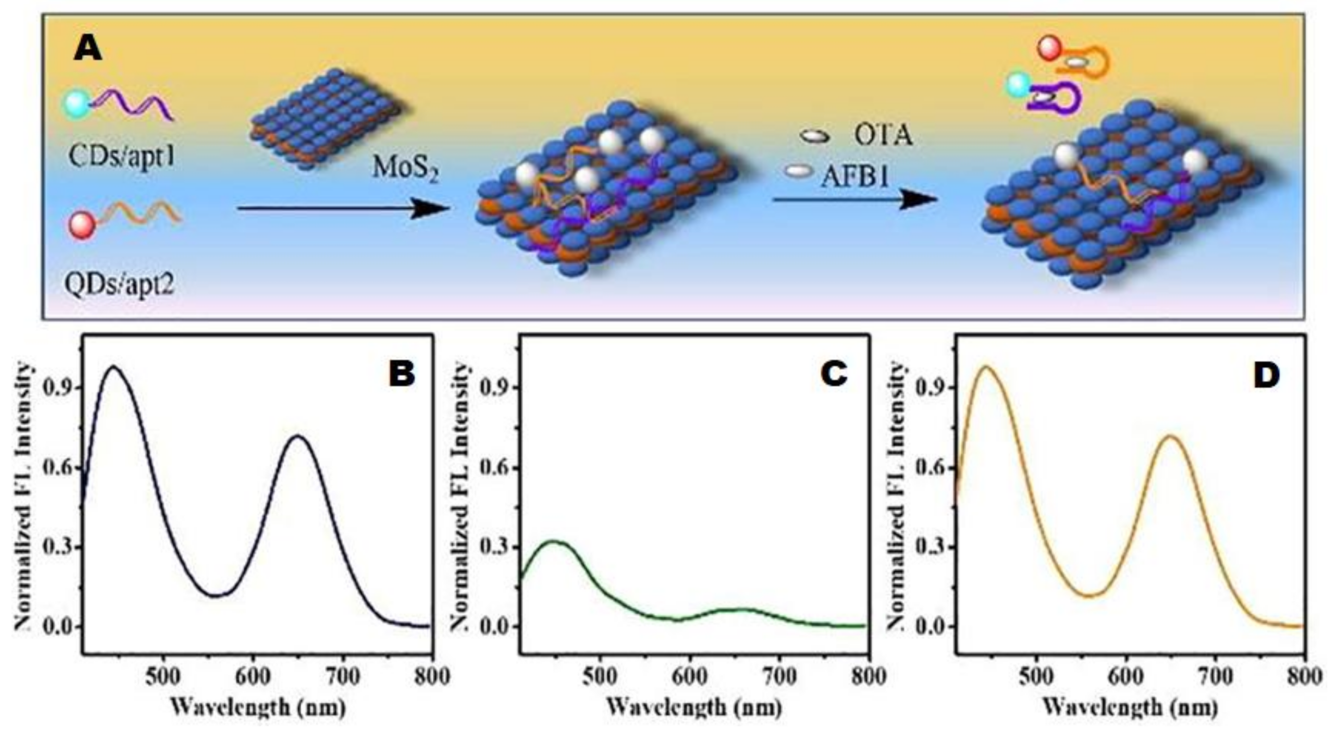
- Qian, J.; Cui, H.; Lu, X.; Wang, C.; An, K.; Hao, N.; Wang, K. Bi-color FRET from two nano-donors to a single nano-acceptor: A universal aptasensing platform for simultaneous determination of dual targets. Chem. Eng. J. 2020, 401, 126017. [Google Scholar] [CrossRef]
- Soleimani, S.; Arkan, E.; Farshadnia, T.; Mahnam, Z.; Jalili, F.; Goicoechea, H.C.; Jalalvand, A.R. The first attempt on fabrication of a nano-biosensing platform and exploiting first-order advantage from impedimetric data: Application to simultaneous biosensing of doxorubicin, daunorubicin and idarubicin. Sens. Bio-Sens. Res. 2020, 29, 100366. [Google Scholar] [CrossRef]
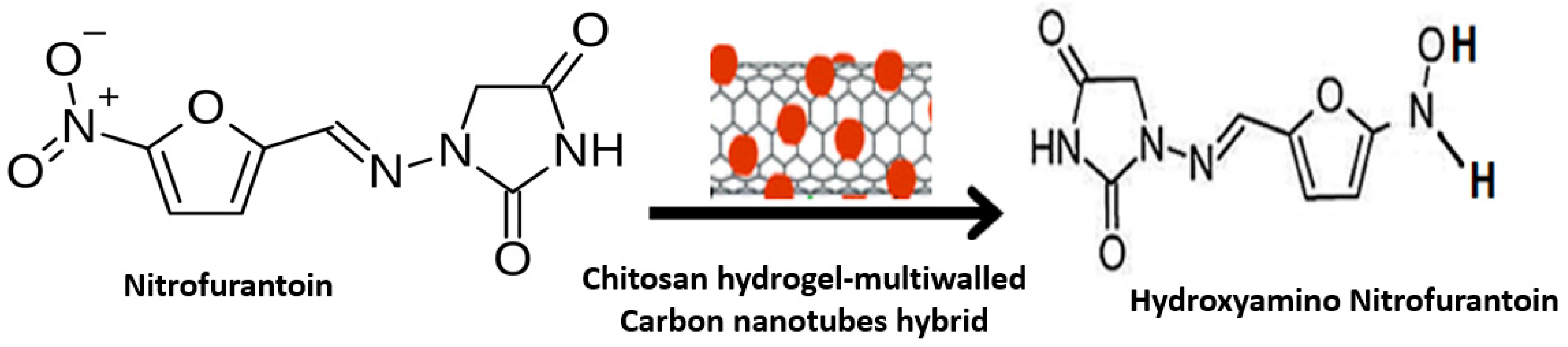
- Velmurugan, S.; Palanisamy, S.; Yang, T.C.-K.; Gochoo, M.; Chen, S.-W. Ultrasonic assisted functionalization of MWCNT and synergistic electrocatalytic effect of nano-hydroxyapatite incorporated MWCNT-chitosan scaffolds for sensing of nitrofurantoin. Ultrason. Sonochemistry 2019, 62, 104863. [Google Scholar] [CrossRef]
- Zhang, F.; Yao, H.; Chu, T.; Zhang, G.; Wang, Y.; Yang, Y. A Lanthanide MOF Thin-Film Fixed with CO3O4 Nano-Anchors as a Highly Efficient Luminescent Sensor for Nitrofuran Antibiotics. Chem. Eur. J. 2017, 23, 10293–10300. [Google Scholar] [CrossRef]
- Meena, S.; Anantharaju, K.; Malini, S.; Dey, A.; Renuka, L.; Prashantha, S.; Vidya, Y. Impact of temperature-induced oxygen vacancies in polyhedron MnFe2O4 nanoparticles: As excellent electrochemical sensor, supercapacitor and active photocatalyst. Ceram. Int. 2020, 47, 14723–14740. [Google Scholar] [CrossRef]
- Meena, S.; Anantharaju, K.S.; Vidya, Y.S.; Renuka, L.; Uma, B.; Sharma, S.C.; More, S.S. Enhanced sunlight driven photocatalytic activity and electrochemical sensing properties of Ce-doped MnFe2O4 nano magnetic ferrites. Ceram. Int. 2020, 47, 14760–14774. [Google Scholar] [CrossRef]
- Pan, Y.; Liu, J.; Yang, K.; Cai, P.; Xiao, H. Novel multi-responsive and sugarcane bagasse cellulose-based nanogels for controllable release of doxorubicin hydrochloride. Mater. Sci. Eng. C 2020, 118, 111357. [Google Scholar] [CrossRef] [PubMed]
- Azad, L.M.; Ehtesabi, H.; Rezaei, A. Smartphone-based fluorometer for pH detection using green synthesized carbon dots. Nano-Struct. Nano-Objects 2021, 26, 100722. [Google Scholar] [CrossRef]
- Haldavnekar, R.; Venkatakrishnan, K.; Tan, B. Boosting the sub-cellular biomolecular cancer signals by self-functionalized tag-free nano sensor. Biosens. Bioelectron. 2021, 190, 113407. [Google Scholar] [CrossRef]
- Saha, P.; Panda, D.; Paul, R.; Dash, J. A DNA nanosensor for monitoring ligand-induced i-motif formation. Org. Biomol. Chem. 2021, 19, 1965–1969. [Google Scholar] [CrossRef] [PubMed]
- Yarman, A.; Kurbanoglu, S.; Zebger, I.; Scheller, F.W. Simple and robust: The claims of protein sensing by molecularly imprinted polymers. Sens. Actuators B Chem. 2020, 330, 129369. [Google Scholar] [CrossRef]
- He, J.-Y.; Shang, X.; Yang, C.-L.; Zuo, S.-Y.; Yuan, R.; Xu, W.-J. Antibody-Responsive Ratiometric Fluorescence Biosensing of Biemissive Silver Nanoclusters Wrapped in Switchable DNA Tweezers. Anal. Chem. 2021, 93, 11634–11640. [Google Scholar] [CrossRef] [PubMed]
- Li, J.; Wu, D.; Yu, Y.; Li, T.; Li, K.; Xiao, M.-M.; Li, Y.; Zhang, Z.-Y.; Zhang, G.-J. Rapid and unamplified identification of COVID-19 with morpholino-modified graphene field-effect transistor nanosensor. Biosens. Bioelectron. 2021, 183, 113206. [Google Scholar] [CrossRef]
- Ramon-Marquez, T.; Medina-Castillo, A.L.; Nagiah, N.; Fernandez-Gutierrez, A.; Fernandez-Sanchez, J.F. A multifunctional material based on co-electrospinning for developing biosensors with optical oxygen transduction. Anal. Chim. Acta 2018, 1015, 66–73. [Google Scholar] [CrossRef] [PubMed]
- Zappi, D.; Gabriele, S.; Gontrani, L.; Dini, D.; Sadun, C.; Marini, F.; Antonelli, M.L. Biologically friendly room temperature ionic liquids and nanomaterials for the development of innovative enzymatic biosensors: Part II. Talanta 2018, 194, 26–31. [Google Scholar] [CrossRef] [PubMed]
- Rezaei, B.; Shoushtari, A.M.; Rabiee, M.; Uzun, L.; Mak, W.C.; Turner, A.P. An electrochemical immunosensor for cardiac Troponin I using electrospun carboxylated multi-walled carbon nanotube-whiskered nanofibres. Talanta 2018, 182, 178–186. [Google Scholar] [CrossRef] [PubMed]
- Riberi, W.I.; Tarditto, L.V.; Zon, M.A.; Arévalo, F.; Fernández, H. Development of an electrochemical immunosensor to determine zearalenone in maize using carbon screen printed electrodes modified with multi-walled carbon nanotubes/polyethyleneimine dispersions. Sens. Actuators B Chem. 2018, 254, 1271–1277. [Google Scholar] [CrossRef]
- Anjum, S.; Narwade, V.; Bogle, K.A.; Khairnar, R.S. Graphite doped Hydroxyapatite nanoceramic: Selective alcohol sensor. Nano-Struct. Nano-Objects 2018, 14, 98–105. [Google Scholar] [CrossRef]
- Taha, S.; Begum, S.; Narwade, V.; Halge, D.; Dadge, J.W.; Mahabole, M.P.; Khairnar, R.S.; Bogle, K.A. Development of alcohol sensor using TiO2-Hydroxyapatite nano-composites. Mater. Chem. Phys. 2019, 240, 122228. [Google Scholar] [CrossRef]
- Su, B.; Zhang, Z.; Sun, Z.; Tang, Z.; Xie, X.; Chen, Q.; Cao, H.; Yu, X.; Xu, Y.; Liu, X.; et al. Fluonanobody-based nanosensor via fluorescence resonance energy transfer for ultrasensitive detection of ochratoxin A. J. Hazard. Mater. 2021, 422, 126838. [Google Scholar] [CrossRef] [PubMed]
- Avdeeva, D.K.; Maksimov, I.V.; Ivanov, M.L.; Yuzhakov, M.M.; Turushev, N.V.; Rybalka, S.A.; Batalov, R.E.; Guo, W.; Filippova, E.B. Results of measurements of the cardiac micropotential energies in the amplitude-time intervals recorded by the nanosensor-based hardware and software complex. Measurement 2020, 173, 108600. [Google Scholar] [CrossRef]
- Zhang, J.; Qin, Z.; Zeng, D.; Xie, C. Metal-oxide-semiconductor based gas sensors: Screening, preparation, and integration. Phys. Chem. Chem. Phys. 2017, 19, 6313–6329. [Google Scholar] [CrossRef]
- Wang, G.-L.; Hu, X.-L.; Wu, X.-M.; Li, Z.-J. Quantum dots-based glucose sensing through fluorescence quenching by bienzyme-catalyzed chromogenic substrate oxidation. Sens. Actuators B Chem. 2014, 205, 61–66. [Google Scholar] [CrossRef]
- Eringen, A.; Edelen, D. On nonlocal elasticity. Int. J. Eng. Sci. 1972, 10, 233–248. [Google Scholar] [CrossRef]
- Srividhya, S.; Raghu, P.; Rajagopal, A.; Reddy, J. Nonlocal nonlinear analysis of functionally graded plates using third-order shear deformation theory. Int. J. Eng. Sci. 2018, 125, 1–22. [Google Scholar] [CrossRef]
- de Sciarra, F.M. Variational formulations and a consistent finite-element procedure for a class of nonlocal elastic continua. Int. J. Solids Struct. 2008, 45, 4184–4202. [Google Scholar] [CrossRef][Green Version]
- Amanatidou, E.; Aravas, N. Mixed finite element formulations of strain-gradient elasticity problems. Comput. Methods Appl. Mech. Eng. 2002, 191, 1723–1751. [Google Scholar] [CrossRef]
- Barretta, R.; Faghidian, S.A.; de Sciarra, F.M.; Pinnola, F.P. Timoshenko nonlocal strain gradient nanobeams: Variational consistency, exact solutions and carbon nanotube Young moduli. Mech. Adv. Mater. Struct. 2019, 28, 1–14. [Google Scholar] [CrossRef]
- Barretta, R.; Fabbrocino, F.; Luciano, R.; De Sciarra, F.M.; Ruta, G. Buckling loads of nano-beams in stress-driven nonlocal elasticity. Mech. Adv. Mater. Struct. 2019, 27, 869–875. [Google Scholar] [CrossRef]
- Shariati, M.; Shishesaz, M.; Sahbafar, H.; Pourabdy, M.; Hosseini, M. A review on stress-driven nonlocal elasticity theory. J. Appl. Comput. Mech. 2021, 52, 535–552. [Google Scholar] [CrossRef]
- McGrath, M.J.; Ni Scanaill, C. Regulations and Standards: Considerations for Sensor Technologies. J. Sens. Technol. 2013, 115–135. [Google Scholar] [CrossRef]
- Ravizza, A.; De Maria, C.; Di Pietro, L.; Sternini, F.; Audenino, A.L.; Bignardi, C. Comprehensive Review on Current and Future Regulatory Requirements on Wearable Sensors in Preclinical and Clinical Testing. Front. Bioeng. Biotechnol. 2019, 7, 313. [Google Scholar] [CrossRef] [PubMed]
- Li, X.; Bao, J.; Sun, J.; Wang, J. Development of circular economy in smart cities based on FPGA and wireless sensors. Microprocess. Microsyst. 2020, 80, 103600. [Google Scholar] [CrossRef]
- Shah, M.; Badwaik, V.; Kherde, Y.; Waghwani, H.K.; Modi, T.; Aguilar, Z.P.; Rodgers, H.; Hamilton, W.; Marutharaj, T.; Webb, C.; et al. Gold nanoparticles: Various methods of synthesis and antibacterial applications. Front. Biosci. 2014, 19, 1320–1344. Available online: https://www.researchgate.net/publication/262843669 (accessed on 31 December 2021). [CrossRef]
- Zielińska, A.; Costa, B.; Ferreira, M.V.; Miguéis, D.; Louros, J.M.S.; Durazzo, A.; Lucarini, M.; Eder, P.; Chaud, M.V.; Morsink, M.; et al. Nanotoxicology and Nanosafety: Safety-By-Design and Testing at a Glance. Int. J. Environ. Res. Public Health 2020, 17, 4657. [Google Scholar] [CrossRef] [PubMed]
- Dahlin, A.B. Size Matters: Problems and Advantages Associated with Highly Miniaturized Sensors. Sensors 2012, 12, 3018–3036. [Google Scholar] [CrossRef]
- Thakkar, S.; Dumee, L.F.; Gupta, M.; Singh, B.R.; Yang, W. Nano–enabled sensors for detection of arsenic in water. Water Res. 2021, 188, 116538. [Google Scholar] [CrossRef] [PubMed]
- Yang, T.; Duncan, T.V. Challenges and potential solutions for nanosensors intended for use with foods. Nature Nanotechnol. 2021, 16, 251–265. [Google Scholar] [CrossRef]
- Surmenev, R.A.; Chernozem, R.V.; Pariy, I.O.; Surmeneva, M.A. A review on piezo-and pyroelectric responses of flexible nano-and micropatterned polymer surfaces for biomedical sensing and energy harvesting applications. Nano Energy 2021, 79, 105442. [Google Scholar] [CrossRef]
- Shi, J.X.; Lei, X.W.; Natsuki, T. Review on Carbon Nanomaterials-Based Nano-Mass and Nano-Force Sensors by Theoretical Analysis of Vibration Behavior. Sensors 2021, 21, 1907. [Google Scholar] [CrossRef] [PubMed]
- Kalyani, N.; Goel, S.; Jaiswal, S. On-site sensing of pesticides using point-of-care biosensors: A review. Environ. Chem. Lett. 2021, 19, 345–354. [Google Scholar] [CrossRef]
- Rangayasami, A.; Kannan, K.; Murugesan, S.; Radhika, D.; Sadasivuni, K.K.; Reddy, K.R.; Raghu, A.V. Influence of nanotechnology to combat against COVID-19 for global health emergency: A review. Sens. Int. 2021, 100079. [Google Scholar] [CrossRef] [PubMed]
- Niu, H.; Zhang, H.; Yue, W.; Gao, S.; Kan, H.; Zhang, C.; Zhang, C.; Pang, J.; Lou, Z.; Wang, L.; et al. Micro-Nano Processing of Active Layers in Flexible Tactile Sensors via Template Methods: A Review. Small 2021, 17, 2100804. [Google Scholar] [CrossRef] [PubMed]
- Sowmya, B.; John, A.; Panda, P.K. A review on metal-oxide based pn and nn heterostructured nano-materials for gas sensing applications. Sens. Int. 2021, 100085. [Google Scholar]
- Alafeef, M.; Moitra, P.; Pan, D. Nano-enabled sensing approaches for pathogenic bacterial detection. Biosens. Bioelectron. 2020, 165, 112276. [Google Scholar] [CrossRef]
- Kah, M.; Tufenkji, N.; White, J.C. Nano-enabled strategies to enhance crop nutrition and protection. Nat. Nanotechnol. 2019, 14, 532–540. [Google Scholar] [CrossRef]
- Singh, G.; Kalia, A. Nano-enabled technological interventions for sustainable production, protection, and storage of fruit crops. In Nanoscience for Sustainable Agriculture; Springer: Cham, Switzerland, 2019; pp. 299–322. [Google Scholar]
- Xin, X.; Judy, J.D.; Sumerlin, B.B.; He, Z. Nano-enabled agriculture: From nanoparticles to smart nanodelivery systems. Environ. Chem. 2020, 17, 413–425. [Google Scholar] [CrossRef]
- Tiwari, S.; Sharma, V.; Mujawar, M.; Mishra, Y.K.; Kaushik, A.; Ghosal, A. Biosensors for epilepsy management: State-of-art and future aspects. Sensors 2019, 19, 1525. [Google Scholar] [CrossRef]


| Year | Reference | Title | Journal | Highlights |
|---|---|---|---|---|
| 2021 | [108] | Nano–Enabled sensors for detection of arsenic in water. | ||
| 2021 | [109] | Challenges and potential solutions for Nano sensors intended for use with foods. | Nature nanotechnology | Provides a critical overview of technical, regulatory, political, legal, economic, environmental health and safety, and ethical hurdles associated with sensors in food industry. |
| 2021 | [110] | A review on piezo- and pyroelectric responses of flexible nano- and micro patterned polymer surfaces for biomedical sensing and energy harvesting applications. | Nano energy | Reveals nano-structuring of the surface of biocompatible bio sensing widely applied in the field of microfluidic nano-actuated devices, smart drug-delivery systems, and multiferroic systems. |
| 2021 | [111] | Review on Carbon Nanomaterials-Based Nano-Mass and Nano-Force Sensors by Theoretical Analysis of Vibration Behavior. | Sensors | Evaluates the developments in nano-mechanical sensors focussing on modeling perspective, continuum mechanical approaches of carbon nanomaterials, symbolic works of CNTs/GSs/carbyne-based nano-mass and nano-force sensors. |
| 2021 | [112] | On-site sensing of pesticides using point-of-care biosensors: A review. | Environmental chemistry letters | Overviews latest biosensors developed, which can be utilized for on-site sensing and optical biosensors are at the forefront of technology with advantages such as easy protocols, simple operation, high sensitivity, broad linearity range, and cost-effectiveness. |
| 2021 | [113] | Influence of nanotechnology to combat against COVID-19 for global health emergency: A review. | Sensors international | Discusses the development of Nano-enabled sensors towards quick immunization improvement of COVID-19. |
| 2021 | [114] | Micro-Nano Processing of Active Layers in Flexible Tactile Sensors via Template Methods: A Review. | Nano-micro-small | Compares the shortcomings and advantages of Sensors via Template Methods to promote the cross-integration of multiple fields and accelerate the development of flexible electronic devices. |
| 2021 | [115] | A review on metal- oxide based p-n and n heterostructured nano-materials for gas sensing applications. | Sensors International | Studies the sensors detecting the morphologies of nano rods, nanosheets, nanobelts, nanoribbons, nanowires, nano flowers, spinel, and their market trends. |
| 2020 | [116] | Nano-enabled sensing approaches for pathogenic bacterial detection. | Biosensors and Bioelectronics | A comprehensive discussion of the commonly adopted techniques for bacterial identification and a prospective outlook of challenges and solutions is presented. |
| 2019 | [117] | Nano-enabled strategies to enhance crop nutrition and protection. | Nature Nanotechnology | Nano-enabled sensing strategies are presented in crop production with a new perspective of profit margin and regulatory aspects in the future agri-business sector. |
| 2019 | [118] | Nano-Enabled Technological Interventions for Sustainable Production, Protection, and Storage of Fruit Crops. | Nanoscience for Sustainable Agriculture | Reviews various aspects of nano interventions of Agro nanotechnology. |
| 2020 | [119] | Nano-enabled agriculture: from nanoparticles to smart Nano delivery systems. | Environmental Chemistry | Extends a systematic study of sensors in food production and plant nutrition. |
| 2019 | [120] | Biosensors for Epilepsy Management: State-of-Art and Future Aspects. | Sensors | Presents highlights on advancements in state-of-art smart nano-enabled bio sensing. |
| 2019 | [118] | Nano-Enabled Technological Interventions for Sustainable Production, Protection, and Storage of Fruit Crops. | Nanoscience for sustainable agriculture | Explores the various aspects of nano interventions through nanosensors in agrinanotechnology. |
| Nanomaterial | Size | Enables Detecting | Negative Impact |
|---|---|---|---|
| Microgel | 200–400 nm | Water retention | Alters water acquisition |
| Nano Biopolymer | 40–1000 nm | Nutrient absorption | Alters nutrient acquisition |
| Multiple emulsions | 65–500 nm | Concentration of peptides | Influences secondary metabolite production |
| Filled microgel | 370–970 nm | Soil conditions | Impedimentation of seed germination |
| Microclusters | 250–460 nm | Pesticide detection | Oxidation of pesticides |
| Nano Material in Sensor | Sensing Methodology | Advantages | Drawbacks | Ref |
|---|---|---|---|---|
| Hydrogel hybridised carbon nanotube | Metabolism of microbial species causes variation in conductance of nanomaterial | Real time detection possible | The composition of malt extract agar used in the study can vary due to metabolite change. | [29] |
| Inorganic semiconductor nanoparticles inserted onto membrane | Membrane potential detection via the quantum confined Stark effect | Simultaneous recording of multiple action potential | The membrane insertion may be uneven. | [30] |
| MoSe2 nano-urchins | Denaturing of target DNA in real life samples of Escherichia coli | Stable and sensitive with insignificant interference | Sensing interface degrades over 14 days. | [34] |
| Prussian blue nanoparticles | H2O2 sensitivity indirectly quantifies glucose level. | Eco friendly material with high degree of correlation coefficient | Gold precursor may be required to enhance the sensitivity. | [36] |
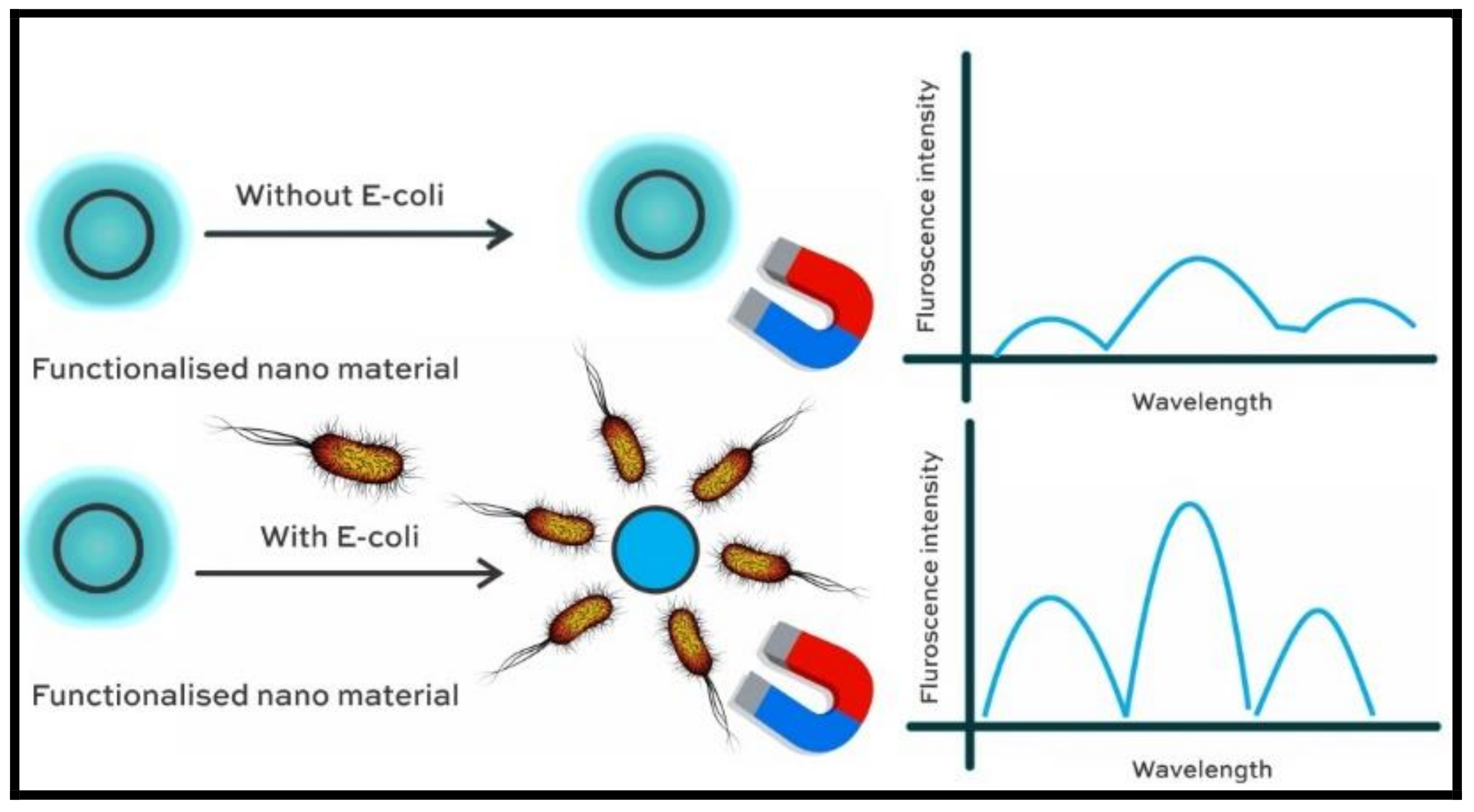
| Aptamer embedded magnetic nanoparticles | Fluorescence emission intensity decreases with intensity of E. Coli | Wide linear range and high selectivity towards adulterated pork samples | Binding properties of aptamer to E. coli requires a better insight. | [40] |
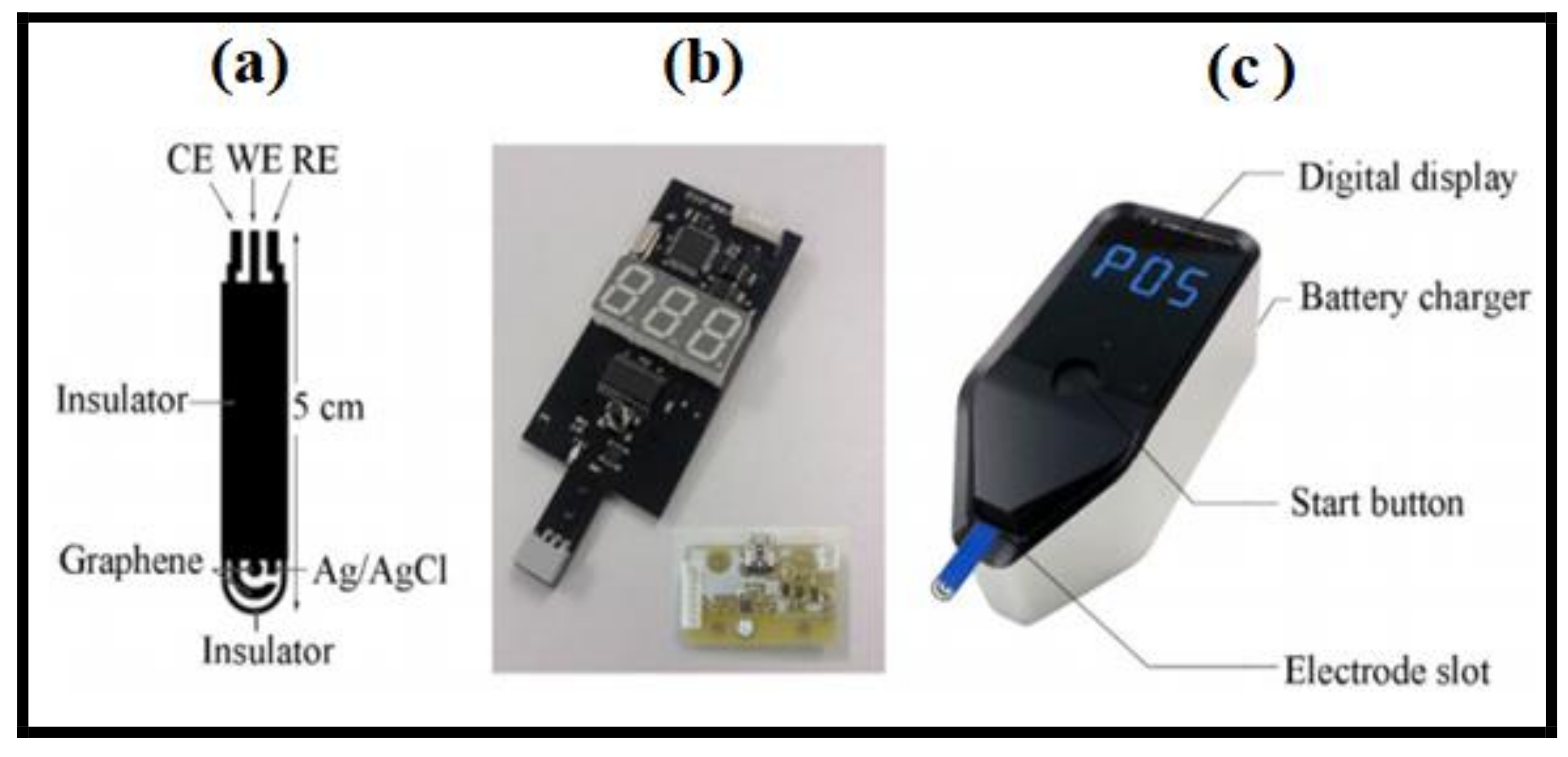
| Screen-printed carbon electrode | Cyclic voltammetry and differential pulse voltammetry | Rapid determination, excellent stability, sensitivity, and good reproducibility | Applicable only in the specific dynamic range and detection limit | [48] |
Publisher’s Note: MDPI stays neutral with regard to jurisdictional claims in published maps and institutional affiliations. |
© 2022 by the authors. Licensee MDPI, Basel, Switzerland. This article is an open access article distributed under the terms and conditions of the Creative Commons Attribution (CC BY) license (https://creativecommons.org/licenses/by/4.0/).
Share and Cite
Malini, S.; Roy, A.; Raj, K.; Raju, K.S.A.; Ali, I.H.; Mahesh, B.; Yadav, K.K.; Islam, S.; Jeon, B.-H.; Lee, S.S. Sensing beyond Senses: An Overview of Outstanding Strides in Architecting Nanopolymer-Enabled Sensors for Biomedical Applications. Polymers 2022, 14, 601. https://doi.org/10.3390/polym14030601
Malini S, Roy A, Raj K, Raju KSA, Ali IH, Mahesh B, Yadav KK, Islam S, Jeon B-H, Lee SS. Sensing beyond Senses: An Overview of Outstanding Strides in Architecting Nanopolymer-Enabled Sensors for Biomedical Applications. Polymers. 2022; 14(3):601. https://doi.org/10.3390/polym14030601
Chicago/Turabian StyleMalini, S., Arpita Roy, Kalyan Raj, K. S. Anantha Raju, Ismat H. Ali, B. Mahesh, Krishna Kumar Yadav, Saiful Islam, Byong-Hun Jeon, and Sean Seungwon Lee. 2022. "Sensing beyond Senses: An Overview of Outstanding Strides in Architecting Nanopolymer-Enabled Sensors for Biomedical Applications" Polymers 14, no. 3: 601. https://doi.org/10.3390/polym14030601
APA StyleMalini, S., Roy, A., Raj, K., Raju, K. S. A., Ali, I. H., Mahesh, B., Yadav, K. K., Islam, S., Jeon, B.-H., & Lee, S. S. (2022). Sensing beyond Senses: An Overview of Outstanding Strides in Architecting Nanopolymer-Enabled Sensors for Biomedical Applications. Polymers, 14(3), 601. https://doi.org/10.3390/polym14030601