
Development and Characterization of Functional Polylactic Acid/Chitosan Porous Scaffolds for Bone Tissue Engineering


Abstract
1. Introduction
2. Materials and Methods
2.1. Materials Used
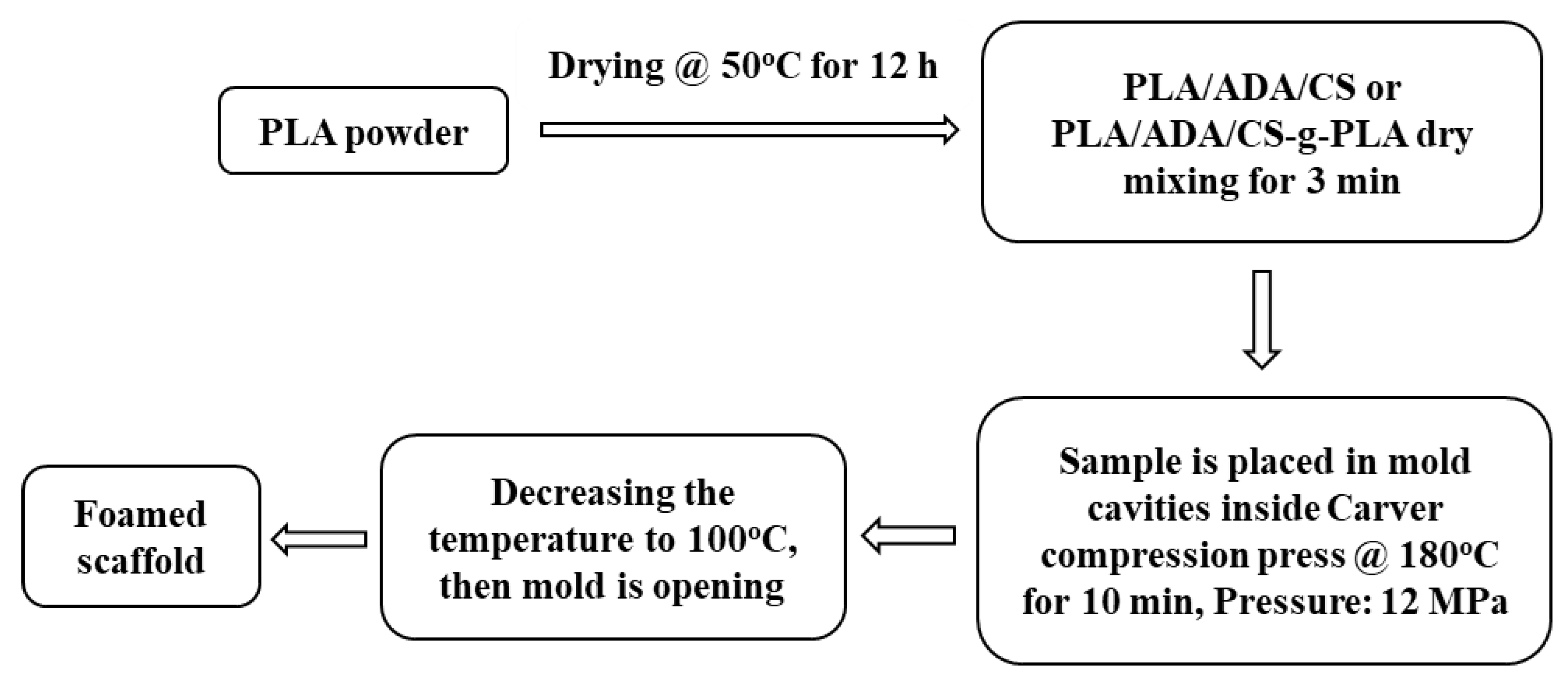
2.2. Composite Samples Preparation and Their Foaming in Open-Cell Porous Scaffolds
2.2.1. Chitosan Grafting with Polylactic Acid
2.2.2. PLA/CS and PLA/CS-g-PLA Composites Composition and Their Corresponding Preparation Steps
2.3. Characterization
2.3.1. Fourier-Transform Infrared (FTIR) Spectroscopy
2.3.2. Thermogravimetric Analysis (TGA)
2.3.3. Scanning Electron Microscopy (SEM) Characterization
2.3.4. Mechanical Characterization in Compression of the Foamed Scaffolds
2.3.5. X-ray Photoelectron Spectroscopy, XPS, Characterization
2.3.6. Biological Characterization
Cells Adhesion Characterization (Hoechst Staining Assay)
Cells Proliferation Quantification (MTT Assay)
3. Results and Discission
3.1. PLA Grafting on Chitosan
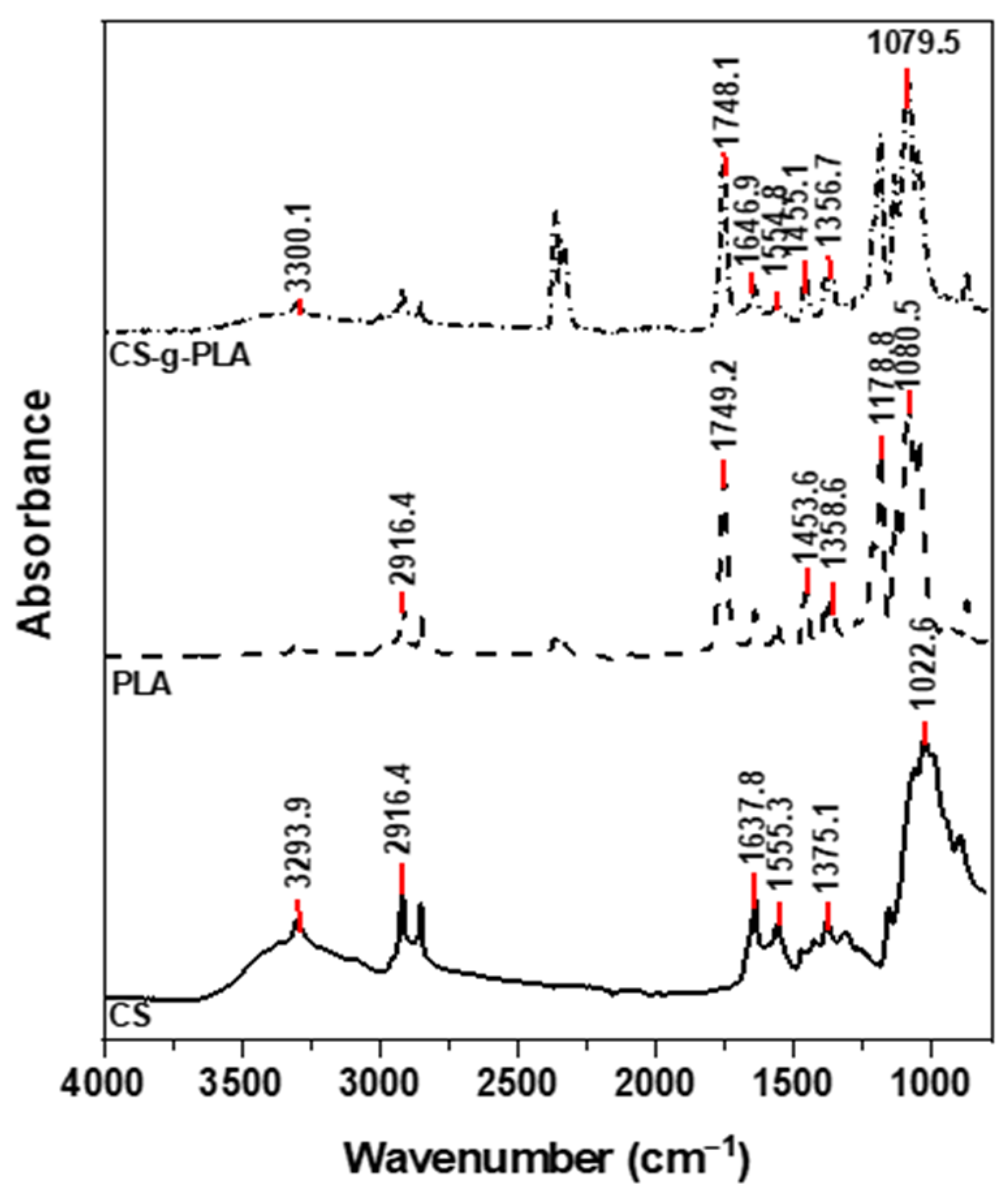
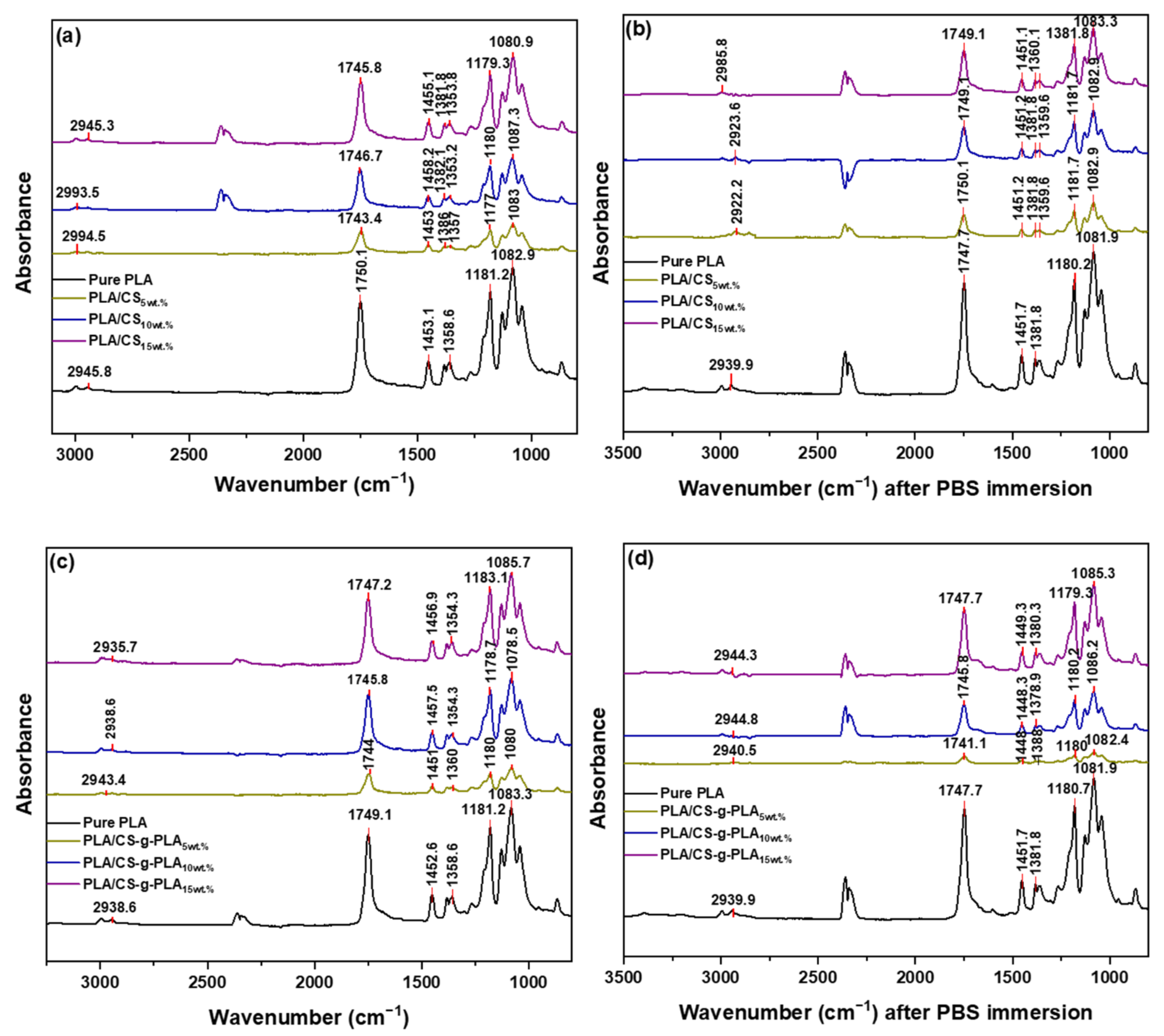
3.1.1. FTIR Characterization
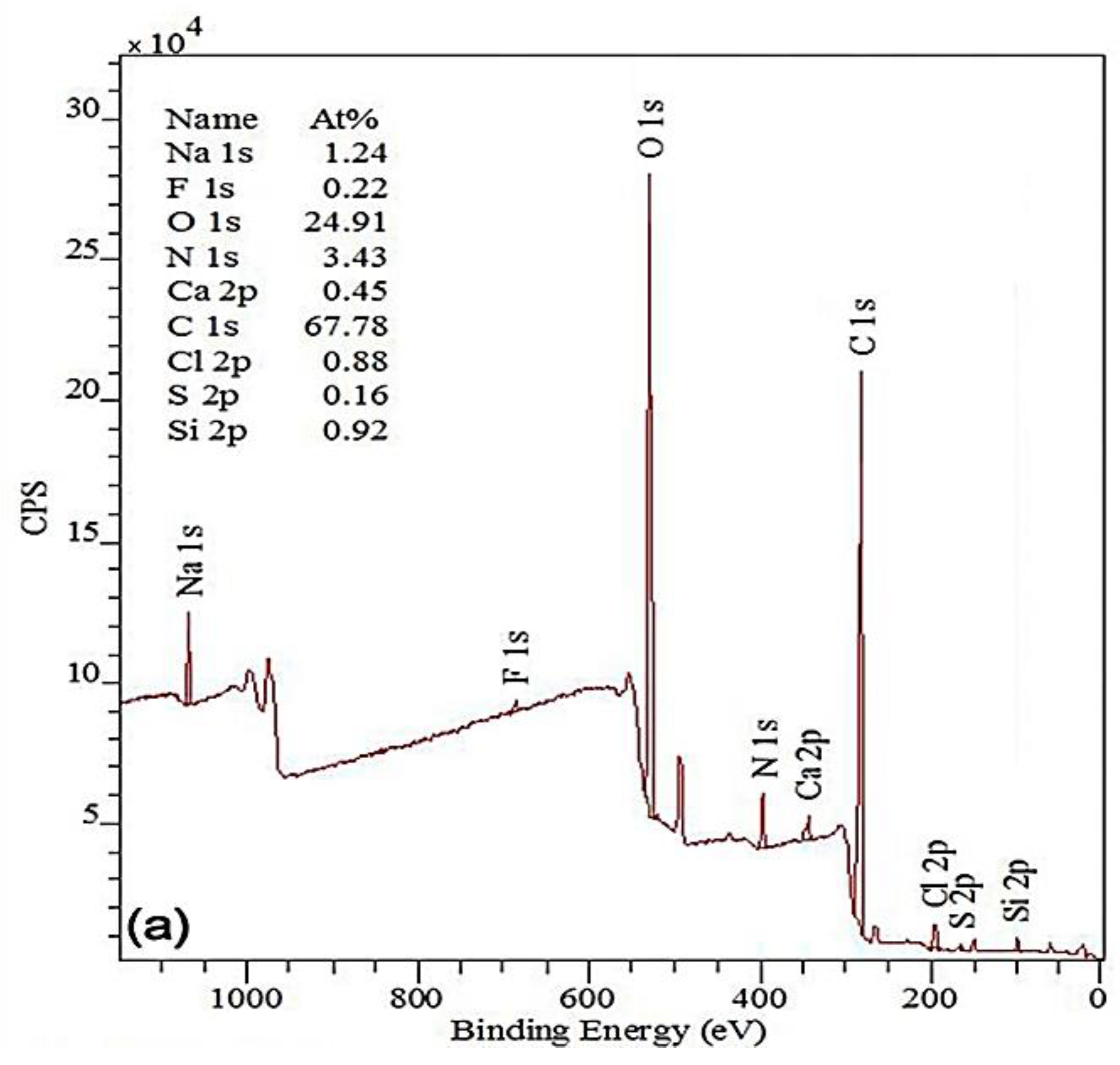
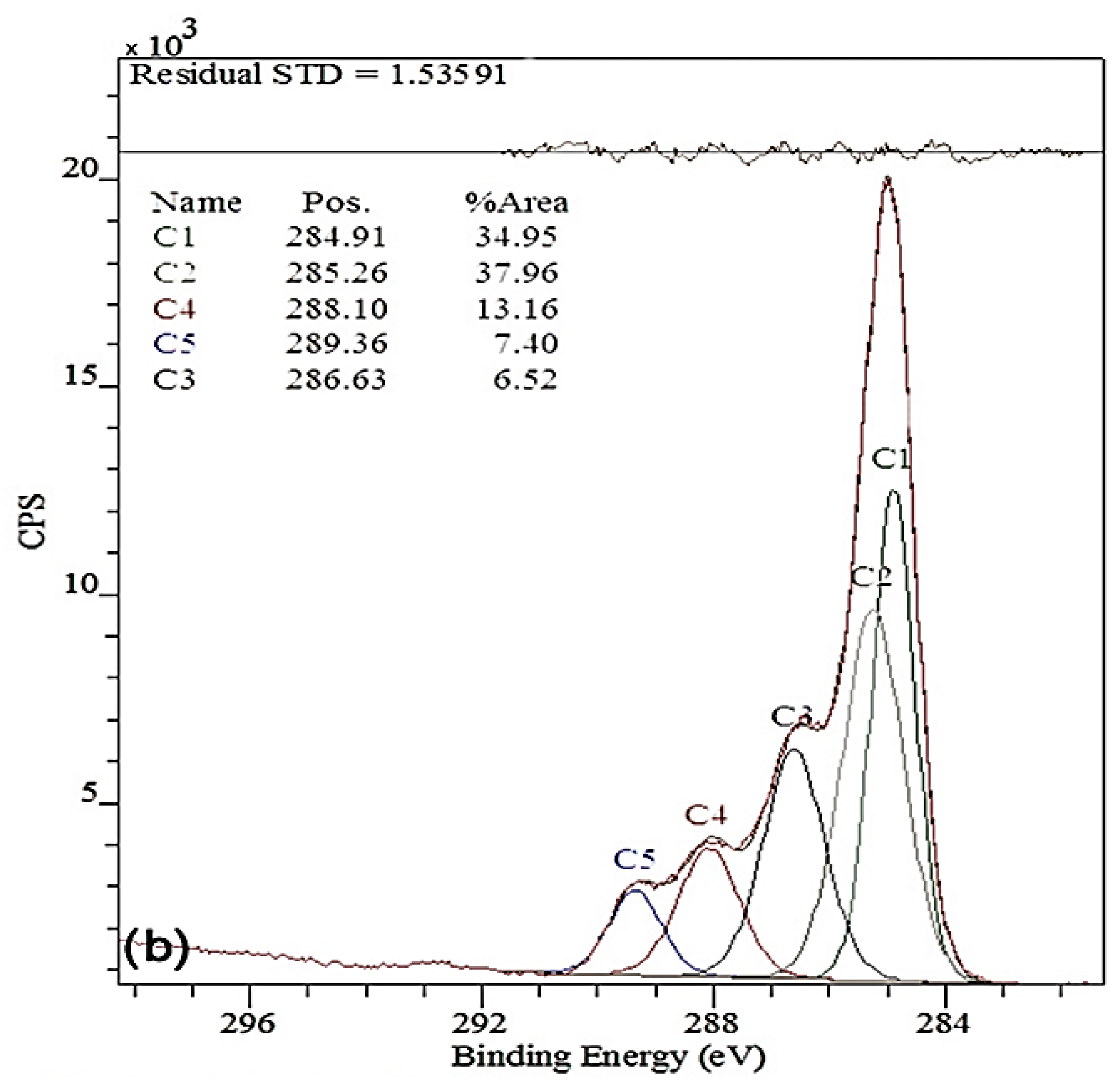
3.1.2. XPS Characterization of CS-g-PLA Copolymer
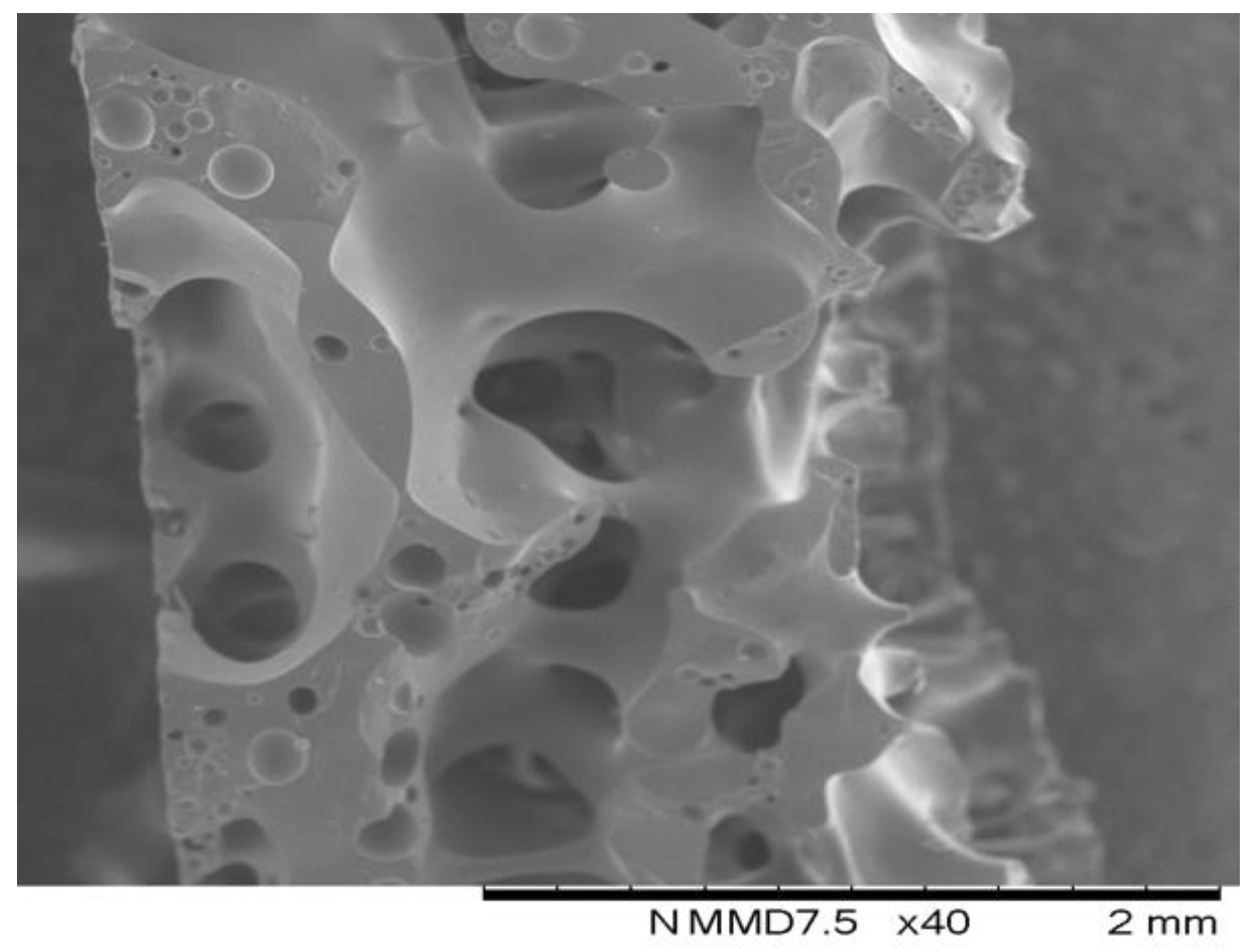
3.2. Morphology of the Open Cell PLA/CS and PLA/CS-g-PLA Composite Scaffolds
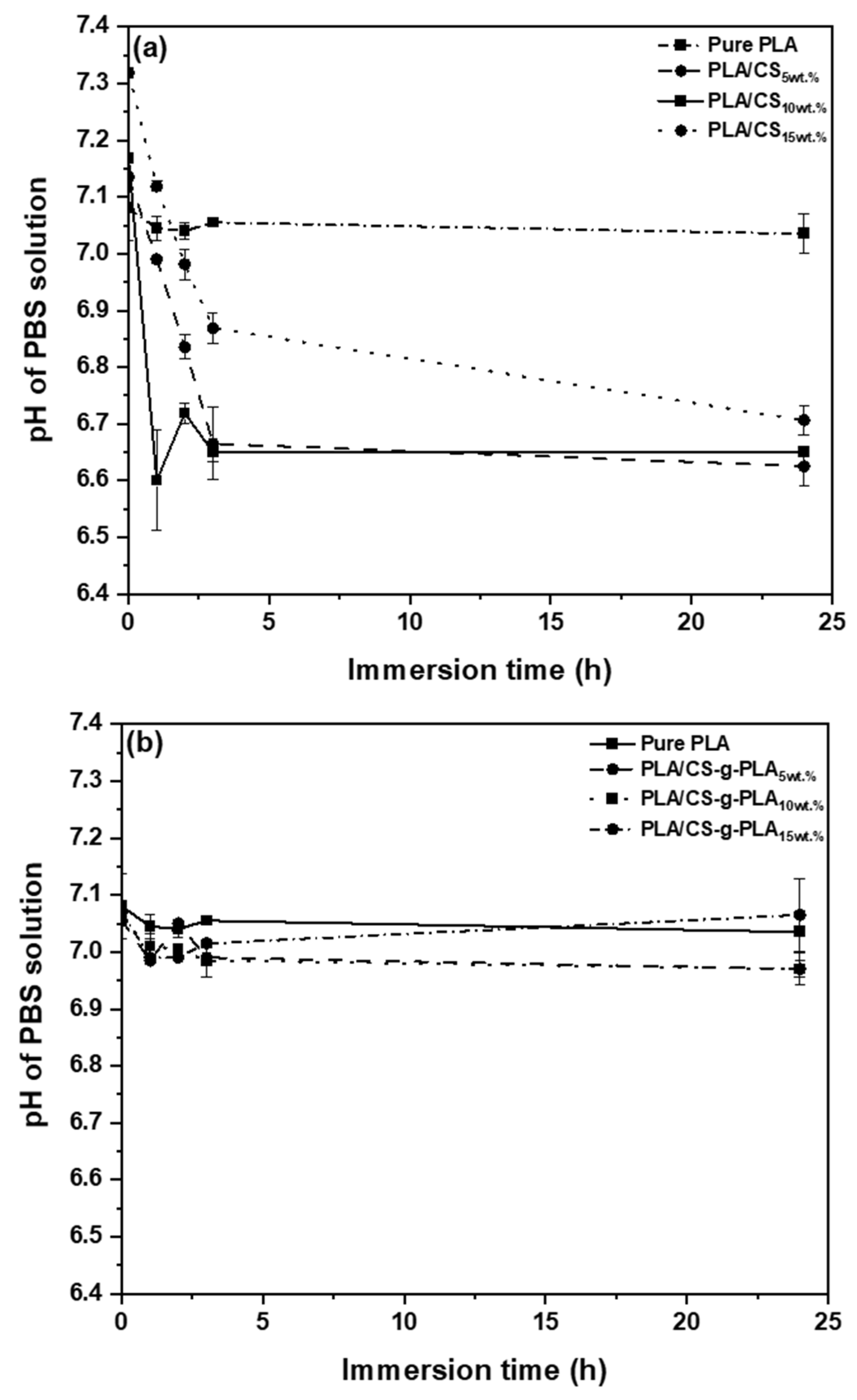
3.3. Hydrolytic Stability of PLA/CS and PLA/CS-g-PLA Porous Composite Scaffolds
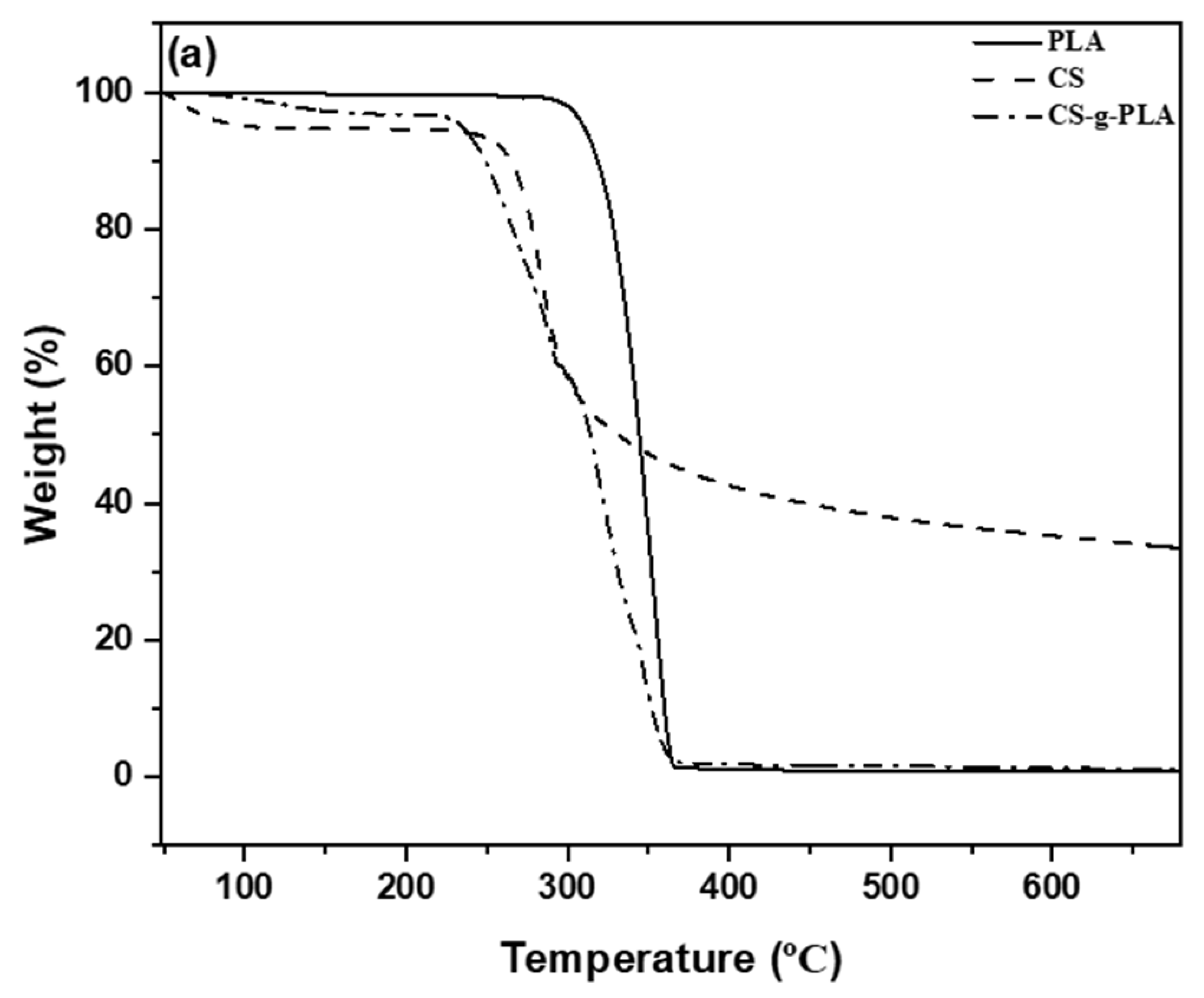
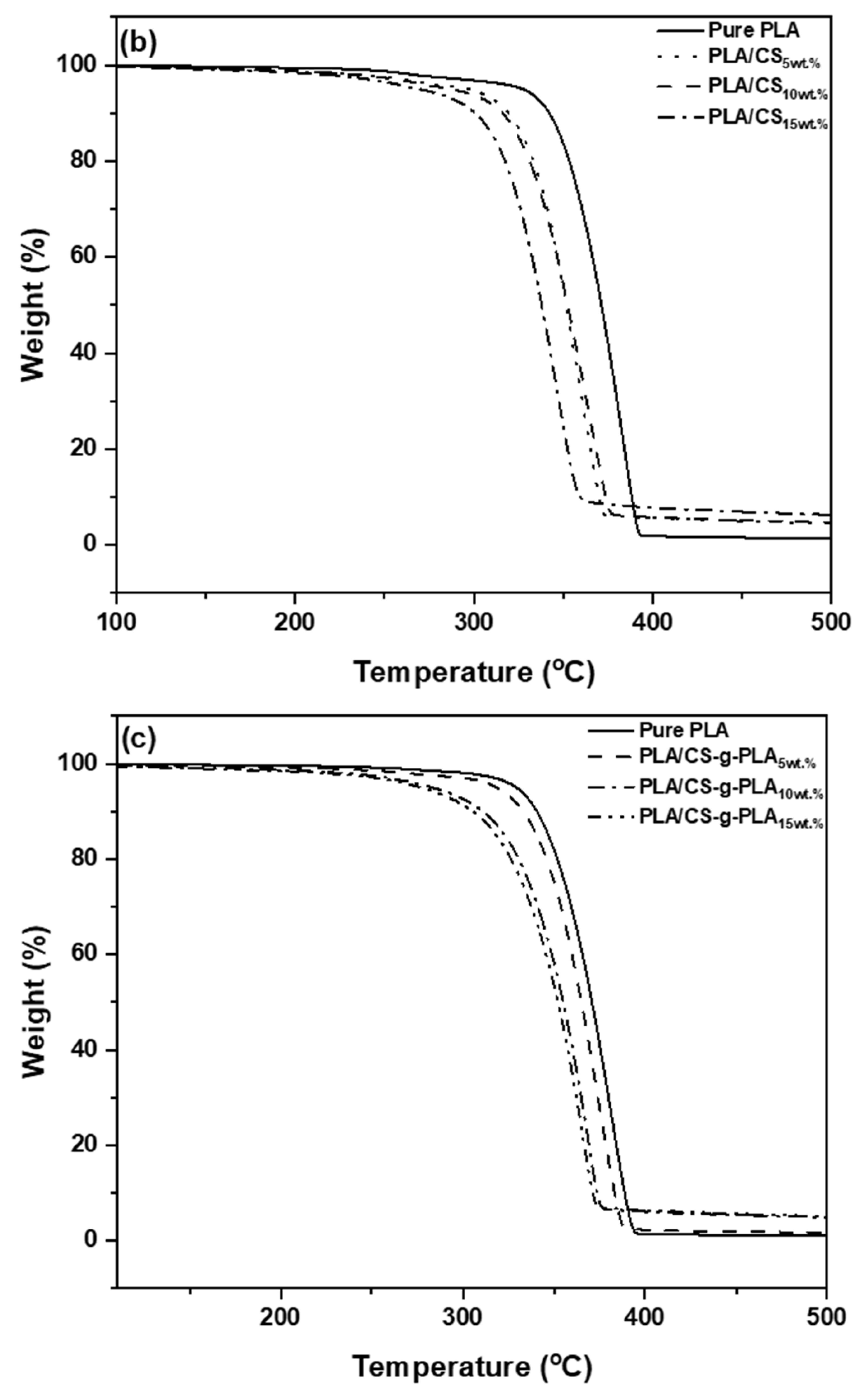
3.4. Structures and Thermal Stability Characterizations of PLA/CS and PLA/CS-g-PLA Porous Scaffolds
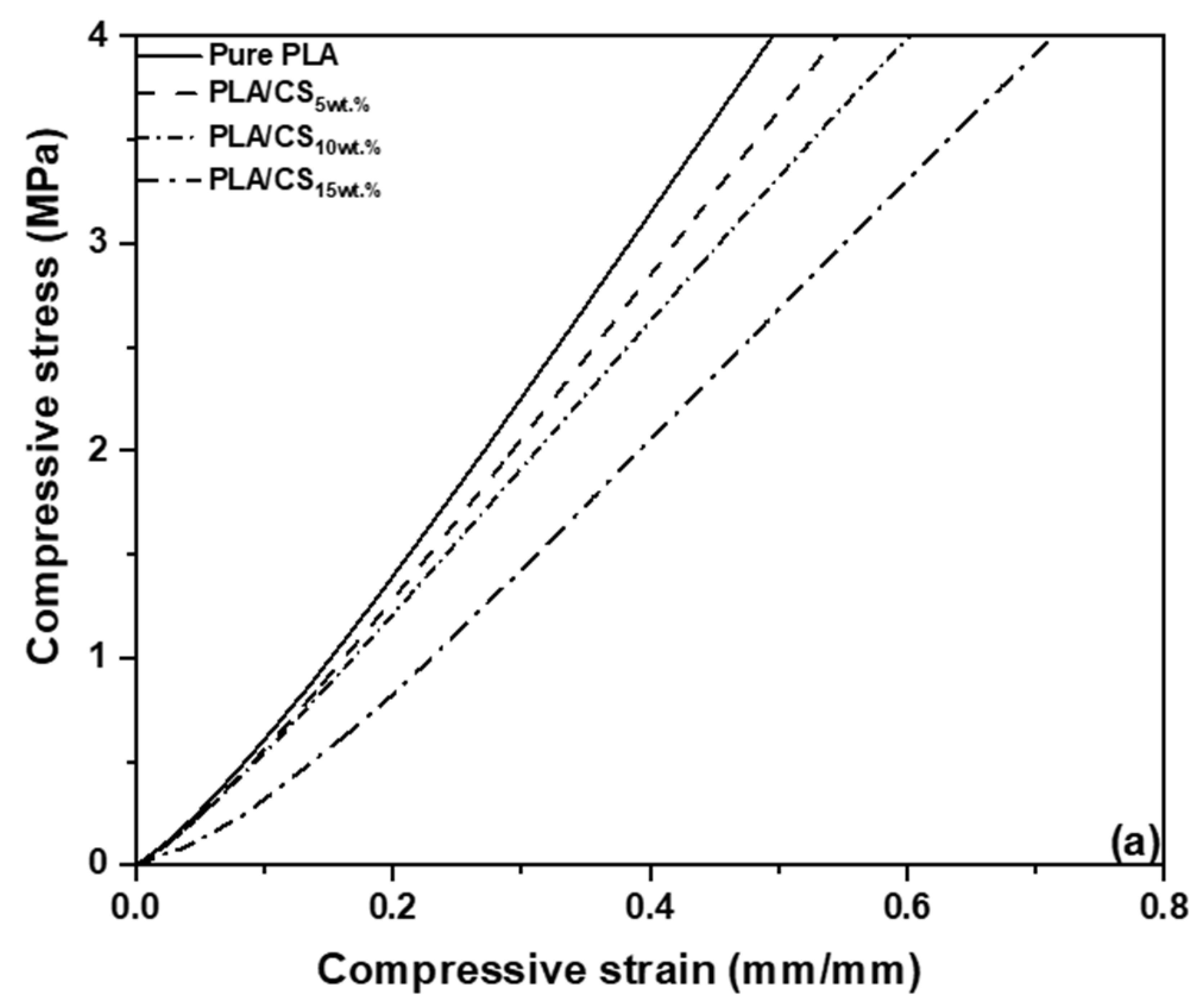
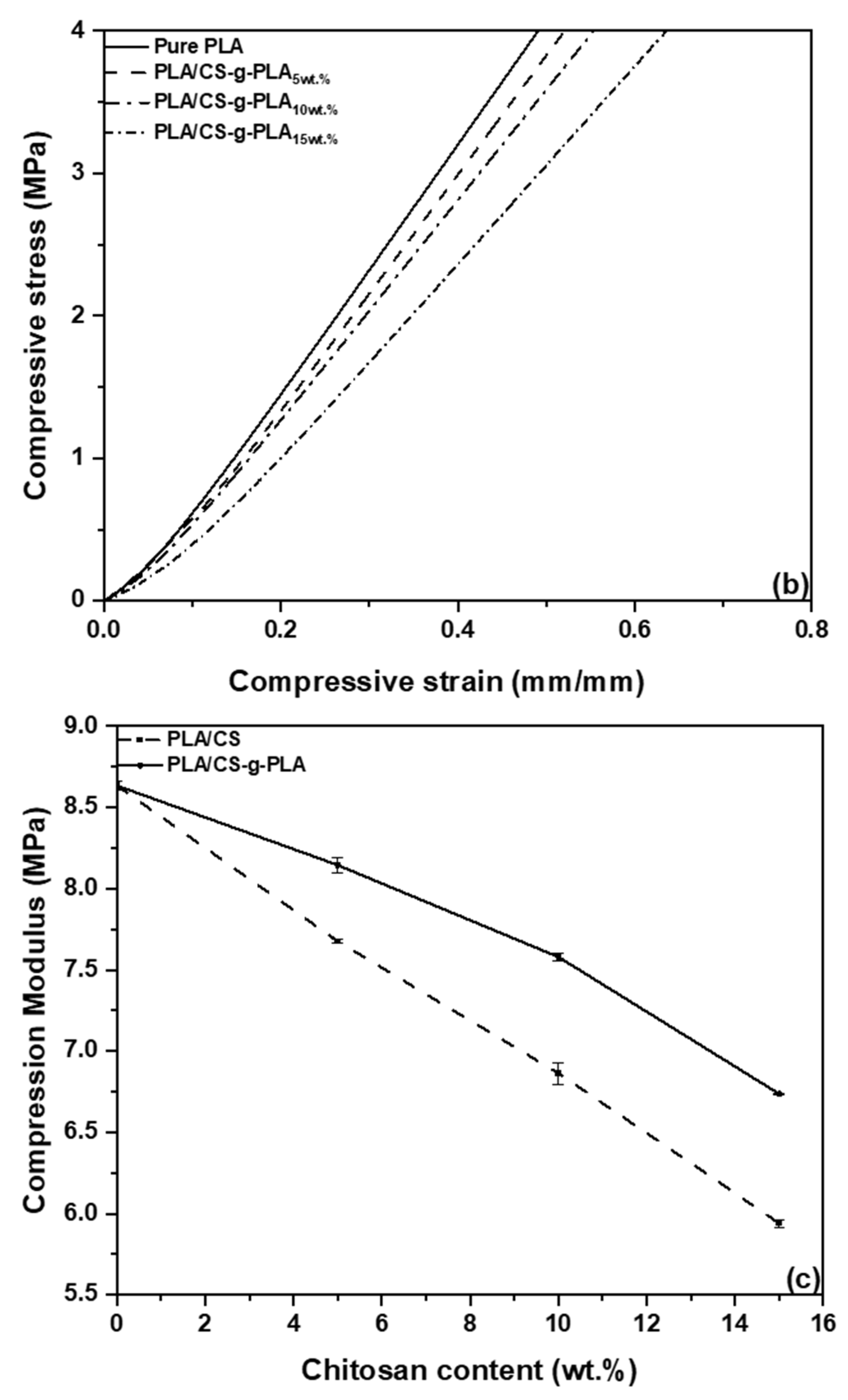
3.5. Mechanical Properties Characterization
3.6. Biological Characterization
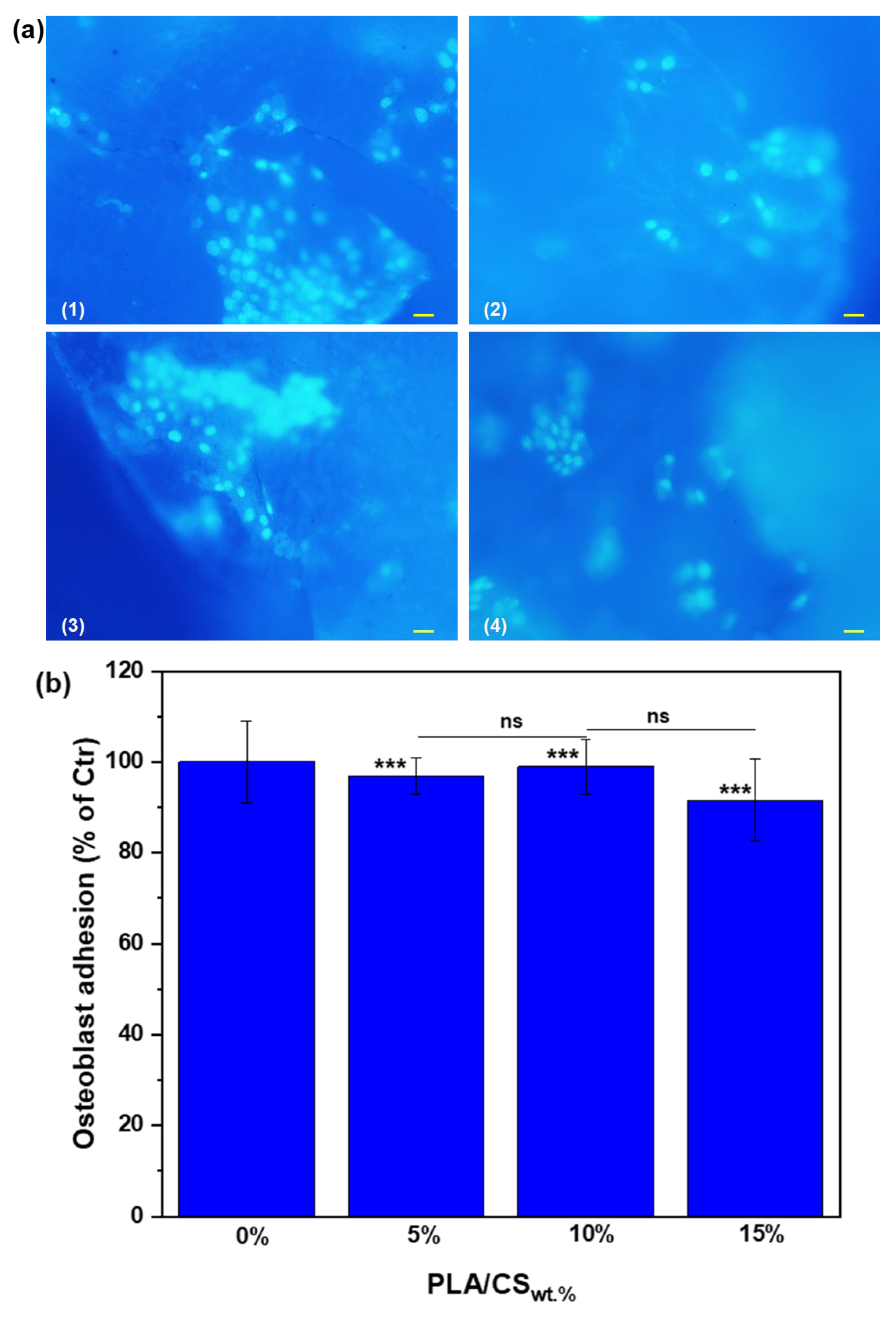
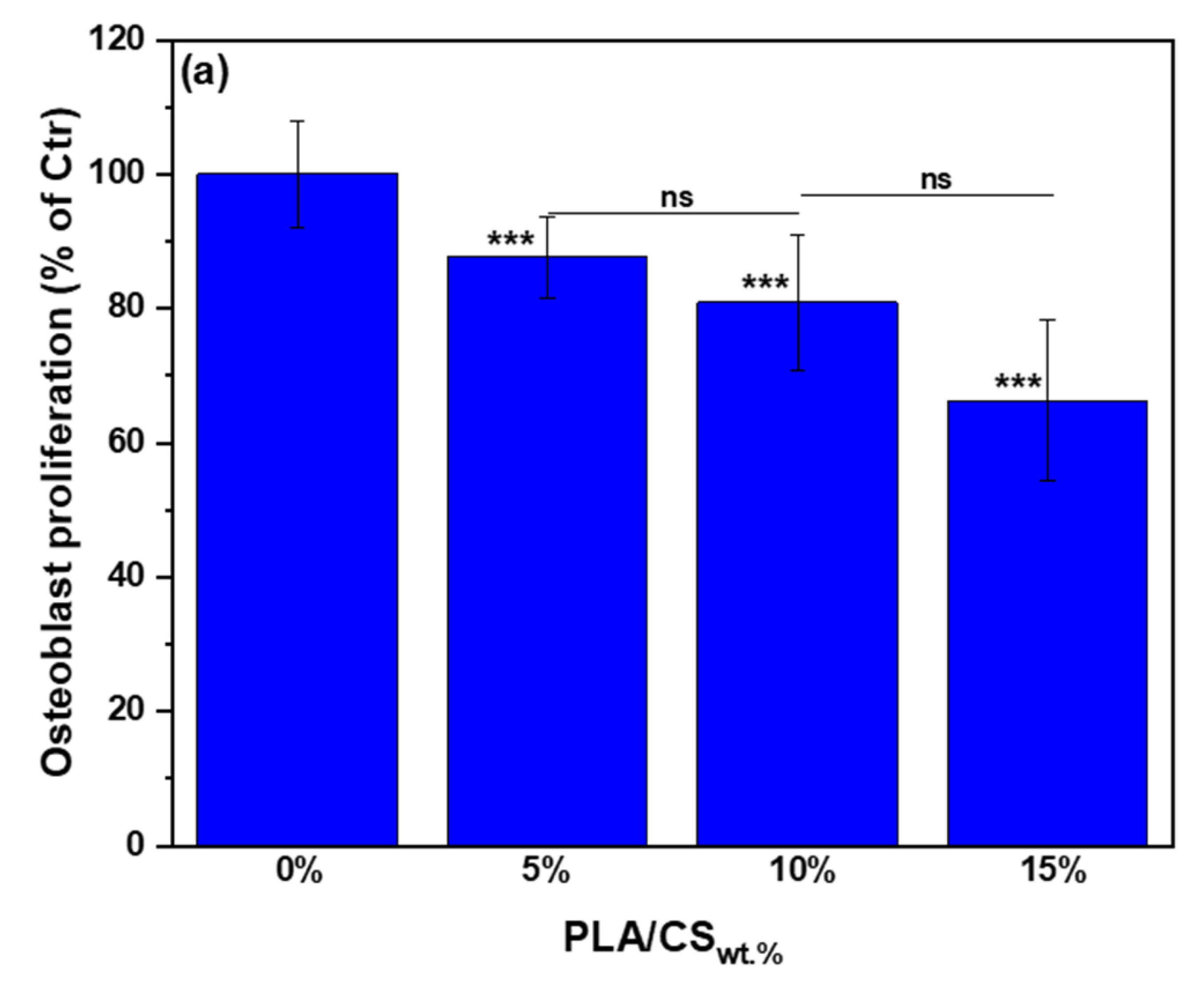
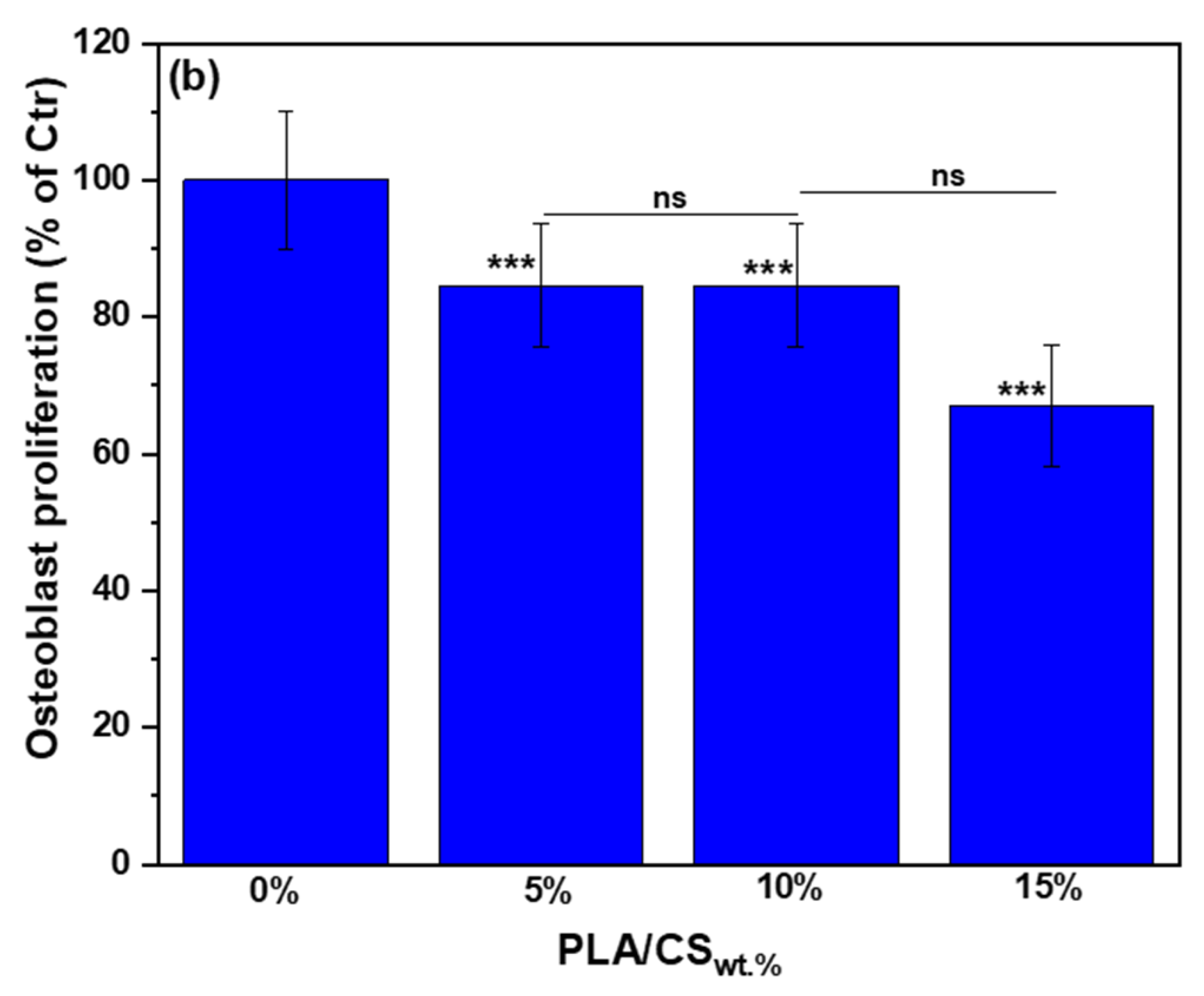
3.6.1. Characterization of Osteoblast Cells Adhesion and Proliferation into PLA/CS Porous Composites Scaffolds
Osteoblast Cells Adhesion
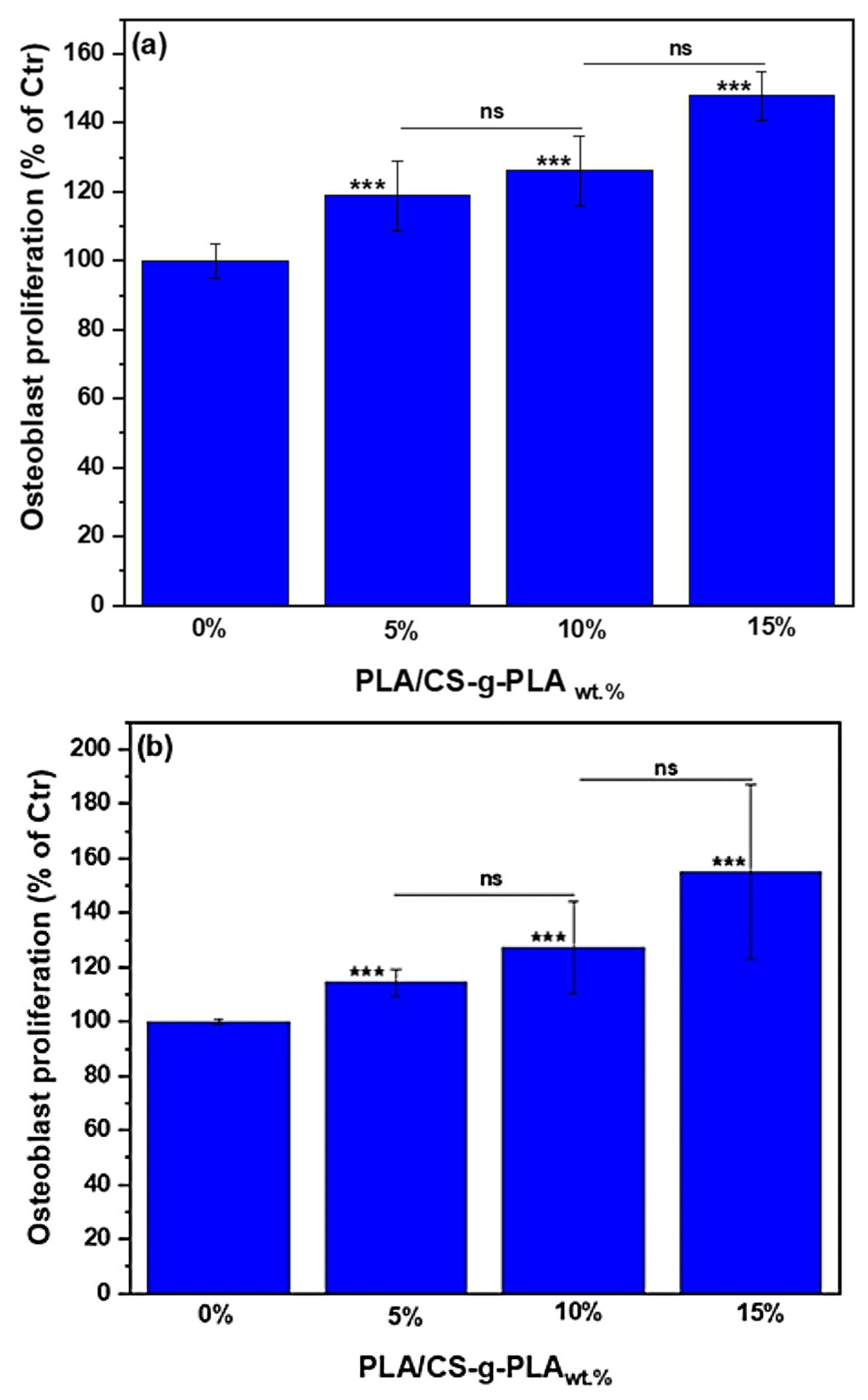
Osteoblast Cells Proliferation
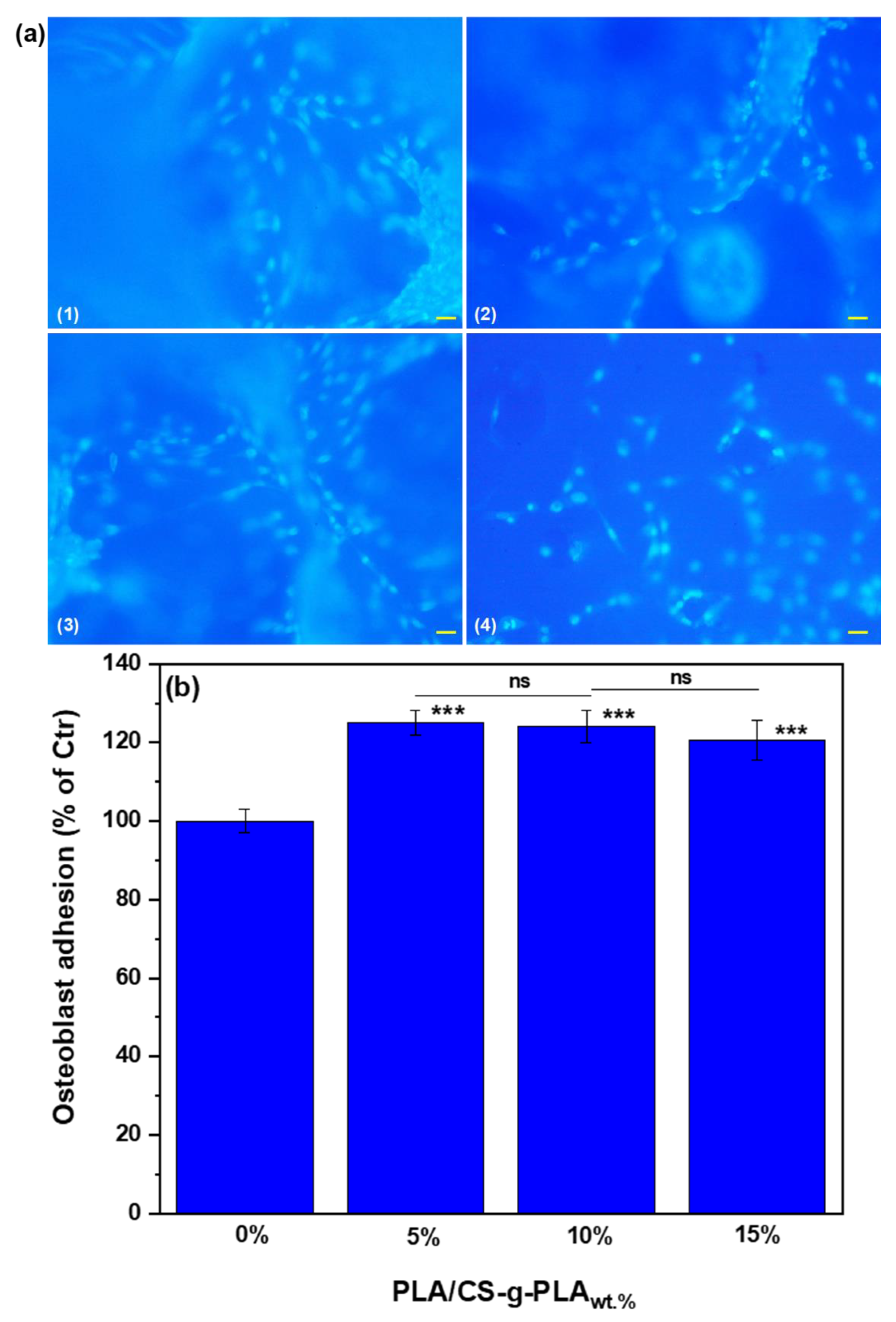
3.6.2. Characterization of Osteoblast Cells Adhesion and Proliferation into PLA/CS-g-PLA Porous Composites Scaffolds
Osteoblast Cells Adhesion
Osteoblast Cells Proliferation
4. Conclusions
Author Contributions
Funding
Institutional Review Board Statement
Data Availability Statement
Conflicts of Interest
References
- Ko, J.; Mohtaram, N.K.; Ahmed, F.; Montgomery, A.; Carlson, M.; Lee, P.C.; Willerth, S.M.; Jun, M.B. Fabrication of Poly (ϵ-Caprolactone) Microfiber Scaffolds with Varying Topography and Mechanical Properties for Stem Cell-Based Tissue Engineering Applications. J. Biomater. Sci. Polym. Ed. 2014, 25, 1–17. [Google Scholar] [CrossRef] [PubMed]
- Chanjuan, D.; Yonggang, Y.L. Application of Collagen Scaffold in Tissue Engineering: Recent Advances and New Perspectives. Polymers 2016, 8, 42. [Google Scholar]
- Su, H.K.; Jung, Y.; Soo, H.K. A Biocompatible Tissue Scaffold Produced by Supercritical Fluid Processing for Cartilage Tissue Engineering. Tissue Eng. Part C Methods 2012, 19, 181–188. [Google Scholar]
- Holland, T.; Mikos, A. Biodegradable Polymeric Scaffolds. Improvements in Bone Tissue Engineering through Controlled Drug Delivery. Adv. Biochem. Eng. Biotechnol. 2006, 102, 161–185. [Google Scholar]
- Shen, H.; Hu, X.; Yang, F.; Bei, J.; Wang, S. The Bioactivity of RhBMP-2 Immobilized Poly(Lactide-Co-Glycolide) Scaffolds. Biomaterials 2009, 30, 3150–3157. [Google Scholar] [PubMed]
- Guntillake, P.; Adhikari, R. Biodegradable Synthetic Polymers for Tissue Engineering. Eur. Cells Mater. 2003, 5, 1–16. [Google Scholar] [CrossRef]
- da Silva, D.; Kaduri, M.; Poley, M.; Adir, O.; Krinsky, N.; Shainsky-Roitman, J.; Schroeder, A. Biocompatibility, Biodegradation and Excretion of Polylactic Acid (PLA) in Medical Implants and Theranostic Systems. Chem. Eng. J. 2018, 340, 9–14. [Google Scholar]
- Munirah, S.; Kim, S.; Ruszymah, B.; Khang, G. The Use of Fibrin and Poly (Lactic-Co-Glycolic Acid) Hybrid Scaffold for Articular Cartilage Tissue Engineering: An in Vivo Analysis. Eur. Cell Mater. 2008, 15, 41–52. [Google Scholar] [CrossRef]
- Swetha, M.; Sahithi, K.; Moorthi, A.; Srinivasan, N.; Ramasamy, K.; Selvamurugan, N. Biocomposites Containing Natural Polymers and Hydroxyapatite for Bone Tissue Engineering. Int. J. Biol. Macromol. 2010, 47, 1–4. [Google Scholar] [PubMed]
- Bhattarai, N.; Edmondson, D.; Veiseh, O.; Matsen, F.; Zhang, M. Electrospun Chitosan—Based Nanofi Bers and Their Cellular Compatibility. Biomaterials 2005, 26, 6176–6184. [Google Scholar] [CrossRef]
- Jayakumar, R.; Prabaharan, M.; Nair, S.; Tamura, H. Novel Chitin and Chitosan Nanofibers in Biomedical Applications. Biotechnol. Adv. 2010, 28, 142–150. [Google Scholar]
- Kim, I.Y.; Seo, S.J.; Moon, H.S.; Yoo, M.K.; Park, I.Y.; Kim, B.C.; Cho, C.S. Chitosan and Its Derivatives for Tissue Engineering Applications. Biotechnol. Adv. 2008, 26, 1–21. [Google Scholar]
- Lee, K.Y.; Jeong, L.; Kang, Y.O.; Lee, S.J.; Park, W.H. Electrospinning of Polysaccharides for Regenerative Medicine. Adv. Drug Deliv. Rev. 2009, 61, 1020–1032. [Google Scholar]
- Seol, Y.; Lee, J.; Park, Y.; Lee, Y.; Rhyu, I.; Lee, S.; Han, S.; Chung, C. Chitosan Sponges as Tissue Engineering Scaffolds for Bone Formation. Biotechnol. Lett. 2004, 26, 1037–1041. [Google Scholar] [CrossRef]
- Venkatesan, J.; Kim, S.K. Chitosan Composites for Bone Tissue Engineering—An Overview. Mar. Drugs 2010, 8, 2252–2266. [Google Scholar] [PubMed]
- Jayakumar, R.; Prabaharan, M.; Sudheesh, K.P.T.; Nair, S.V.; Tamura, H. Biomaterials based on chitin and chitosan in wound dressing applications. Biotechnol. Adv. 2011, 29, 322–337. [Google Scholar] [PubMed]
- Mao, J.S.; Cui, Y.L.; Wang, X.H.; Sun, Y.; Yin, Y.J.; Zhao, H.M.; De Yao, K. A preliminary study on chitosan and gelatin polyelectrolyte complex cytocompatibility by cell cycle and apoptosis analysis. Biomaterials 2004, 25, 3973–3981. [Google Scholar]
- Freier, T.; Koh, H.S.; Kazazian, K.; Shoichet, M.S. Controlling cell adhesion and degradation of chitosan films by N-acetylation. Biomaterials 2005, 26, 5872–5878. [Google Scholar] [PubMed]
- Ding, K.; Wang, Y.; Wang, H.; Yuan, L.; Tan, M.; Shi, X.; Chen, H. 6-O-Sulfated Chitosan Promoting the Neural Differentiation of Mouse Embryonic Stem Cells. ACS Appl. Mater. Interfac. 2014, 6, 20043–20050. [Google Scholar]
- Kazachenko, A.S.; Akman, F.; Malyar, Y.N.; Issaoui, N.; Vasilieva, N.Y.; Karacharov, A.A. Synthesis optimization, DFT and physicochemical study of chitosan sulfates. J. Mol. Struct. 2021, 1245, 131083. [Google Scholar]
- Polo-Corrales, L.; Latorre-Esteves, M.; Ramirez-Vick, J. Scaffold Design for Bone Regeneration. J. Nanosci. Nanotechnol. 2014, 14, 15–56. [Google Scholar] [PubMed]
- Abbasi, N.; Hamlet, S.; Love, R.M.; Nguyen, N.-T. Porous Scaffolds for Bone Regeneration. J. Sci. Adv. Mater. Devices 2020, 5, 1–9. [Google Scholar]
- Ramtani, S. Mechanical Modeling of Cell/ECM and Cell/Cell Interactions during the Contraction of a Fibroblast-Populated Collagen Microsphere: Theory and Model Simulation. J. Biomech. 2004, 37, 1709–1718. [Google Scholar] [PubMed]
- Venugopal, J.; Prabhakaran, M.; Zhang, Y.; Low, S.; Choon, A.; Ramakrishna, S. Biomimetic Hydroxyapatite-Containing Composite Nanofibrous Substrates for Bone Tissue Engineering. Philos. Trans. A Math. Phys. Eng. Sci. 2010, 368, 2065–2081. [Google Scholar] [CrossRef] [PubMed]
- Wagoner Johnson, A.; Herschler, B. A Review of the Mechanical Behavior of CaP and CaP/Polymer Composites for Applications in Bone Replacement and Repair. Acta Biomater. 2011, 7, 16–30. [Google Scholar]
- Ramay, H.; Zhang, M. Biphasic Calcium Phosphate Nanocomposite Porous Scaffolds for Load-Bearing Bone Tissue Engineering. Biomaterials 2004, 25, 5171–5180. [Google Scholar]
- Syahrom, A.; Abdul Kadir, M.R.; Abdullah, J.; Andreas, Q. Mechanical and Microarchitectural Analyses of Cancellous Bone through Experiment and Computer Simulation. Med. Biol. Eng. Comput. 2011, 49, 1393–1403. [Google Scholar]
- Stratton, S.; Shelke, N.B.; Hoshing, K.; Kumbar, S.G. Bioactive Polymeric Scaffolds for Tissue Engineering. Bioact. Mater. 2016, 1, 96–108. [Google Scholar]
- Chang, K.Y.; Hung, L.H.; Chu, I.; Ko, C.S.; Lee, Y.D. The Application of Type II Collagen and Chondroitin Sulfate Grafted PCL Porous Scaffold in Cartilage Tissue Engineering. J. Biomed. Mater. Res. Part A 2010, 92, 712–723. [Google Scholar]
- Gerçek, I.; Tıǧlı, R.; Gümüs, M. A Novel Scaffold Based on Formation and Agglomeration of PCL Microbeads by Freeze-Drying. J. Biomed. Mater. Res. Part A 2008, 86, 1012–1022. [Google Scholar]
- Lobos, J.; Iasella, S.; Rodriguez-Perez, M.A.; Velankar, S.S. Improving the Stability of Polylactic Acid Foams by Interfacially Adsorbed Particles. Polym. Eng. Sci. 2016, 56, 9–17. [Google Scholar] [CrossRef]
- Shelke, N.B.; Anderson, M.; Idrees, S.; Nip, M.J.; Donde, S.; Yu, X. Handbook of Polyester Drug Delivery Systems; Pan Stanford: Redwood City, CA, USA, 2016; pp. 595–649. [Google Scholar]
- Kolesky, D.B.; Truby, R.L.; Gladman, A.; Busbee, T.A.; Homan, K.A.; Lewis, J.A. 3D Bioprinting of Vascularized, Heterogeneous Cell-Lagen Tissue Constructs. Adv. Mater. 2014, 26, 3124–3130. [Google Scholar] [CrossRef] [PubMed]
- Mighri, N.; Mao, J.; Mighri, F.; Ajji, A.; Rouabhia, M. Chitosan-Coated Collagen Membranes Promotechondrocyte Adhesion, Growth, and Interleukin-6 Secretion. Materials 2015, 8, 7673–7689. [Google Scholar] [CrossRef] [PubMed]
- Mighri, N.; Mao, J.; Park, H.J.; Mighri, F.; Rouabhia, M.; Ajji, A.; Zhang, Z. Chondrocytes Adhesion and Proliferation Following Culture on Amino Acid Treated Chitosan Coated Collagen Scaffold. Can. J. Chem. Eng. 2010, 96, 2236–2242. [Google Scholar]
- Li, J.; Kong, M.; Cheng, X.J.; Dang, Q.F.; Zhou, X.; Wei, Y.N.; Chen, X.G. Preparation of Biocompatible Chitosan Grafted Poly (Lactic Acid) Nanoparticles. Int. J. Biol. Macromol. 2012, 51, 221–227. [Google Scholar] [CrossRef]
- Abubaker Osman, M.; Virgilio, N.; Rouabhia, M.; Mighri, F. Polylactide (PLA) Foaming: Design of Experiments for Cell Size Optimization. Mater. Sci. Appl. 2022, 13, 63–77. [Google Scholar]
- Zhan, S.; Liu, N.; Wang, W.; Chen, S.; Wang, J. Preparation and Characterization of Chitosan-Graft-Poly (L-Lactic Acid) Microparticles. Polym. Eng. Sci. 2016, 56, 1432–1436. [Google Scholar] [CrossRef]
- Li, J.; Kong, M.; Cheng, X.J.; Li, J.J.; Liu, W.F.; Chen, X.G. A Facile Method for Preparing Biodegradable Chitosan Derivatives with Low Grafting Degree of Poly (Lactic Acid). Int. J. Biol. Macromol. 2011, 49, 1016–1021. [Google Scholar] [CrossRef] [PubMed]
- Arnett, T.R. Extracellular PH Regulates Bone Cell Function. J. Nutr. 2008, 138, 415S–418S. [Google Scholar] [CrossRef]
- Arnett, T.R.; Dempster, D.W. Effect of PH on Bone Resorption by Rat Osteoclasts In Vitro. Endocrinology 1986, 119, 119–124. [Google Scholar] [CrossRef]
- Kaysinger, K.K.; Ramp, W.K. Extracellular PH Modulates the Activity of Cultured Human Osteoblasts. J. Cell. Biochem. 1998, 68, 83–89. [Google Scholar] [CrossRef]
- Rinaudo, M. Chitin and Chitosan: Properties and Application. Prog. Polym. Sci. 2006, 31, 603–632. [Google Scholar] [CrossRef]
- Kumar, M.N.R. A Review of Chitin and Chitosan Applications. React. Funct. Polym. 2000, 46, 1–27. [Google Scholar] [CrossRef]
- Di Martino, A.; Sittinger, M.; Risbud, M.V. Chitosan: A Versatile Biopolymer for Orthopaedic Tissue Engineering. Biomaterials 2015, 26, 5983–5990. [Google Scholar] [CrossRef]
- Zhang, Z.; Cui, H. Biodegradability and Biocompatibility Study of Poly (Chitosan-g-Lactic Acid) Scaffolds. Molecules 2012, 17, 3243–3258. [Google Scholar] [CrossRef]
- Niu, X.; Feng, Q.; Wang, M.; Guo, X.; Zheng, Q. In Vitro Degradation and Release Behavior of Porous Poly (Lactic Acid) Scaffolds Containing Chitosan Microspheres as a Carrier for BMP-2-Derived Synthetic Peptide. Polym. Degrad. Stab. 2009, 94, 176–182. [Google Scholar] [CrossRef]
- Athanasiou, K.A.; Darling, E.M.; Hu, J.C. Articular Cartilage Tissue Engineering. Synth. Lect. Tissue Eng. 2009, 1, 1–182. [Google Scholar]
- Cai, K.; Yao, K.; Cui, Y.L.; Lin, S.; Yang, Z.; Li, X.; Xie, H.; Qing, T.; Luo, J. Surface Modification of Poly (d,l-Lactic Acid) with Chitosan and Its Effects on the Culture of Osteoblasts In Vitro. J. Biomed. Mater. Res. 2002, 60, 398–404. [Google Scholar] [CrossRef]
- Ho, M.H.; Liao, M.H.; Lin, Y.L.; Lai, C.H.; Lin, P.I.; Chen, R.M. Improving Effects of Chitosan Nanofiber Scaffolds on Osteoblast Proliferation and Maturation. Int. J. Nanomed. 2014, 9, 4293–4304. [Google Scholar]
- Torres-Hernández, Y.G.; Ortega-Díaz, G.M.; Téllez-Jurado, L.; Castrejón-Jiménez, N.S.; Altamirano-Torres, A.; García-Pérez, B.E.; Balmori-Ramírez, H. Biological Compatibility of a Polylactic Acid Composite Reinforced with Natural Chitosan Obtained from Shrimp Waste. Materials 2018, 11, 1465. [Google Scholar] [CrossRef]
- Chen, P.; Liu, L.; Pan, J.; Mei, J.; Li, C.; Zheng, Y. Biomimetic Composite Scaffold of Hydroxyapatite/Gelatin-Chitosan Core-Shell Nanofibers for Bone Tissue Engineering. Mater. Sci. Eng. C Mater. Biol. Appl. 2019, 97, 325–335. [Google Scholar] [PubMed]
- O’Brien, F.J.; Harley, B.A.; Yannas, I.V.; Gibson, L.J. The Effect of Pore Size on Cell Adhesion in Collagen-GAG Scaffolds. Biomaterials 2005, 26, 433–441. [Google Scholar] [PubMed]


| Sample Designation | PLA (wt.%) | ADA (wt.%) | CS or CS-g-PLA (wt.%) |
|---|---|---|---|
| PLA/CS5wt.% | 88.1 | 6.9 | 5.0 |
| PLA/CS10wt.% | 83.1 | 6.9 | 10.0 |
| PLA/CS15wt.% | 78.1 | 6.9 | 15.0 |
| PLA/CS-g-PLA5wt.% | 88.1 | 6.9 | 5.0 |
| PLA/CS-g-PLA10wt.% | 83.1 | 6.9 | 10.0 |
| PLA/CS-g-PLA15wt.% | 78.1 | 6.9 | 15.0 |
Publisher’s Note: MDPI stays neutral with regard to jurisdictional claims in published maps and institutional affiliations. |
© 2022 by the authors. Licensee MDPI, Basel, Switzerland. This article is an open access article distributed under the terms and conditions of the Creative Commons Attribution (CC BY) license (https://creativecommons.org/licenses/by/4.0/).
Share and Cite
Osman, M.A.; Virgilio, N.; Rouabhia, M.; Mighri, F. Development and Characterization of Functional Polylactic Acid/Chitosan Porous Scaffolds for Bone Tissue Engineering. Polymers 2022, 14, 5079. https://doi.org/10.3390/polym14235079
Osman MA, Virgilio N, Rouabhia M, Mighri F. Development and Characterization of Functional Polylactic Acid/Chitosan Porous Scaffolds for Bone Tissue Engineering. Polymers. 2022; 14(23):5079. https://doi.org/10.3390/polym14235079
Chicago/Turabian StyleOsman, Miada Abubaker, Nick Virgilio, Mahmoud Rouabhia, and Frej Mighri. 2022. "Development and Characterization of Functional Polylactic Acid/Chitosan Porous Scaffolds for Bone Tissue Engineering" Polymers 14, no. 23: 5079. https://doi.org/10.3390/polym14235079
APA StyleOsman, M. A., Virgilio, N., Rouabhia, M., & Mighri, F. (2022). Development and Characterization of Functional Polylactic Acid/Chitosan Porous Scaffolds for Bone Tissue Engineering. Polymers, 14(23), 5079. https://doi.org/10.3390/polym14235079