Article

Thermally Treated Berberine-Loaded SA/PVA/PEO Electrospun Microfiber Membranes for Antibacterial Wound Dressings

1
School of Textile, Garment & Design, Changshu Institute of Technology, Changshu 215500, China
2
School of Textile Science and Engineering, Tiangong University, Tianjin 300387, China
3
College of Engineering and Technology, Jiyang College of Zhejiang A & F University, Shaoxing 312000, China
4
Bioresource Technology Division, School of Industrial Technology, Universiti Sains Malaysia, Penang 11800, Malaysia
5
Renewable Biomass Transformation Cluster, School of Industrial Technology, Universiti Sains Malaysia, Penang 11800, Malaysia
6
Bioresources and Biorefinery Laboratory, Faculty of Science and Technology, Universiti Kebangsaan Malaysia, Bangi 43600, Malaysia
*
Authors to whom correspondence should be addressed.
These authors contributed equally to this work.
Polymers 2022, 14(21), 4473; https://doi.org/10.3390/polym14214473
Submission received: 30 September 2022 / Revised: 17 October 2022 / Accepted: 18 October 2022 / Published: 22 October 2022
(This article belongs to the Section Polymer Fibers)

Abstract

:
This study aimed to develop a safe and advanced antibacterial material of electrospun microfiber membranes (MFMs) for wound dressings. Combinations of several materials were investigated; thermal treatment and electrospinning techniques were used to form the best quality of MFMs to suit its end applications. By comparing the fiber morphology, diameter changes, and fracture strength, the suitable ratio of raw materials and thermal treatment were obtained before and after adding Trition X-100 as a surfactant for MFMs of sodium alginate/polyvinyl alcohol/polyethylene oxide (SA/PVA/PEO). The electrospinning solution was mixed with berberine as an antibacterial substance; meanwhile, calcium chloride (CaCl2) was used as the crosslinking agent. The antibacterial properties, water dissolution resistance, water content, and fracture strength were thoroughly investigated. The results showed that the antibacterial rates of MFMs with different mass fractions of berberine (0, 3, and 5 wt.%) to Escherichia coli (E. coli) were 14.7, 92.9, and 97.2%, respectively. The moisture content and fracture strength of MFMs containing 5 wt.% berberine were 72.0% and 7.8 MPa, respectively. In addition, the produced MFMs embodied great water dissolution resistance. Berberine-loaded SA/PVA/PEO MFMs could potentially serve as an antibacterial wound dressing substrate with low cost and small side effects.

1. Introduction

With the growth of the geriatric population, chronic wound care problems are increasingly serious. Traditional wound dressings such as bandages, cotton wool, and gauzes limit swelling capacity, have problems with moisture vapor permeability, and promote dryness and adherence to the wound surface, causing considerable pain upon removal and secondary damage [1,2]. A good wound dressing must possess antibacterial activity, a thin layer, and high mechanical strength, as well as maintaining the wound’s moist environment, allowing gas exchange, being non-adherent, and acting as a barrier to avoid pathogen penetration [3,4,5]. Therefore, developing high-performance antibacterial wound dressings has an important significance in the application. For this reason, microfiber membranes (MFMs) are good candidates to fulfill these requirements. With high surface area, substantial mechanical strength, and proper materials selection, developing a high-performance antibacterial wound dressing is viable. In the exploration of potential MFMs, a profound understanding of techniques and materials must first be tackled.
Microfibers can be prepared via various processing techniques such as self-assembly, template synthesis, phase separation, electrospinning, etc. [6,7]. Among these, the most direct and simple method is the electrospinning process. Electrospinning process is widely used in the preparation of wound dressings due to its simple operation and low cost, and it does not change the characteristics of the solution itself [8]. The obtained microfibers usually possess larger specific surface area, which is good for better contiguity with the wound, and smaller pore size to prevent instant dryness [9,10,11]. In addition, MFMs prepared by electrospinning have high porosity and good air permeability, which can provide a moist environment on the wound surface and facilitate cell respiration [12,13].
The electrostatic spinning device comprises three parts: a high-voltage power supply, a syringe pump with a capillary syringe, and a collection device. The syringe needle is connected with the polymer to the anode end of the high voltage power supply, and then the voltage is turned on to charge the polymer. In the case of an external electric field force, the charged polymer droplets will be stretched and deformed, forming a “Taylor cone” through the combined action of electric field force and surface tension. As the electric field strength increases and the voltage reaches a certain threshold, the charge repulsion on the polymer surface breaks the surface tension and viscosity, forming a jet. Finally, the jet falls on the receiving plate to form the MFM [14,15].
Currently, wound dressings are commonly divided into natural and synthetic materials. Wound dressings made from bioresource materials are more favorable due to renewability, biocompatibility, and biodegradability. Extracted sodium alginate (SA) from algae is among the bioresources often employed in wound dressings applications [16,17,18]. Alginate is formed by β-D-mannuronic acid (unit M) and α-L-guluronic acid (unit G), and the order of the units determines the physical and chemical properties [19]. The main products of SA wound dressings are hydrogel, film, and microfiber [20,21,22]. SA has been shown to have the capability to imitate the structure of human extracellular matrix (ECM) while steering cells’ behavior and functionality [23,24]. Microfibers are soft and porous, which can effectively absorb wound exudates, making them suitable carriers for drug delivery applications. SA microfiber wound dressings prepared by electrospinning are non-toxic and difficult to adhere to the skin, have good air permeability, and exudate absorption can quickly stop bleeding [25,26]. Hence, the availability and renewability of SA, combined with the low production cost of the electrospinning process, can provide a substitute for the long reigning synthetic materials in wound dressing applications.
In an aqueous solution, the macromolecular chain of SA is a rigid and worm-like-structure, which eventually makes the electrospinning process become very challenging. Thus, a material with macromolecular chains of strong hydrogen bonds and good entanglement such as polyvinyl alcohol (PVA) is required to overcome this. With the addition of PVA to the SA solution, the effect of electrostatic spinning on the formation of microfibers has substantially improved [27,28,29]. Nevertheless, with the increase of SA content, macromolecules of SA are prone to break the hydrogen bond of PVA macromolecules, resulting in a considerable decline of mechanical properties [30]. Hence, proper SA to PVA ratios must be scrutinized.
Polyethylene oxide (PEO) is a water-soluble polymer with better entanglement than PVA, non-toxic, and easy to degrade [31]. PEO can be used to adjust the electrical conductivity and MFMs morphology of the SA electrospinning solution [32]. In addition, PEO is a thermoplastic polymer with a melting point of about 65 °C, while the melting points of both SA and PVA are above 150 °C [33,34,35]. The PEO thermal behavior implies improved molten adhesion for MFMs and subsequently affects the mechanical properties of MFMs.
With the electrospinning process, a solution with high surface tension and high electrical conductivity could decrease the efficiency of the process and the quality of the MFMs produced. Hence, to solve this predicament, Triton X-100 is a perfect surfactant to be mixed with the electrospinning solution, as it can substantially decrease electrical conductivity and surface tension [36,37]. Triton X-100 also exemplifies low cytotoxicity, thus making it suitable for medical applications. Furthermore, the addition of Triton X-100 in the electrospinning solution enhances SA efficiency while ensuring the mechanical properties of MFMs [38].
Good antibacterial property is necessary for wound dressings [39]. An antibacterial substance in the MFMs will extend shelf life by protecting it from being colonized by bacteria and by exhibiting an antibacterial property. Berberine is a quaternary ammonium alkaloid isolated from Coptis chinensis that possesses an antibacterial property. It has significant bacteriostatic effects on various pathogenic microorganisms such as Staphylococcus aureus, Escherichia coli, and Pseudomonas aeruginosa [40,41]. In contemplating obtaining antibacterial properties, traditional wound dressings have been developed by combining dressing materials with antibiotics and with metal ions with antibacterial properties, such as silver compounds and nano zinc [42,43]. However, excessive use of antibiotics often occurs, which causes drug resistance in bacteria and causes side effects to other tissues and organs. Furthermore, concerns have been expressed about the development of bacterial resistance to metals ions. Additionally, most metal ions and particles are heavy metals, and they are potentially harmful to human organs [44,45,46]. Dissimilarly, berberine has fewer side effects compared with antibiotics and metal antibacterial particles. It does not make bacteria resistant to drugs, nor does it carry the risk of heavy metals. Nonetheless, the incorporation of berberine in the MFMs’ network has its downside. Based on the proposed materials, the produced MFMs are naturally prone to rapid condensation and dissolve in water, restricting their use in wound dressing applications [47]. Hence, crosslinking is one of the simplest approaches to reduce the solubility [48,49,50].
In this study, SA, PVA, PEO, and Trition X-100 were used as raw materials to produce MFMs via the electrospinning process. The fiber morphology, diameter changes, and fracture strength of SA/PVA/PEO MFMs before and after adding Trition X-100 were investigated. Suitable ratios of each raw material and treatment temperatures were examined. On this basis, different amounts of berberine as an antibacterial agent were added to the electrospinning solution to prepare the MFMs. The produced MFMs have low production cost, good antibacterial property, and small side effects. The produced MFMs could prevent harm to the human body caused by conventional metal-based antibacterial substances and resolve bacterial resistance instigated by antibiotics.

2. Materials and Methods

2.1. Materials

SA was provided by Tianjin Fuchen Chemical Reagent Factory; PVA (viscosity: 20.5–24.5 cps, pH: 5–7, degree of alcoholysis: 87–89 mole%) was provided by Business Guide-Sha; PEO (molecular weight: 1,000,000) was provided by Guangzhou Lihou Trading Co., Ltd.; Trition X-100 (molecular weight 647W) was provided by Beijing Solaibao Technology Co., Ltd. (Beijing, China); Berberine (active ingredient: 98%) was provided by Xi’an Xiaocao Plant Technology Co., Ltd. (Xi’an China); Beef Cream, Peptone, and Agar were supplied by Beijing Aobaxing Biotechnology Co., Ltd. (Beijing, China); calcium chloride (CaCl2) was provided by Tianjin Fengchuan Chemical Reagent Technology Co., Ltd. (Tianjin, China); analytically pure sodium chloride (NaCl) were supplied by Tianjin Chemical Reagent Factory (Tianjin, China).

2.2. Equipment

The device for preparing microfibers in this experiment was a self-assembled horizontal electrospinning device shown in Figure 1. It mainly consisted of a constant flow pump, collector plate, high voltage DC power supply, syringe, and magnetic stirrer. In the process of electrostatic spinning, the magnetic stirrer drives the collector plate to rotate, producing even microfibers distribution. The collector plate was connected to the high-voltage power supply’s negative pole, and the needle on the transmitter was attached to the positive pole of the power supply. A plexiglass plate was installed outside the generator to avoid the influence of the external environment during the spinning process.

2.3. Preparation of SA/PVA/PEO Microfiber Membranes

The SA (2 wt.%), PVA (10 wt.%), and PEO (3 wt.%) solutions were prepared by dissolving them separately in distilled water. SA/PVA/PEO solutions containing two volume ratios (4:3:3 and 5:3:2) of dissolved 2 wt.% SA, 10 wt.% PVA, and 3 wt.% PEO were prepared [38,51]. In addition, SA/PVA electrospinning solutions with volume ratios of 4:6 and 5:5 was prepared for comparison. The prepared solution was loaded into the generator, and the receiving distance was adjusted. Then, the power supply was switched on to adjust the voltage and flow. The receiving disk speed was set to 300 r/min. The spinning time was controlled to 4 h per piece. Trition X-100 (0.5 wt.%) was added to the spinning solutions to prepare the MFMs.

2.4. Morphology Observation

The foil containing the MFMs was cut into 10 mm× 5 mm samples, and the morphologies of the electrospinning fibers were observed under TM-1000 SEM (Hitachi, Tokyo, Japan) with ×6000 magnification. The diameter of the electrospinning fiber was measured by Image J.

2.5. Preparation and Treatment of Berberine-Loaded SA/PVA/PEO Microfiber Membranes

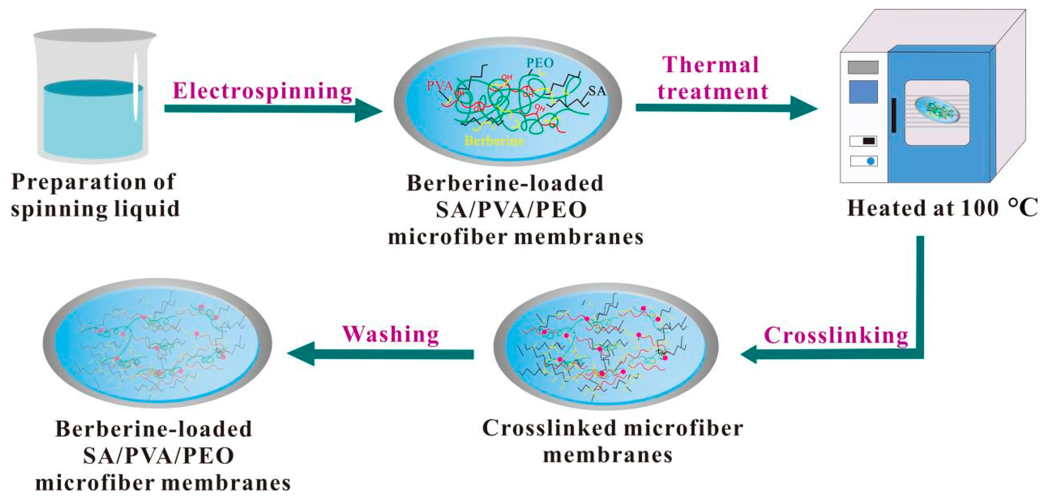
The whole preparation process was translated in Figure 2. Berberine (mass fractions of 0, 3, and 5 wt.%) was added to the optimal ratio of SA/PVA/PEO solution prior to the electrospinning process to form MFMs. A 16 kV voltage was supplied, and the feed rate for the polymer solution was adjusted to a constant rate of 0.6 mL/h. The magnetic stirrer’s speed and spinning time were set to 200 r/min and 48 h, respectively. The produced MFMs were then thermally treated in an electric blast drying oven at 100 and 120 °C for 2 h. The basic experimental scheme is shown in Table 1. Afterward, the MFMs were immersed in an ethanol solution of 4 wt.% CaCl2, which acts as crosslinking exchange and ionic crosslinking media, for 4 h. The crosslinking treatment is used to improve the water dissolution resistance of medical dressings. Finally, the berberine-loaded SA/PVA/PEO MFMs were washed with ethanol solution and dried at 20 °C for 24 h.

2.6. Antibacterial Activity Test of Berberine-Loaded SA/PVA/PEO Microfiber Membranes

“GB/T 20944.3-2008-Part 3: Shake flask method” was applied to assess the antimicrobial property of microfiber membranes [52]. The antibacterial rate of SA/PVA/PEO MFMs containing different amounts of berberine to E. coli was tested. The circular bacteria were inoculated from cultured E. coli (3rd to 10th generation) in test tubes and inoculated on nutrient agar plates and cultured at 37 °C for 18 h. A typical colony was inoculated in 20 mL nutritious broth and was incubated under 130 r/min for 18 h. Then the inoculation suspension was prepared and determined by spectrophotometer; the counted living bacteria were 3 × 10⁵ CFU/mL to 4 × 10⁵ CFU/mL. The MFMs with 0, 3, and 5 wt.% berberine were cut into 5 mm × 5 mm samples and sterilized by ultraviolet irradiation. Samples were then mixed with E. coli suspension for shock culture, and the E. coli suspension without samples was used as the counter sample. Finally, every mixture mentioned above was diluted for 104, 105, and 106 times, respectively, and placed in a constant temperature biochemical incubator at 37 °C for 24 h. The plate with the appropriate dilution ratio between 30 CFU and 300 CFU was selected.

2.7. Water Dissolution Resistance Test of Berberine-Loaded SA/PVA/PEO Microfiber Membranes

According to the liquid absorption rate measurement method, MFMs were cut into 3 cm × 3 cm samples and placed in deionized water [53]. Then the dissolution status of the MFMs before and after crosslinking treatment was observed.

2.8. Moisture Content Test of Berberine-Loaded SA/PVA/PEO Microfiber Membranes

Moisture content was measured under the standards of the YY/T 0471.1-2004 [54]. First, the MFMs were dried in an oven at 50 °C for 12 h. Then, they were cut into 1 cm × 1 cm and referred to as G0. Next, they were soaked in phosphate buffer saline (PBS) for 2 min and referred to as G1. PBS was used to mimic the humoral environment, and MFMs can be biodegradable in phosphate-buffered brine [55,56]. The formulation of PBS buffer is shown in Table 2. The moisture content was calculated by Equation (1).
  S = ( G 1 G 0 )   /   G 0 × 100 % .
where S is the moisture content rate (%), G1 is the mass of the wet sample (g), and G0 is the mass of the dry sample (g).

2.9. Fracture Strength Test of Berberine-Loaded SA/PVA/PEO Microfiber Membranes

Fracture testing was performed by Instron 3369 universal strength machine (INSTRON Co., Ltd., Boston, USA). Samples were cut into 90 mm × 10 mm and placed between the jaw with a gauge length of 30 mm. On each sample, three points were selected, and thickness values were measured with a thickness gauge. The fracture strength was calculated by Equation (2).
P = F   /   ( w × d ) .
where P is the fracture strength (MPa), F is the fracture force (cN), w is the width of the sample (mm), and d is the thickness of the sample (mm).

3. Results and Discussion

3.1. The Best Ratio of SA/PVA/PEO with a Good Electrostatic Spinning Effect

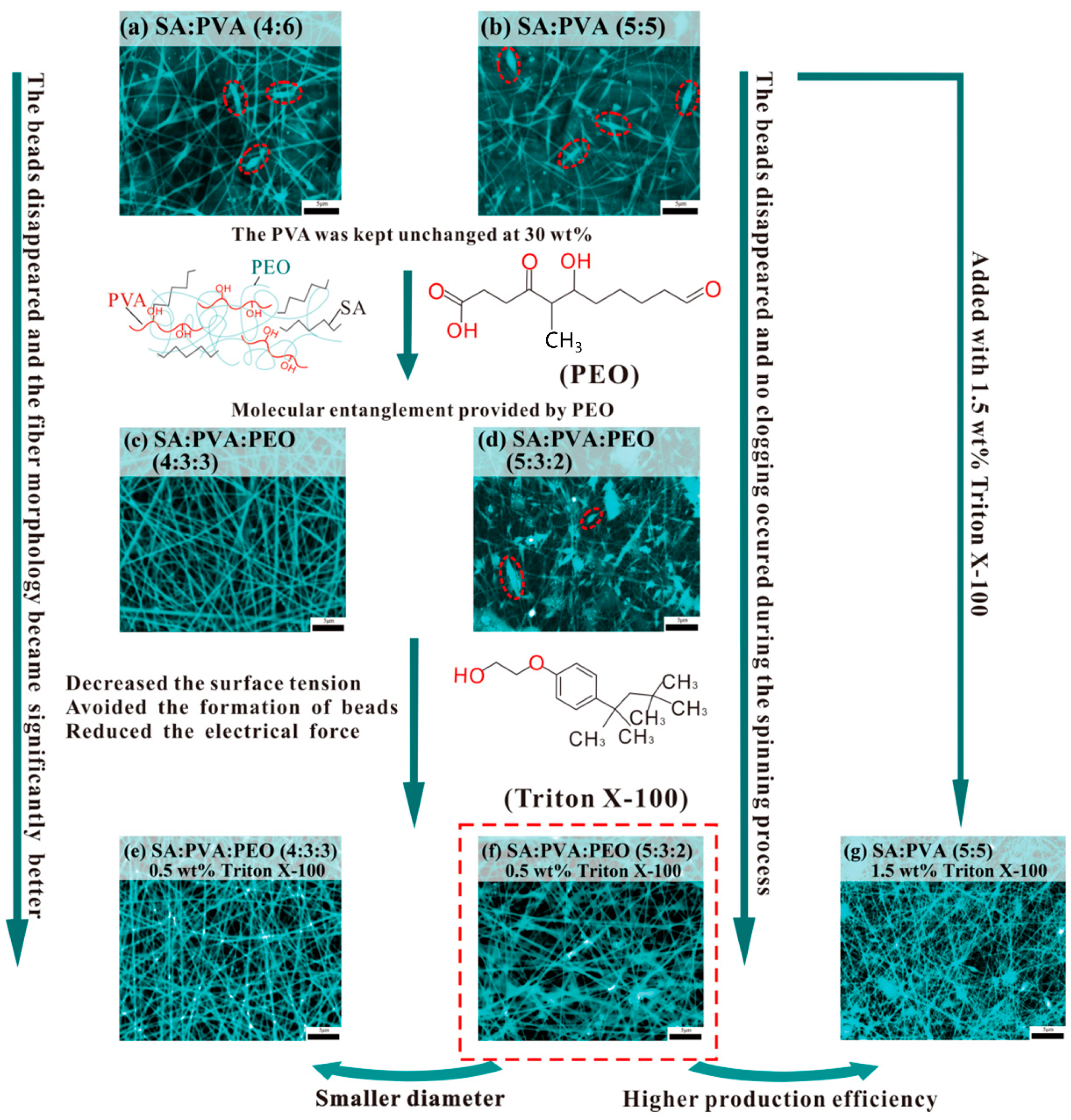
There are apparent differences in morphological structures between MFMs made with SA/PVA and with SA/PVA/PEO. In Figure 3a of SA/PVA 4:6 and Figure 3b of SA-PVA 5:5 MFMs, the formation of beads is visibly observed. Meanwhile, there are no beads formed for SA/PVA/PEO 4:3:3 MFMs, as observed in Figure 3c. The reason is that PEO contributes a better degree of entanglement between macromolecules, thus improving the electrospinning performances of the SA/PVA solution system [57]. Nevertheless, when the ratios between SA/PVA/PEO change to 5:3:2, respectively, very thin microfibers formed with obvious clogging and beads are observed in Figure 3d. With the mass fraction of PEO decreased and that of SA increased, the improvement effect of PEO gradually weakened. Meanwhile, as the mass fraction of SA increased, and the viscosity of the spinning solution gradually decreased, it resulted in insufficient entanglement of macromolecular chains in the spinning solution and discontinuous jet flow [58]. Apparent differences between solutions with and without Triton X-100 are noted. With the addition of 0.5 wt.% Triton X-100 surfactant on SA/PVA/PEO of 4:3:3, microfibers’ structures have improved with a clear reduction of beads, and no clogging is formed, as seen in Figure 3e. For Figure 3f, at the same 0.5 wt.% Triton X-100 surfactant, reduction of beads and fluent spinning are also noticeable for SA:PVA:PEO 5:3:2, in comparison to similar solutions without Triton X-100 in Figure 3d. The reason is that Triton X-100 in solution affected the circularity of beads and improved the homogeneity of fiber corresponding to the decrease in electrical conductivity and the surface tension [59]. In addition, the SA/PVA/PEO 5:3:2 microfibers added to with 0.5 wt.% Triton X-100 of Figure 3f have given better production efficiency than the SA/PVA 5:5 microfibers added to with 1.5 wt.% Triton X-100 of Figure 3g. This has also proven that PEO can effectively promote the preparation of alginate wound dressings by electrospinning.
Other than morphological observations, an investigation on the effects of different materials on microfibers’ diameter was performed. The diameter of SA/PVA/PEO 4:3:3 microfibers with Triton X-100 is reduced from 276 ± 64.6 nm to 216 ± 30.24 nm, compared to that without Triton X-100, as shown in Figure 4. Meanwhile, the fiber diameter unevenness is significantly reduced, and the coefficient of variation of fiber diameter is reduced from 23.4 to 14.0%. Therefore, the addition of surfactants can improve the uniformity of microfibers, subsequently improving the quality of microfibers [60]. Nevertheless, as revealed in Figure 3, with 0.5 wt.% Triton X-100, SA/PVA/PEO 5:3:2 produces smaller microfiber diameters than SA/PVA/PEO 4:3:3, which is due to the decreased mass fraction of PEO and reduced chain entanglement.

3.2. Thermal Treatment to SA/PVA/PEO Microfiber Membranes

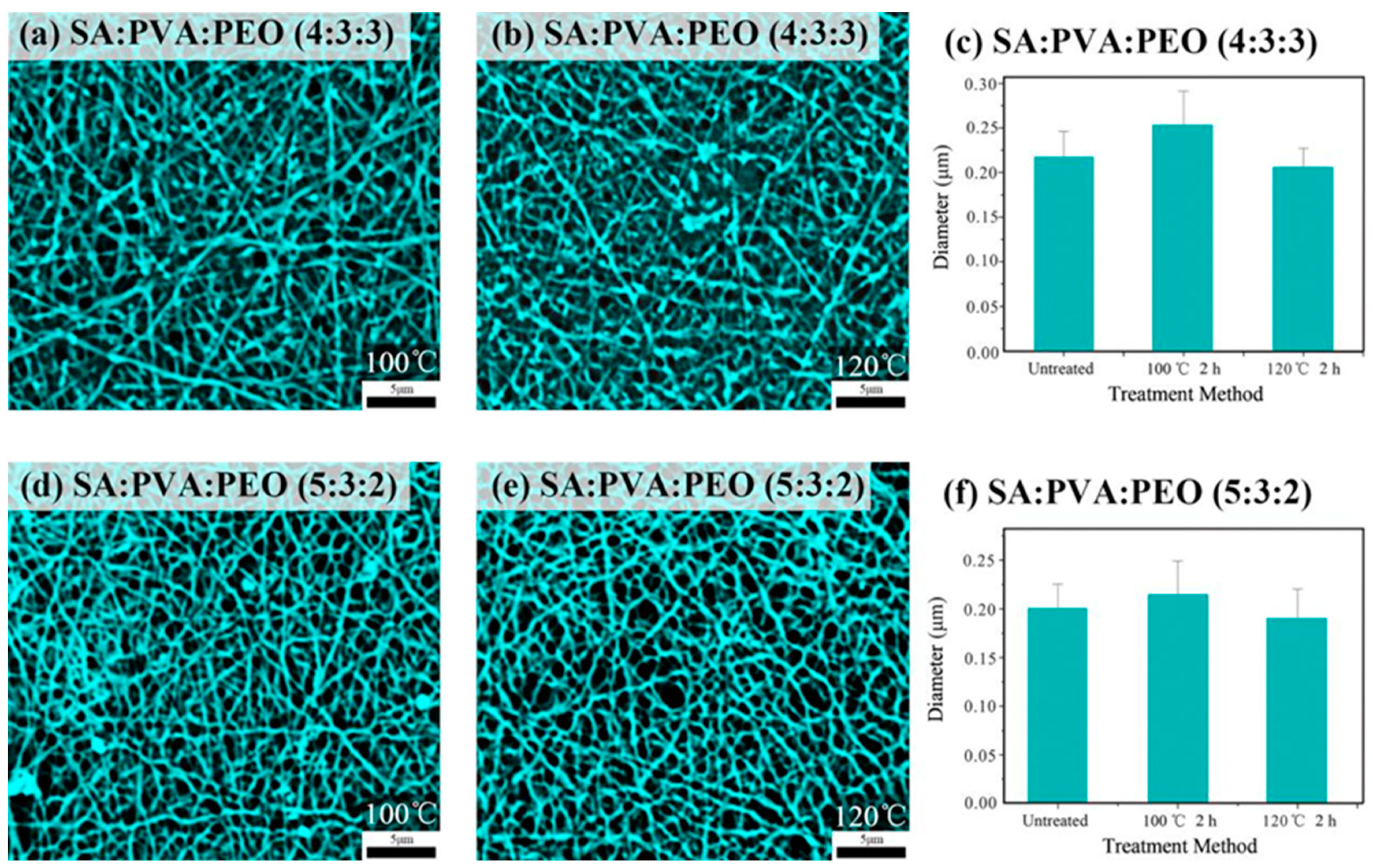
Figure 5 shows the SEM morphology and fiber diameter distribution of thermally treated SA/PVA/PEO microfibers at different ratios of 4:3:3 and 5:3:2. It can be observed in Figure 5 that all MFMs showed different degrees of melting adhesion due to the melted PEO. When the microfibers of SA/PVA/PEO 4:3:3 and 5:3:2 were treated at 100 °C for 2 h, the diameter substantially increased compared to the untreated. However, as the temperature rises to 120 °C with a similar duration, the fiber diameter noticeably reduced. It is believed that the PEO in MFMs melted and flowed out slowly at 100 °C [34,61,62], most of which are glued to the outer layer of microfibers, and some might have flowed into the voids of the fibers. After thermal treatment, the PEO re-solidified and caused a substantial increase in microfibers’ diameters [63]. Meanwhile, at 120 °C, the PEO was prone to decompose and be volatilized [64], which eventually decreased fibers’ diameters compared with before treatment and the microfibers treated at 100 °C.
As shown in Figure 6a,b, trend changes are similar to what is revealed in Figure 5c,f. The average fracture strength of the untreated SA/PVA/PEO 4:3:3 MFMs is 11.7 MPa. After being thermally treated at 100 °C, point bonding occurred between the fibers due to the solidified melted PEO, which eventually improved the fracture strength of the MFMs. However, when the treatment temperature was increased to 120 °C, the fracture strength of the MFMs decreased due to the thermal decomposition and volatilization of PEO. Similarly, the fracture strength of SA/PVA/PEO 5:3:2 MFMs treated at 100 °C is greatly improved. Furthermore, changes in diameter size between untreated and treated MFMs showed great influence on the fracture strength of MFMs. As the diameter increases, the fracture strength will be increased too. This intertwined coincidence suggests that, to some extent, with the presence of PEO and a suitable temperature, improvements of fracture strength are feasible.

3.3. Antibacterial Efficacy of Berberine-Loaded SA/PVA/PEO Microfiber Membranes

Table 3 shows the antibacterial properties of MFMs containing different weight fractions of berberine (3 and 5 wt.%) against E. coli. Without berberine, the antibacterial rate of MFMs was only 14.7%, indicating a non-antibacterial characteristic. When 3 and 5 wt.% berberine were added, the inhibition rates of the MFMs against E. coli were 92.9 and 97.2%, respectively. Therefore, it can be deduced that berberine exhibited substantial antibacterial activity [41].

3.4. Water Dissolution Resistance of Berberine-Loaded SA/PVA/PEO Microfiber Membranes

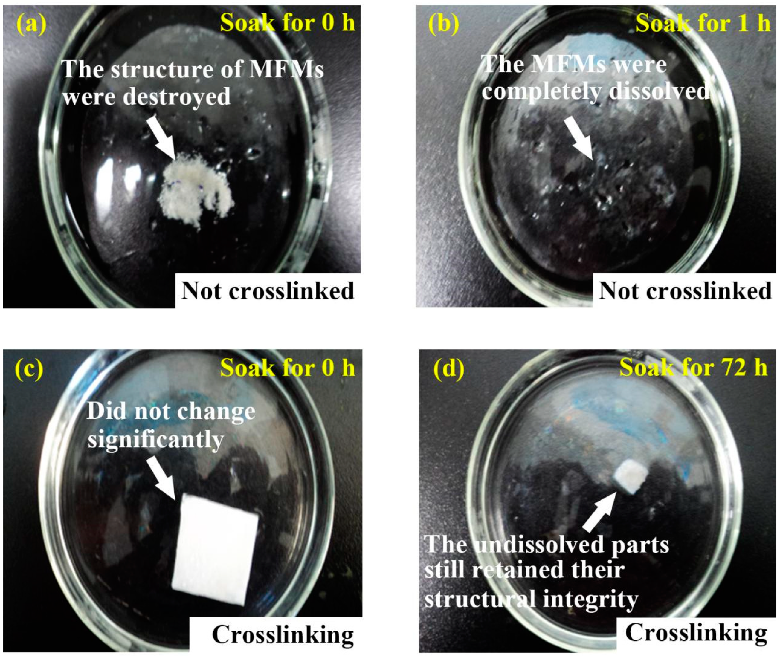
Figure 7 shows the water dissolution resistance of the MFMs before and after crosslinking treatment. It can be seen from Figure 7a that, upon soaking, the structure of non-crosslinked MFMs is clearly destroyed. Within 1 h, the MFMs are wholly dissolved, as seen in Figure 7b. However, as seen in Figure 7c, the crosslinked MFMs with CaCl2 structurally do not change upon soaking in water. Then, after about 72 h, as seen in Figure 7d, most of the crosslinked MFMs are dissolved and the surface area decreased by more than 80%. The undissolved parts of crosslinked MFMs still preserved their structural integrity. The reason is that SA can be crosslinked with most multivalent cations, which leads to reticular structure and reduces the degree of freedom of SA polymer chains. Ca2+ is crosslinked between the polymer chains and generated a three-dimensional structure to inhibit the flow of free water molecules. Therefore, the water solubility of MFMs is reduced, and the water dissolution resistance is increased [65,66,67].

3.5. Moisture Absorption and Fracture Strength of Berberine-Loaded SA/PVA/PEO Microfiber Membranes

Cells cannot live without water, and microfiber wound dressings should have a certain moisture content to meet the requirement of wound cells’ growth environment [53]. The ideal wound dressing should be able to maintain a high moisture content [68]. The moisture content of MFMs with 5 wt.% berberine is 71.98%. The moisture content of the MFMs implies that it could maintain the moist environment for cell growth and promote cell growth and reproduction. Ass can be seen from Figure 8, the fracture strength of SA/PVA/PEO MFMs containing 5 wt.% berberine reached 7.8 MPa, which is higher than that of SA/PVA/PEO MFMs 5:3:2 without berberine. Berberine is a quaternary ammonium salt with considerable molecular weight and hard skeleton, enhancing the interaction between the polymer side chains and improving the mechanical properties of MFMs [69].

4. Conclusions

The addition of PEO improved the electrostatic spinning performance of SA and the mechanical performance of MFMs. With the addition of 0.5 wt.% Trition X-100, the proportion of SA solution in the spinning system has increased up to 50%. The SA/PVA/PEO 5:3:2 MFMs showed good fiber morphology with no clogging formation during spinning. The fracture strength of the MFMs was improved substantially by thermal treatment at 100 °C for 2 h. When different weight fractions of berberine, 3 and 5 wt.%, were added to SA/PVA/PEO MFMs, an excellent antibacterial property was observed, as well as decent moisture content, improved structural strength, and good water dissolution resistance. Berberine-loaded SA/PVA/PEO MFMs have the potential to be a new type of antibacterial wound dressing substrate with values that should be considered in the wound dressing field.

Author Contributions

Data curation, validation, and writing—original draft preparation, J.Z., Y.L. and H.L.; writing—review and editing, K.M.S. and S.Z.; funding acquisition, resources, supervision, and writing—review and editing, H.W. and C.W. All authors have read and agreed to the published version of the manuscript.

Funding

This research was funded by the National Natural Science Foundation of China, grant number 11802205.

Institutional Review Board Statement

Not applicable.

Data Availability Statement

Not applicable.

Conflicts of Interest

The authors declare no conflict of interest. This study does not include experiments conducted on humans or animals.

References

  1. Fu, R.; Li, C.; Yu, C.; Xie, H.; Shi, S.; Li, Z.; Wang, Q.; Lu, L. A novel electrospun membrane based on moxifloxacin hydrochloride/poly(vinyl alcohol)/sodium alginate for antibacterial wound dressings in practical application. Drug Deliv. 2016, 23, 818–829. [Google Scholar] [CrossRef] [PubMed]
  2. Souza, J.M.; Henriques, M.; Teixeira, P.; Fernandes, M.M.; Fangueiro, R.; Zille, A. Comfort and Infection Control of Chitosan-impregnated Cotton Gauze as Wound Dressing. Fibers Polym. 2019, 20, 922–932. [Google Scholar] [CrossRef]
  3. Kim, Y.; Doh, S.J.; Lee, G.D.; Kim, C.; Im, J.N. Composite Nonwovens Based on Carboxymethyl Cellulose for Wound Dressing Materials. Fibers Polym. 2019, 20, 2048–2056. [Google Scholar] [CrossRef]
  4. Lee, G.D.; Doh, S.J.; Kim, Y.; Im, J.N. Preparation and Characterization of Carboxymethyl Chitosan Nonwovens for Wound Dressing. Fibers Polym. 2020, 21, 1894–1905. [Google Scholar] [CrossRef]
  5. Gharibi, R.; Yeganeh, H.; Abdali, Z. Preparation of antimicrobial wound dressings via thiolene photopolymerization reaction. J. Mater. Sci. 2018, 53, 1581–1595. [Google Scholar] [CrossRef]
  6. Jeong, Y.H.; Lee, J. Fabrication of Microfiber Patterns with Ivy Shoot-Like Geometries Using Improved Electrospinning. Materials 2016, 9, 266. [Google Scholar] [CrossRef] [PubMed] [Green Version]
  7. Liu, W.; Xu, Z.; Sun, L.; Guo, P.; Zeng, C.; Wang, C.; Zhang, L. Polymerization-induced phase separation fabrication: A versatile microfluidic technique to prepare microfibers with various cross sectional shapes and structures. Chem. Eng. J. 2017, 315, 25–34. [Google Scholar] [CrossRef]
  8. Liu, M.; Duan, X.P.; Li, Y.M.; Yang, D.P.; Long, Y.Z. Electrospun nanofibers for wound healing. Mat. Sci. Eng. C-Mater. 2017, 76, 1413–1423. [Google Scholar] [CrossRef]
  9. Mahamuni-Badiger, P.P.; Patil, P.M.; Patel, P.R.; Dhanavade, M.J.; Badiger, M.V.; Marathe, Y.N.; Bohara, R.A. Electrospun poly(3-hydroxybutyrate-co-3-hydroxyvalerate)/polyethylene oxide (PEO) microfibers reinforced with ZnO nanocrystals for antibacterial and antibiofilm wound dressing applications. New J. Chem. 2020, 44, 9754–9766. [Google Scholar] [CrossRef]
  10. Thorvaldsson, A.; Edvinsson, P.; Glantz, A.; Rodriguez, K.; Walkenström, P.; Gatenholm, P. Superhydrophobic behaviour of plasma modified electrospun cellulose nanofiber-coated microfibers. Cellulose 2012, 19, 1743–1748. [Google Scholar] [CrossRef]
  11. Barhate, R.; Koeppl, S.; Ramakrishna, S. Porous nano- and microfibrous polymeric membrane material for catalytic support. Chem. Eng. Res. Des. 2011, 89, 621–630. [Google Scholar] [CrossRef]
  12. Lanno, G.-M.; Ramos, C.; Preem, L.; Putrinš, M.; Laidmäe, I.; Tenson, T.; Kogermann, K. Antibacterial Porous Electrospun Fibers as Skin Scaffolds for Wound Healing Applications. ACS Omega 2020, 5, 30011–30022. [Google Scholar] [CrossRef] [PubMed]
  13. Zhao, Y.-T.; Zhang, J.; Gao, Y.; Liu, X.-F.; Liu, J.-J.; Wang, X.-X.; Xiang, H.-F.; Long, Y.-Z. Self-powered portable melt electrospinning for in situ wound dressing. J. Nanobiotechnol. 2020, 18, 111. [Google Scholar] [CrossRef] [PubMed]
  14. Xue, J.; Wu, T.; Dai, Y.; Xia, Y. Electrospinning and Electrospun Nanofibers: Methods, Materials, and Applications. Chem. Rev. 2019, 119, 5298–5415. [Google Scholar] [CrossRef]
  15. Karayeğen, G.; Koçum, I.C.; Serdaroğlu, D.; Doğan, M. Aligned polyvinylpyrrolidone nanofibers with advanced electrospinning for biomedical applications. Bio-Med. Mater. Eng. 2018, 29, 685–697. [Google Scholar] [CrossRef]
  16. Chen, K.; Wang, F.; Liu, S.; Wu, X.; Xu, L.; Zhang, D. In situ reduction of silver nanoparticles by sodium alginate to obtain silver-loaded composite wound dressing with enhanced mechanical and antimicrobial property. Int. J. Biol. Macromol. 2020, 148, 501–509. [Google Scholar] [CrossRef]
  17. Li, S.; Li, L.; Guo, C.; Qin, H.; Yu, X. A promising wound dressing material with excellent cytocompatibility and proangiogenesis action for wound healing: Strontium loaded Silk fibroin/Sodium alginate (SF/SA) blend films. Int. J. Biol. Macromol. 2017, 104, 969–978. [Google Scholar] [CrossRef]
  18. Wu, C.; Zhang, Z.; Zhou, K.; Chen, W.; Tao, J.; Li, C.; Xin, H.; Song, Y.; Ai, F. Preparation and characterization of borosilicate-bioglass-incorporated sodium alginate composite wound dressing for accelerated full-thickness skin wound healing. Biomed. Mater. 2020, 15, 055009. [Google Scholar] [CrossRef]
  19. Oh, S.-T.; Kim, W.-R.; Kim, S.-H.; Chung, Y.-C.; Park, J.-S. The preparation of polyurethane foam combined with pH-sensitive alginate/bentonite hydrogel for wound dressings. Fibers Polym. 2011, 12, 159–165. [Google Scholar] [CrossRef]
  20. Abdel-Mohsen, A.M.; Jancar, J.; Massoud, D.; Fohlerova, Z.; Elhadidy, H.; Spotz, Z.; Hebeish, A. Novel chitin/chitosan-glucan wound dressing: Isolation, characterization, antibacterial activity and wound healing properties. Int. J. Pharm. 2016, 510, 86–99. [Google Scholar] [CrossRef]
  21. He, Y.; Li, Y.; Sun, Y.; Zhao, S.; Feng, M.; Xu, G.; Zhu, H.; Ji, P.; Mao, H.; He, Y.; et al. A double-network polysaccharide-based composite hydrogel for skin wound healing. Carbohydr. Polym. 2021, 261, 117870. [Google Scholar] [CrossRef] [PubMed]
  22. Sarwar, M.S.; Ghaffar, A.; Huang, Q. Development and characterization of sodium alginate/poly(sodium 4-styrenesulfonate) composite films for release behavior of ciprofloxacin hydrogen chloride monohydrate. Polym. Polym. Compos. 2021, 29, S143–S153. [Google Scholar] [CrossRef]
  23. Hu, W.-W.; Yu, H.-N. Coelectrospinning of chitosan/alginate fibers by dual-jet system for modulating material surfaces. Carbohydr. Polym. 2013, 95, 716–727. [Google Scholar] [CrossRef] [PubMed]
  24. Bonino, C.A.; Krebs, M.D.; Saquing, C.D.; Jeong, S.I.; Shearer, K.L.; Alsberg, E.; Khan, S.A. Electrospinning alginate-based nanofibers: From blends to crosslinked low molecular weight alginate-only systems. Carbohydr. Polym. 2011, 85, 111–119. [Google Scholar] [CrossRef]
  25. Sharma, A.; Mittal, A.; Puri, V.; Kumar, P.; Singh, I. Curcumin-loaded, alginate–gelatin composite fibers for wound healing applications. 3 Biotech 2020, 10, 464. [Google Scholar] [CrossRef]
  26. Lu, J.-W.; Zhu, Y.-L.; Guo, Z.-X.; Hu, P.; Yu, J. Electrospinning of sodium alginate with poly(ethylene oxide). Polymer 2006, 47, 8026–8031. [Google Scholar] [CrossRef]
  27. Vicini, S.; Mauri, M.; Vita, S.; Castellano, M. Alginate and alginate/hyaluronic acid membranes generated by electrospinning in wet conditions: Relationship between solution viscosity and spinnability. J. Appl. Polym. Sci. 2018, 135, 46390. [Google Scholar] [CrossRef]
  28. Shen, W.; Hsieh, Y.-L. Biocompatible sodium alginate fibers by aqueous processing and physical crosslinking. Carbohydr. Polym. 2014, 102, 893–900. [Google Scholar] [CrossRef] [Green Version]
  29. Paduraru, A.; Ghitulica, C.; Trusca, R.; Surdu, V.A.; Neacsu, I.A.; Holban, A.M.; Birca, A.C.; Iordache, F.; Vasile, B.S. Antimicrobial Wound Dressings as Potential Materials for Skin Tissue Regeneration. Materials 2019, 12, 1859. [Google Scholar] [CrossRef] [Green Version]
  30. Saquing, C.D.; Tang, C.; Monian, B.; Bonino, C.A.; Manasco, J.L.; Alsberg, E.; Khan, S.A. Alginate–Polyethylene Oxide Blend Nanofibers and the Role of the Carrier Polymer in Electrospinning. Ind. Eng. Chem. Res. 2013, 52, 8692–8704. [Google Scholar] [CrossRef]
  31. Surendhiran, D.; Cui, H.; Lin, L. Encapsulation of Phlorotannin in Alginate/PEO blended nanofibers to preserve chicken meat from Salmonella contaminations. Food Packag. Shelf Life 2019, 21, 100346. [Google Scholar] [CrossRef]
  32. Gutierrez-Gonzalez, J.; García-Cela, E.; Magan, N.; Rahatekar, S.S. Electrospinning alginate/polyethylene oxide and curcumin composite nanofibers. Mater. Lett. 2020, 270, 127662. [Google Scholar] [CrossRef]
  33. Luo, X.; Li, J.; Lin, X. Effect of gelatinization and additives on morphology and thermal behavior of corn starch/PVA blend films. Carbohydr. Polym. 2012, 90, 1595–1600. [Google Scholar] [CrossRef]
  34. Martín-Alfonso, J.E.; Cuadri, A.A.; Franco, J.M. Development and Characterization of Novel Fibers Based on Potato Protein/Polyethylene Oxide Through Electrospinning. Fibers Polym. 2019, 20, 1586–1593. [Google Scholar] [CrossRef]
  35. Song, Q.; Li, A.; Shi, L.; Qian, C.; Feric, T.G.; Fu, Y.; Zhang, H.; Li, Z.; Wang, P.; Li, Z.; et al. Thermally stable, nano-porous and eco-friendly sodium alginate/attapulgite separator for lithium-ion batteries. Energy Storage Mater. 2019, 22, 48–56. [Google Scholar] [CrossRef]
  36. Fang, W.; Yang, S.; Yuan, T.-Q.; Charlton, A.; Sun, R.-C. Effects of Various Surfactants on Alkali Lignin Electrospinning Ability and Spun Fibers. Ind. Eng. Chem. Res. 2017, 56, 9551–9559. [Google Scholar] [CrossRef] [Green Version]
  37. Yao, L.; Haas, T.W.; Guiseppi-Elie, A.; Bowlin, G.L.; Simpson, D.G.; Wnek, G.E. Electrospinning and Stabilization of Fully Hydrolyzed Poly(Vinyl Alcohol) Fibers. Chem. Mater. 2003, 15, 1860–1864. [Google Scholar] [CrossRef]
  38. Dou, X.; Wang, Q.; Li, Z.; Ju, J.; Wang, S.; Hao, L.; Sui, K.; Xia, Y.; Tan, Y. Seaweed-Derived Electrospun Nanofibrous Membranes for Ultrahigh Protein Adsorption. Adv. Funct. Mater. 2019, 29, 1905610. [Google Scholar] [CrossRef]
  39. Zheng, X.; Zhang, Y.; Zou, L.; Wang, Y.; Zhou, X.; Yao, L.; Wang, Z.; Li, C.; Qiu, Y. Robust ZIF-8/alginate fibers for the durable and highly effective antibacterial textiles. Colloids Surf. B 2020, 193, 111127. [Google Scholar] [CrossRef]
  40. Ni, W.-J.; Ding, H.-H.; Tang, L.-Q. Berberine as a promising anti-diabetic nephropathy drug: An analysis of its effects and mechanisms. Eur. J. Pharmacol. 2015, 760, 103–112. [Google Scholar] [CrossRef]
  41. Jin, J.; Xu, M.; Liu, Y.; Ji, Z.; Dai, K.; Zhang, L.; Wang, L.; Ye, F.; Chen, G.; Lv, Z. Alginate-based composite microspheres coated by berberine simultaneously improve hemostatic and antibacterial efficacy. Colloids Surf. B 2020, 194, 111168. [Google Scholar] [CrossRef] [PubMed]
  42. Tong, W.Y.; bin Abdullah, A.Y.K.; binti Rozman, N.A.S.; bin Wahid, M.I.A.; Hossain, M.; Ring, L.C.; Lazim, Y.; Tan, W.-N. Antimicrobial wound dressing film utilizing cellulose nanocrystal as drug delivery system for curcumin. Cellulose 2018, 25, 631–638. [Google Scholar] [CrossRef]
  43. Zhong, Y.; Xiao, H.; Seidi, F.; Jin, Y. Natural Polymer-Based Antimicrobial Hydrogels without Synthetic Antibiotics as Wound Dressings. Biomacromolecules 2020, 21, 2983–3006. [Google Scholar] [CrossRef] [PubMed]
  44. Dragan, E.S.; Dinu, M.V. Advances in porous chitosan-based composite hydrogels: Synthesis and applications. React. Funct. Polym. 2020, 146, 104372. [Google Scholar] [CrossRef]
  45. Subhedar, A.; Bhadauria, S.; Ahankari, S.; Kargarzadeh, H. Nanocellulose in biomedical and biosensing applications: A review. Int. J. Biol. Macromol. 2021, 166, 587–600. [Google Scholar] [CrossRef]
  46. Pang, Q.; Zheng, X.; Luo, Y.; Ma, L.; Gao, C. A photo-cleavable polyprodrug-loaded wound dressing with UV-responsive antibacterial property. J. Mater. Chem. B 2017, 5, 8975–8982. [Google Scholar] [CrossRef]
  47. Hedayati, N.; Montazer, M.; Mahmoudirad, M.; Toliyat, T. Ketoconazole and Ketoconazole/β-cyclodextrin performance on cotton wound dressing as fungal skin treatment. Carbohydr. Polym. 2020, 240, 116267. [Google Scholar] [CrossRef]
  48. Nataraj, D.; Reddy, R.; Reddy, N. Crosslinking electrospun poly (vinyl) alcohol fibers with citric acid to impart aqueous stability for medical applications. Eur. Polym. J. 2020, 124, 109484. [Google Scholar] [CrossRef]
  49. Jin, Y.; Yang, D.; Zhou, Y.; Ma, G.; Nie, J. Photocrosslinked electrospun chitosan-based biocompatible nanofibers. J. Appl. Polym. Sci. 2008, 109, 3337–3343. [Google Scholar] [CrossRef]
  50. Xu, X.; Yue, Y.; Cai, D.; Song, J.; Han, C.; Liu, Z.; Wang, D.; Xiao, J.; Wu, H. Aqueous Solution Blow Spinning of Seawater-Stable Polyamidoxime Nanofibers from Water-Soluble Precursor for Uranium Extraction from Seawater. Small Methods 2020, 4, 2000558. [Google Scholar] [CrossRef]
  51. Islam, S.; Karim, M.R. Fabrication and characterization of poly(vinyl alcohol)/alginate blend nanofibers by electrospinning method. Colloids Surf. A Physicochem. Eng. Asp. 2010, 366, 135–140. [Google Scholar] [CrossRef]
  52. Tian, Y.; Liu, X.; Zheng, X.; Wang, L. Antimicrobial Properties of Flax Fibers in the Enzyme Retting Process. Fibres Text. East. Eur. 2016, 24, 15–17. [Google Scholar] [CrossRef] [Green Version]
  53. Kalaycıoğlu, Z.; Kahya, N.; Adımcılar, V.; Kaygusuz, H.; Torlak, E.; Akın-Evingür, G.; Erim, F.B. Antibacterial nano cerium oxide/chitosan/cellulose acetate composite films as potential wound dressing. Eur. Polym. J. 2020, 133, 109777. [Google Scholar] [CrossRef]
  54. YY/T 0471.1-2004; Test Methods for Primary Wound Dressing—Part 1: Aspects of Absorbency. National Medical Products Administration: Beijing, China, 2004.
  55. Wu, D.; Wei, W.; Li, H.; Wang, X.; Wang, T.; Tang, S.; Li, Q.; Yao, Y.; Pan, Y.; Wei, J. Blended films containing polybutyrolactam and chitosan for potential wound dressing applications. J. Appl. Polym. Sci. 2018, 135, 46511. [Google Scholar] [CrossRef]
  56. Pourhojat, F.; Sohrabi, M.; Shariati, S.; Mahdavi, H.; Asadpour, L. Evaluation of poly ε-caprolactone electrospun nanofibers loaded with Hypericum perforatum extract as a wound dressing. Res. Chem. Intermed. 2017, 43, 297–320. [Google Scholar] [CrossRef]
  57. Safi, S.; Morshed, M.; Hosseini Ravandi, S.A.; Ghiaci, M. Study of electrospinning of sodium alginate, blended solutions of sodium alginate/poly(vinyl alcohol) and sodium alginate/poly(ethylene oxide). J. Appl. Polym. Sci. 2007, 104, 3245–3255. [Google Scholar] [CrossRef]
  58. Ahire, J.; Robertson, D.; van Reenen, A.; Dicks, L. Surfactin-loaded polyvinyl alcohol (PVA) nanofibers alters adhesion of Listeria monocytogenes to polystyrene. Mater. Sci. Eng. C 2017, 77, 27–33. [Google Scholar] [CrossRef]
  59. Yu, D.-G.; Chatterton, N.P.; Yang, J.-H.; Wang, X.; Liao, Y.-Z. Coaxial Electrospinning with Triton X-100 Solutions as Sheath Fluids for Preparing PAN Nanofibers. Macromol. Mater. Eng. 2012, 297, 395–401. [Google Scholar] [CrossRef]
  60. Beigmoradi, R.; Samimi, A.; Mohebbi-Kalhori, D. Fabrication of polymeric nanofibrous mats with controllable structure and enhanced wetting behavior using one-step electrospinning. Polymer 2018, 143, 271–280. [Google Scholar] [CrossRef]
  61. Yang, R.; Li, H.M.; Jiang, J.; Zhou, D.S. Study on isothermal crystallization kinetics of polyvinyl oxide in restricted state by high-speed scanning calorimetry. Acta Polym. Sin. 2018, 9, 1228–1235. (In Chinese) [Google Scholar] [CrossRef]
  62. Martín-Alfonso, J.; Cuadri, A.; Greiner, A. The combined effect of formulation and pH on properties of polyethylene oxide composite fiber containing egg albumen protein. Int. J. Biol. Macromol. 2018, 112, 996–1004. [Google Scholar] [CrossRef]
  63. Medeiros, G.B.; de Souza, P.R.; Retamiro, K.M.; Nakamura, C.V.; Muniz, E.C.; Corradini, E. Experimental design to evaluate properties of electrospun fibers of zein/poly (ethylene oxide) for biomaterial applications. J. Appl. Polym. Sci. 2021, 138, 50898. [Google Scholar] [CrossRef]
  64. Yang, K.; Chi, Q.; Wang, X.; Jiang, Y.; Li, F.; Xue, B. The role of halloy site on crystallinity, ion conductivity, thermal and mechanical properties of poly(ethylene-oxide)/halloysite nanocomposites. J. Polym. Res. 2019, 26, 138. [Google Scholar] [CrossRef]
  65. Shahzad, A.; Khan, A.; Afzal, Z.; Umer, M.F.; Khan, J.; Khan, G.M. Formulation development and characterization of cefazolin nanoparticles-loaded cross-linked films of sodium alginate and pectin as wound dressings. Int. J. Biol. Macromol. 2019, 124, 255–269. [Google Scholar] [CrossRef] [PubMed]
  66. Wang, Q.; Ju, J.; Tan, Y.; Hao, L.; Ma, Y.; Wu, Y.; Zhang, H.; Xia, Y.; Sui, K. Controlled synthesis of sodium alginate electrospun nanofiber membranes for multi-occasion adsorption and separation of methylene blue. Carbohydr. Polym. 2019, 205, 125–134. [Google Scholar] [CrossRef] [PubMed]
  67. Díez-García, I.; Santamaría-Echart, A.; Eceiza, A.; Tercjak, A. Synthesis and characterization of environmentally-friendly waterborne poly(urethane-urea)s. Eur. Polym. J. 2018, 99, 240–249. [Google Scholar] [CrossRef]
  68. Akrami-Hasan-Kohal, M.; Tayebi, L.; Ghorbani, M. Curcumin-loaded naturally-based nanofibers as active wound dressing mats: Morphology, drug release, cell proliferation, and cell adhesion studies. New J. Chem. 2020, 44, 10343–10351. [Google Scholar] [CrossRef]
  69. Samadian, H.; Zamiri, S.; Ehterami, A.; Farzamfar, S.; Vaez, A.; Khastar, H.; Alam, M.; Ai, A.; Derakhshankhah, H.; Allahyari, Z.; et al. Electrospun cellulose acetate/gelatin nanofibrous wound dressing containing berberine for diabetic foot ulcer healing: In vitro and in vivo studies. Sci. Rep. 2020, 10, 8312. [Google Scholar] [CrossRef]
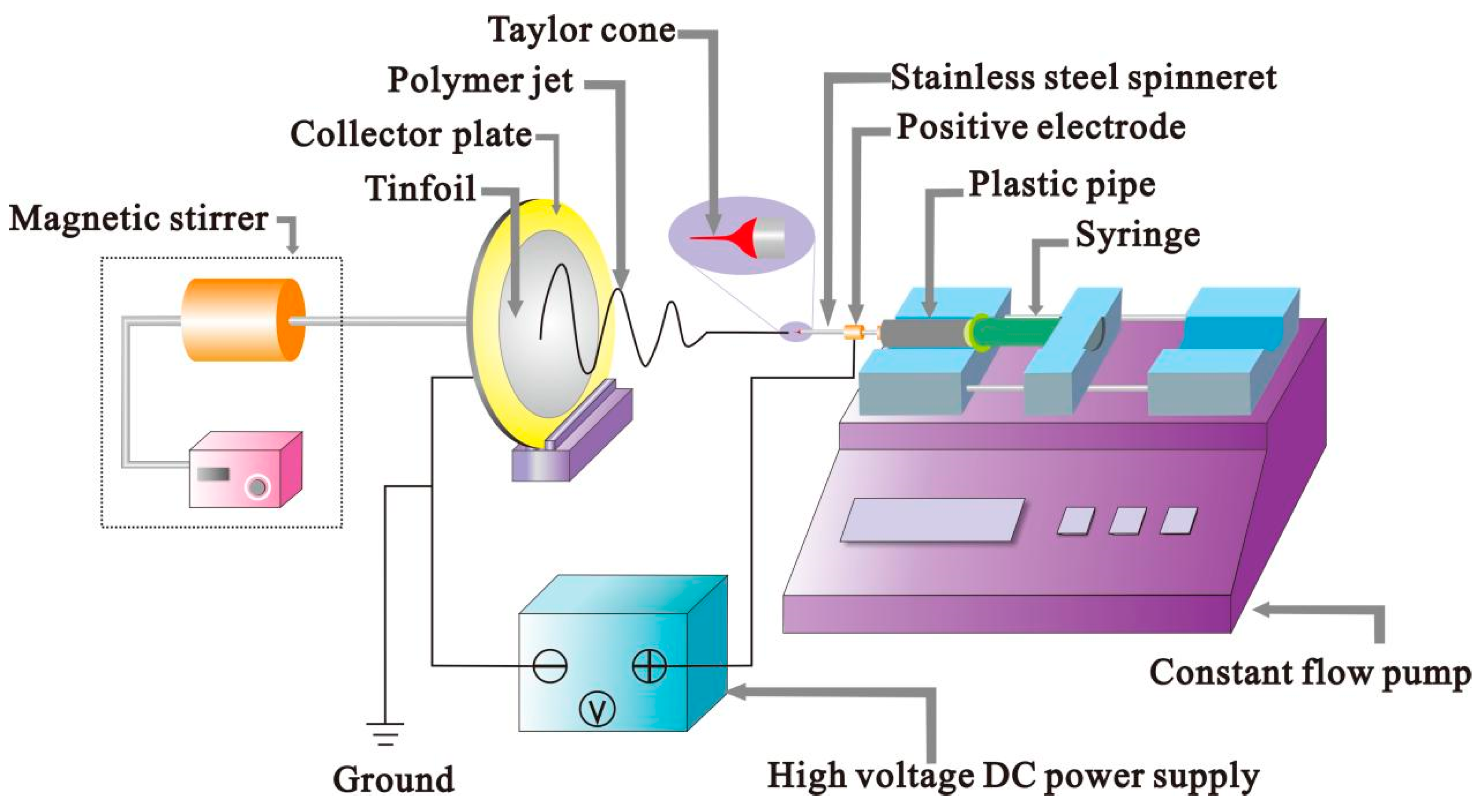
Figure 1. Connection diagram of electrostatic spinning equipment.
Polymers 14 04473 g001
Figure 2. Preparation of berberine-loaded SA/PVA/PEO MFMs.
Polymers 14 04473 g002
Figure 3. (ag) The SEM image (×6000) of seven series of MFMs. (a) The SA/PVA MFM in the ratio of 4:6. (b) The SA/PVA MFM in the ratio of 5:5. (c) The SA/PVA/PEO MFM in the ratio of 4:3:3. (d) The SA/PVA/PEO MFM in the ratio of 5:3:2. (e) The SA/PVA/PEO MFM in the ratio of 4:3:3 containing 0.5 wt% Triton X-100. (f) The SA/PVA/PEO MFM in the ratio of 5:3:2 containing 0.5 wt% Triton X-100. (g) The SA/PVA MFM in the ratio of 5:5 containing 1.5 wt.% Triton X-100.
Polymers 14 04473 g003
Figure 4. Average diameter distribution of three types of MFMs (TX: Triton X-100).
Polymers 14 04473 g004
Figure 5. Surface morphological structure of SA/PVA/PEO 4:3:3 MFMs treated at (a) 100 °C and (b) 120 °C. (c) Average diameter of SA/PVA/PEO 4:3:3 MFMs treated at different temperatures. Surface morphological structure of SA/PVA/PEO 5:3:2 MFMs treated at (d) 100 °C and (e) 120 °C. (f) Average diameter of SA/PVA/PEO 5:3:2 MFMs treated at different temperatures.
Polymers 14 04473 g005
Figure 6. (a) Fracture strength of SA/PVA/PEO 4:3:3 MFMs treated at different temperatures (b) Fracture strength of SA/PVA/PEO 5:3:2 MFMs treated at different temperatures.
Polymers 14 04473 g006
Figure 7. Water dissolution resistance of non-crosslinked SA/PVA/PEO 5:3:2 MFMs (a) soaked for 0 h, and (b) soaked for 1 h. Water dissolution resistance of crosslinked SA/PVA/PEO 5:3:2 MFMs with CaCl2 (c) soaked for 0 h, and (d) soaked for 72 h.
Polymers 14 04473 g007
Figure 8. Fracture strength comparison between SA/PVA/PEO MFMs 5:3:2 without berberine and SA/PVA/PEO containing 5 wt.% berberine.
Polymers 14 04473 g008
Table 1. Thermal treatment of SA/PVA/PEO MFMs.
The Ratio of SA/PVA/PEOProcessing Temperature (°C)Processing Time (h)
4:3:31002
4:3:31202
5:3:21002
5:3:21202
Table 2. Mixing ratios of 1 L PBS buffer.
Serial NumberChemicalsWeight (g)
1NaCl8.0
2KCl0.2
3Na2HPO4·12H2O1.96
4KH2PO40.24
Table 3. Bacteriostatic rates of SA/PVA/PEO 5:3:2 MFMs containing different mass fractions of berberine.
Sample NameBacterial Colony Count after Antibacterial (cfu·mL−1)Bacteriostatic Rate (%)
Control sample394-
SA MFMs with 0% berberine33614.7
SA MFMs with 3% berberine2892.9
SA MFMs with 5% berberine1197.2
Publisher’s Note: MDPI stays neutral with regard to jurisdictional claims in published maps and institutional affiliations.