

Tissue Engineering with Stem Cell from Human Exfoliated Deciduous Teeth (SHED) and Collagen Matrix, Regulated by Growth Factor in Regenerating the Dental Pulp

 ,
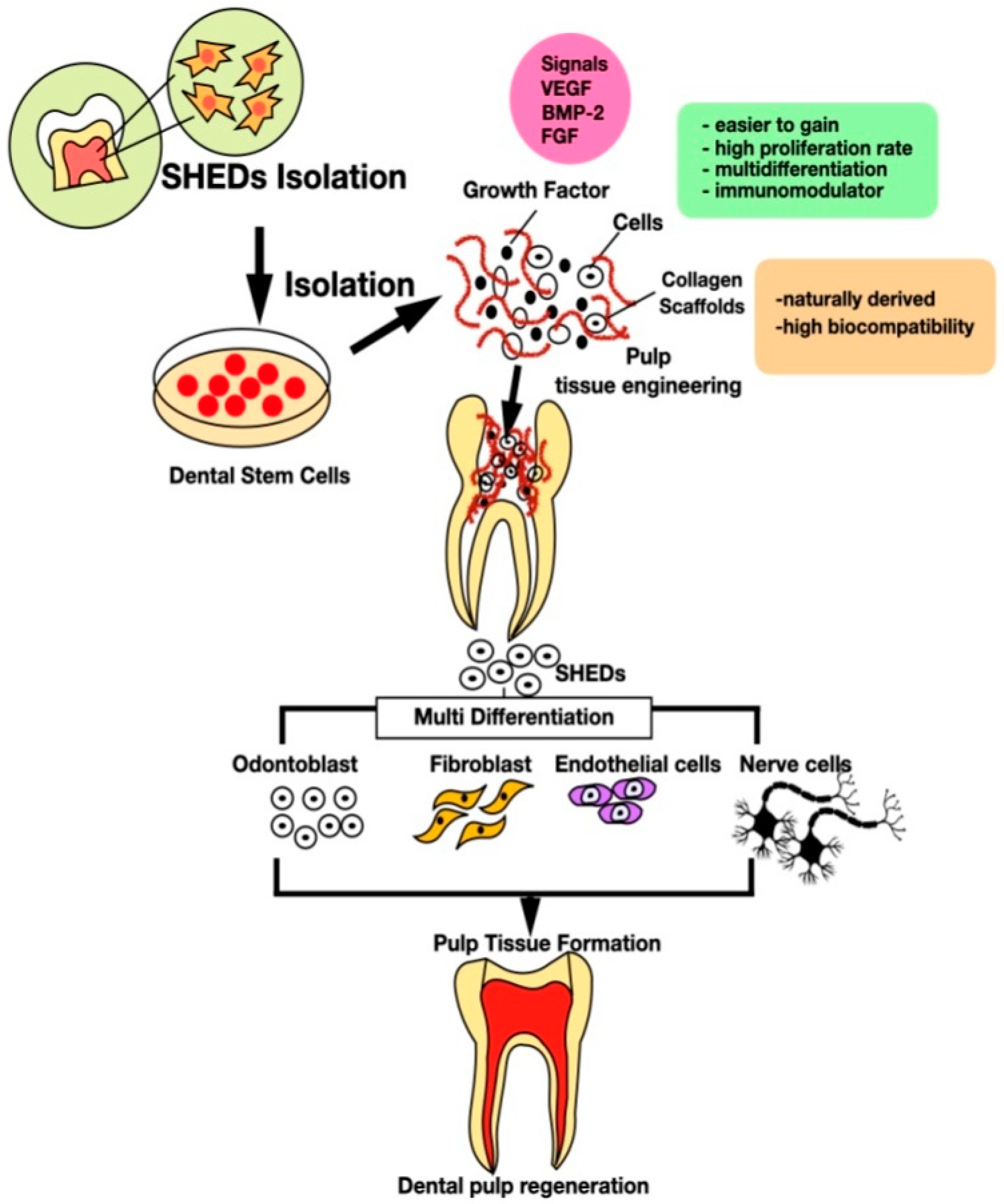
,  , , ,
, , ,
Abstract
1. Introduction
2. Tissue Engineering (TE) in Endodontic Treatment
3. Stem Cells
3.1. Totipotent Stem Cells
3.2. Pluripotent Stem Cells
3.3. Multipotent Stem Cells
3.4. Unipotent Stem Cell
3.5. Induced Pluripotent Cells
4. Stem Cells from Human Exfoliated Deciduous Teeth (SHED)
5. Collagen Scaffold
6. Growth Factor as Regulator
7. Dentin Pulp Regeneration
8. Conclusions
Author Contributions
Funding
Institutional Review Board Statement
Informed Consent Statement
Acknowledgments
Conflicts of Interest
References
- Lu, Y.; Liu, Z.; Huang, J.; Liu, C. Therapeutic effect of one-time root canal treatment for irreversible pulpitis. J. Int. Med. Res. 2019, 48, 0300060519879287. [Google Scholar] [CrossRef]
- Guan, X.; Zhou, Y.; Yang, Q.; Zhu, T.; Chen, X.; Deng, S.; Zhang, D. Vital pulp therapy in permanent teeth with irreversible pulpitis caused by caries: A prospective cohort study. J. Pers. Med. 2021, 11, 1125. [Google Scholar] [CrossRef] [PubMed]
- Rechenberg, D.K.; Galicia, J.C.; Peters, O.A. Biological markers for pulpal inflammation: A systematic review. PLoS ONE 2016, 11, e0167289. [Google Scholar] [CrossRef] [PubMed]
- Andreasen, F.M.; Kahler, B. Pulpal response after acute dental injury in the permanent dentition: Clinical implications—A review. J. Endod. 2015, 41, 299–308. [Google Scholar] [CrossRef]
- Goldberg, M. Root Canal Treatment (RCT): From Traditional Endodontic Therapies to Innovating Pulp Regeneration. J. Dent. Oral Disord. Ther. 2016, 4, 1–6. [Google Scholar] [CrossRef]
- Eliyas, S.; Jalili, J.; Martin, N. Restoration of the root canal treated tooth. Br. Dent. J. 2015, 218, 53–62. [Google Scholar] [CrossRef] [PubMed]
- Mazumdar, P.; Choudhury, S.R. Moisture Analysis of Endodontically Treated and Sound Teeth Using Moisture Analyser and Indirect Gravimetric Analysis. J. Evol. Med. Dent. Sci. 2020, 9, 3721–3725. [Google Scholar] [CrossRef]
- Ahmed, G.M.; Abouauf, E.A.; Abubakr, N.; Dörfer, C.E.; El-Sayed, K.F. Tissue Engineering Approaches for Enamel, Dentin, and Pulp Regeneration: An Update. Stem Cells Int. 2020, 2020, 5734539. [Google Scholar] [CrossRef] [PubMed]
- Gong, T.; Heng, B.C.; Lo, E.C.M.; Zhang, C. Current Advance and Future Prospects of Tissue Engineering Approach to Dentin/Pulp Regenerative Therapy. Stem Cells Int. 2016, 2016, 9204574. [Google Scholar] [CrossRef] [PubMed]
- Xie, Z.; Shen, Z.; Zhan, P.; Yang, J.; Huang, Q.; Huang, S.; Chen, L.; Lin, Z. Functional dental pulp regeneration: Basic research and clinical translation. Int. J. Mol. Sci. 2021, 22, 8991. [Google Scholar] [CrossRef]
- Huang, G.T.J.; Liu, J.; Zhu, X.; Yu, Z.; Li, D.; Chen, C.; Azim, A.A. Pulp/Dentin Regeneration: It Should Be Complicated. J. Endod. 2020, 46, S128–S134. [Google Scholar] [CrossRef] [PubMed]
- Bakhtiar, H.; Mazidi, S.A.; Mohammadi Asl, S.; Ellini, M.R.; Moshiri, A.; Nekoofar, M.H.; Dummer, P.M.H. The role of stem cell therapy in regeneration of dentine-pulp complex: A systematic review. Prog. Biomater. 2018, 7, 249–268. [Google Scholar] [CrossRef] [PubMed]
- Shah, R.; Hiremutt, D.; Jajoo, S.; Kamble, A. Dental tissue engineering: Future of regenerative dentistry. J. Dent. Res. Sci. Dev. 2016, 3, 31. [Google Scholar] [CrossRef]
- Kwack, K.H.; Lee, H.W. Clinical Potential of Dental Pulp Stem Cells in Pulp Regeneration: Current Endodontic Progress and Future Perspectives. Front. Cell Dev. Biol. 2022, 10, 734. [Google Scholar] [CrossRef] [PubMed]
- Mitsiadis, T.A.; Orsini, G. Stem Cell-Based Approaches in Dentistry. Eur. Cells Mater. 2015, 30, 248–257. [Google Scholar] [CrossRef]
- Narang, S.; Sehgal, N. Stem cells: A potential regenerative future in dentistry. Indian J. Hum. Genet. 2012, 18, 150–154. [Google Scholar] [CrossRef][Green Version]
- Nakajima, K.; Kunimatsu, R.; Ando, K.; Hiraki, T.; Rikitake, K.; Tsuka, Y.; Abe, T.; Tanimoto, K. Success rates in isolating mesenchymal stem cells from permanent and deciduous teeth. Sci. Rep. 2019, 9, 16764. [Google Scholar] [CrossRef]
- Shetty, R.M.; Prasad, P.; Shetty, S.; Patil, V.; Ramprasad, V.; Deoghare, A.; Boddun, M. SHED (Stem Cells from Human Exfoliated Deciduous teeth)—A new source of stem cells in dentistry. Chhattisgarh J. Helath Sci. 2013, 1, 66–68. [Google Scholar]
- Rosa, V.; Dubey, N.; Islam, I.; Min, K.S.; Nör, J.E. Pluripotency of Stem Cells from Human Exfoliated Deciduous Teeth for Tissue Engineering. Stem Cells Int. 2016, 2016, 5957806. [Google Scholar] [CrossRef] [PubMed]
- Taguchi, T.; Yanagi, Y.; Yoshimaru, K.; Zhang, X.Y.; Matsuura, T.; Nakayama, K.; Kobayashi, E.; Yamaza, H.; Nonaka, K.; Ohga, S.; et al. Regenerative medicine using stem cells from human exfoliated deciduous teeth (SHED): A promising new treatment in pediatric surgery. Surg. Today 2019, 49, 316–322. [Google Scholar] [CrossRef]
- Jindal, L.; Bhat, N.; Vyas, D.; Thakur, K.; Mehta, S. Stem Cells from Human Exfoliated Deciduous Teeth (SHED)—Turning Useless into Miracle: A Review Article. Acta Sci. Dent.Sci. 2019, 3, 49–54. [Google Scholar] [CrossRef]
- Wang, H.; Zhong, Q.; Yang, T.; Qi, Y.; Fu, M.; Yang, X.; Xiao, L.; Ling, Q.; Liu, S.; Zhao, Y. Comparative characterization of SHED and DPSCs during extended cultivation in vitro. Mol. Med. Rep. 2018, 17, 6551–6559. [Google Scholar] [CrossRef]
- Dong, C.; Lv, Y. Application of collagen scaffold in tissue engineering: Recent advances and new perspectives. Polymers 2016, 8, 42. [Google Scholar] [CrossRef] [PubMed]
- Joseph, N.V. Mesenchymal Stem Cells Seeded Decellularized Tendon Scaffold for Tissue Engineering. Curr. Stem Cell Res. Ther. 2021, 16, 155–164. [Google Scholar]
- Fiorillo, L.; Cervino, G.; Galindo-Moreno, P.; Herford, A.S.; Spagnuolo, G.; Cicciù, M. Growth Factors in Oral Tissue Engineering: New Perspectives and Current Therapeutic Options. Biomed. Res. Int. 2021, 2021, 8840598. [Google Scholar] [CrossRef] [PubMed]
- Duncan, H.F.; Kobayashi, Y.; Shimizu, E. Growth Factors and Cell Homing in Dental Tissue Regeneration. Curr. Oral Health Rep. 2018, 5, 276–285. [Google Scholar] [CrossRef]
- Nosrat, A.; Kim, J.R.; Verma, P.; Chand, P.S. Tissue engineering considerations in dental pulp regeneration. Iran Endod. J. 2013, 9, 30–39. [Google Scholar] [PubMed]
- Siddiqui, Z.; Acevedo-Jake, A.M.; Griffith, A.; Kadincesme, N.; Dabek, K.; Hindi, D.; Kim, K.K.; Kobayashi, Y.; Shimizu, E.; Kumar, V. Cells and material-based strategies for regenerative endodontics. Bioact. Mater. 2022, 14, 234–249. [Google Scholar] [CrossRef] [PubMed]
- Retana-Lobo, C. Dental Pulp Regeneration: Insights from Biological Processes. Odovtos Int. J. Dent. Sci. 2017, 20, 10–16. [Google Scholar] [CrossRef]
- Machla, F.; Angelopoulos, I.; Epple, M.; Chatzinikolaidou, M.; Bakopoulou, A. Biomolecule-Mediated Therapeutics of the Dentin–Pulp Complex: A Systematic Review. Biomolecules 2022, 12, 285. [Google Scholar] [CrossRef]
- Nakashima, M.; Iohara, K.; Murakami, M.; Nakamura, H.; Sato, Y.; Ariji, Y.; Matsushita, K. Pulp regeneration by transplantation of dental pulp stem cells in pulpitis: A pilot clinical study. Stem Cell Res. Ther. 2017, 8, 1–13. [Google Scholar] [CrossRef] [PubMed]
- Chandra Mouli, P.E.; Manoj Kumar, S.; Senthil, B.; Parthiban, S.; Priya, R.; Subha, R. Stem cells in dentistry—A review. J. Pharm. Sci. Res. 2012, 4, 1872–1876. [Google Scholar] [CrossRef]
- Schmalz, G.; Smith, A.J. Pulp development, repair, and regeneration: Challenges of the transition from traditional dentistry to biologically based therapies. J. Endod. 2014, 40 (Suppl. S4), S2–S5. [Google Scholar] [CrossRef] [PubMed]
- Saskianti, T.; Ramadhani, R.; Budipramana, E.S.; Pradopo, S.; Suardita, K. Potential Proliferation of Stem Cell from Human Exfoliated Deciduous Teeth (SHED) in Carbonate Apatite and Hydroxyapatite Scaffold. J. Int. Dent. Med. Res. 2017, 10, 350–353. [Google Scholar]
- Kumar, K.P.; Manjunath, S. Regenerative Endodontics: An Update. J. Int. Acad. Res. Multidiscip. 2014, 2, 139–147. [Google Scholar]
- Farzin, A.; Bahrami, N.; Mohamadnia, A.; Mousavi, S.; Gholami, M.; Ai, J.; Moayeri, R.S. Scaffolds in Dental Tissue Engineering: A Review. Arch. Neurosci. 2019, 7, 1–5. [Google Scholar] [CrossRef]
- Cui, D.; Yu, S.; Zhou, X.; Liu, Y.; Gan, L.; Pan, Y.; Zheng, L.; Wan, M. Roles of Dental Mesenchymal Stem Cells in the Management of Immature Necrotic Permanent Teeth. Front. Cell Dev. Biol. 2021, 1030. [Google Scholar] [CrossRef]
- Bjørge, I.M.; Kim, S.Y.; Mano, J.F.; Kalionis, B.; Chrzanowski, W. Extracellular vesicles, Exosomes and Shedding Vesicles in Regenerative Medicine—A new paradigm for tissue repair. Biomater. Sci. 2017, 6, 60–78. [Google Scholar] [CrossRef]
- Sreelatha, A.; Nair, U.; Thavarajah, R.; Ranganathan, K. Saliva and dental practice. J. Dr. NTR Univ. Health Sci. 2012, 1, 72–76. [Google Scholar] [CrossRef]
- Wang, S.; Qu, X.; Zhao, R.C. Clinical applications of mesenchymal stem cells. J. Hematol. Oncol. 2012, 5, 19. [Google Scholar] [CrossRef]
- Abdullah, M.F. DPSCs and SHED in Tissue Engineering and Regenerative Medicine. Open Stem Cell J. 2013, 4, 1–6. [Google Scholar] [CrossRef]
- Aljamie, M.; Alessa, L.; Noah, R.; Elsayed, L. Dental Pulp Stem Cells, a New Era in Regenerative Medicine: A Literature Review. Open J. Stomatol. 2016, 6, 155–163. [Google Scholar] [CrossRef][Green Version]
- Olaru, M.; Sachelarie, L.; Calin, G. Hard dental tissues regeneration—Approaches and challenges. Materials 2021, 14, 2558. [Google Scholar] [CrossRef]
- Romito, A.; Cobellis, G. Pluripotent stem cells: Current understanding and future directions. Stem Cells Int. 2016, 2016, 9451492. [Google Scholar] [CrossRef]
- Obuli Ganesh Kishore, S.; Don, K.R.; Jothi Priya, A. Therapeutic potential of stem cells from human exfoliated deciduous teeth(Shed)—A review. Indian J. Forensic Med. Toxicol. 2020, 14, 4624–4629. [Google Scholar] [CrossRef]
- Mirzaei, H.; Sahebkar, A.; Shiri, L.; Moridikia, A.; Nazari, S.; Nahand, J.S.; Salehi, H.; Stenvang, J.; Masoudifar, A.; Mirzaei, H.R.; et al. Therapeutic application of multipotent stem cells. J. Cell Physiol. 2018, 233, 2815–2823. [Google Scholar] [CrossRef] [PubMed]
- Horst, O.V.; Chavez, M.G.; Jheon, A.H.; Desai, T.; Klein, O.D. Stem Cell and Biomaterials Research in Dental Tissue Engineering and Regeneration. Dent. Clin. N. Am. 2012, 56, 495–520. [Google Scholar] [CrossRef] [PubMed]
- Sohni, A.; Verfaillie, C.M. Mesenchymal stem cells migration homing and tracking. Stem Cells Int. 2013, 2013, 14–16. [Google Scholar] [CrossRef]
- Ma, S.; Xie, N.; Li, W.; Yuan, B.; Shi, Y.; Wang, Y. Immunobiology of mesenchymal stem cells. Cell Death Differ. 2014, 21, 216–225. [Google Scholar] [CrossRef]
- Gao, F.; Chiu, S.M.; Motan, D.A.L.; Zhang, Z.; Chen, L.; Ji, H.L.; Tse, H.F.; Fu, Q.L.; Lian, Q. Mesenchymal stem cells and immunomodulation: Current status and future prospects. Cell Death Dis. 2016, 7, e2062. [Google Scholar] [CrossRef]
- Jacobs, S.A.; Roobrouck, V.D.; Verfaillie, C.M.; Van Gool, S.W. Immunological characteristics of human mesenchymal stem cells and multipotent adult progenitor cells. Immunol. Cell Biol. 2013, 91, 32–39. [Google Scholar] [CrossRef] [PubMed]
- Zhao, Q.; Ren, H.; Han, Z. Mesenchymal stem cells: Immunomodulatory capability and clinical potential in immune diseases. J. Cell Immunother. 2016, 2, 3–20. [Google Scholar] [CrossRef]
- Kim, J.Y.; Kim, M.R.; Kim, S.J. Modulation of osteoblastic/odontoblastic differentiation of adult mesenchymal stem cells through gene introduction: A brief review. J. Korean Assoc. Oral Maxillofac. Surg. 2013, 39, 55. [Google Scholar] [CrossRef] [PubMed][Green Version]
- Osman, Z.F.; Ahmad, A.; Noordin, K.B.A.A. Naturally derived scaffolds for dental pulp regeneration: A review. Gulhane Med. J. 2019, 61, 81–88. [Google Scholar] [CrossRef]
- Botelho, J.; Cavacas, M.A.; Machado, V.; Mendes, J.J.; Cavacas, M.A.; Machado, V. Dental stem cells: Recent progresses in tissue engineering and regenerative medicine medicine. Ann. Med. 2017, 49, 644–651. [Google Scholar] [CrossRef]
- Feter, Y.; Afiana, N.S.; Chandra, J.N.; Abdullah, K.; Shafira, J.; Sandra, F. Dental Mesenchymal Stem Cell: Its role in tooth development, types, surface antigens and differentiation potential. Mol. Cell Biomed. Sci. 2017, 1, 50. [Google Scholar] [CrossRef]
- Smojver, I.; Katalinić, I.; Bjelica, R.; Gabric, D.; Matisic, V.; Molnar, V.; Primorac, D. Mesenchymal Stem Cells Based Treatment in Dental Medicine: A Narrative Review. Int. J. Mol. Sci. 2022, 23, 1662. [Google Scholar] [CrossRef]
- Anggrarista, K.N.; Cecilia, P.H.N.A.; Saskianti, T.S.M. SHED, PRF, and Chitosan as Three-Dimensional of Tissue Engineering for Dental Pulp Regeneration. Dent.Hypotheses 2021, 12, 43–46. [Google Scholar]
- Mori, G.; Brunetti, G.; Ballini, A.; Benedetto, A.D.; Tarantino, U.; Colucci, S.; Grano, M. Biological characteristics of dental stem cells for tissue engineering. Key Eng. Mater. 2013, 541, 51–59. [Google Scholar] [CrossRef]
- Miura, M.; Gronthos, S.; Zhao, M.; Lu, B.; Fisher, L.W.; Robey, P.G.; Shi, S. SHED: Stem cells from human exfoliated deciduous teeth. Proc. Natl. Acad. Sci. USA 2003, 100, 5807–5812. [Google Scholar] [CrossRef] [PubMed]
- Leyendecker, A., Jr.; Gomes Pinheiro, C.C.; Lazzaretti Fernandes, T.; Franco Bueno, D. The use of human dental pulp stem cells for in vivo bone tissue engineering: A systematic review. J. Tissue Eng. 2018, 9, 2041731417752766. [Google Scholar] [CrossRef] [PubMed]
- Cao, L.; Su, H.; Si, M.; Xu, J.; Chang, X.; Jiajia, L.; Zhai, Y. Tissue Engineering in Stomatology: A Review of Potential Approaches for Oral Disease Treatments. Front. Bioeng. Biotechnol. 2021, 9, 662418. [Google Scholar] [CrossRef]
- Karim, E.; Analysis, F.T.A.C.; Potential, O. A Comparative Analysis of the Osteogenic Potential of Dental Mesenchymal Stem Cells. Stem Cells Dev. 2019, 28, 1050–1058. [Google Scholar]
- Prahasanti, C.; Ulfah, N.; Kusuma, I.I.; Hayati, N. Transforming Growth Factor-β 1 and Runt-related Transcription Factor 2 as Markers of Osteogenesis in Stem Cells from Human Exfoliated Deciduous Teeth Enriched Bone Grafting. Contemp. Clin. Dent. 2019, 9, 574–576. [Google Scholar] [CrossRef] [PubMed]
- Elsayed, I.S.M. Mitigation of the urban heat island of the city of Kuala Lumpur, Malaysia. Middle East J. Sci. Res. 2012, 11, 1602–1613. [Google Scholar] [CrossRef]
- Han, Y.; Zang, L.; Zhang, C.; Dissanayaka, W.L. Guiding Lineage Specific Differention of SHED for Target Tissue/Organ Regeneration. Curr. Stem Cell Res. Ther. 2021, 16, 518–584. [Google Scholar] [CrossRef] [PubMed]
- Esrefoglu, M. Exfoliated Deciduous Teeth Pulp Stem Cells: Data on Experimental and Clinical Potential. J. Gastroenterol. Hepatol. Res. 2021, 10, 3548–3553. Available online: http://www.ghmet.org/index.php/joghr/article/view/3194 (accessed on 6 August 2022).
- Khazaei, S.; Khademi, A.; Torabinejad, M.; Nasr Esfahani, M.H.; Khazaei, M.; Razavi, S.M. Improving pulp revascularization outcomes with buccal fat autotransplantation. J. Tissue Eng. Regen. Med. 2020, 14, 1227–1235. [Google Scholar] [CrossRef]
- Vu, H.T.; Han, M.; Lee, J.; Kim, J.S.; Shin, J.S.; Yoon, J.Y.; Park, J.H.; Dashnyam, K.; Knowles, J.C.; Lee, H.H.; et al. Investigating the Effects of Conditioned Media from Stem Cells of Human Exfoliated Deciduous Teeth on Dental Pulp Stem Cells. Biomedicines. 2022, 10, 906. [Google Scholar] [CrossRef] [PubMed]
- de Cara, S.P.H.M.; Origassa, C.S.T.; de Sá Silva, F.; Moreira, M.S.N.A.; de Almeida, D.C.; Pedroni, A.C.F.; Carvalho, G.L.; Cury, D.P.; Camara, N.O.S.; Marques, M.M. Angiogenic properties of dental pulp stem cells conditioned medium on endothelial cells in vitro and in rodent orthotopic dental pulp regeneration. Heliyon. 2019, 5, e01560. [Google Scholar] [CrossRef] [PubMed]
- Bento, L.W.; Zhang, Z.; Imai, A.; Nor, F.; Dong, Z.; Shi, S.; Araujo, F.B.; Nor, J.E. Endothelial differentiation of SHED requires MEK1/ERK signaling. J. Dent. Res. 2013, 92, 51–57. [Google Scholar] [CrossRef] [PubMed]
- Yildirim, S.; Zibandeh, N.; Genc, D.; Ozcan, E.M.; Goker, K.; Akkoc, T. The comparison of the immunologic properties of stem cells isolated from human exfoliated deciduous teeth, dental pulp, and dental follicles. Stem Cells Int. 2016, 2016, 11–13. [Google Scholar] [CrossRef]
- Bhandi, S.; Khatani, A.A.; Sumayli, H.A.; Sabyei, M.Y.; Zailai, A.M.A.; Sumaili, M.A.; Hakami, H.I.; Jafer, M.A.; Vyas, N.; Baeshen, H.A.; et al. Comparative analysis of cytokines and growth factors in the conditioned media of stem cells from the pulp of deciduous, young, and old permanent tooth. Saudi J. Biol. Sci. 2021, 28, 3559–3565. [Google Scholar] [CrossRef] [PubMed]
- Diogenes, A.; Henry, M.A.; Teixeira, F.B.; Hargreaves, K.M. An update on clinical regenerative endodontics. Br. Dent. J. 2013, 215, 289. [Google Scholar] [CrossRef]
- Wu, D.T.; Munguia-Lopez, J.G.; Cho, Y.W.; Ma, X.; Song, V.; Zhu, Z.; Tran, S.D. Polymeric scaffolds for dental, oral, and craniofacial regenerative medicine. Molecules. 2021, 26, 7043. [Google Scholar] [CrossRef] [PubMed]
- Albuquerque, M.T.P.; Valera, M.C.; Nakashima, M.; Nör, J.E.; Bottino, M.C. Tissue-engineering-based strategies for regenerative endodontics. J. Dent. Res. 2014, 93, 1222–1231. [Google Scholar] [CrossRef] [PubMed]
- Gupte, M.J.; Ma, P.X. Nanofibrous scaffolds for dental and craniofacial applications. J. Dent. Res. 2012, 91, 227–234. [Google Scholar] [CrossRef] [PubMed]
- Hagar, M.N.; Yazid, F.; Luchman, N.A.; Hisham, S.; Ariffin, Z. Comparative evaluation of osteogenic differentiation potential of stem cells derived from dental pulp and exfoliated deciduous teeth cultured over granular hydroxyapatite based scaffold. BMC Oral Health. 2021, 21, 1–13. [Google Scholar] [CrossRef] [PubMed]
- Dayı, B.; Sezlev Bilecen, D.; Eröksüz, H.; Yalçın, M.; Hasırcı, V. Evaluation of a collagen-bioaggregate composite scaffold in the repair of sheep pulp tissue. Eur. Oral Res. 2021, 55, 152–161. [Google Scholar] [CrossRef] [PubMed]
- Galler, K.M.; D’Souza, R.N.; Hartgerink, J.D.; Schmalz, G. Scaffolds for dental pulp tissue engineering. Adv. Dent. Res. 2011, 23, 333–339. [Google Scholar] [CrossRef] [PubMed]
- Rosa, V. What and where are the stem cells for Dentistry? Singap. Dent. J. 2013, 34, 13–18. [Google Scholar] [CrossRef]
- Bottino, M.C.; Pankajakshan, D.; Nör, J.E. Advanced Scaffolds for Dental Pulp and Periodontal Regeneration. Dent. Clin. N. Am. 2017, 61, 689–711. [Google Scholar] [CrossRef]
- Rosa, V.; Zhang, Z.; Grande, R.H.M.; Nör, J.E. Dental pulp tissue engineering in full-length human root canals. J. Dent. Res. 2013, 92, 970–975. [Google Scholar] [CrossRef] [PubMed]
- Moussa, D.G.; Aparicio, C. Present and future of tissue engineering scaffolds for dentin-pulp complex regeneration. J. Tissue Eng. Regen. Med. 2019, 13, 58–75. [Google Scholar] [CrossRef] [PubMed]
- Annibali, S.; Cristalli, M.P.; Tonoli, F.; Polimeni, A. Stem cells derived from human exfoliated deciduous teeth: A narrative synthesis of literature. Eur. Rev. Med. Pharmacol. Sci. 2014, 18, 2863–2881. [Google Scholar] [PubMed]
- Xie, F.; He, J.; Chen, Y.; Hu, Z.; Qin, M.; Hui, T. Multi-lineage differentiation and clinical application of stem cells from exfoliated deciduous teeth. Hum. Cell. 2020, 33, 295–302. [Google Scholar] [CrossRef] [PubMed]
- Arany, P.R.; Huang, G.X.; Gadish, O.; Feliz, J.; Weaver, J.C.; Kim, J.; Yuen, W.W.; Mooney, D.J. Multi-lineage MSC differentiation via engineered morphogen fields. J. Dent. Res. 2014, 93, 1250–1257. [Google Scholar] [CrossRef] [PubMed]
- Gupta, S.; Sharma, C.; Dinda, A.K.; Ray, A.K.; Mishra, N.C. Tooth tissue engineering: Potential and piffalls. J. Biomim. Biomater. Tissue Eng. 2012, 12, 59–81. [Google Scholar] [CrossRef]
- y Baena, A.R.; Casasco, A.; Monti, M. Hypes and Hopes of Stem Cell Therapies in Dentistry: A Review. Stem Cell Rev.Rep. 2022, 18, 1294–1308. [Google Scholar] [CrossRef] [PubMed]
- Kerkis, I.; Caplan, A.I. Stem cells in dental pulp of deciduous teeth. Tissue Eng. Part B Rev. 2012, 18, 129–138. [Google Scholar] [CrossRef] [PubMed]
- Hara, K.; Yamada, Y.; Nakamura, S.; Umemura, E.; Ito, K.; Ueda, M. Potential characteristics of stem cells from human exfoliated deciduous teeth compared with bone marrow-derived mesenchymal stem cells for mineralized tissue-forming cell biology. J. Endod. 2011, 37, 1647–1652. [Google Scholar] [CrossRef] [PubMed]
- Koh, B.; Sulaiman, N.; Nursyazwani, S.; Ramli, R.; Yunus, S.S.; Idrus, R.; Arifin, S.H.Z.; Wahab, R.M.A.; Yasid, M.D. Mesenchymal stem cells: A comprehensive methods for odontoblastic induction. Biol. Proc.Online 2021, 23, 18. [Google Scholar] [CrossRef] [PubMed]
- Kim, J.K.; Baker, J.; Nor, J.E.; Hill, E.E. MTor plays an important role in odontoblast differentiation. J. Endod. 2011, 37, 1081–1085. [Google Scholar] [CrossRef] [PubMed]
- Dahake, P.T.; Panpaliya, N.P.; Kale, Y.J.; Dadpe, M.V.; Kendre, S.B.; Bogar, C. Response of stem cells from human exfoliated deciduous teeth (SHED) to three bioinductive materials—An in vitro experimental study. Saudi Dent. J. 2020, 32, 43–51. [Google Scholar] [CrossRef] [PubMed]
- Fujii, H.; Matsubara, K.; Sakai, K.; Ito, M.; Ohno, K.; Ueda, M.; Yamamoto, A. Dopaminergic differentiation of stem cells from human deciduous teeth and their therapeutic benefits for Parkinsonian rats. Brain Res. 2015, 1613, 59–72. [Google Scholar] [CrossRef]
- Nourbakhsh, N.; Soleimani, M.; Taghipour, Z.; Karbalaie, K.; Mousavi, S.B.; Talebi, A.; Nadali, F.; Tanhaei, S.; Kiyani, G.A.; Nematollahi, M.; et al. Induced in vitro differentiation of neural-like cells from human exfoliated deciduous teeth-derived stem cells. Int. J. Dev. Biol. 2011, 55, 189–195. [Google Scholar] [CrossRef]
- Liu, J.; Zhang, Z.Y.; Yu, H.; Yang, A.P.; Hu, P.F.; Liu, Z.; Wang, M. Long noncoding RNA C21orf121/bone morphogenetic protein 2/microRNA-140-5p gene network promotes directed differentiation of stem cells from human exfoliated deciduous teeth to neuronal cells. J. Cell Biochem. 2019, 120, 1464–1476. [Google Scholar] [CrossRef]
- Cordeiro, M.M.; Dong, Z.; Kaneko, T.; Zhang, Z.; Miyazawa, M.; Shi, S.; Smith, A.J.; Nor, J.E. Dental Pulp Tissue Engineering with Stem Cells from Exfoliated Deciduous Teeth. J. Endod. 2008, 34, 962–969. [Google Scholar] [CrossRef]
- Demarco, F.F.; Casagrande, L.; Zhang, Z.; Dong, Z.; Tarquinio, S.B.; Zeitlin, B.D.; Shi, S.; Smith, A.J.; Nor, J.E. Effects of morphogen and scaffold porogen on the differentiation of dental pulp stem cells. J. Endod. 2010, 36, 1805–1811. [Google Scholar] [CrossRef]
- Kodonas, K.; Gogos, C.; Papadimitriou, S.; Kouzi-Koliakou, K.; Tziafas, D. Experimental formation of dentin-like structure in the root canal implant model using cryopreserved swine dental pulp progenitor cells. J. Endod. 2012, 38, 913–919. [Google Scholar] [CrossRef]
- Wang, Y.; Zhao, Y.; Jia, W.; Yang, J.; Ge, L. Preliminary study on dental pulp stem cell-mediated pulp regeneration in canine immature permanent teeth. J. Endod. 2013, 39, 195–201. [Google Scholar] [CrossRef] [PubMed]
- Iohara, K.; Murakami, M.; Nakata, K.; Nakashima, M. Age-dependent decline in dental pulp regeneration after pulpectomy in dogs. Exp. Gerontol. 2014, 52, 39–45. [Google Scholar] [CrossRef]
- Qu, T.; Jing, J.; Jiang, Y.; Tailor, R.J.; Feng, J.Q.; Geiger, B.; Liu, X. Magnesium-containing nanostructured hybrid scaffolds for enhanced dentin regeneration. Tissue Eng. Part A. 2014, 20, 2422–2433. [Google Scholar] [CrossRef] [PubMed]
- Murakami, M.; Hayashi, Y.; Iohara, K.; Osako, Y.; Hirose, Y.; Nakashima, M. Trophic effects and regenerative potential of mobilized mesenchymal stem cells from bone marrow and adipose tissue as alternative cell sources for pulp/dentin regeneration. Cell Transplant. 2015, 24, 1753–1765. [Google Scholar] [CrossRef]
- Kwon, Y.S.; Lee, S.H.; Hwang, Y.C.; Rosa, V.; Lee, K.W.; Min, K.S. Behaviour of human dental pulp cells cultured in a collagen hydrogel scaffold cross-linked with cinnamaldehyde. Int. Endod. J. 2017, 50, 58–66. [Google Scholar] [CrossRef]
- Piva, E.; Tarlé, S.A.; Nör, J.E.; Zou, D.; Hatfield, E.; Guinn, T.; Eubanks, E.J.; Kaigler, D. Dental Pulp Tissue Regeneration Using Dental Pulp Stem Cells Isolated and Expanded in Human Serum. J. Endod. 2017, 43, 568–574. [Google Scholar] [CrossRef]
- Widbiller, M.; Driesen, R.B.; Eidt, A.; Lambrichts, I.; Hiller, K.; Buchalla, W.; Schmalz, G.; Galle, K. Cell Homing for Pulp Tissue Engineering with Endogenous Dentin Matrix Proteins. J. Endod. 2018, 44, 956–962.e2. [Google Scholar] [CrossRef] [PubMed]
- Chang, C.C.; Lin, T.A.; Wu, S.Y.; Lin, C.P.; Chang, H.H. Regeneration of Tooth with Allogenous, Autoclaved Treated Dentin Matrix with Dental Pulpal Stem Cells: An In Vivo Study. J. Endod. 2020, 46, 1256–1264. [Google Scholar] [CrossRef]
- Chen, H.; Fu, H.; Wu, X.; Duan, Y.; Zhang, S.; Hu, H.; Liao, Y.; Wang, T.; Yang, Y.; Chen, G.; et al. Regeneration of pulpo-dentinal-like complex by a group of unique multipotent CD24a+ stem cells. Sci. Adv. 2020, 6, 1–15. [Google Scholar] [CrossRef]
- Jang, J.H.; Moon, J.H.; Kim, S.G.; Kim, S.Y. Pulp regeneration with hemostatic matrices as a scaffold in an immature tooth minipig model. Sci. Rep. 2020, 10, 12536. [Google Scholar] [CrossRef]
- Gl, S.P.; Ramalingam, S.; Udhayakumar, Y. Human dental pulp stem cells and its applications in regenerative medicine—A literature review. J.Glob. Oral Health 2019, 1, 59–67. [Google Scholar] [CrossRef]
- Cooper, P.R.; Holder, M.J.; Smith, A.J. Inflammation and regeneration in the dentin-pulp complex: A double-edged sword. J. Endod. 2014, 40 (Suppl. S4), S46–S51. [Google Scholar] [CrossRef] [PubMed]
- Janebodin, K.; Chavanachat, R.; Hays, A. Silencing VEGFR-2 Hampers Odontoblastic Differentiation of Dental Pulp Stem Cells. Front. Cell Dev. Biol. 2021, 9, 1–14. [Google Scholar] [CrossRef]
- Hu, L.; Liu, Y.; Wang, S. Stem cell based tooth and periodontal regeneration. Oral Dis. 2018, 24, 696–705. [Google Scholar] [CrossRef]
- Morotomi, T.; Washio, A.; Kitamura, C. Current and future options for dental pulp therapy. Jpn. Dent. Sci. Rev. 2019, 55, 5–11. [Google Scholar] [CrossRef] [PubMed]
- Malik, Z.; Roth, D.M.; Eaton, F.; Theodor, J.M.; Graf, D. Mesenchymal Bmp7 Controls Onset of Tooth Mineralization: A Novel Way to Regulate Molar Cusp Shape. Front. Physiol. 2020, 11, 1–13. [Google Scholar] [CrossRef]
- Vera, C.; Tapia, V.; Vega, M.; Romero, C. Role of nerve growth factor and its TRKA receptor in normal ovarian and epithelial ovarian cancer angiogenesis. J.Ovarian Res. 2014, 7, 1–8. [Google Scholar] [CrossRef]
- Wang, H.; Wang, R.; Thrimawithana, T.; Little, P.J.; Xu, J.; Feng, Z.; Zheng, W. The Nerve Growth Factor Signaling and Its Potential as Therapeutic Target for Glaucoma. Biomed. Res. Int. 2014, 2014, 759473. [Google Scholar] [CrossRef] [PubMed]
- Zha, K.; Yang, Y.; Tian, G.; Sun, Z.; Yang, Z.; Li, X.; Sui, X.; Liu, S.; Zhao, J.; Guo, Q. Nerve growth factor (NGF) and NGF receptors in mesenchymal stem/stromal cells: Impact on potential therapies. Stem Cells Transl. Med. 2021, 10, 1008–1020. [Google Scholar] [CrossRef]


| Article (Author, Year) | Type of Stem Cell | Type of Scaffold | Types of Studies | Evaluation Technique | Outcome |
|---|---|---|---|---|---|
| Cordeiro, 2008 [98] | SHED | Poly-L-lactic acid (PLLA) | In-vivo (mice) | Transmission electron microscopy and immunohistochemistry | Odontoblast and endothelial-like cells can be differentiated from SHED |
| Demarco, 2010 [99] | DPSC | Poly-L-lactic acid (PLLA) | In-vivo (mice) | Immunohistochemistry | Differentiation was determined by evaluation of three putative odontoblastic markers (DSPP, DMP1, and MEPE) |
| Kodonas, 2012 [100] | DPSCs |
| In vivo (mini-pigs) | Histological and immunohistochemistry | The formation of new organic matrix deposits and odontoblast-like cell differentiation occurred. |
| Rosa, 2013 [83] | SHED |
| In vivo (mice) | Histological and immunohistochemistry | Differentiation and proliferative activity to form microvessels and cellular density, expressed odontoblastic differentiation markers(DSPP, DMP-1, MEPE). |
| Wang Y, 2013 [101] | DPSC | Gelfoam | In vivo (dog) | Radiographic and histologic analyses | Generating pulp-like tissues containing dentin-like tissue and blood vessels. |
| Iohara K, 2014 [102] | DPSC | Atelocollagen | In vivo (dog) | Immunohistochemically evaluated | Regenerated pulp-like loose connective tissue with vasculature. Odontoblastlike cells attached to the dentinal wall, angiogenesis and re-innervation |
| Qu, 2014 [103] | DPSC |
| In vitro In vivo (mice) | Immunohistochemical X-ray SEM ALP activity | NF-gelatin/MgP act better as scaffold than Nf-gelatin |
| Murakami, 2015 [104] | DPSCs/BMMSCs/ ADSCs | Atelocollagen | In-vivo (dog) | Immunohistochemistry | Neovascularization occurs, and nerve fibers form in the regenerated pulp tissue. The MDPSC transplantation showed a higher area of vascularization and innervation compared to the MBMSC and MADSC. |
| Y. S. Kwon, 2015 [105] | DPSC | Collagen hydrogel scaffold cross-linked with cinnamaldehyde (CA) | In vitro | Real-time polymerase chain reaction (PCR) gene expression analysis | Cross-linking of collagen scaffolds with CA is a new strategy for regenerative endodontic therapy regarding hDPC attachment, proliferation and differentiation. |
| Piva, 2017 [106] | DPSC | Medical-grade poly(L-lactide) (PLLA) | In vivo (mice) | Histology and Immunohistochemistry | Capable of differentiating into endothelial cells, |
| Widbiller, 2018 [107] | Extraction of dentin matrix protein (eDMP) |
| In vivo (mice) | Histological and immunohistochemistry | eDMP + fibrin and fibrin sealant increased tissue formation than PRGF and SAP |
| Chang, 2020 [108] | DPSC | Autoclaved treated dentin matrix (a-TDM) | In vivo (mice and goats) | ALP activity spectrophotometer immunohistochemistry | a-TDM + DPSC effective in proliferating and differentiate |
| Chen H, 2020 [109] | DSC | Matrigel | In vivo (mice) | H&E staining | Microvessel formation, which resembled the natural pulp tissue. |
| Jang JH, 2020 [110] | DPSC |
| In vivo (mini- pig) | Radiographic and histologic |
|
Publisher’s Note: MDPI stays neutral with regard to jurisdictional claims in published maps and institutional affiliations. |
© 2022 by the authors. Licensee MDPI, Basel, Switzerland. This article is an open access article distributed under the terms and conditions of the Creative Commons Attribution (CC BY) license (https://creativecommons.org/licenses/by/4.0/).
Share and Cite
Sugiaman, V.K.; Djuanda, R.; Pranata, N.; Naliani, S.; Demolsky, W.L.; Jeffrey. Tissue Engineering with Stem Cell from Human Exfoliated Deciduous Teeth (SHED) and Collagen Matrix, Regulated by Growth Factor in Regenerating the Dental Pulp. Polymers 2022, 14, 3712. https://doi.org/10.3390/polym14183712
Sugiaman VK, Djuanda R, Pranata N, Naliani S, Demolsky WL, Jeffrey. Tissue Engineering with Stem Cell from Human Exfoliated Deciduous Teeth (SHED) and Collagen Matrix, Regulated by Growth Factor in Regenerating the Dental Pulp. Polymers. 2022; 14(18):3712. https://doi.org/10.3390/polym14183712
Chicago/Turabian StyleSugiaman, Vinna K., Rudy Djuanda, Natallia Pranata, Silvia Naliani, Wayan L. Demolsky, and Jeffrey. 2022. "Tissue Engineering with Stem Cell from Human Exfoliated Deciduous Teeth (SHED) and Collagen Matrix, Regulated by Growth Factor in Regenerating the Dental Pulp" Polymers 14, no. 18: 3712. https://doi.org/10.3390/polym14183712
APA StyleSugiaman, V. K., Djuanda, R., Pranata, N., Naliani, S., Demolsky, W. L., & Jeffrey. (2022). Tissue Engineering with Stem Cell from Human Exfoliated Deciduous Teeth (SHED) and Collagen Matrix, Regulated by Growth Factor in Regenerating the Dental Pulp. Polymers, 14(18), 3712. https://doi.org/10.3390/polym14183712