Review

Titanium or Biodegradable Osteosynthesis in Maxillofacial Surgery? In Vitro and In Vivo Performances

Department of Oral and Maxillofacial Surgery, University Medical Centre Groningen, Hanzeplein 1, 9713 GZ, P.O. Box 30001, 9700 RB Groningen, The Netherlands
*
Author to whom correspondence should be addressed.
Polymers 2022, 14(14), 2782; https://doi.org/10.3390/polym14142782
Submission received: 30 May 2022 / Revised: 30 June 2022 / Accepted: 2 July 2022 / Published: 7 July 2022
(This article belongs to the Section Polymer Applications)

Abstract

:
Osteosynthesis systems are used to fixate bone segments in maxillofacial surgery. Titanium osteosynthesis systems are currently the gold standard. However, the disadvantages result in symptomatic removal in up to 40% of cases. Biodegradable osteosynthesis systems, composed of degradable polymers, could reduce the need for removal of osteosynthesis systems while avoiding the aforementioned disadvantages of titanium osteosyntheses. However, disadvantages of biodegradable systems include decreased mechanical properties and possible foreign body reactions. In this review, the literature that focused on the in vitro and in vivo performances of biodegradable and titanium osteosyntheses is discussed. The focus was on factors underlying the favorable clinical outcome of osteosyntheses, including the degradation characteristics of biodegradable osteosyntheses and the host response they elicit. Furthermore, recommendations for clinical usage and future research are given. Based on the available (clinical) evidence, biodegradable copolymeric osteosyntheses are a viable alternative to titanium osteosyntheses when applied to treat maxillofacial trauma, with similar efficacy and significantly lower symptomatic osteosynthesis removal. For orthognathic surgery, biodegradable copolymeric osteosyntheses are a valid alternative to titanium osteosyntheses, but a longer operation time is needed. An osteosynthesis system composed of an amorphous copolymer, preferably using ultrasound welding with well-contoured shapes and sufficient mechanical properties, has the greatest potential as a biocompatible biodegradable copolymeric osteosynthesis system. Future research should focus on surface modifications (e.g., nanogel coatings) and novel biodegradable materials (e.g., magnesium alloys and silk) to address the disadvantages of current osteosynthesis systems.

Graphical Abstract

1. Introduction

Most of the osteosynthesis systems applied in oral and maxillofacial surgery (OMF-surgery) consist of plates and screws. In maxillofacial traumatology, osteosynthesis systems are used for fixation of bone segments after anatomical reduction of dislocated or mobile fractures. In orthognathic surgery, they are used for fixation of osteotomy segments in a predetermined position to treat maxillofacial deformities.
Titanium osteosynthesis systems are currently the gold standard for maxillofacial fracture treatment and orthognathic surgery. The titanium plate and screw combination has excellent mechanical and handling properties, providing adequate bone stability with clinically acceptable plate and screw dimensions without the need for rigid maxillomandibular fixation [1,2,3,4,5,6,7]. However, the disadvantages of titanium systems include temperature sensitivity [7], tactile sensation of plates and screws [8], possible growth restrictions [9], hampering of imaging and radiotherapy [10,11,12], presence of titanium particles in surrounding tissue [13,14,15], high elastic modulus causing stress shielding of the underlying bone [12], and potential mutagenicity [7].
The potential of using biodegradable biomaterials to increase health-care quality and reduce costs has led to a substantial increase in interest in biomaterials by researchers as well as clinicians over recent years [16,17,18]. Commercially available biodegradable osteosynthesis systems are commonly composed of synthetic polymers (e.g., polylactide; Table 1). However, in the last decade, researchers have also focused on novel classes of biodegradable materials for osteosynthesis systems such as degradable metals (e.g., magnesium, zinc, iron, and their alloys) and natural polymers (e.g., silk) [18]. Biodegradable osteosynthesis systems composed of synthetic (co)polymers can reduce the need for removal of osteosynthesis systems in a second operation while also avoiding most of the aforementioned disadvantages of titanium osteosyntheses [19]. Biodegradable systems have, however, their own limitations including decreased mechanical properties [20], palpability due to bulkiness [19], and possible foreign body reactions [21].
Over the last few decades, both the titanium and biodegradable osteosynthesis systems have been improved [18,22]. Examples of improvements are the adaption of the production process of titanium systems (e.g., to increase or decrease the elastic modulus of the plates) [23,24], modulating biodegradable polymer compositions (e.g., using L- and D-chirality of lactic acid, or by copolymerization with different homopolymer ratios) [18,20,25], and using ultrasound pin welding of thermoplastic pins for plate fixation instead of using conventional screws [20]. These improvements affect the mechanical properties of the system as well as the host response and, in turn, the complications that arise as a result [18,20]. In addition, recent research provides new insights into long-term host response to biodegradable biomaterials [18,25,26]. Currently, there is no overview that summarizes and discusses these novel insights to guide clinical usage and future research of these osteosynthesis systems.
This review explores advances gathered from in vitro and in vivo studies (i.e., including the clinical performances) on biodegradable and titanium osteosyntheses in maxillofacial surgery. First, the osteosynthesis concepts in maxillofacial surgery are briefly summarized. Next, the factors underlying the favorable clinical outcome of biodegradable osteosyntheses as an alternative to titanium osteosyntheses based on pre-clinical evidence are reviewed, including the physico-chemical and mechanical properties of the (co)polymers as well as the degradation characteristics and the host response they elicit. Then, the clinical outcomes of both types of osteosyntheses are compared and discussed based on the available evidence followed by clinical recommendations. Finally, leads for future research are given and future directions for the clinical translation of novel biomaterials are discussed. Together, these aspects will guide evidence-based selection of osteosynthesis systems in maxillofacial surgery and target future research.
Table 1. Commercially available biodegradable osteosynthesis systems for maxillofacial surgery.
Brand NameManufacturerCompositionIndicationBiodegradation DurationRefs
Homopolymer (first generation)
Biofix SR-PGABionx Implants (Tampere, Finland)100% SR PGAMidface and mandible fractures and osteotomiesLM: 36 months [27,28]
Biofix SR-PLLABionx Implants (Tampere, Finland)100% SR PLLAMidface and mandible fractures and osteotomiesLM: >54 months [27,28]
FIXORB-MXTeijin Medical Technologies Co., Ltd. (Osaka, Japan)100% PLLAMidface and mandible fractures and osteotomiesLM: >3 years [22]
GrandFixGunze (Kyoto, Japan)100% PLLAMidface and mandible fractures and osteotomiesLM: >3 years[22,29,30,31]
Copolymer (second generation)
BioSorb FXConMed Linvatec Biomaterials Ltd. (Tampere, Finland)70% SR PLLA, 30% SR PDLLAMidface fractures and osteotomies, and mandibular symphysis facturesSEM with EDX: >4 years [20,25]
DeltaStryker (Kalamazoo, MI, USA)85% PLLA, 10% PGA, 5% PDLAMidface fractures and osteotomiesVisual inspection: 8–13 months [18,32]
Inion CPSInion Oy (Tampere, Finland)70–78.5% PLLA, 16–24% PDLLA, 4% TMC 1Midface and mandible fractures and osteotomiesSEM with EDX: >4 years [20,25]
Inion CPS BabyInion Oy (Tampere, Finland)82% PLLA, 12% PGA, 6% TMCCranial reconstructions, including midface and mandibular fracture fixation, in pediatric patientsUltrasonography: 2–3 years [33,34]
LactoSorbBiomet Microfixation (Jacksonville, FL, USA)82% PLLA, 18% PGAMidface fractures and osteotomiesSEM with EDX: >4 years [18,20,25]
MacroporeMedtronic, Inc. (Minneapolis, MN, USA)70% PLLA, 30% PDLLAMidface fractures and osteotomiesUnknown [20]
MacroSorbMedtronic, Inc. (Minneapolis, MN, USA)70% PLLA, 30% PDLLAMidface and mandible fractures and osteotomiesLM: >12 months [27,35]
PolymaxSynthes (Oberdorf, Switzerland)70% PLLA, 30% PDLLAMidface and mandible fractures and osteotomiesLM: >12 months [20,27,35]
Polymax RAPIDSynthes (Oberdorf, Switzerland)85% PLLA, 15% PGAMidface and mandible fractures and osteotomiesUnknown [27]
RapidSorbDePuy Synthes (West Chester, PA, USA)70% PLLA, 30% PDLLAMidface fractures and osteotomiesIn vitro: 12 months [20,22]
ResomerEvonik Industries (Darmstad, Germany)50% PLLA, 50% PDLLAMidface fractures and osteotomiesUnknown [27]
ResorbXKLS Martin Group (Gebrüder Martin GmbH & Co., Tuttlingen, Germany)100% PDLLAMidface fractures and osteotomiesLM: 12–30 months [18,20]
SonicWeld RxKLS Martin Group (Gebrüder Martin GmbH & Co., Tuttlingen, Germany)100% PDLLAMidface fractures and osteotomiesSEM with EDX: >4 years [20,25]
SonicWeld xGKLS Martin Group (Gebrüder Martin GmbH & Co., Tuttlingen, Germany)85% PLLA, 15% PGAMidface fractures and osteotomiesLM: 12–14 months [18,20]
Biocomposite (third generation)
OsteotransMXTeijin Medical Technologies Co., Ltd. (Osaka, Japan)Plate: 60% PLLA, 40% uHA
Screw: 70% PLLA, 30% uHA
Midface and mandible fractures and osteotomiesLM: 5.5 years[20,22,36,37]
1 The manufacturer does not publicly report the exact composition of the copolymers. PLLA, poly-L-lactic acid; PDLLA, poly-D,L-lactic acid; TMC, trimethylene carbonate; SR: self-reinforced; PGA, poly-glycolic acid; uHA, unsintered hydroxyapatite; LM, light microscopy; SEM, scanning electron microscopy; EDX, energy-dispersive X-ray analysis.

2. Pre-Clinical Evidence

A biodegradable osteosynthesis system should meet two intertwined criteria to be used as an osteosynthesis system: (1) the biomaterial needs to be biocompatible with the host tissue and (2) the mechanical properties should be sufficient for stable fixation of fracture or osteotomy segments during the surgical procedure (primary stability) and during the degradation of the biomaterial, with a gradual transfer of stress to the healing bone [18].

2.1. Biocompatibility

2.1.1. Initial Host Response

Implanted materials evoke an initial host response after implantation that includes inflammation, proliferation and tissue remodeling, and, in the case of biodegradable biomaterials, is affected by the degradation products [18]. This host response is mediated by both the innate and adaptive immune systems. Macrophages are the most important innate immune cells during the host response and also play a main role in the outcome of biodegradable implants [18]. The phenotype of macrophages ranges from pro-inflammatory M1 macrophages to anti-inflammatory M2 macrophages [38,39]. After tissue injury, M1 macrophages secrete several inflammatory mediators such as interleukin-1 (IL-1) and tumor necrosis factor-α (TNF-α) to initiate the healing process [18,40]. After the initial inflammatory phase, macrophages switch to a wound-healing phenotype (M2a), secreting growth factors (e.g., platelet-derived growth factor) that promote angiogenesis and cell proliferation [40,41]. Subsequently, macrophages switch to an anti-inflammatory phenotype (M2c) and produce anti-inflammatory cytokines (e.g., IL-10) that leads to the inhibition of the inflammatory response [42].
The adaptive immune system is also involved in the host response. Through antigen presentation, macrophages and dendritic cells can activate CD4+ T-cells of the adaptive immune system. T helper 1 (TH1) cells can induce M1 macrophages by producing interferon-γ and IL-2 [43]. Subsequently, M1 macrophages can produce cytokines and chemokines (e.g., IL-12, CXC-chemokine ligand 9) that intensify the TH1 response by recruiting additional TH1 cells [18]. In contrast to TH1 cells, TH2 cells produce anti-inflammatory cytokines (e.g., IL-4 and IL-10) that induce polarization of macrophages towards M2 macrophages. M2 macrophages in turn secrete cytokines (e.g., CC-chemokine ligand 17) that recruit additional TH2 cells that temper the inflammatory response [43]. Imbalances of M1 over M2 macrophages or prominent presence of M1 macrophages may lead to (chronic) foreign body reactions (e.g., a sterile abscess formation with fibrous encapsulation) [18]. Therefore, it is essential that a well-controlled and timely switch of M1 to M2 macrophages occurs as this then leads to implant degradation and tissue remodeling, to eventually replace the implant by host tissue (biodegradable systems) or to controlled fibrous encapsulation (titanium systems) [18].

2.1.2. Synthetic Biodegradable Polymers

The most commonly used (co)polymers in biodegradable osteosynthesis systems consist of poly(α-esters) such as poly(L-lactic acid) (PLLA), poly(D,L-lactic acid) (PDLLA), poly(lactic-co-glycolic acid) (PLGA), or poly(L-co-D,L-lactic acid-co-trimethylene carbonate) (P(LLA-co-DLLA-co-TMC)) (Table 1) [18].

Biodegradation

Synthetic polymers undergo biodegradation via two different modes depending on the rates of bond cleavages and water diffusion into the polymer: bulk and surface degradation. In bulk degradation, the degradation occurs in the complete implant resulting in a decrease in molecular weight and molecular strength with time. Since the complete implant degrades at a similar rate, disintegration of the implant with generation of polymeric debris can occur. In contrast, surface degradation occurs on the surface of the implant, resulting in a decrease in size and mass of the implant with time. Here, the molecular weight and mechanical properties of the material remain relatively unchanged [18].
Extracellular degradation of poly(α-esters) occurs through hydrolysis (two phases), enzymatic degradation, and oxidation. During hydrolysis, cleavage of the ester bonds by water results in oligomers and monomers, such as lactic acid and glycolic acid (primary hydrolysis) [44,45], that can enter the tricarboxylic-acid cycle (secondary hydrolysis) to form carbon dioxide and water that can be excreted in the lungs or via urine. Secondary hydrolysis is the rate-limiting step and depends highly on the crystallinity and hydrophobicity of the intermediate products [16]. Enzymes secreted by macrophages and derived from the blood can contribute to hydrolysis through extracellular hydrolysis [18]. Macrophages can also phagocytize biomaterial particles. In addition, inflammatory cells (e.g., macrophages and neutrophils) can induce depolymerization of polymers by oxidation via the release of reactive oxygen species [46]. Macrophages can also undergo fusion to improve their efficiency and form multinucleated giant cells [47] which can remain for up to 24 months after implantation [25]. Although the phagocytosis capacity of multinucleated giant cells is reduced compared to macrophages, the capacity of extracellular degradation is increased by secreting higher concentrations of enzymes and reactive oxygen species into the interface between the multinucleated giant cells and implant [47].

Late Host Response

Biodegradable osteosynthesis systems should, preferably, be completely resorbed within 12 months [17]. However, foreign body reactions to polymeric biodegradable materials remain a major concern, even years after implantation [26]. Factors that influence foreign-body reactions are implant related (e.g., polymer composition, crystallinity, geometry, and surface topology), recipient related (e.g., blood supply), and plate location related (e.g., epiperiosteal versus subperiosteal) [18,48,49].
The progression of the host response is affected by the acidic degradation products of the poly(α-esters). A lowering in pH intensifies the inflammatory response that results in fibrous encapsulation of the implant [50,51]. Furthermore, the acidic degradation products are autocatalytic, resulting in progressive degradation of the remaining polymers and an increase in the inflammatory response. Additionally, bulk degradation leads to fragmentation of the polymer that may result in phagocytized particles within the fibrous tissue [18]. Demineralization of the surrounding bone can occur whenever the degradation occurs too quickly and the surrounding tissue fails to eliminate the degradation products [52]. Therefore, the possibility to induce a foreign body reaction is dependent on an equilibrium between the levels of degradation products, the degree of fibrous encapsulation, and the ability of the host to eliminate the degradation products [18]. Short-term foreign body reactions are mainly caused by fast-degrading polymers (e.g., PGA) [53] while delayed foreign body reactions are often associated with slow-degrading polymers (e.g., PLLA) with high crystallinity and crystalline degradation fragments [21,54,55]. Foreign body reactions to polymeric biodegradable materials can occur to particle sizes of <2 µm, even years after the implantation (Table 2) [26].
Currently, two main hypotheses regarding the etiology of foreign body reactions to these synthetic polymeric biomaterials exist. After implantation, the biodegradable polymers are encapsulated by fibrous tissue that acts as a semi-permeable membrane [48]. The first hypothesis is that, as the polymer degradation continues over time, the size of the polymeric fragments decreases while the number of particles increases. These particles cannot pass the semi-permeable membrane. Subsequently, the osmotic pressure within the area surrounded by the fibrous layer increases and this results in a clinically observable swelling that, without an intervention, remains [21]. An alternative hypothesis is that, eventually, the acidic polymeric fragments become small enough to pass the membrane. This results in a decrease in pH of the surrounding tissues which then causes excessive sterile inflammation [56,57] accompanied by phagocytosis of any residual fragments [48]. However, since crystalline fragments are stable and more resistant to further hydrolytic degradation, they accumulate in the macrophages and multinucleated giant cells, and then remain in situ. Furthermore, extra- and intracellular residual fragments can lead to the accumulation of crystalline oligomeric stereo-complexes over time that are resistant to further hydrolytic degradation [18,58]. These two hypotheses could also occur simultaneously.
Differences in vascularization also contribute to inducing foreign body reactions. Sufficient vascularization is necessary for adequate bone healing, but it is also essential to eliminate the acidic degradation products of the hydrolyzed poly-α-esters (e.g., polylactide), thereby affecting the equilibrium between the levels of degradation products and the ability of the host to eliminate the degradation products [18]. Accumulation of acidic degradation products may result in decreased pH [18], bone demineralization [52], and may damage the surrounding cells such as macrophages [59,60,61]. Whenever micromovements are present, fibrous encapsulation can entrap the acidic degradation products, resulting in reduced elimination of the degradation products [18]. The acidic degradation products have an autocatalytic effect and cause further degradation of the remaining polymer resulting in a vicious circle that eventually leads to a more severe inflammatory reaction [18]. Since the mandible has lesser vascularization and is exposed to higher forces, mandibular osteosyntheses are more prone to these (accumulating) effects compared to those in other parts of the facial skeleton.
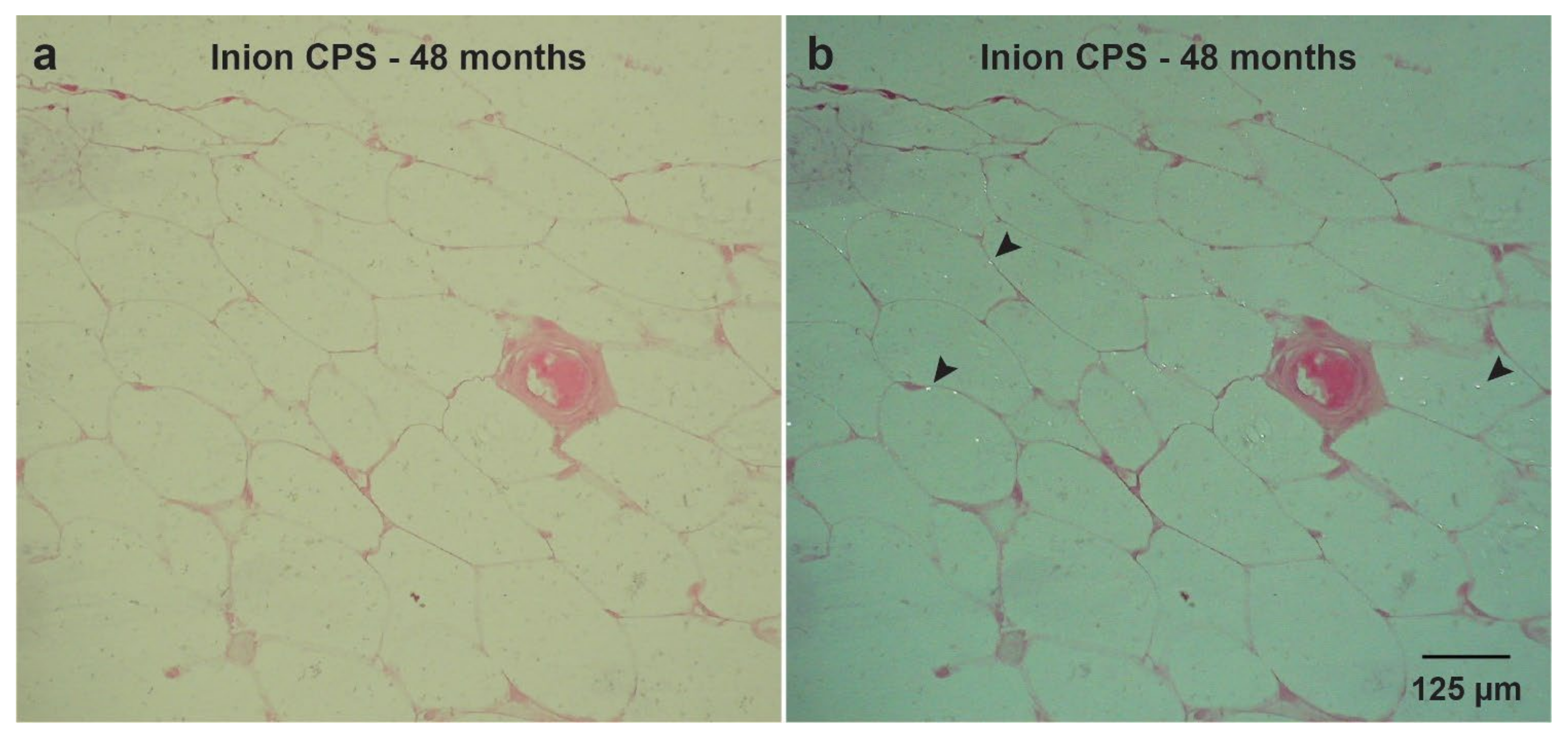
In a recent study, the long-term (i.e., up to 4-year follow-up) biocompatibility and degradation of four commonly used biodegradable copolymeric osteosynthesis systems was compared using a goat model [25]. The study included the BioSorb FX [poly(70LLA-co-30DLLA)], Inion CPS [poly([70–78.5]LLA-co-[16–24]DLLA-co-4TMC)], SonicWeld Rx [poly(DLLA)], and LactoSorb [poly(82LLA-co-18GA)] biodegradable osteosynthesis systems. The copolymer of the SonicWeld Rx system was the only one that was amorphous; all the other assessed systems were semi-crystalline. All the biodegradable systems were safe to use and well-tolerated. The SonicWeld Rx system showed the most predictable degradation profile. In addition, together with the LactoSorb system, new bone percentages similar to negative controls were observed after 18 months while the two other included systems reached these levels after 36 months (Figure 1). However, nanoscale residual polymeric fragments, predominately accumulated in adipocytes (Figure 2), were observed at every system’s assessment (Figure 3).
Since the crystalline regions of synthetic (co)polymers, the intermediate degradation products and the crystalline oligomeric stereo-complexes that can be formed in vivo over time are hydrophobic [17,58,62], this could explain the remarkable accumulation of polymeric birefringent fragments in adipocytes within the medullary bone cavity up to 4-year follow-up (Figure 2) [25]. Similar birefringent fragments, derived from as-polymerized PLLA, were observed in a case report [21] and experimental studies up to the 5-year follow-up [63,64]. Such particles were found intracellular after 3 and 4.5 years of implantation, although the particles decreased in size over time [63]. Crystalline fragments derived from as-polymerized PLLA can induce foreign body reactions even up to 5.7 years after implantation [21,65]. Another clinical study that focused on the efficacy of an osteosynthesis system composed of unsintered hydroxyapatite/PLLA composite, with a 12-month follow-up, showed that the removed symptomatic systems included up to 65% crystalline regions in the explanted polymers [66]. In a study that implanted the Resorb X osteosynthesis system (PDLLA) at the condyle of sheep mandibles, no foreign body reactions and complete bone formation were observed after 12 months [67]. Another study showed complete bone formation 18 months after implanting the LactoSorb system in the maxillofacial area of Göttingen minipigs without signs of foreign body reactions [68]. In contrast, after implanting the Inion CPS system in sheep, the system was surrounded by a fibrous capsule with granulomatous foreign body reactions after 52 weeks [69]. In the literature, foreign body reactions have predominately been reported for biodegradable osteosyntheses with a high proportion (i.e., >70%) of PLLA [18,21,55,70] or poly(glycolic acid) (PGA) [18]. More amorphous copolymers such as PDLLA (e.g., 50LLA/50DLA ratio) are more hydrophilic, and degrade and resorb more quickly and predictably [71]. These findings, as well as those of different (pre-)clinical studies [18,25,26,72], emphasize that the (co)polymers used in biodegradable systems should be completely amorphous. Future research should focus on amorphous (co)polymers with a minimum follow-up of ≥24 months so that a proper degradation assessment can be performed. Furthermore, it remains unknown whether the observed nanoparticles after 4-year follow-up [25] may be harmful in the long run (i.e., >4 years). Since microplastics have been shown to be toxic in vitro, with a potential impact on human health (e.g., effects on the gastrointestinal tract, lungs, immune system, and blood components) [73,74], the effects of the observed nanoparticles need further research.
Other than (co)polymer composition, the geometry and surface topography of the implanted materials also affect biocompatibility in vivo [72]. Thick biomaterials, especially with points and sharp edges, can increase the risk of foreign body reactions [26,75,76]. In contrast, thinner biomaterials, as well as smaller sized polymeric particles used to engineer a biomaterial, allow for quicker degradation and a lower risk of foreign body reactions [72,77,78]. A smooth well-contoured shape without acute angles induced macrophage polarization towards macrophages with an immune regulatory phenotype [79,80]. In vivo biocompatibility of medical devices, such as implants, can be significantly improved by tuning the spherical dimensions [72]. Furthermore, low implant volume reduces the amount of acidic degradation products and thus reduces the risk of (late) foreign body reactions [18]. The fact that screws possess acute angles, while welded pins do not, may explain the favorable degradation profile of the SonicWeld Rx system compared to the BioSorb FX, Inion CPS and LactoSorb biodegradable systems [18,25,81,82]. Novel biodegradable system development should incorporate geometry and surface topography into the design-phase as these characteristics are tunable and may be efficient ways to decrease foreign body reaction risk, hasten degradation, enhance quicker bone formation, and balance the degradation and regeneration equilibrium (Table 2) [26].
Table 2. Different aspects of biodegradable osteosynthesis systems accompanied with the ideal properties and the potential solutions to accomplish these properties.
AspectIdeal PropertiesMethodPotential SolutionsRefs
Surgical handlingEasy perioperative adaptation of plates3D engineeringPatient specific osteosynthesis systems [18,26,83]
Production processPlate adaption at room temperature [20]
No risk of perioperative screw breakageAlternative application methodUltrasound welding of thermoplastic pins instead of using conventional screws [20]
Elastic modulus of materialsEnough elastic modulus to avoid micromovements, but not stiffer than bone to avoid stress-shielding of the underlying boneProduction processCreate composites to tailor the elastic modulus to the application of interest [26]
Self-reinforcing of polymers to increase the elastic modulus of systems [20]
Alternative application methodUltrasound welding of thermoplastic pins to increase the maximum tensile load and stiffness, and side-bending stiffness [20]
Bacterial infectionPreventing bacterial adhesion to implant surface CoatingHydrophobic coatings [26]
Eliminating surrounding bacteria without antibioticsSurface modificationAdjusting the nano-scale surface topography (e.g., pillars on the surface) [84]
Eliminating surrounding bacteria with local antibioticsPolymer coating containing stabilized gas bubbles loaded with antibiotics that can be released locally using ultrasound [85]
Foreign body response (FBR)Materials that do not elicit an FBRSelection of materialsMaterials with non-toxic degradation products (e.g., derived from silk) [18]
Production processAvoid thick materials, especially with points and sharp edges [26,75,76]
Tailor the host response so that FBR are avoidedProduction processAvoid particle sizes < 2 µm [26]
Avoid micromovements (max. 28–150 µm), that can result in fibrous encapsulation of the implantSelection of materials, production process, and 3D engineeringOsteosynthesis system with material properties that matches with the mechanical properties of the target tissue (e.g., by using ultrasound welding) [26]
Degradation profilePredictable degradation, preferably after 3–12 months3D engineeringThinner materials degrade quicker [17]
Production processBalance the degradation and regeneration equilibrium by, e.g., using L- and D-chirality or by copolymerization [25,26]

2.1.3. Biodegradable Metals

Biodegradable metals are promising alternatives to polymeric osteosynthesis systems due to their mechanical properties that are closer to bone than (co)polymeric materials [18] and their less harmful degradation products. The tensile strength, elastic modulus, axial pull-out force, and maximum torque of magnesium alloys are higher than that of (co)polymers, but lower than that of titanium alloys [18,86]. To date, three biodegradable metal groups have been researched to be used for biodegradable osteosynthesis systems, i.e., magnesium (Mg), iron (Fe), and zinc (Zn) and their alloys [18]. Mg-based biodegradable metals have been studied most extensively. The available research for Fe- and Zn-based degradable metals is limited due to the low degradation rate of Fe-based metals while Zn-based metals have been introduced only recently [18].

Biodegradation

Biodegradable metal degradation is driven by anodic and cathodic reactions that result in the production of oxides, hydroxides and/or hydrogen gas [18,87]. Once biodegradable metals come into contact with body fluids, they are oxidized into metal cations combined with producing electrons via an anodic reaction. The electrons generated by implanting Mg-based biodegradable metals are consumed by cathodic reactions with water to form hydrogen gas and hydroxide. For Fe- and Zn-based metals, oxygen reduction only produces hydroxide without hydrogen gas. Hydroxide then reacts with the adjacent metal to form a metal-hydroxide layer on the surface of the implant. The protective layer can be eroded by high levels of chloride ions in the body fluids resulting in continuation of the degradation process. However, in Fe-based biodegradable metals, the protective layer consists of Fe(OH)2, Fe(OH)3, and Fe3O4, that inhibits further degradation. As a result, the degradation rate of Fe-based metals is very slow [18]. These ongoing reactions cause an oversaturation of calcium and phosphate ions in the surrounding body fluids that result in a layer of calcium-phosphate on the metal-oxide layer, that is able to induce bone formation [18].
A major challenge of biodegradable metals, particularly Mg-based materials, is the unpredictable degradation profile in vivo with subcutaneous emphysema due to the accumulation of hydrogen gas [18]. The degradation rate of biodegradable metals can be controlled by tailoring the microstructure, surface properties and coatings of the materials. For example, a recent study included gallium (i.e., a bone resorption inhibitor) in a magnesium alloy and showed promising results with inhibition of bone porosity formation, mechanical properties matching cortical bone, and low corrosion rate resulting in less hydrogen gas formation compared to other available magnesium alloys for orthopedic surgery [88]. In addition, surface modifications and coatings can be used to tune the degradation rates. For example, Mg-alloys and polymers can be combined to form Mg–polymer composites. These composites include high strength and elastic modulus derived from biodegradable metals while the surrounding biodegradable polymer matrix improves the corrosion resistance of the underlying metal [89].

Late Host Response

The degradation products of degradable metals such as hydroxide ions, hydrogen gas, metal-oxides, abraded particles, and calcium-phosphate affect the host response [90,91]. In a bone environment, the formation of the calcium-phosphate layer induces new bone deposition, making it a unique feature as base material for an osteosynthesis system. In addition, the Mg-ions can induce new bone formation in cortical bone by increasing calcitonin gene-related peptide 1 levels in periosteum-derived stem cells [92]. However, current Mg-based biodegradable metals often show a burst release of Mg-ions that can lead to excess formation of hydrogen gas resulting in gas pockets, tissue displacement, and subcutaneous emphysema. The fast degradation rate can also induce osteolysis, hemolysis, and rapid reduction of the mechanical properties [93].

2.1.4. Silk

Silk is the most recent addition to biodegradable materials [18]. Silk is a natural biodegradable polymer that is usually derived from the silkworm Bombyx mori. Although the evidence is still limited to pre-clinical evidence, the current evidence shows excellent biocompatibility and unique mechanical properties combined with easily and environmentally friendly processing into mechanically robust three-dimensional bulk materials with excellent machinability [18,94]. To date, it is the only natural polymer that has been used to prepare an osteosynthesis system [94].

Biodegradation

As with most natural polymers, silk is degraded enzymatically, e.g., by protease XIV, matrix metalloproteinase and collagenase [44]. These enzymes cleave silk protein chains into peptide fragments with decreased molecular weight and strength [95]. Immune cells, especially macrophages and FBGCs, play an important role during degradation of silk. Immune cells mediate silk degradation through (1) phagocytosis and (2) extracellular degradation mediated by proteolytic enzymes derived from macrophages and FBGCs. The degradation products are tightly packed aggregates or amino acids for metabolism [18]. The degradation time depends on implant-related factors (e.g., molecular weight, porosity, crystallinity, and surface topography) and host-related factors (e.g., species and implantation site). The degradation times can be tailored from minutes to years by controlling the material variables such as molecular weight, surface topography, β-sheet content, and porosity [18]. Although in vivo research in animal studies showed complete silk degradation, a thorough understanding of the degradation pathways and clearing mechanisms as well as degradation in humans is still lacking [18].

Late Host Response

After implantation of silk materials, a mild inflammatory response occurs that decreases within a few weeks. This host response involves recruitment and activation of macrophages and the formation of FBGCs. The silk implant can be degraded and replaced by host tissue (e.g., bone), but it can also be integrated within the tissue or encapsulated by fibrous tissue. There is currently limited data regarding the short- and long-term host response in vivo. In the currently only available study that prepared a silk-based osteosynthesis systems for fracture fixation in maxillofacial surgery, the in vivo assessment of the 4- and 8-week host response by rats showed more favorable mechanical properties than biodegradable synthetic polymers and excellent biocompatibility accompanied with bone remodeling [94]. These results are promising but additional research is necessary to unravel the complete degradation pathways as well as the host responses that this natural-derived polymer elicits.

2.1.5. Titanium and Its Alloys

Late Host Response

Titanium osteosynthesis systems are commonly made of pure titanium or titanium alloys [20,96]. The most frequently used titanium alloy for maxillofacial osteosynthesis systems consists of 90% titanium, 6% aluminum, and 4% vanadium (Ti6Al4V, also called titanium alloy grade 5) [96,97,98,99]. However, although titanium and its alloys are presumed to be completely bioinert, there is growing evidence that wearing of particles occurs that can accumulate in surrounding tissues and different organs of which the consequences are still largely unknown [96,98,100].
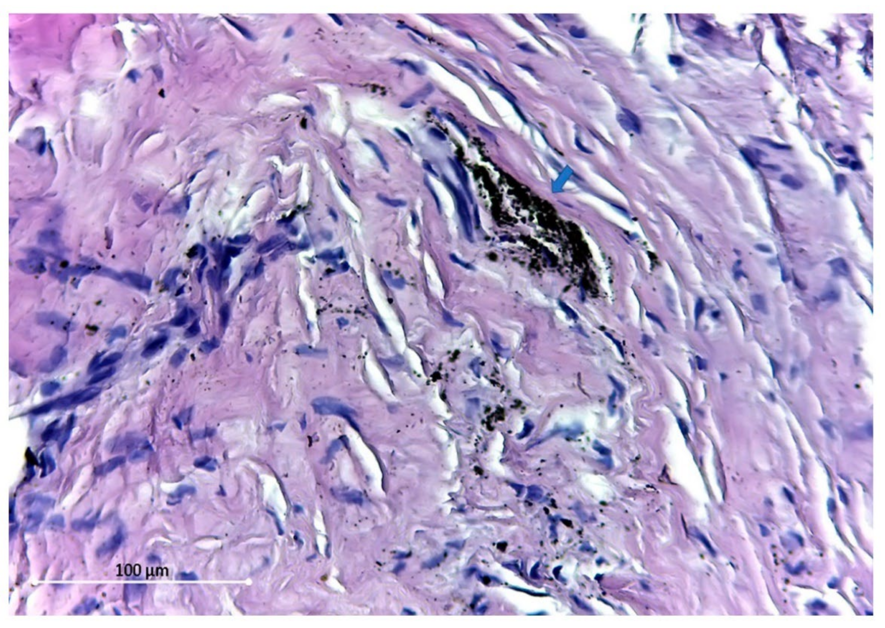
In a study that explanted titanium osteosynthesis plates from patients that underwent craniofacial surgery, titanium particles (7.9 to 31.8 µg/gram of dry tissue) could be detected in the regional soft tissue and lymph nodes after 24-month follow-up [13]. Similarly, a recent study showed that the tissue surrounding titanium plates after fracture and osteotomy fixation contained 1.03 and 1.09 ppm titanium particles, respectively [14]. Meningaud et al. revealed a large variation in titanium levels within the surrounding tissue (4–8000 µg/gram) after titanium fixation of osteotomies, but concluded that almost all of these particles were produced at the moment of applying the osteosynthesis system [101]. Other studies reported on the presence of dark-grey pigmentation accompanied with fibrosis of the surrounding tissue and macrophages containing intra-cellular titanium particles (Figure 4) [15,102,103]. Zaffe et al. have also shown the presence of titanium in the surrounding tissue as well as that erythrocytes and lymphocytes contained titanium particles [104]. In addition, explanted osteosynthesis plates analyzed with scanning electron microscopy showed defects and irregularities most likely due to in vivo substance loss [102]. Titanium debris has also been found throughout the body suggesting hematogenous dissemination, with traceable amounts of titanium particles within the liver, spleen, and lymphatic system [98,100].
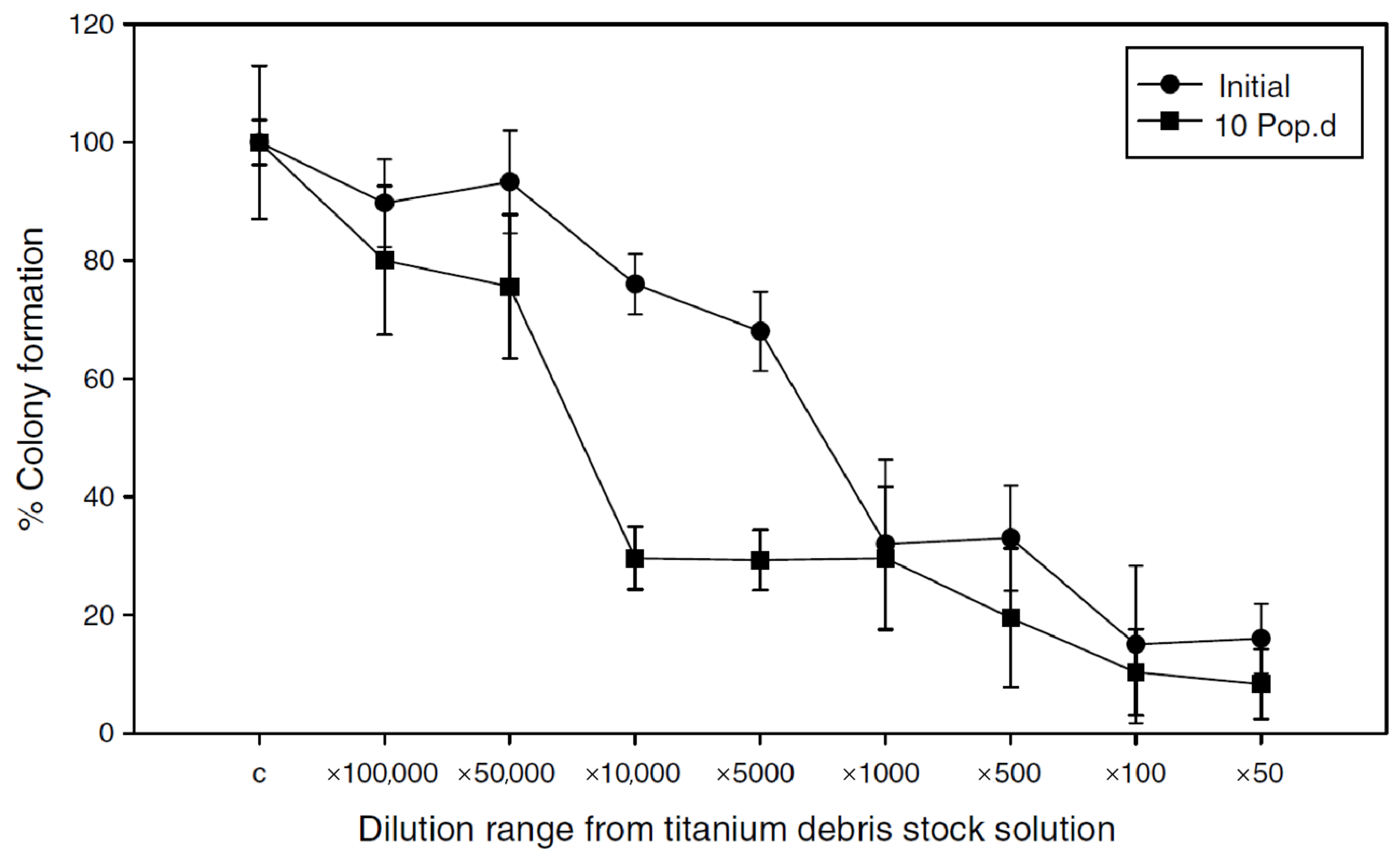
To determine the effect of such titanium particles, Coen et al. assessed the cytotoxicity of Ti6Al4V particles on human fibroblast cells in vitro, and showed chromosomal instability, reproductive failure and decreased clonogenic survival 10 generations postexposure (Figure 5) [105]. Studies that analyzed the periosteum surrounding titanium plates as well as blood samples in patients after mandibular fracture fixation showed redox abnormalities, and increased oxidative stress and damage [106,107]. Furthermore, an association between aluminum and the pathogenesis of Alzheimer’s disease has been suggested. In addition, increased levels of circulating aluminum are associated with microcytic anemia and osteomalacia [98,108,109]. These findings indicated that there is a need for long-term epidemiological studies that assess the effect of these particles in the long run.
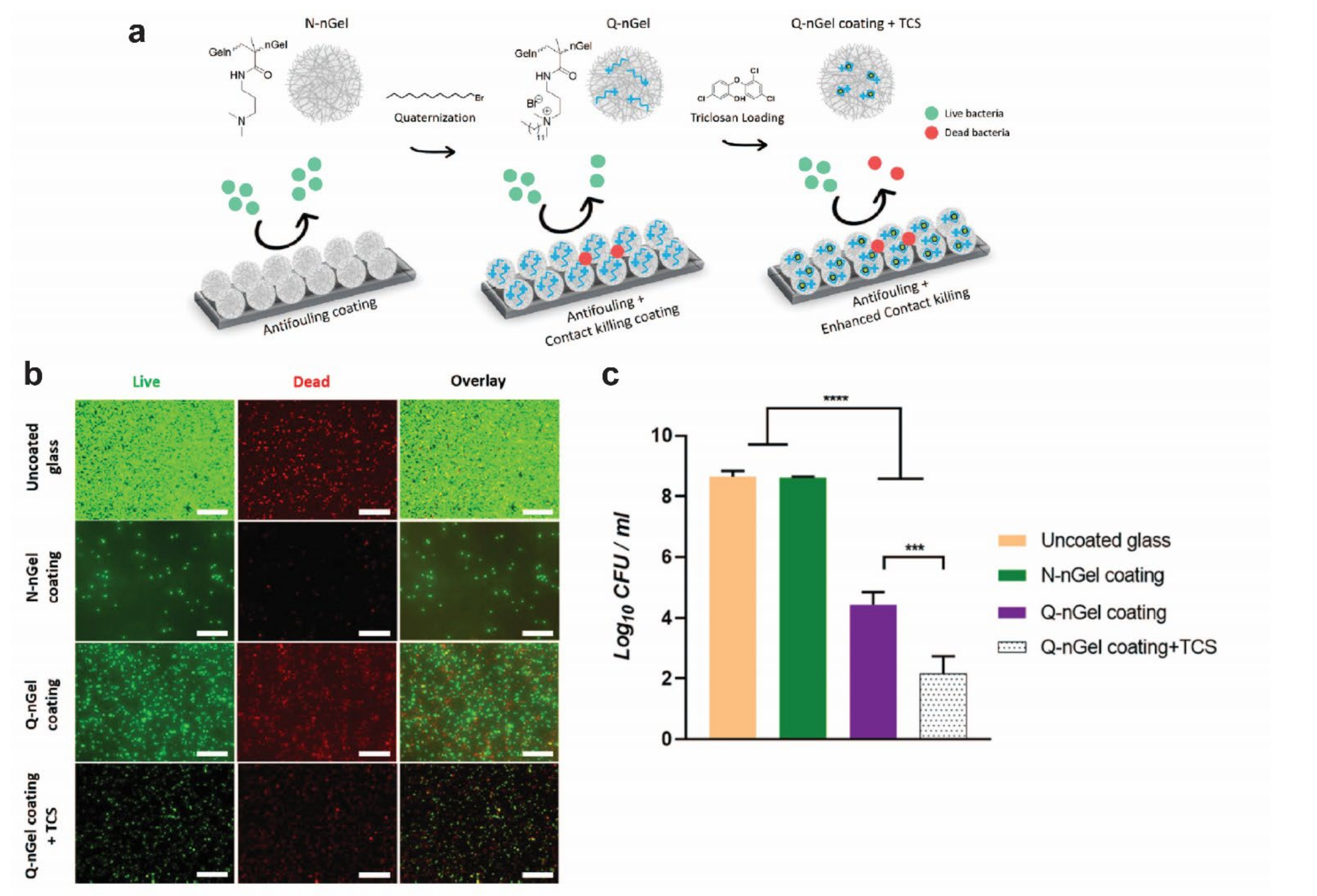
Surface modifications (e.g., oxygen plasma immersion ion implantation) have been proposed to reduce metal ion release from the implant (Table 3) [110]. In addition, they are an important aspect of biocompatibility [98,110]. Titanium, without surface modifications, has a positively charged surface and will, therefore, tend to covalently bond to negatively charged proteins such as fibronectin [111]. Fibronectin promotes bacterial adhesion and, thus, increases the risk of infection [112]. Besides bonding to autologous proteins, most of the cell surface of bacterial species (e.g., Staphylococcus aureus, the most common etiological pathogen of infections surrounding osteosyntheses [113]) is negatively charged, and thus also adheres to positively charged surfaces such as titanium [114]. By modifying the surface charge, adhesion of various bacteria (e.g., Staphylococcus aureus and Escherichia coli) is inhibited and, ideally, the risk of infection is reduced [110]. These properties of titanium systems can also be tuned by other surface modifications (Table 3 and Figure 6).
Table 3. Different aspects of titanium osteosynthesis systems accompanied with the ideal properties and the potential solutions to accomplish these properties.
AspectIdeal PropertiesMethodsPotential SolutionsRefs
Surgical handlingEasy perioperative adaptation of plates3D engineeringPatient specific osteosynthesis systems [115,116]
Production processAdaption of the production process to alter the mechanical properties of plates (e.g., lower stiffness) [20,117,118,119,120]
No risk of perioperative screw breakage3D engineeringAdjusting the screw head to improve the grip on the screws [20]
Elastic modulusEnough elastic modulus to avoid micromovements, but not stiffer than bone to avoid stress-shielding of the underlying boneProduction processAdaption of the production process to alter the mechanical properties of plates [20,117,118,119,120]
Bacterial infectionPreventing bacterial adhesion to implant surfaceCoatingHydrophobic coatings [26]
(Nano)gel coatings [121,122]
Surface modificationPlasma immersion ion implantation (surface modification) [110,123,124]
Physical vapor deposition [125,126]
Increasing surface energy by acid etching [127]
Eliminating surrounding bacteria without antibioticsCoatingTitanium Nitride (TiN) coating [128,129]
Surface modificationAdjusting the nano-scale surface topography (e.g., pillars on the surface) [84]
Plasma immersion ion implantation [110,130]
Physical vapor deposition [131]
Laser surface modification [132]
Anodization [133,134]
Micro-Arc oxidation [135,136]
Eliminating surrounding bacteria with local antibioticsCoatingPolymer coating containing stabilized gas bubbles loaded with antibiotics that can be released locally using ultrasound [85]
(Nano)gel coatings [122,137]
Surface modificationChemical vapor deposition [138]
OsteogenesisImproving bone growth surrounding the implantCoating(Nano)gel coatings [137]
Surface modificationPlasma spraying with hydroxyapatite [139,140,141,142,143]
Plasma immersion ion implantation [144,145]
Physical vapor deposition [146,147]
Chemical vapor deposition [148]
Increasing surface energy by acid etching [127]
Laser surface modification [132,149,150]
Anodization [151]
Wear resistanceNo wearing of titanium (alloy) particlesCoatingTitanium Nitride (TiN) coating [152,153]
Surface modificationPlasma immersion ion implantation [110]
Physical vapor deposition [98]
Laser surface modification [150,154]
Anodization [134,155,156]

2.2. Mechanical Properties

2.2.1. Minimally Required Mechanical Properties

Several studies assessed the mechanical forces surrounding osteosyntheses applied to maxillofacial fractures [157,158,159,160,161,162,163], osteotomies [164,165] and reconstructions [166], so that the minimally required mechanical properties of an osteosynthesis system can be estimated. After maxillofacial trauma, the reported bite force increases up to 64 N by the second postoperative fracture fixation day, 92 N after 1 week, 187 N after 4 weeks, and up to 373 N at the 3-month follow-up [157]. Other studies focusing on trauma patients showed that 100 N forces were measured after 4 weeks of fixation [159,161]. The mechanical forces around maxillofacial osteotomies have been reported to increase from 21 ± 14 N (i.e., after 1 week) to 65 ± 43 N (i.e., after 6 weeks) [160] while other studies reported forces ranging from 82.5 to 132 N [164,165]. The masticatory forces after mandibular reconstructions ranged from 28 to 186 N [166]. However, the mechanical stress surrounding osteosynthesis systems is multi-factorial and is affected by the location of the fracture [1], differences in interfragmentary stability [1], mandibular height [1], degree and direction of movement [167], and preoperative masticatory forces [159,168,169]. Load-sharing osteosynthesis allows sharing of the load between bone segments and the osteosynthesis system (e.g., fractures with interfragmentary stability) whereas in load-bearing osteosynthesis, the complete load at the fracture site is carried by the osteosynthesis system without interfragmentary stability [1,170]. In a load-bearing situation, the osteosynthesis system is exposed to substantially higher loads and, thus, the biomechanical requirements for an optimal osteosynthesis system are higher compared to load-sharing osteosyntheses [70,171]. Although it would be of high clinical value to determine the exact cut-off value of the transition from load-sharing to load-bearing osteosyntheses, this is currently unknown. Since the mandible is exposed to considerably higher biomechanical forces compared to the maxilla [1], load-bearing osteosynthesis of the mandible requires even higher mechanical properties of the used osteosynthesis system compared to load-bearing osteosynthesis of the maxilla or load-sharing osteosynthesis of the mandible [172,173]. Furthermore, as bone healing progresses, the forces will be shared by the osteosynthesis system and the underlying healing bone. Thus, it remains difficult to estimate the least mechanical properties an osteosynthesis system has to meet. Therefore, researchers have mainly focused on relative differences between the available osteosynthesis systems [20].

2.2.2. Mechanical Properties of Osteosynthesis Systems
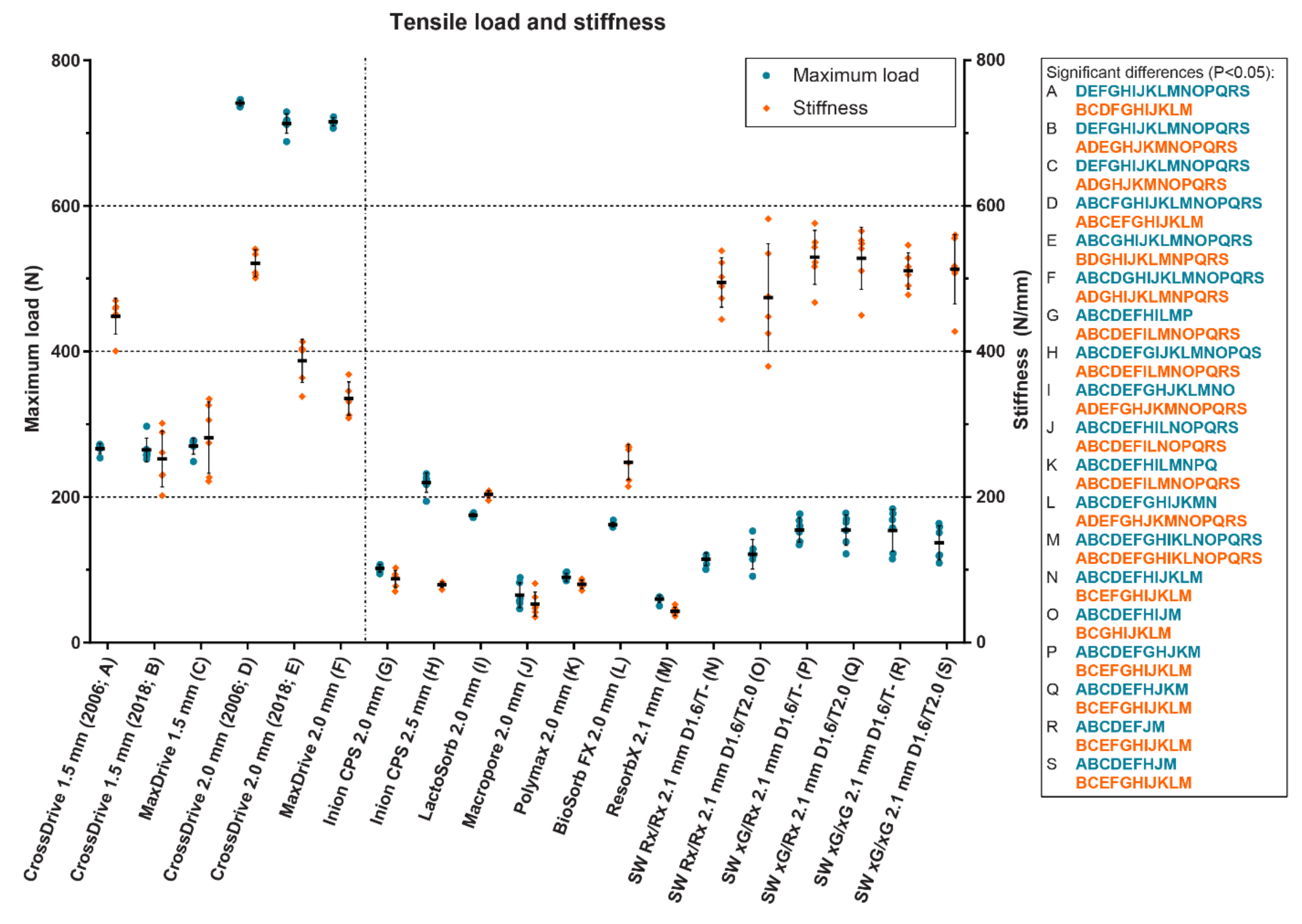
The mechanical properties of osteosynthesis systems depend on several factors including composition (i.e., titanium (alloys) or (co-)polymers), the production processes (e.g., stamping versus laser cutting of titanium systems) [117,118,174], dimensions, polymer self-reinforcement [175], the application method (i.e., screws or ultrasound welded pins) [176], ageing, and sterilization methods [177,178,179]. The tensile, bending and torsional stiffness of an osteosynthesis system are a more clinically relevant outcome than maximum tensile load since this affects adequate fixation and bone healing (i.e., malunion and non-union) [180] while maximum tensile load is only relevant whenever the bone segments are already separated by more than a few millimeters. In the latter case, this will certainly result in compromised bone healing or malunion.
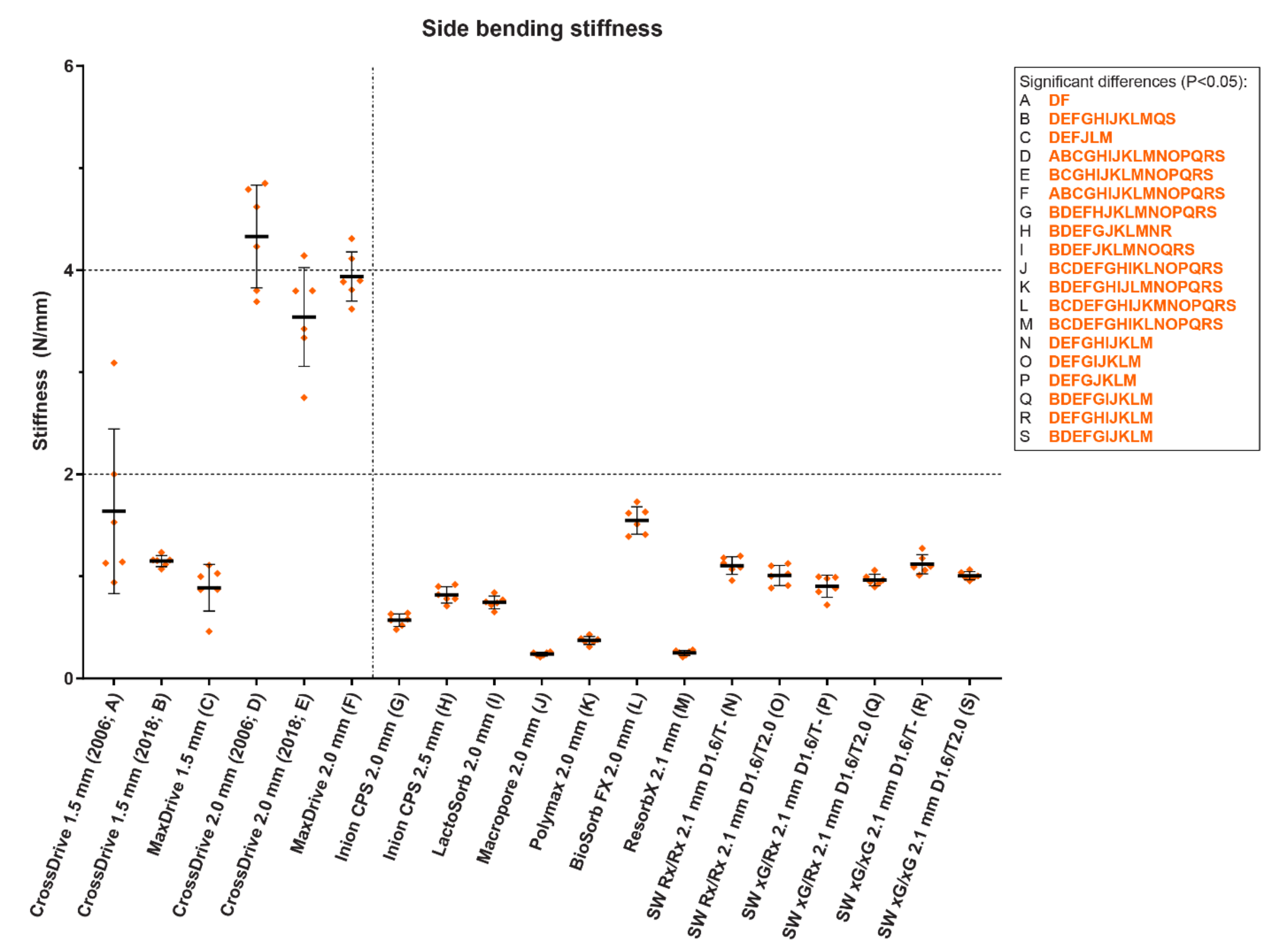
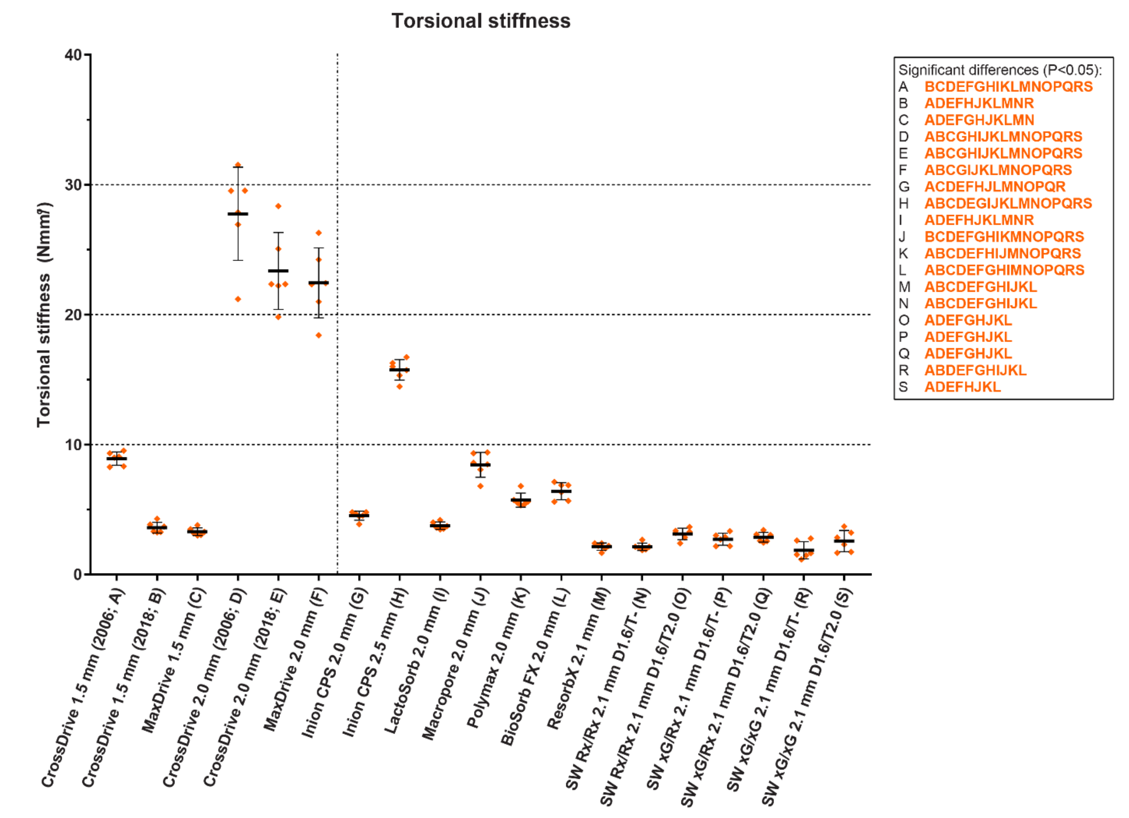
In a recent in vitro study, the maximum tensile load as well as the tensile, bending and torsional stiffness of 13 biodegradable and 6 titanium straight, four-hole osteosynthesis systems derived from static mechanical tests of the initial materials were assessed and compared (Figure 7, Figure 8 and Figure 9) [20]. The titanium systems’ tensile loads were higher than those of the biodegradable systems. The bending stiffness of the 1.5 mm titanium systems was comparable to all the biodegradable systems whereas the 2.0 mm system’s bending stiffness was higher. Regarding the biodegradable systems, Inion CPS 2.5 mm had the highest tensile load and torsional stiffness, SonicWeld 2.1 mm the highest tensile stiffness, and BioSorbFX 2.0 mm the highest bending stiffness. Regarding the titanium systems, the CrossDrive (2006) systems had the highest tensile, bending and torsional stiffness. It must be noted, though, that although high mechanical osteosynthesis properties are sought for adequate fixation, the extreme stiffness of the titanium systems can be a disadvantage due to the stress shielding of the underlying bone [12]. Stress shielding occurs when the underlying bone is exposed to less stress than it should endure, leading to an increase in osteoclast activity and bone resorption, that can, in turn, lead to decreased bone density and aseptic loosening [98,181]. This has led to the development of new titanium osteosynthesis systems with a lower elastic modulus to reduce stress shielding of the underlying bone by adjusting the production process (Figure 7, Figure 8 and Figure 9 and Table 3) [20,23,24].
Within the limitations of finite element analyses (e.g., assuming the masticatory forces are fixed), three-dimensional analyses indicated that the biomechanical stresses surrounding osteosynthesis systems remain far below the threshold of their ultimate strength of both biodegradable and titanium osteosynthesis systems [163,172,182,183]. In addition, the empirical evidence of fracture [19] and osteotomy [184] osteosyntheses shows that the efficacy of titanium and biodegradable osteosyntheses is similar (e.g., absence of malunion), indicating that the less favorable mechanical properties of biodegradable osteosynthesis are still sufficient to achieve similar healing outcomes. However, as also observed from the empirical evidence, the mechanical properties of biodegradable osteosyntheses of mandibular osteotomies may be insufficient to avoid micromovements [184]. Future research should also focus on these micromovements since they play an important role in developing foreign body reactions [26].
Finite element analyses also demonstrated that the stress surrounding conventional screws is much larger compared to those of plates, indicating that material complications may arise from the screws rather than the plates (e.g., screw loosening or fractures) [182]. The positive effect of ultrasound welding of biodegradable, thermoplastic pins instead of using conventional screws was demonstrated by the superior mechanical properties of the SonicWeld Rx (PDLLA with thermoplastic pins) compared to the Resorb X system (identical system with screws) [20]. Additionally, ultrasound welding caused a shift of the weakest link of the complete osteosynthesis system from the screw-plate interface to the plate itself. Therefore, ultrasound welding may reduce screw-related material complications, but this has to be investigated by future research.
Figure 7. The tensile load and stiffness of 13 biodegradable and 6 titanium osteosynthesis systems commonly used in oral and maxillofacial surgery. The characters in blue and orange represent significant pairwise differences in maximum load and stiffness, respectively, between the corresponding systems using a one-way analysis of variance adjusted for multiple testing. The titanium CrossDrive (2006) plates consisted of 100% titanium produced by stamping of plates. The titanium CrossDrive (2018) and MaxDrive consisted of 100% titanium produced by milling of plates. The titanium CrossDrive (2006 and 2018) and MaxDrive screws consisted of a Ti6Al4V alloy. The composition of each biodegradable system is described in Table 1. Error bars: mean values  ±  standard deviation. Ti6Al4V, 90% titanium, 6% aluminum and 4% vanadium alloy; SW, SonicWeld; D, drill diameter (mm); T, tap diameter (mm). The dotted line separates the titanium (left) and biodegradable systems (right). Reprinted with permission from [20].
Polymers 14 02782 g007
Figure 8. The side bending stiffness of 13 biodegradable and 6 titanium osteosynthesis systems commonly used in oral and maxillofacial surgery. The characters in blue and orange represent significant pairwise differences in maximum load and stiffness, respectively, between the corresponding systems using a one-way analysis of variance adjusted for multiple testing. The titanium CrossDrive (2006) plates consisted of 100% titanium produced by stamping of plates. The titanium CrossDrive (2018) and MaxDrive consisted of 100% titanium produced by milling of plates. The titanium CrossDrive (2006 and 2018) and MaxDrive screws consisted of a Ti6Al4V alloy. The composition of each biodegradable system is described in Table 1. Error bars: mean values  ±  standard deviation. Ti6Al4V, 90% titanium, 6% aluminum and 4% vanadium alloy; SW, SonicWeld; D, drill diameter (mm); T, tap diameter (mm). The dotted line separates the titanium (left) and biodegradable systems (right). Reprinted with permission from [20].
Polymers 14 02782 g008
Figure 9. The torsional stiffness of 13 biodegradable and 6 titanium osteosynthesis systems commonly used in oral and maxillofacial surgery. The characters in blue and orange represent significant pairwise differences in maximum load and stiffness, respectively, between the corresponding systems using a one-way analysis of variance adjusted for multiple testing. The titanium CrossDrive (2006) plates consisted of 100% titanium produced by stamping of plates. The titanium CrossDrive (2018) and MaxDrive consisted of 100% titanium produced by milling of plates. The titanium CrossDrive (2006 and 2018) and MaxDrive screws consisted of a Ti6Al4V alloy. The composition of each biodegradable system is described in Table 1. Error bars: mean values  ±  standard deviation. Ti6Al4V, 90% titanium, 6% aluminum and 4% vanadium alloy; SW, SonicWeld; D, drill diameter (mm); T, tap diameter (mm). The dotted line separates the titanium (left) and biodegradable systems (right). Reprinted with permission from [20].
Polymers 14 02782 g009

3. Clinical Evidence

3.1. Biodegradable Versus Titanium Osteosyntheses: Efficacy and Symptomatic Removal

In theory, the primary advantage of biodegradable compared to titanium osteosynthesis is the reduced, or even eliminated, need for symptomatic device removal while having similar efficacy (i.e., adequate bone healing without malunion). In a recent systematic review with meta-analyses, the available clinical evidence (i.e., randomized controlled trials [RCTs], and prospective and retrospective controlled studies without language or period restrictions) from patients treated for maxillofacial fractures (i.e., Le Fort I, cranial, zygomaticomaxillary complex and mandibular fractures) with load-sharing biodegradable versus titanium osteosyntheses was compared [19]. Following a sensitive and thorough literature search that focused on all relevant clinical endpoints, the meta-analysis of RCTs demonstrated similar efficacy and morbidity between the two systems, but symptomatic osteosynthesis removal was significantly lower in the biodegradable compared to the titanium group [19]. Other studies that focused on cohorts of patients who had undergone load-sharing biodegradable fixation of midface and malar fractures [66,185,186], naso-orbital-ethmoid fractures [187], and mandibular body, parasymphysis and symphysis fractures [37,66,183], also showed adequate internal fixation, long-term stability, and biocompatibility with biodegradable systems. Similar results were observed in non-comparative cohorts that included adults with isolated orbital floor fractures [186] and combined orbital floor and medial wall fractures [187] treated with biodegradable mesh plates. In addition, another systematic review that focused on the composite of the complications (e.g., infection, palpability, dehiscence, material-related complications, and exposure) encountered with biodegradable versus titanium osteosynthesis of zygomatic and mandibular fractures revealed significantly fewer complications from biodegradable compared to titanium osteosyntheses, although the analysis had a risk of bias due to substantial methodological heterogeneity of the included studies [188]. Symptomatic biodegradable osteosynthesis system removal in these studies (0 to 8.6%) was comparable to that of the patients with mandibular, Le Fort I, and zygomatic fractures in a multicenter randomized controlled trial (RCT) that compared titanium (KLS Martin CrossDrive system) with biodegradable osteosynthesis (Inion CPS system) with a median follow-up of 99 (78;113) months [8].
Similar to the abovementioned results, a recent systematic review with meta-analyses that compared biodegradable versus titanium osteosyntheses in adults with dentofacial deformities treated with orthognathic surgery (i.e., Le Fort I, bilateral sagittal split (BSSO), and intraoral vertical ramus osteotomies (IVRO), with and without concurrent genioplasty) also demonstrated similar efficacy and morbidity between biodegradable and titanium osteosynthesis systems [184]. Non-comparative cohort studies that focused on the stability of maxillary [189,190,191], mandibular [189,190,191], or bimaxillary osteotomies [189,190,191,192,193], and post-operative complications [191,194,195], also showed predictable skeletal stability with post-operative complications rates similar to those reported in the literature after titanium osteosyntheses [196,197,198].
In contrast to the abovementioned trauma population [19], a non-significant difference in symptomatic biodegradable and titanium osteosynthesis system removal was observed in the aforementioned orthognathic population [184]. The main reasons for biodegradable osteosynthesis removal in both populations were chronic inflammation and discomfort [19,184]. Similar results were observed in the aforementioned RCT with long-term follow-up, but in-depth analyses revealed that all the removals among the biodegradable group were due to clinical problems in the mandible, and were only seen after fixation of osteotomies [8]. Different reviews noted that mandibular osteotomies are associated with significantly more complications and higher symptomatic biodegradable and titanium osteosyntheses removal compared to maxillary osteotomies, and compared to fracture fixation [188,199,200,201]. A large retrospective cohort study (n = 685 patients) that focused on the efficacy and complications of biodegradable osteosyntheses and symptomatic removal of the systems also showed that mandibular osteotomies are associated with more complications and higher symptomatic removal rates compared to other osteotomies [202]. In addition, earlier reviews also showed more complications and symptomatic osteosynthesis removal after titanium fixation of mandibular osteotomies compared to other osteotomies [199,200]. Together, these results indicate that biodegradable osteosynthesis is a viable alternative to titanium osteosynthesis for fixation of both fractures and osteotomies. However, among the studied trauma population, the symptomatic biodegradable osteosynthesis systems removal rates are lower compared to the titanium osteosyntheses group whereas the biodegradable and titanium osteosyntheses groups have similar symptomatic osteosyntheses removal rates after orthognathic surgery.
An important aspect of successful biodegradable osteosyntheses (i.e., adequate bone healing and stability, and lack of foreign body reactions) is the biomechanical perspective. The biomechanical differences between fixation of fractures and osteotomies may explain the beneficial effect of biodegradable systems compared to titanium systems after fracture fixation versus osteotomy fixation. Fracture fixation with interfragmentary stability ensures load-sharing osteosyntheses whereas in osteotomies, interfragmentary stability is absent and thus the complete load at the osteotomy is carried by the osteosynthesis system (i.e., load-bearing osteosyntheses) [1,203]. Since the mandible is exposed to considerably higher biomechanical forces compared to the maxilla, this effect is even more pronounced in mandibular osteosyntheses. This is supported by the empirical findings that symptomatic osteosynthesis removal after fixation of mandibular fractures and osteotomies is significantly higher compared to those of other parts of the facial skeleton, both after titanium and biodegradable osteosyntheses [188,199,200,201,202,204]. Biomechanical forces that are not sufficiently counteracted can result in micromovements surrounding the osteosyntheses that may result in disturbed bone healing or foreign body reactions [1,18,26,205,206,207,208]. Animal studies have shown that these micromovements should be limited to 28–150 µm to avoid fibrosis and accompanying foreign body reactions [26,205,206,207,208]. Therefore, it is important that the mechanical properties of biodegradable implants are sufficient for the intended clinical application to ensure (primary) stability for adequate bone healing and to avoid micromovements and the accompanying risk of foreign body reactions (Table 2).
The clinical evidence of biodegradable metals is limited. A clinical study that compared magnesium (MgYREZr alloy; WE43) with titanium compression screws (Ti6Al7Nb alloy) for the fixation of mandibular condylar head fractures showed similar results after 18-month follow-up, with limited formation of hydrogen gas within the first 4 months [86]. These and other intermediate-term clinical outcomes [209] indicate that tuning the composition and structure of magnesium alloys are promising techniques to achieve biodegradable metal osteosyntheses in maxillofacial surgery. Although the degradation profiles of biodegradable magnesium alloys look promising [210,211,212], future research should focus on in vivo studies with long-term follow-up.

3.2. Biodegradable Versus Titanium Osteosyntheses: Secondary Advantages

At first glance, the non-significant difference in the proportion of symptomatic removal of biodegradable and titanium osteosyntheses in orthognathic surgery seems to negate the benefits of biodegradable osteosyntheses. However, asymptomatic biodegradable osteosynthesis systems will eventually be resorbed while titanium osteosynthesis will remain in situ until removed surgically. Therefore, titanium osteosynthesis systems have a life-time risk of, e.g., late infection or palpability complaints. Furthermore, biodegradable osteosyntheses have other (secondary) advantages, besides the most obvious benefit of less device removal compared to titanium systems, including no interference with radiographic imaging and radiotherapy, a more gradual transfer of stress to the healing bone (i.e., less stress shielding), and less system palpability in the long-term [8,9,19,176,213,214,215]. Whereas 17–80% of the patients undergo a second operation for elective titanium osteosynthesis removal due to their awareness of the presence of a foreign body (i.e., the titanium system) [216,217,218], asymptomatic biodegradable systems are generally eventually resorbed, thus forestalling such elective removals. When asked prior to surgery, the vast majority of patients (i.e., >95%), therefore, prefer biodegradable over titanium osteosyntheses in both maxillofacial traumatology and orthognathic surgery [219,220]. Despite the similar symptomatic removal rates in orthognathic surgery, the other benefits of biodegradable over titanium systems (e.g., no temperature sensitivity, no possible growth restrictions, and no hampering of imaging and radiotherapy) could also be valid reasons to choose biodegradable osteosynthesis systems. All these aspects should therefore be addressed preoperatively when informing the patients to ensure well-informed decision making.
Similarly, after fracture fixation in pediatric patients, common practice is to electively remove all the titanium systems due to possible later growth disturbances [221,222] and plate migration [223,224,225], whereas only the symptomatic biodegradable systems (12%) are removed [19]. In a recent systematic review including pediatric upper- and mid-facial fractures, the biodegradable osteosyntheses showed significantly fewer complications and symptomatic osteosynthesis system removal rates compared to titanium osteosyntheses [226]. Comparable rates were observed after applying biodegradable osteosynthesis for mandibular fracture fixation [227,228] and in craniofacial surgery [229,230] in pediatric patients. Biodegradable osteosyntheses would thus overall result in lower osteosynthesis removal compared to titanium osteosyntheses from pediatric patients. It must be noted, though, that there is currently no conclusive evidence whether growth disturbances actually occur with titanium systems and, thus, there is controversy in the current literature regarding the elective removal of titanium osteosynthesis systems from pediatric fracture patients [223,224,231,232]. Therefore, this subject needs to be addressed by future research. OMF-surgeons should inform pediatric patients and/or their caregivers about the above preoperatively to guide the shared-decision making.

3.3. Certainty of the Current Evidence

An important aspect when assessing efficacy, morbidity and symptomatic osteosynthesis removal in clinical studies is the duration of follow-up. The studies included in the most recent and comprehensive systematic reviews that focused on biodegradable versus titanium osteosyntheses in maxillofacial traumatology [19] and orthognathic surgery [184] predominately had 2-year follow-ups. Although this is sufficient follow-up to assess efficacy (e.g., adequate bone healing), symptomatic osteosynthesis removal can occur later on (i.e., >2-year follow-up) due to osteosynthesis system palpability, thermal sensitivity or foreign body reactions [8,53,65,69,233,234,235]. In a cohort study with a follow-up of 67 months, 7% of the included patients underwent symptomatic biodegradable osteosynthesis removal between 24 and 67 months [186]. In an RCT with a follow-up of 99 months, 3% and 5% of the patients in the biodegradable and titanium group, respectively, underwent a secondary surgical procedure to remove symptomatic osteosynthesis system removal between the 24-month and final follow-up [8]. Similarly, titanium system removals due to infection and discomfort occurred after the 4- [201] and 5.5-year follow-ups [235]. Therefore, future research should also include long-term follow-up assessments (e.g., ≥5-year follow-ups) in the pre-specified protocols.
Since significant differences in biocompatibility and degradation profiles as well as in mechanical properties are observed between copolymeric biodegradable osteosynthesis systems, future clinical research should focus on a specific biodegradable system that has proven to be biocompatible in the long-term as well as having the most favorable mechanical properties for specific surgical indications. In addition, there is currently no evidence to support or refute the use of biodegradable osteosynthesis in load-bearing fracture fixation. Since current evidence suggests that load-sharing fixation of fractures using biodegradable osteosynthesis is feasible, the next step would be to focus on load-bearing biodegradable osteosynthesis of fractures. Finally, systematic reviews should include tools to assess the degree of clinical heterogeneity [236], and should perform network meta-analyses to assess the most preferred biodegradable system. This would allow conclusions regarding the efficacy of specific biodegradable osteosynthesis systems based on clinical evidence.

4. Clinical Recommendations: Titanium or Biodegradable Osteosyntheses?

Current pre-clinical and clinical evidence indicates that biodegradable copolymeric osteosynthesis is a viable alternative to titanium osteosynthesis for fixation of both fractures and osteotomies, with similar efficacy [19,184]. Fixation of fractures also leads to significantly lower symptomatic device removal, thereby achieving the primary advantage of biodegradable osteosyntheses. Based on both the biological and biomechanical perspectives [20,25], a biodegradable osteosynthesis system composed of amorphous copolymers (e.g., PDLLA), preferably using ultrasound welding with a well-contoured shape without acute angles, and that has sufficient mechanical properties has the greatest potential as a biocompatible biodegradable copolymeric osteosynthesis system. Therefore, for midface fractures, the SonicWeld Rx 2.1 mm system is recommended [20,25]. The Inion CPS 2.5 mm system is recommended for mandibular fractures. Whenever the patient or surgeon prefers a titanium osteosynthesis system, 1.5 (e.g., midface fractures) and 2.0 mm titanium systems (e.g., mandibular fractures) from several suppliers are recommended [1,20,170].
Although copolymeric biodegradable and titanium osteosyntheses of osteotomies result in similar efficacy, both groups also have similar symptomatic osteosyntheses removal rates. Therefore, the primary advantage of biodegradable osteosyntheses in this population is neglected. Titanium systems are, therefore, still preferred due to lower risk of perioperative screw breakage, lower operation time, and better perioperative handling compared to biodegradable systems [19,171,184]. For maxillary and mandibular osteotomies, 1.5 and 2.0 mm titanium systems are recommended, respectively. Biodegradable osteosyntheses in orthognathic surgery may still be used, but only whenever the patient and/or surgeon prefer biodegradable over titanium systems, e.g., on the basis on secondary advantages (e.g., no temperature sensitivity, no possible growth restrictions, and no hampering of imaging). Then, the SonicWeld Rx 2.1 mm and Inion CPS 2.5 mm systems are also recommended for maxillary and mandibular osteotomies [20,25], respectively.
There is currently no evidence to support or refute the use of biodegradable osteosynthesis in load-bearing fracture fixation (e.g., a comminuted fracture) or osteosyntheses in reconstructive surgery (e.g., anatomical defects after an oncological resection) [19]. However, since the biomechanical requirements for these surgical procedures are comparable or even higher compared to osteosyntheses of osteotomies [1,20,166], titanium osteosyntheses remain preferred over biodegradable osteosyntheses until (empirical) evidence becomes available that supports the use of biodegradable osteosyntheses for these surgical procedures.

5. Future Perspectives

5.1. Overcoming the Disadvantages of Current Osteosynthesis Systems

5.1.1. Biodegradable (Co)Polymeric Systems

In Table 2, different important aspects of biodegradable osteosynthesis systems have been summarized accompanied with the ideal properties and the potential solutions to accomplish these properties.
Both in maxillofacial trauma treatment and orthognathic surgery, the risk of perioperative screw breakage and perioperative time is significantly higher in the biodegradable compared to titanium osteosyntheses group [19,184]. Perioperative biodegradable screw breakage is a commonly reported complaint [237,238] and, together with the need to pre-tap burr holes for biodegradable screws (i.e., a time-consuming extra step), it is the lowest rated perioperative handling aspect of biodegradable systems by OMF-surgeons [238].
Screw breakage is more likely when the difference between the torque applied to the screws for adequate fixation (i.e., hand-tight) and the maximum allowed torque (i.e., torque up to screw breakage) is small [239,240]. The difference between hand-tight and maximum torque is much smaller for biodegradable screws composed of synthetic polymers compared to titanium screws [20,239,240] and, thus, explains the higher risk of perioperative biodegradable screw breakage. Biodegradable screws composed of degradable metals or silk have more better mechanical properties that can be prepared as self-tapping screws as well as that screw breakage occurs less often than when using screws of synthetic materials [94,209].
An alternative to biodegradable screws is biodegradable pins that are inserted via ultrasonic welding (e.g., the SonicWeld systems), thereby diminishing the risk of perioperative screw breakage (Table 2). This also obviates the need to pre-tap the burr holes. Ultrasound welding has been shown to be easy to use and reduces the time needed to apply the osteosynthesis systems by up to 50% compared to the same biodegradable system with screws [229,230,241].
Besides the advantages in perioperative handling, systems with ultrasound pin welding have significantly better mechanical properties compared to an identical system with conventional screws [20,176]. A positive effect of ultrasound welding was demonstrated by the superior mechanical properties of the SonicWeld Rx (PDLLA with thermoplastic pins) compared to the Resorb X system (identical system with screws; Figure 7, Figure 8 and Figure 9) [20].
Improvements in mechanical properties may also result in smaller biodegradable osteosynthesis devices, thereby reducing issues regarding palpability of the system by patients, and stress-free closure of the incision by OMF-surgeons. In addition, low implant volume reduces the amount of acidic degradation products and thus reduces the risk of (late) foreign body reactions [18]. Studies have also shown that the geometry of the implant also affects the host response. A smooth, well-contoured shape without acute angles induces macrophage polarization towards M2 macrophages (i.e., towards wound repair and an immune regulatory phenotype) whereas implants with acute angles and non-contoured shapes increase the risk of foreign body reactions to biomaterials [25,79,80]. Since screws possess acute angles and welded pins are smooth without acute angles, welded pins may also contribute to a more biocompatible osteosynthesis system compared to a similar system with screws [18].
Besides ultrasound welding, patient-specific osteosynthesis systems will also contribute to osteosynthesis systems with superior mechanical properties while reducing the total volume of the system, i.e., the system can be strengthened where necessary while removing excess material [18,83,242,243]. This will also reduce the amount of degradation products and, thus, the risk of foreign body reactions [18].
Other novel developments such as different types of coatings to prevent bacterial adhesion, surface topography adjustments to eliminate surrounding bacteria without the need for antibiotics, and coatings with antibiotics that can be released locally using ultrasound are promising developments to reduce infection risk (Table 2). These leads should be included in future research.

5.1.2. Titanium Systems

In Table 3, different important aspects of titanium osteosynthesis systems have been summarized accompanied with the ideal properties and the potential solutions to accomplish these properties.
Titanium osteosynthesis systems have been improved to overcome the associated disadvantages. The titanium alloy production process can be altered in such a way to increase or decrease the elastic modulus of the titanium osteosynthesis plates [23,24]. By increasing the elastic modulus, the titanium plates can be thinner while maintaining sufficient mechanical properties for adequate bone healing. Another advantage of thinner systems is that they may reduce the tactile sensation of the osteosynthesis systems for patients which, in turn, could reduce symptomatic osteosynthesis removal rates. In addition, reducing the volume of the titanium osteosynthesis systems reduces imaging and radiotherapy restrictions. On the other hand, decreasing the elastic modulus of existing osteosynthesis plates would address the potential issue of stress shielding of the underlying bone [23,24] as well as improve the perioperative handling of the systems (Table 3).
Besides the adjustments in mechanical properties, different types of surface modifications and coatings have been introduced to decrease infection risk, improve osteogenesis surrounding the implant, and reduce wearing of particles [26,110]. Surface modifications, such as adjusting the nano-scale surface topography (e.g., pillars on the surface) [84], can lead to the elimination of surrounding bacteria. Another promising surface modification is oxygen plasma immersion ion implantation. Here, the implant surface is modified by targeting it with specific ions (e.g., TiO2) to gain specific properties including inhibiting various bacteria from adhering (e.g., Staphylococcus aureus and Escherichia coli) and reducing metal ion release from the implant [110]. Finally, different types of coatings have also been introduced to prevent bacterial adhesion to implant surfaces, diminishing the need for antibiotics (Figure 6 and Table 3) [26].

5.2. Outlook

Besides the currently available biodegradable synthetic copolymeric systems (e.g., PDLLA), novel biodegradable systems composed of degradable metals (e.g., magnesium and zinc alloys) [87,244,245] or natural polymers (e.g., amorphous silk fibers derived from the silkworm Bombyx mori) are being developed [18].
Biodegradable metals are promising alternatives to polymeric osteosynthesis but a major challenge of biodegradable metals is the unpredictable degradation profile in vivo [18]. Therefore, although the short-term degradation profiles of biodegradable magnesium and zinc alloys look promising [210,211,212], research should focus on controlling the degradation rates in vivo and assessing the long-term outcomes of biodegradable metal systems in a clinical setting.
Silk is the most recent addition to biodegradable materials for osteosynthesis systems [18]. The preliminary fracture fixation results are promising [94]. However, no data are currently available on the short- and long-term effects of in vivo produced degradation products. Future research should focus on these aspects as well as on the degradation pathways in vivo and the comparison of the biocompatibility and safety profiles with other available biodegradable materials.
Besides material composition, the microstructure of biomaterials, material morphology, geometry, internal structure, surface topology, porosity, and coatings require more attention as these are important factors that contribute to the host response (Table 2 and Table 3) [18,246]. Surface modifications (e.g., polarity and charge) have been shown to influence cellular behaviour [247,248,249,250] and that surface coatings can have antimicrobial effects [85]. Tuning the spherical dimensions of biomaterials increases their biocompatibility [72]. Furthermore, bioactive molecules can be incorporated into biomaterials [251] which may be useful for incorporating antibiotics into osteosynthesis systems used in revision surgery following an infection. These factors may improve the next-generation (biodegradable) osteosynthesis systems so that the host responses are influenced and the risk of surgical site infections is decreased (Table 2) [72].
Research should also aim to assess the least required mechanical properties for an osteosynthesis system for specific cases (e.g., by developing in silico models). Ideally, these insights should be incorporated into a validated model in which osteotomy and fracture parameters (e.g., presence of interfragmentary stability, gap width, and mandibular height) can be easily adjusted. In addition, three-dimensional printing technologies such as stereolithography and selective laser sintering [252] means that patient-specific biodegradable osteosynthesis systems are now feasible [83,253]. Applying such new design methods could lead to implants with a better fit, stress resistance and dimensions (Table 2 and Table 3) [26]. An excellent example is a patient-specific osteoinductive implant made by stereolithography to repair orbital floor defects that has shown promising results [83]. Constructing and validating in silico models would also contribute to, and accelerate, the translation to patient-specific biodegradable osteosyntheses systems for maxillary and/or mandibular fractures and osteotomies [18].

6. Conclusions

In this review, the current literature that compared the in vitro and in vivo performances (i.e., including the clinical performances) of different biodegradable and titanium osteosynthesis systems was discussed. It was shown that, based on current pre-clinical and clinical evidence, biodegradable copolymeric osteosyntheses are a viable alternative to titanium osteosyntheses when applied to treat maxillofacial trauma, with similar efficacy and significantly lower symptomatic osteosynthesis removal, but with higher perioperative screw breakage. For orthognathic surgery, biodegradable copolymeric osteosynthesis is also a valid alternative to titanium osteosyntheses, but with longer operation times compared to titanium osteosyntheses. Furthermore, it was shown that an osteosynthesis system composed of an amorphous copolymer (e.g., PDLLA), preferably using ultrasound welding with a well-contoured shape without acute angles, and that has sufficient mechanical properties (e.g., the SonicWeld Rx 2.1 mm system) has the greatest potential as a biocompatible biodegradable copolymeric osteosynthesis system. Future research should focus on surface modifications (e.g., nanogel coatings and surface topography) of titanium and biodegradable osteosynthesis systems to improve surgical handling, osteogenesis, and wear resistance. Finally, novel biodegradable materials (e.g., magnesium alloys and silk) are promising candidates for the development of next-generation biodegradable osteosynthesis systems.

Author Contributions

B.G.: Conceptualization, Investigation, Writing-Original Draft, Visualization; N.B.V.B.: Conceptualization, Writing-Review and Editing, Supervision; A.V.: Conceptualization, Writing-Review and Editing, Supervision; R.R.M.B.: Conceptualization, Writing-Review and Editing, Supervision; B.V.M.: Conceptualization, Writing-Review and Editing, Supervision. All authors have read and agreed to the published version of the manuscript.

Funding

This research received no external funding.

Informed Consent Statement

Not applicable.

Acknowledgments

The authors would like to thank H.J. Makken, visual designer, for designing the graphical abstract.

Conflicts of Interest

The authors declare no competing interest.

References

  1. Haerle, F.; Champy, M.; Terry, B.C. Atlas of Craniomaxillofacial Osteosynthesis; Thieme: Stuttgart, Germany, 2009. [Google Scholar]
  2. Champy, M.; Lodde, J.P.; Wilk, A. A propos des ostèosynthéses fronto-malaires par plaques vissées. Rev. Stomat. 1975, 76, 483–488. [Google Scholar]
  3. Champy, M.; Lodde, J.P.; Jaeger, J.H.; Wilk, A. Ostéosynthèses mandibulaires selon la technique de Michelet. I Bases biomécaniques. Rev. Stomatol. Chir. Maxillofac. 1976, 77, 569–576. [Google Scholar] [PubMed]
  4. Champy, M.; Lodde, J.P.; Jaeger, J.H.; Wilk, A.; Gerber, J.C. Ostéosynthèses mandibulaires selon la technique de Michelet. II Présentation d’un nouveau matérial. Résultats. Rev. Stomatol. Chir. Maxillofac. 1976, 77, 252–255. [Google Scholar] [PubMed]
  5. Champy, M.; Lodde, J.P.; Muster, D.; Wilk, A.; Gastelo, L. Osteosynthesis using miniaturized screws on plates in facial and cranial surgery. Indications and results in 400 cases. Ann. Chir. Plast. 1977, 22, 261–264. [Google Scholar] [PubMed]
  6. Champy, M.; Lodde, J.P. Étude des contraintes dans la mandibule fracturee chez l’homme: Measures theoriques et verification par jauges extensometriques in situ. Rev. Stomatol. 1977, 78, 545. [Google Scholar]
  7. Buijs, G.J.; Stegenga, B.; Bos, R.R. Efficacy and safety of biodegradable osteofixation devices in oral and maxillofacial surgery: A systematic review. J. Dent. Res. 2006, 85, 980–989. [Google Scholar] [CrossRef]
  8. Gareb, B.; Van Bakelen, N.B.; Buijs, G.J.; Jansma, J.; De Visscher, J.G.A.M.; Hoppenreijs, T.J.M.; Bergsma, J.E.; Van Minnen, B.; Stegenga, B.; Bos, R.R.M. Comparison of the long-term clinical performance of a biodegradable and a titanium fixation system in maxillofacial surgery: A multicenter randomized controlled trial. PLoS ONE 2017, 12, e0177152. [Google Scholar] [CrossRef]
  9. Yaremchuk, M.J.; Posnick, J.C. Resolving controversies related to plate and screw fixation in the growing craniofacial skeleton. J. Craniofac. Surg. 1995, 6, 525–538. [Google Scholar] [CrossRef]
  10. Postlethwaite, K.R.; Philips, J.G.; Booth, S.; Shaw, J.; Slater, A. The effects of small plate osteosynthesis on postoperative radiotherapy. Br. J. Oral Maxillofac. Surg. 1989, 27, 375–378. [Google Scholar] [CrossRef]
  11. Alpert, B.; Seligson, D. Removal of asymptomatic bone plates used for orthognathic surgery and facial fractures. J. Oral Maxillofac. Surg. 1996, 54, 618–621. [Google Scholar] [CrossRef]
  12. Viljanen, J.; Kinnunen, J.; Bondestam, S.; Majola, A.; Rokkanen, P.; Törmälä, P. Bone changes after experimental osteotomies fixed with absorbable self-reinforced poly-l-lactide screws or metallic screws studied by plain radiographs, quantitative computed tomography and magnetic resonance imaging. Biomaterials 1995, 16, 1353–1358. [Google Scholar] [CrossRef]
  13. Jorgenson, D.S.; Mayer, M.H.; Ellenbogen, R.G.; Centeno, J.A.; Johnson, F.B.; Mullick, F.G.; Manson, P.N. Detection of titanium in human tissues after craniofacial surgery. Plast. Reconstr. Surg. 1997, 99, 976–981. [Google Scholar] [CrossRef]
  14. Opris, H.; Armencea, G.; Manea, A.; Mitre, I.; Baciut, M.; Onișor, F.; Imre-Lucaci, F.; Vulpoi, A.; Vacaras, S.; Simion, B.; et al. Titanium Periimplant Tissue Alterations: A Prospective Cohort Plate Retrieval Study. Appl. Sci. 2021, 11, 6315. [Google Scholar] [CrossRef]
  15. Armencea, G.; Gheban, D.; Onisor, F.; Mitre, I.; Manea, A.; Trombitas, V.; Lazar, M.; Baciut, G.; Baciut, M.; Bran, S. Histological Change in Soft Tissue Surrounding Titanium Plates after Jaw Surgery. Materials 2019, 12, 3205. [Google Scholar] [CrossRef] [Green Version]
  16. Da Silva, D.; Kaduri, M.; Poley, M.; Adir, O.; Krinsky, N.; Shainsky-Roitman, J.; Schroeder, A. Biocompatibility, biodegradation and excretion of polylactic acid (PLA) in medical implants and theranostic systems. Chem. Eng. J. 2018, 340, 9–14. [Google Scholar] [CrossRef]
  17. Vacaras, S.; Baciut, M.; Lucaciu, O.; Dinu, C.; Baciut, G.; Crisan, L.; Hedesiu, M.; Crisan, B.; Onisor, F.; Armencea, G.; et al. Understanding the basis of medical use of poly-lactide-based resorbable polymers and composites–a review of the clinical and metabolic impact. Drug Metab. Rev. 2019, 51, 570–588. [Google Scholar] [CrossRef]
  18. Li, C.; Guo, C.; Fitzpatrick, V.; Ibrahim, A.; Zwierstra, M.J.; Hanna, P.; Lechtig, A.; Nazarian, A.; Lin, S.J.; Kaplan, D.L. Design of biodegradable, implantable devices towards clinical translation. Nat. Rev. Mater. 2020, 5, 61–81. [Google Scholar] [CrossRef]
  19. Gareb, B.; van Bakelen, N.B.; Dijkstra, P.U.; Vissink, A.; Bos, R.R.M.; Van Minnen, B. Biodegradable versus titanium osteosyntheses in maxillofacial traumatology: A systematic review with meta-analysis and trial sequential analysis. Int. J. Oral Maxillofac. Surg. 2020, 49, 914–931. [Google Scholar] [CrossRef]
  20. Gareb, B.; Roossien, C.C.; Van Bakelen, N.B.; Verkerke, G.J.; Vissink, A.; Bos, R.R.M.; Van Minnen, B. Comparison of the mechanical properties of biodegradable and titanium osteosynthesis systems used in oral and maxillofacial surgery. Sci. Rep. 2020, 10, 18143. [Google Scholar] [CrossRef]
  21. Bergsma, J.E.; de Bruijn, W.C.; Rozema, F.R.; Bos, R.R.; Boering, G. Late degradation tissue response to poly(L-lactide) bone plates and screws. Biomaterials 1995, 16, 25–31. [Google Scholar] [CrossRef] [Green Version]
  22. Kanno, T.; Sukegawa, S.; Furuki, Y.; Nariai, Y.; Sekine, J. Overview of innovative advances in bioresorbable plate systems for oral and maxillofacial surgery. Jpn. Dent. Sci. Rev. 2018, 54, 127–138. [Google Scholar] [CrossRef] [PubMed]
  23. Niinomi, M. Low Modulus Titanium Alloys for Inhibiting Bone Atrophy; InTech: Rijeka, Croatia, 2011. [Google Scholar]
  24. Kang, D.H.; Kim, T.W. Mechanical behavior and microstructural evolution of commercially pure titanium in enhanced multi-pass equal channel angular pressing and cold extrusion. Mater. Des. 2010, 31, S54–S60. [Google Scholar] [CrossRef]
  25. Gareb, B.; van Bakelen, N.B.; Driessen, L.; Buma, P.; Kuipers, J.; Grijpma, D.W.; Vissink, A.; Bos, R.R.; van Minnen, B. Biocompatibility and degradation comparisons of four biodegradable copolymeric osteosynthesis systems used in maxillofacial surgery: A goat model with four years follow-up. Bioact. Mater. 2022, 17, 439–456. [Google Scholar] [CrossRef] [PubMed]
  26. Dewey, M.J.; Harley, B.A.C. Biomaterial design strategies to address obstacles in craniomaxillofacial bone repair. RSC Adv. 2021, 11, 17809–17827. [Google Scholar] [CrossRef]
  27. On, S.W.; Cho, S.W.; Byun, S.H.; Yang, B.E. Bioabsorbable osteofixation materials for maxillofacial bone surgery: A review on polymers and magnesium-based materials. Biomedicines 2020, 8, 300. [Google Scholar] [CrossRef]
  28. Bötman, O.M.; Laitinen, O.M.; Tynninen, O.; Salminen, S.T.; Pihlajamäki, H.K. Tissue restoration after resorption of polyglycolide and poly-laevo-lactic acid screws. J. Bone Jt. Surg.—Ser. B 2005, 87, 1575–1580. [Google Scholar] [CrossRef] [Green Version]
  29. Oiwa, H.; Ishida, R.; Sudo, K. Sternal closure with reabsorbable pin and cord in pediatric less invasive cardiac surgery. Ann. Thorac. Surg. 2004, 78, 358–359. [Google Scholar] [CrossRef]
  30. Saito, T.; Iguchi, A.; Sakurai, M.; Tabayashi, K. Biomechanical study of a Poly-L-Lactide (PLLA) sternal pin in sternal closure after cardiothoracic surgery. Ann. Thorac. Surg. 2004, 77, 684–687. [Google Scholar] [CrossRef]
  31. Tatsumi, A.; Kanemitsu, N.; Nakamura, T.; Shimizu, Y. Bioabsorbable poly-L-lactide costal coaptation pins and their clinical application in thoracotomy. Ann. Thorac. Surg. 1999, 67, 765–768. [Google Scholar] [CrossRef]
  32. Ricalde, P.; Posnick, J.C. Degradation rate of delta (resorbable) internal fixation: Report of 2 cases. J. Oral Maxillofac. Surg. 2004, 62, 250–255. [Google Scholar] [CrossRef]
  33. Losken, H.W.; van Aalst, J.A.; Mooney, M.P.; Godfrey, V.L.; Burt, T.; Teotia, S.; Dean, S.B.; Moss, J.R.; Rahbar, R. Biodegradation of Inion fast-absorbing biodegradable plates and screws. J. Craniofac. Surg. 2008, 19, 748–756. [Google Scholar] [CrossRef]
  34. Degala, S.; Shetty, S.; Ramya, S. Fixation of zygomatic and mandibular fractures with biodegradable plates. Ann. Maxillofac. Surg. 2013, 3, 25. [Google Scholar] [CrossRef] [Green Version]
  35. Landes, C.A.; Kriener, S. Resorbable plate osteosynthesis of sagittal split osteotomies with major bone movement. Plast. Reconstr. Surg. 2003, 111, 1828–1840. [Google Scholar] [CrossRef]
  36. Shikinami, Y.; Matsusue, Y.; Nakamura, T. The complete process of bioresorption and bone replacement using devices made of forged composites of raw hydroxyapatite particles/poly l-lactide (F-u-HA/PLLA). Biomaterials 2005, 26, 5542–5551. [Google Scholar] [CrossRef]
  37. Sukegawa, S.; Kanno, T.; Kawai, H.; Shibata, A.; Takahashi, Y.; Nagatsuka, H.; Furuki, Y. Long-Term Bioresorption of Bone Fixation Devices Made from Composites of Unsintered Hydroxyapatite Particles and Poly-L-Lactide. J. Hard Tissue Biol. 2015, 24, 219–224. [Google Scholar] [CrossRef] [Green Version]
  38. Mantovani, A.; Biswas, S.K.; Galdiero, M.R.; Sica, A.; Locati, M. Macrophage plasticity and polarization in tissue repair and remodelling. J. Pathol. 2013, 229, 176–185. [Google Scholar] [CrossRef]
  39. Sica, A.; Mantovani, A. Macrophage plasticity and polarization: In Vivo veritas. J. Clin. Investig. 2012, 122, 787–795. [Google Scholar] [CrossRef]
  40. Wynn, T.A.; Vannella, K.M. Macrophages in Tissue Repair, Regeneration, and Fibrosis. Immunity 2016, 44, 450–462. [Google Scholar] [CrossRef] [Green Version]
  41. Willenborg, S.; Lucas, T.; van Loo, G.; Knipper, J.A.; Krieg, T.; Haase, I.; Brachvogel, B.; Hammerschmidt, M.; Nagy, A.; Ferrara, N.; et al. CCR2 recruits an inflammatory macrophage subpopulation critical for angiogenesis in tissue repair. Blood 2012, 120, 613–625. [Google Scholar] [CrossRef] [Green Version]
  42. Shouval, D.S.; Biswas, A.; Goettel, J.A.; McCann, K.; Conaway, E.; Redhu, N.S.; Mascanfroni, I.D.; Al Adham, Z.; Lavoie, S.; Ibourk, M.; et al. Interleukin-10 receptor signaling in innate immune cells regulates mucosal immune tolerance and anti-inflammatory macrophage function. Immunity 2014, 40, 706–719. [Google Scholar] [CrossRef] [Green Version]
  43. Biswas, S.K.; Mantovani, A. Macrophage plasticity and interaction with lymphocyte subsets: Cancer as a paradigm. Nat. Immunol. 2010, 11, 889–896. [Google Scholar] [CrossRef] [PubMed]
  44. Nair, L.S.; Laurencin, C.T. Biodegradable polymers as biomaterials. Prog. Polym. Sci. 2007, 32, 762–798. [Google Scholar] [CrossRef]
  45. Middleton, J.C.; Tipton, A.J. Synthetic biodegradable polymers as orthopedic devices. Biomaterials 2000, 21, 2335–2346. [Google Scholar] [CrossRef]
  46. Xie, F.; Zhang, T.; Bryant, P.; Kurusingal, V.; Colwell, J.M.; Laycock, B. Degradation and stabilization of polyurethane elastomers. Prog. Polym. Sci. 2019, 90, 211–268. [Google Scholar] [CrossRef]
  47. Xia, Z.; Triffitt, J.T. A review on macrophage responses to biomaterials. Bio. Mater. 2006, 1, R1–R9. [Google Scholar] [CrossRef]
  48. Chandorkar, Y.; Ravikumar, K.; Basu, B. The Foreign Body Response Demystified. ACS Biomater. Sci. Eng. 2019, 5, 19–44. [Google Scholar] [CrossRef]
  49. Amini, A.R.; Wallace, J.S.; Nukavarapu, S.P. Short-term and long-term effects of orthopedic biodegradable implants. J. Long. Term. Eff. Med. Implants 2011, 21, 93–122. [Google Scholar] [CrossRef] [Green Version]
  50. Kim, M.S.; Ahn, H.H.; Na Shin, Y.; Cho, M.H.; Khang, G.; Lee, H.B. An in vivo study of the host tissue response to subcutaneous implantation of PLGA-and/or porcine small intestinal submucosa-based scaffolds. Biomaterials 2007, 28, 5137–5143. [Google Scholar] [CrossRef]
  51. Jones, K.S. Effects of biomaterial-induced inflammation on fibrosis and rejection. Semin. Immunol. 2008, 20, 130–136. [Google Scholar] [CrossRef]
  52. Böstman, O.; Pihlajamäki, H. Clinical biocompatibility of biodegradable orthopaedic implants for internal fixation: A review. Biomaterials 2000, 21, 2615–2621. [Google Scholar] [CrossRef]
  53. Bostman, O.M.; Pihlajamaki, H.K. Adverse tissue reactions to bioabsorbable fixation devices. Clin. Orthop. Relat. Res. 2000, 371, 216–227. [Google Scholar] [CrossRef] [Green Version]
  54. Seino, D.; Fukunishi, S.; Yoshiya, S. Late foreign-body reaction to PLLA screws used for fixation of acetabular osteotomy. J. Orthop. Traumatol. 2007, 8, 188–191. [Google Scholar] [CrossRef] [Green Version]
  55. Jeon, H.B.; Kang, D.H.; Gu, J.H.; Oh, S.A. Delayed Foreign Body Reaction Caused by Bioabsorbable Plates Used for Maxillofacial Fractures. Arch. Plast. Surg. 2016, 43, 40–45. [Google Scholar] [CrossRef] [Green Version]
  56. Van Bakelen, N.B.; Buijs, G.J.; Jansma, J.; de Visscher, J.G.A.M.; Hoppenreijs, T.J.; Bergsma, J.E.; Stegenga, B.; Bos, R.R.M. Comparison of biodegradable and titanium fixation systems in maxillofacial surgery: A two-year multi-center randomized controlled trial. J. Dent. Res. 2013, 92, 1100–1105. [Google Scholar] [CrossRef]
  57. Grizzi, I.; Garreau, H.; Li, S.; Vert, M. Hydrolytic degradation of devices based on poly[m-lactic acid) size- dependence. Biomateriak 1995, 16, 305–311. [Google Scholar] [CrossRef]
  58. Vert, M.; Li, S.; Garreau, H. New insights on the degradation of bioresorbable polymeric devices based on lactic and glycolic acids. Clin. Mater. 1992, 10, 3–8. [Google Scholar] [CrossRef]
  59. Athanasiou, K.A.; Niederauer, G.G.; Agrawal, C.M. Sterilization, toxicity, biocompatibility and clinical applications of polylactic acid/polyglycolic acid copolymers. Biomaterials 1996, 17, 93–102. [Google Scholar] [CrossRef]
  60. Athanasiou, K.A.; Agrawal, C.M.; Barber, F.A.; Burkhart, S.S. Orthopaedic applications for PLA-PGA biodegradable polymers. Arthroscopy 1998, 14, 726–737. [Google Scholar] [CrossRef]
  61. Gredes, T.; Kunath, F.; Gedrange, T.; Kunert-Keil, C. Bone Regeneration after Treatment with Covering Materials Composed of Flax Fibers and Biodegradable Plastics: A Histological Study in Rats. Bio. Res. Int. 2016, 2016, 5146285. [Google Scholar] [CrossRef] [Green Version]
  62. Vert, M.; Mauduit, J.; Li, S. Biodegradation of PLA/GA polymers: Increasing complexity. Biomaterials 1994, 15, 1209–1213. [Google Scholar] [CrossRef]
  63. Laitinen, O.; Pihlajamäki, H.; Sukura, A.; Böstman, O. Transmission electron microscopic visualization of the degradation and phagocytosis of a poly-L-lactide screw in cancellous bone: A long-term experimental study. J. Bio. Mater. Res. 2002, 61, 33–39. [Google Scholar] [CrossRef] [PubMed]
  64. Pihlajamäki, H.; Böstman, O.; Tynninen, O.; Laitinen, O. Long-term tissue response to bioabsorbable poly-l-lactide and metallic screws: An experimental study. Bone 2006, 39, 932–937. [Google Scholar] [CrossRef] [PubMed]
  65. Bergsma, E.J.; Rozema, F.R.; Bos, R.R.; de Bruijn, W.C. Foreign body reactions to resorbable poly(L-lactide) bone plates and screws used for the fixation of unstable zygomatic fractures. J. Oral Maxillofac. Surg. 1993, 51, 666–670. [Google Scholar] [CrossRef]
  66. Sukegawa, S.; Kanno, T.; Katase, N.; Shibata, A.; Takahashi, Y.; Furuki, Y. Clinical evaluation of an unsintered hydroxyapatite/poly-l-lactide osteoconductive composite device for the internal fixation of maxillofacial fractures. J. Craniofac. Surg. 2016, 27, 1391–1397. [Google Scholar] [CrossRef] [PubMed] [Green Version]
  67. Rasse, M.; Moser, D.; Zahl, C.; Gerlach, K.L.; Eckelt, U.; Loukota, R. Resorbable poly(d,l)lactide plates and screws for osteosynthesis of condylar neck fractures in sheep. Br. J. Oral Maxillofac. Surg. 2007, 45, 35–40. [Google Scholar] [CrossRef]
  68. Wiltfang, J.; Merten, H.-A.; Schultze-Mosgau, S.; Schrell, U.; Wénzel, D.; Keler, P. Biodegradable Miniplates (LactoSorb): Long-Term Results in Infant Minipigs and Clinical Results. J. Craniofac. Surg. 2000, 11, 239–243. [Google Scholar] [CrossRef]
  69. Nieminen, T.; Rantala, I.; Hiidenheimo, I.; Keränen, J.; Kainulainen, H.; Wuolijoki, E.; Kallela, I. Degradative and mechanical properties of a novel resorbable plating system during a 3-year follow-up in vivo and in vitro. J. Mater. Sci. Med. 2008, 19, 1155–1163. [Google Scholar] [CrossRef]
  70. Buijs, G.J.; van der Houwen, E.B.; Stegenga, B.; Bos, R.R.M.; Verkerke, G.J. Mechanical Strength and Stiffness of Biodegradable and Titanium Osteofixation Systems. J. Oral Maxillofac. Surg. 2007, 65, 2148–2158. [Google Scholar] [CrossRef]
  71. Xue, A.S.; Koshy, J.C.; Weathers, W.M.; Wolfswinkel, E.M.; Kaufman, Y.; Sharabi, S.E.; Brown, R.H.; Hicks, M.J.; Hollier, L.H., Jr. Local foreign-body reaction to commercial biodegradable implants: An in vivo animal study. Craniomaxillofac. Trauma Reconstr. 2014, 7, 27–34. [Google Scholar] [CrossRef] [Green Version]
  72. Veiseh, O.; Doloff, J.C.; Ma, M.; Vegas, A.J.; Tam, H.H.; Bader, A.R.; Li, J.; Langan, E.; Wyckoff, J.; Loo, W.S.; et al. Size- and shape-dependent foreign body immune response to materials implanted in rodents and non-human primates. Nat. Mater. 2015, 14, 643–651. [Google Scholar] [CrossRef] [Green Version]
  73. Shi, Q.; Tang, J.; Liu, R.; Wang, L. Toxicity in vitro reveals potential impacts of microplastics and nanoplastics on human health: A review. Crit. Rev. Environ. Sci. Technol. 2021, 195, 1528. [Google Scholar] [CrossRef]
  74. Hwang, J.; Choi, D.; Han, S.; Jung, S.Y.; Choi, J.; Hong, J. Potential toxicity of polystyrene microplastic particles. Sci. Reports 2020, 10, 7391. [Google Scholar] [CrossRef]
  75. Vallés, G.; González-Melendi, P.; González-Carrasco, J.L.; Saldaña, L.; Sánchez-Sabaté, E.; Munuera, L.; Vilaboa, N. Differential inflammatory macrophage response to rutile and titanium particles. Biomaterials 2006, 27, 5199–5211. [Google Scholar] [CrossRef]
  76. Stankevich, K.S.; Gudima, A.; Filimonov, V.D.; Klüter, H.; Mamontova, E.M.; Tverdokhlebov, S.I.; Kzhyshkowska, J. Surface modification of biomaterials based on high-molecular polylactic acid and their effect on inflammatory reactions of primary human monocyte-derived macrophages: Perspective for personalized therapy. Mater. Sci. Eng. C 2015, 51, 117–126. [Google Scholar] [CrossRef]
  77. Bose, S.; Roy, M.; Bandyopadhyay, A. Recent advances in bone tissue engineering scaffolds. Trends Biotechnol. 2012, 30, 546–554. [Google Scholar] [CrossRef] [Green Version]
  78. Deshayes, S.; Kasko, A.M. Polymeric biomaterials with engineered degradation. J. Polym. Sci. Part A Polym. Chem. 2013, 51, 3531–3566. [Google Scholar] [CrossRef]
  79. Matlaga, B.F.; Yasenchak, L.P.; Salthouse, T.N. Tissue response to implanted polymers: The significance of sample shape. J. Bio. Mater. Res. 1976, 10, 391–397. [Google Scholar] [CrossRef]
  80. Salthouse, T.N. Some aspects of macrophage behavior at the implant interface. J. Bio. Mater. Res. 1984, 18, 395–401. [Google Scholar] [CrossRef]
  81. Soundararajan, A.; Muralidhar R, J.; Dhandapani, R.; Radhakrishnan, J.; Manigandan, A.; Kalyanasundaram, S.; Sethuraman, S.; Subramanian, A. Surface topography of polylactic acid nanofibrous mats: Influence on blood compatibility. J. Mater. Sci. Mater. Med. 2018, 29, 145. [Google Scholar] [CrossRef]
  82. Mann, G.S.; Singh, L.P.; Kumar, P.; Singh, S.; Prakash, C. On briefing the surface modifications of polylactic acid: A scope for betterment of biomedical structures. J. Thermoplast. Compos. Mater. 2019, 34, 977–1005. [Google Scholar] [CrossRef]
  83. Guillaume, O.; Geven, M.A.; Varjas, V.; Varga, P.; Gehweiler, D.; Stadelmann, V.A.; Smidt, T.; Zeiter, S.; Sprecher, C.; Bos, R.R.; et al. Orbital floor repair using patient specific osteoinductive implant made by stereolithography. Biomaterials 2020, 233, 119721. [Google Scholar] [CrossRef] [PubMed]
  84. Lee, S.W.; Phillips, K.S.; Gu, H.; Kazemzadeh-Narbat, M.; Ren, D. How microbes read the map: Effects of implant topography on bacterial adhesion and biofilm formation. Biomaterials 2021, 268, 120595. [Google Scholar] [CrossRef] [PubMed]
  85. Keskin, D.; Zu, G.; Forson, A.M.; Tromp, L.; Sjollema, J.; van Rijn, P. Nanogels: A novel approach in antimicrobial delivery systems and antimicrobial coatings. Bioact. Mater. 2021, 6, 3634–3657. [Google Scholar] [CrossRef] [PubMed]
  86. Kozakiewicz, M.; Gabryelczak, I.; Bielecki-Kowalski, B. Clinical Evaluation of Magnesium Alloy Osteosynthesis in the Mandibular Head. Materials 2022, 15, 711. [Google Scholar] [CrossRef] [PubMed]
  87. Shuai, C.; Li, S.; Peng, S.; Feng, P.; Lai, Y.; Gao, C. Biodegradable metallic bone implants. Mater. Chem. Front. 2019, 3, 544–562. [Google Scholar] [CrossRef]
  88. Bazhenov, V.; Koltygin, A.; Komissarov, A.; Li, A.; Bautin, V.A.; Khasenova, R.; Anishchenko, A.; Seferyan, A.; Komissarova, J.; Estrin, Y. Gallium-containing magnesium alloy for potential use as temporary implants in osteosynthesis. J. Magnes. Alloys 2020, 8, 352–363. [Google Scholar] [CrossRef]
  89. Sezer, N.; Evis, Z.; Kayhan, S.M.; Tahmasebifar, A.; Koç, M. Review of magnesium-based biomaterials and their applications. J. Magnes. Alloys 2018, 6, 23–43. [Google Scholar] [CrossRef]
  90. Gu, X.; Zheng, Y.; Cheng, Y.; Zhong, S.; Xi, T. In vitro corrosion and biocompatibility of binary magnesium alloys. Biomaterials 2009, 30, 484–498. [Google Scholar] [CrossRef]
  91. Seitz, J.M.; Eifler, R.; Bach, F.W.; Maier, H.J. Magnesium degradation products: Effects on tissue and human metabolism. J. Bio. Mater. Res. Part A 2014, 102, 3744–3753. [Google Scholar] [CrossRef]
  92. Zhang, Y.; Xu, J.; Ruan, Y.C.; Yu, M.K.; O’Laughlin, M.; Wise, H.; Chen, D.; Tian, L.; Shi, D.; Wang, J.; et al. Implant-derived magnesium induces local neuronal production of CGRP to improve bone-fracture healing in rats. Nat. Med. 2016, 22, 1160–1169. [Google Scholar] [CrossRef] [Green Version]
  93. Sheikh, Z.; Najeeb, S.; Khurshid, Z.; Verma, V.; Rashid, H.; Glogauer, M. Biodegradable Materials for Bone Repair and Tissue Engineering Applications. Materials 2015, 8, 5744–5794. [Google Scholar] [CrossRef]
  94. Perrone, G.S.; Leisk, G.G.; Lo, T.J.; Moreau, J.E.; Haas, D.S.; Papenburg, B.J.; Golden, E.B.; Partlow, B.P.; Fox, S.E.; Ibrahim, A.M.S.; et al. The use of silk-based devices for fracture fixation. Nat. Commun. 2014, 5, 3385. [Google Scholar] [CrossRef] [Green Version]
  95. Numata, K.; Cebe, P.; Kaplan, D.L. Mechanism of Enzymatic Degradation of Beta-sheet Crystals. Biomaterials 2010, 31, 2926. [Google Scholar] [CrossRef] [Green Version]
  96. Riviș, M.; Roi, C.; Roi, A.; Nica, D.; Văleanu, A.; Rusu, L.-C. The Implications of Titanium Alloys Applied in Maxillofacial Osteosynthesis. Appl. Sci. 2020, 10, 3203. [Google Scholar] [CrossRef]
  97. Gupta, K.; Laubscher, R.F. Sustainable machining of titanium alloys: A critical review. Proc. Inst. Mech. Eng. Part B J. Eng. Manuf. 2017, 231, 2543–2560. [Google Scholar] [CrossRef]
  98. Quinn, J.; McFadden, R.; Chan, C.W.; Carson, L. Titanium for Orthopedic Applications: An Overview of Surface Modification to Improve Biocompatibility and Prevent Bacterial Biofilm Formation. iScience 2020, 23, 101745. [Google Scholar] [CrossRef]
  99. Elias, C.N.; Lima, J.H.C.; Valiev, R.; Meyers, M.A. Biomedical applications of titanium and its alloys. JOM 2008, 60, 46–49. [Google Scholar] [CrossRef]
  100. Urban, R.M.; Jacobs, J.J.; Tomlinson, M.J.; Gavrilovic, J.; Black, J.; Peoc’H, M. Dissemination of wear particles to the liver, spleen, and abdominal lymph nodes of patients with hip or knee replacement. J. Bone Jt. Surg.—Ser. A 2000, 82, 457–476. [Google Scholar] [CrossRef]
  101. Meningaud, J.-P.; Poupon, J.; Bertrand, J.-C.; Chenevier, C.; Galliot-Guilley, M.; Guilbert, F. Dynamic study about metal release from titanium miniplates in maxillofacial surgery. Int. J. Oral Maxillofac. Surg. 2001, 30, 185–188. [Google Scholar] [CrossRef]
  102. Acero, J.; Calderon, J.; Salmeron, J.I.; Verdaguer, J.J.; Concejo, C.; Somacarrera, M.L. The behaviour of titanium as a biomaterial: Microscopy study of plates and surrounding tissues in facial osteosynthesis. J. Cranio-Maxillofac. Surg. 1999, 27, 117–123. [Google Scholar] [CrossRef]
  103. Lalor, P.A.; Revell, P.A. T-lymphocytes and titanium aluminium vanadium (TiAlV) alloy: Evidence for immunological events associated with debris deposition. Clin. Mater. 1993, 12, 57–62. [Google Scholar] [CrossRef]
  104. Zaffe, D.; Bertoldi, C.; Consolo, U. Element release from titanium devices used in oral and maxillofacial surgery. Biomaterials 2003, 24, 1093–1099. [Google Scholar] [CrossRef]
  105. Coen, N.; Kadhim, M.A.; Wright, E.G.; Case, C.P.; Mothersill, C.E. Particulate debris from a titanium metal prosthesis induces genomic instability in primary human fibroblast cells. Br. J. Cancer 2003, 88, 548–552. [Google Scholar] [CrossRef] [PubMed] [Green Version]
  106. Borys, J.; Maciejczyk, M.; Antonowicz, B.; Kretowski, A.; Waszkiel, D.; Bortnik, P.; Czarniecka-Bargłowska, K.; Kocisz, M.; Szulimowska, J.; Czajkowski, M.; et al. Exposure to Ti4Al4V titanium alloy leads to redox abnormalities, oxidative stress, and oxidative damage in patients treated for mandible fractures. Oxid. Med. Cell. Longev. 2018, 2018, 3714725. [Google Scholar] [CrossRef]
  107. Borys, J.; Maciejczyk, M.; Krȩtowski, A.J.; Antonowicz, B.; Ratajczak-Wrona, W.; Jabłońska, E.; Załęski, P.; Waszkiel, D.; Ładny, J.R.; Żukowski, P.; et al. The redox balance in erythrocytes, plasma, and periosteum of patients with titanium fixation of the jaw. Front. Physiol. 2017, 8, 386. [Google Scholar] [CrossRef] [Green Version]
  108. Kawahara, M.; Kato-Negishi, M. Link between aluminum and the pathogenesis of Alzheimer’s disease: The integration of the aluminum and amyloid cascade hypotheses. Int. J. Alzheimer’s Dis. 2011, 2011, 276393. [Google Scholar] [CrossRef] [Green Version]
  109. Schifman, R.B.; Luevano, D.R. Aluminum Toxicity: Evaluation of 16-Year Trend Among 14 919 Patients and 45 480 Results. Arch. Pathol. Lab. Med. 2018, 142, 742–746. [Google Scholar] [CrossRef] [Green Version]
  110. Xue, T.; Attarilar, S.; Liu, S.; Liu, J.; Song, X.; Li, L.; Zhao, B.; Tang, Y. Surface modification techniques of titanium and its alloys to functionally optimize their biomedical properties: Thematic review. Front. Bioeng. Biotechnol. 2020, 8, 1261. [Google Scholar] [CrossRef]
  111. Do Nascimento, R.M.; de Carvalho, V.R.; Govone, J.S.; Hernandes, A.C.; da Cruz, N.C. Effects of negatively and positively charged Ti metal surfaces on ceramic coating adhesion and cell response. J. Mater. Sci. Mater. Med. 2017, 28, 33. [Google Scholar] [CrossRef] [Green Version]
  112. Ribeiro, M.; Monteiro, F.J.; Ferraz, M.P. Infection of orthopedic implants with emphasis on bacterial adhesion process and techniques used in studying bacterial-material interactions. Biomatter 2012, 2, 176–194. [Google Scholar] [CrossRef] [Green Version]
  113. Arciola, C.R.; Campoccia, D.; Ehrlich, G.D.; Montanaro, L. Biofilm-Based Implant Infections in Orthopaedics. Adv. Exp. Med. Biol. 2015, 830, 29–46. [Google Scholar]
  114. Katsikogianni, M.; Missirlis, Y.F.; Harris, L.; Douglas, J. Concise review of mechanisms of bacterial adhesion to biomaterials and of techniques used in estimating bacteria-material interactions. Eur. Cells Mater. 2004, 8, 37–57. [Google Scholar] [CrossRef]
  115. Kraeima, J.; Glas, H.H.; Witjes, M.J.H.; Schepman, K.P. Patient-specific pre-contouring of osteosynthesis plates for mandibular reconstruction: Using a three-dimensional key printed solution. J. Cranio-Maxillofac. Surg. 2018, 46, 1037–1040. [Google Scholar] [CrossRef]
  116. Kraeima, J.; Schepers, R.; Spijkervet, F.; Maal, T.; Baan, F.; Witjes, M.; Jansma, J. Splintless surgery using patient-specific osteosynthesis in Le Fort I osteotomies: A randomized controlled multi-centre trial. Int. J. Oral Maxillofac. Surg. 2020, 49, 454–460. [Google Scholar] [CrossRef]
  117. Gupta, R.K.; Kumar, V.A.; Mathew, C.; Rao, G.S. Strain hardening of Titanium alloy Ti6Al4V sheets with prior heat treatment and cold working. Mater. Sci. Eng. A 2016, 662, 537–550. [Google Scholar] [CrossRef]
  118. Koike, M.; Greer, P.; Owen, K.; Lilly, G.; Murr, L.E.; Gaytan, S.M.; Martinez, E.; Okabe, T.H. Evaluation of Titanium Alloys Fabricated Using Rapid Prototyping Technologies—Electron Beam Melting and Laser Beam Melting. Materials 2011, 4, 1776–1792. [Google Scholar] [CrossRef]
  119. Venkatesh, B.D.; Chen, D.L.; Bhole, S.D. Effect of heat treatment on mechanical properties of Ti-6Al-4V ELI alloy. Mater. Sci. Eng. A 2009, 506, 117–124. [Google Scholar] [CrossRef]
  120. Yang, F.; Pi, Z.Q.; Zhao, Q.Y.; Raynova, S.; Liu, Q.; Sharp, K.; Brandt, M.; Bolzoni, L.; Qian, M. Strong and Ductile Ti-6Al-4V Alloy Produced by Hot Pressing of Ti-6Al-4V Swarf. JOM 2019, 71, 1056–1061. [Google Scholar] [CrossRef]
  121. Guo, L.; Feng, W.; Liu, X.; Lin, C.; Li, B.; Qiang, Y. Sol-gel synthesis of antibacterial hybrid coatings on titanium. Mater. Lett. 2015, 160, 448–451. [Google Scholar] [CrossRef] [Green Version]
  122. Keskin, D.; Tromp, L.; Mergel, O.; Zu, G.; Warszawik, E.; van der Mei, H.C.; van Rijn, P. Highly Efficient Antimicrobial and Antifouling Surface Coatings with Triclosan-Loaded Nanogels. ACS Appl. Mater. Interfaces 2020, 12, 57721–57731. [Google Scholar] [CrossRef]
  123. Yu, L.; Tian, Y.; Qiao, Y.; Liu, X. Mn-containing titanium surface with favorable osteogenic and antimicrobial functions synthesized by PIII&D. Colloids Surf. B Biointerfaces 2017, 152, 376–384. [Google Scholar] [PubMed] [Green Version]
  124. Shanaghi, A.; Chu, P.K. Investigation of corrosion mechanism of NiTi modified by carbon plasma immersion ion implantation (C-PIII) by electrochemical impedance spectroscopy. J. Alloys Compd. 2019, 790, 1067–1075. [Google Scholar] [CrossRef]
  125. Fabritius, M.; Al-Munajjed, A.A.; Freytag, C.; Jülke, H.; Zehe, M.; LeMarchand, T.; Arts, J.J.; Schumann, D.; Alt, V.; Sternberg, K. Antimicrobial silver multilayer coating for prevention of bacterial colonization of orthopedic implants. Materials 2020, 13, 1415. [Google Scholar] [CrossRef] [PubMed] [Green Version]
  126. Bergemann, C.; Zaatreh, S.; Wegner, K.; Arndt, K.; Podbielski, A.; Bader, R.; Prinz, C.; Lembke, U.; Nebe, J.B. Copper as an alternative antimicrobial coating for implants—An in vitro study. World J. Transplant. 2017, 7, 193–202. [Google Scholar] [CrossRef]
  127. Jäger, M.; Jennissen, H.P.; Dittrich, F.; Fischer, A.; Köhling, H.L. Antimicrobial and Osseointegration Properties of Nanostructured Titanium Orthopaedic Implants. Materials 2017, 10, 1302. [Google Scholar] [CrossRef] [Green Version]
  128. Scarano, A.; Piattelli, M.; Vrespa, G.; Caputi, S.; Piattelli, A. Bacterial adhesion on titanium nitride-coated and uncoated implants: An in vivo human study. J. Oral Implantol. 2003, 29, 80–85. [Google Scholar] [CrossRef]
  129. Zhao, J.; Cai, X.M.; Tang, H.Q.; Liu, T.; Gu, H.Q.; Cui, R.Z. Bactericidal and biocompatible properties of TiN/Ag multilayered films by ion beam assisted deposition. J. Mater. Sci. Mater. Med. 2008, 20, 101–105. [Google Scholar] [CrossRef]
  130. Soares, T.P.; Garcia, C.S.; Roesch-Ely, M.; da Costa, M.E.M.; Giovanela, M.; Aguzzoli, C. Cytotoxicity and antibacterial efficacy of silver deposited onto titanium plates by low-energy ion implantation. J. Mater. Res. 2018, 33, 2545–2553. [Google Scholar] [CrossRef]
  131. Ewald, A.; Glückermann, S.K.; Thull, R.; Gbureck, U. Antimicrobial titanium/silver PVD coatings on titanium. Bio. Eng. Online 2006, 5, 22. [Google Scholar] [CrossRef] [Green Version]
  132. Chan, C.W.; Carson, L.; Smith, G.C.; Morelli, A.; Lee, S. Enhancing the antibacterial performance of orthopaedic implant materials by fibre laser surface engineering. Appl. Surf. Sci. 2017, 404, 67–81. [Google Scholar] [CrossRef] [Green Version]
  133. Tsuchiya, H.; Shirai, T.; Nishida, H.; Murakami, H.; Kabata, T.; Yamamoto, N.; Watanabe, K.; Nakase, J. Innovative antimicrobial coating of titanium implants with iodine. J. Orthop. Sci. 2012, 17, 595–604. [Google Scholar] [CrossRef]
  134. Feng, W.; Geng, Z.; Li, Z.; Cui, Z.; Zhu, S.; Liang, Y.; Liu, Y.; Wang, R.; Yang, X. Controlled release behaviour and antibacterial effects of antibiotic-loaded titania nanotubes. Mater. Sci. Eng. C 2016, 62, 105–112. [Google Scholar] [CrossRef]
  135. Sedelnikova, M.; Komarova, E.; Sharkeev, Y.; Tolkacheva, T.; Khlusov, I.; Litvinova, L.; Yurova, K.; Shupletsova, V. Comparative investigations of structure and properties of micro-arc wollastonite-calcium phosphate coatings on titanium and zirconium-niobium alloy. Bioact. Mater. 2017, 2, 177–184. [Google Scholar] [CrossRef]
  136. Li, Y.; Wang, W.; Liu, H.; Lei, J.; Zhang, J.; Zhou, H.; Qi, M. Formation and in vitro/in vivo performance of “cortex-like” micro/nano-structured TiO2 coatings on titanium by micro-arc oxidation. Mater. Sci. Eng. C 2018, 87, 90–103. [Google Scholar] [CrossRef]
  137. Yu, S.; Li, Z.; Han, L.; Zhao, Y.; Fu, T. Biocompatible MgO Film on Titanium Substrate Prepared by Sol-gel Method. Xiyou Jinshu Cailiao Yu Gongcheng/Rare Met. Mater. Eng. 2018, 47, 2663–2667. [Google Scholar]
  138. Martin, T.P.; Kooi, S.E.; Chang, S.H.; Sedransk, K.L.; Gleason, K.K. Initiated chemical vapor deposition of antimicrobial polymer coatings. Biomaterials 2006, 28, 909–915. [Google Scholar] [CrossRef]
  139. Shahali, H.; Jaggessar, A.; Yarlagadda, P.K.D.V. Recent Advances in Manufacturing and Surface Modification of Titanium Orthopaedic Applications. Procedia Eng. 2017, 174, 1067–1076. [Google Scholar] [CrossRef] [Green Version]
  140. Sun, L. Thermal Spray Coatings on Orthopedic Devices: When and How the FDA Reviews Your Coatings. J. Therm. Spray Technol. 2018, 27, 1280–1290. [Google Scholar] [CrossRef] [Green Version]
  141. Fox, K.E.; Tran, N.L.; Nguyen, T.A.; Nguyen, T.T.; Tran, P.A. Surface modification of medical devices at nanoscale—Recent development and translational perspectives. Biomater. Transl. Med. A Biomater. Approach 2019, 163–189. [Google Scholar] [CrossRef]
  142. Guimond-Lischer, S.; Ren, Q.; Braissant, O.; Gruner, P.; Wampfler, B.; Maniura-Weber, K. Vacuum plasma sprayed coatings using ionic silver doped hydroxyapatite powder to prevent bacterial infection of bone implants. Biointerphases 2016, 11, 011012. [Google Scholar] [CrossRef] [Green Version]
  143. Ullah, I.; Siddiqui, M.A.; Liu, H.; Kolawole, S.K.; Zhang, J.; Zhang, S.; Ren, L.; Yang, K. Mechanical, Biological, and Antibacterial Characteristics of Plasma-Sprayed (Sr,Zn) Substituted Hydroxyapatite Coating. ACS Biomater. Sci. Eng. 2020, 6, 1355–1366. [Google Scholar] [CrossRef] [PubMed]
  144. Lai, H.; Qiao, S.; Cao, H.; Zhao, X.; Lo, H.; Zhuang, L.; Gu, Y.; Shi, J.-Y.; Liu, X. Ag-plasma modification enhances bone apposition around titanium dental implants: An animal study in Labrador dogs. Int. J. Nanomed. 2015, 10, 653–664. [Google Scholar] [CrossRef] [PubMed] [Green Version]
  145. Hanawa, T.; Kamiura, Y.; Yamamoto, S.; Kohgo, T.; Amemiya, A.; Ukai, H.; Murakami, K.; Asaoka, K. Early bone formation around calcium-ion-implanted titanium inserted into rat tibia. J. Bio. Mater. Res. 1997, 36, 131–136. [Google Scholar] [CrossRef]
  146. Hong, Z.; Mello, A.; Yoshida, T.; Luan, L.; Stern, P.H.; Rossi, A.; Ellis, D.E.; Ketterson, J.B. Osteoblast proliferation on hydroxyapatite coated substrates prepared by right angle magnetron sputtering. J. Bio. Mater. Res. Part A 2010, 93, 878–885. [Google Scholar] [CrossRef] [PubMed]
  147. Hauschild, G.; Hardes, J.; Gosheger, G.; Stoeppeler, S.; Ahrens, H.; Blaske, F.; Wehe, C.; Karst, U.; Höll, S. Evaluation of osseous integration of PVD-silver-coated hip prostheses in a canine model. Bio. Res. Int. 2015, 2015, 292406. [Google Scholar] [CrossRef]
  148. Giavaresi, G.; Ambrosio, L.; Battiston, G.A.; Casellato, U.; Gerbasi, R.; Finia, M.; Aldini, N.N.; Martini, L.; Rimondini, L.; Giardino, R. Histomorphometric, ultrastructural and microhardness evaluation of the osseointegration of a nanostructured titanium oxide coating by metal-organic chemical vapour deposition: An in vivo study. Biomaterials 2004, 25, 5583–5591. [Google Scholar] [CrossRef]
  149. Chien, C.S.; Liao, T.Y.; Hong, T.F.; Kuo, T.Y.; Chang, C.H.; Yeh, M.L.; Lee, T.M. Surface microstructure and bioactivity of hydroxyapatite and fluorapatite coatings deposited on Ti-6Al-4V substrates using Nd-YAG laser. J. Med. Biol. Eng. 2014, 34, 109–115. [Google Scholar] [CrossRef]
  150. Chen, J.; Bly, R.; Saad, M.; AlKhodary, M.; El-Backly, R.; Cohen, D.; Kattamis, N.; Fatta, M.; Moore, W.; Arnold, C.; et al. In-vivo study of adhesion and bone growth around implanted laser groove/RGD-functionalized Ti-6Al-4V pins in rabbit femurs. Mater. Sci. Eng. C 2011, 31, 826–832. [Google Scholar] [CrossRef]
  151. Popat, K.C.; Leoni, L.; Grimes, C.A.; Desai, T.A. Influence of engineered titania nanotubular surfaces on bone cells. Biomaterials 2007, 28, 3188–3197. [Google Scholar] [CrossRef]
  152. Chouirfa, H.; Bouloussa, H.; Migonney, V.; Falentin-Daudré, C. Review of titanium surface modification techniques and coatings for antibacterial applications. Acta Biomater. 2019, 83, 37–54. [Google Scholar] [CrossRef]
  153. Van Hove, R.P.; Sierevelt, I.N.; Van Royen, B.J.; Nolte, P.A. Titanium-Nitride Coating of Orthopaedic Implants: A Review of the Literature. Bio. Res. Int. 2015, 2015, 485975. [Google Scholar] [CrossRef] [Green Version]
  154. Mukherjee, S.; Dhara, S.; Saha, P. Enhancing the biocompatibility of Ti6Al4V implants by laser surface microtexturing: An in vitro study. Int. J. Adv. Manuf. Technol. 2015, 76, 5–15. [Google Scholar] [CrossRef]
  155. Roy, P.; Berger, S.; Schmuki, P. TiO2 nanotubes: Synthesis and applications. Angew. Chem.—Int. Ed. 2011, 50, 2904–2939. [Google Scholar] [CrossRef]
  156. Kim, K.; Lee, B.A.; Piao, X.H.; Chung, H.J.; Kim, Y.J. Surface characteristics and bioactivity of an anodized titanium surface. J. Periodontal Implant Sci. 2013, 43, 198–205. [Google Scholar] [CrossRef] [Green Version]
  157. Gerlach, K. Elektrische Messverfahren zur Bestimmung der Belastbarkeit des Unterkiefers bei Patienten mit vollbezahntem Gebiss. Dtsch. Zahnarztl. Z. 1986, 39, 146–149. [Google Scholar]
  158. Tams, J.; van Loon, J.-P.; Otten, E.; Rozema, F.R.; Bos, R.R.M. A three-dimensional study of bending and torsion moments for different fracture sites in the mandible: An in vitro study. Int. J. Oral Maxillofac. Surg. 1997, 26, 383–388. [Google Scholar] [CrossRef]
  159. Tams, J.; Van Loon, J.P.; Rozema, F.R.; Otten, E.; Bos, R.R.M. A three-dimensional study of loads across the fracture for different fracture sites of the mandible. Br. J. Oral Maxillofac. Surg. 1996, 34, 400–405. [Google Scholar] [CrossRef]
  160. Pistner, H.; Kukiz, P. Chewing forces after orthodontic therapy, osteotomies and fractures of the mandible. Dtsch. Zahnarztl. Z. 1998, 53, 528–531. [Google Scholar]
  161. Tate, G.S.; Ellis, E.; Throckmorton, G. Bite forces in patients treated for mandibular angle fractures: Implications for fixation recommendations. J. Oral Maxillofac. Surg. 1994, 52, 734–736. [Google Scholar] [CrossRef]
  162. Feller, K.U.; Richter, G.; Schneider, M.; Eckelt, U. Combination of microplate and miniplate for osteosynthesis of mandibular fractures: An experimental study. Int. J. Oral Maxillofac. Surg. 2002, 31, 78–83. [Google Scholar] [CrossRef]
  163. Wu, W.; Zhou, J.; Xu, C.-T.; Zhang, J.; Jin, Y.-J.; Sun, G.-L. Biomechanical evaluation of maxillary Lefort Ι fracture with bioabsorbable osteosynthesis internal fixation. Dent. Traumatol. 2014, 30, 447–454. [Google Scholar] [CrossRef] [PubMed]
  164. Maurer, P.; Holweg, S.; Knoll, W.D.; Schubert, J. Study by finite element method of the mechanical stress of selected biodegradable osteosynthesis screws in sagittal ramus osteotomy. Br. J. Oral Maxillofac. Surg. 2002, 40, 76–83. [Google Scholar] [CrossRef] [PubMed]
  165. Maurer, P.; Holweg, S.; Schubert, J. Finite-element-analysis of different screw-diameters in the sagittal split osteotomy of the mandible. J. Cranio-Maxillofac. Surg. 1999, 27, 365–372. [Google Scholar] [CrossRef] [PubMed]
  166. Steffen, C.; Sellenschloh, K.; Polster, V.; Heyland, M.; Vollmer, M.; Morlock, M.M.; Heiland, M.; Huber, G.; Rendenbach, C. Biomechanical comparison of polylactide-based versus titanium miniplates in mandible reconstruction in vitro. J. Stomatol. Oral Maxillofac. Surg. 2019, 121, 377–382. [Google Scholar] [CrossRef]
  167. Huang, S.-F.; Lo, L.-J.; Lin, C.-L. Biomechanical interactions of different mini-plate fixations and maxilla advancements in the Le Fort I Osteotomy: A finite element analysis. Comput. Methods Biomech. Bio. Eng. 2016, 19, 1704–1713. [Google Scholar] [CrossRef]
  168. Proffit, W.R.; Turvey, T.A.; Fields, H.W.; Phillips, C. The effect of orthognathic surgery on occlusal force. J. Oral Maxillofac. Surg. 1989, 47, 457–463. [Google Scholar] [CrossRef]
  169. Tams, J.; Otten, B.; Van Loon, J.P.; Bos, R.R.M. A computer study of fracture mobility and strain on biodegradable plates used for fixation of mandibular fractures. J. Oral Maxillofac. Surg. 1999, 57, 973–981. [Google Scholar] [CrossRef]
  170. Ehrenfeld, M.; Manson, P.N.; Prein, J. Principles of Internal Fixation of the Craniomaxillofacial Skeleton: Trauma and Orthognathic Surgery; Thieme: Stuttgart, Germany, 2012. [Google Scholar]
  171. Buijs, G.; van Bakelen, N.; Jansma, J.; de Visscher, J.; Hoppenreijs, T.; Bergsma, J.; Stegenga, B.; Bos, R. A randomized clinical trial of biodegradable and titanium fixation systems in maxillofacial surgery. J. Dent. Res. 2012, 91, 299–304. [Google Scholar] [CrossRef]
  172. Lee, J.H.; Han, H.S.; Kim, Y.C.; Lee, J.Y.; Lee, B.K. Stability of biodegradable metal (Mg–Ca–Zn alloy) screws compared with absorbable polymer and titanium screws for sagittal split ramus osteotomy of the mandible using the finite element analysis model. J. Cranio-Maxillofac. Surg. 2017, 45, 1639–1646. [Google Scholar] [CrossRef]
  173. Chang, L.-R.; Chen, C.-C.; Jeng, S.F.; Chen, Y.-R.; Hwang, L.-C.; Lin, T.-S. Investigation of a Modified Novel Technique in Bilateral Sagittal Splitting Osteotomy Fixation: Finite Element Analysis and in Vitro Biomechanical Test. Bio. Res. Int. 2020, 2020, 8707389. [Google Scholar] [CrossRef]
  174. Mutombo, K.; Rossouw, P.; Govender, G. Mechanical Properties of Mill-Annealed Ti6Al4V Investment Cast. Mater. Sci. Forum 2011, 690, 69–72. [Google Scholar] [CrossRef]
  175. Ashammakhi, N.; Peltoniemi, H.; Waris, E.; Suuronen, R.; Serlo, W.; Kellomäki, M.; Törmälä, P.; Waris, T. Developments in Craniomaxillofacial Surgery: Use of Self-Reinforced Bioabsorbable Osteofixation Devices. Plast. Reconstr. Surg. 2001, 108, 167–180. [Google Scholar] [CrossRef]
  176. Buijs, G.J.; van der Houwen, E.B.; Stegenga, B.; Verkerke, G.J.; Bos, R.R.M. Mechanical Strength and Stiffness of the Biodegradable SonicWeld Rx Osteofixation System. J. Oral Maxillofac. Surg. 2009, 67, 782–787. [Google Scholar] [CrossRef] [Green Version]
  177. Gorna, K.; Gogolewski, S. The effect of gamma radiation on molecular stability and mechanical properties of biodegradable polyurethanes for medical applications. Polym. Degrad. Stab. 2003, 79, 465–474. [Google Scholar] [CrossRef]
  178. Grabow, N.; Schlun, M.; Sternberg, K.; Hakansson, N.; Kramer, S.; Schmitz, K.-P. Mechanical properties of laser cut poly(L-lactide) micro-specimens: Implications for stent design, manufacture, and sterilization. J. Biomech. Eng. 2005, 127, 25–31. [Google Scholar] [CrossRef]
  179. Gogolewski, S.; Mainil-Varlet, P. Effect of thermal treatment on sterility, molecular and mechanical properties of various polylactides. 2. Poly(L/D-lactide) and poly(L/DL-lactide). Biomaterials 1997, 18, 251–255. [Google Scholar] [CrossRef]
  180. Engroff, S.L.; Blanchaert, R.H.; Von Fraunhofer, J.A. Mandibulotomy Fixation: A Laboratory Analysis. J. Oral Maxillofac. Surg. 2003, 61, 1297–1301. [Google Scholar] [CrossRef]
  181. Arabnejad, S.; Johnston, B.; Tanzer, M.; Pasini, D. Fully porous 3D printed titanium femoral stem to reduce stress-shielding following total hip arthroplasty. J. Orthop. Res. 2017, 35, 1774–1783. [Google Scholar] [CrossRef] [Green Version]
  182. Park, B.; Jung, B.T.; Kim, W.H.; Lee, J.-H.; Kim, B.; Lee, J.-H. The Stability of Hydroxyapatite/Poly-L-Lactide Fixation for Unilateral Angle Fracture of the Mandible Assessed Using a Finite Element Analysis Model. Materials 2020, 13, 228. [Google Scholar] [CrossRef] [Green Version]
  183. Song, I.S.; Choi, J.; Kim, S.R.; Lee, J.H. Stability of bioresorbable plates following reduction of mandibular body fracture: Three-dimensional analysis. J. Cranio-Maxillofac. Surg. 2019, 47, 1752–1757. [Google Scholar] [CrossRef]
  184. Gareb, B.; van Bakelen, N.B.; Dijkstra, P.U.; Vissink, A.; Bos, R.R.M.; van Minnen, B. Efficacy and morbidity of biodegradable versus titanium osteosyntheses in orthognathic surgery: A systematic review with meta-analysis and trial sequential analysis. Eur. J. Oral Sci. 2021, 29, e12800. [Google Scholar] [CrossRef] [PubMed]
  185. Hayashi, M.; Muramatsu, H.; Sato, M.; Tomizuka, Y.; Inoue, M.; Yoshimoto, S. Surgical treatment of facial fracture by using unsintered hydroxyapatite particles/poly l-lactide composite device (OSTEOTRANS MX®): A clinical study on 17 cases. J. Cranio-Maxillofac. Surg. 2013, 41, 783–788. [Google Scholar] [CrossRef] [PubMed]
  186. Landes, C.; Ballon, A.; Ghanaati, S.; Tran, A.; Sader, R. Treatment of Malar and Midfacial Fractures With Osteoconductive Forged Unsintered Hydroxyapatite and Poly-L-Lactide Composite Internal Fixation Devices. J. Oral Maxillofac. Surg. 2014, 72, 1328–1338. [Google Scholar] [CrossRef] [PubMed]
  187. Kanno, T.; Karino, M.; Yoshino, A.; Koike, T.; Ide, T.; Tatsumi, H.; Tsunematsu, K.; Yoshimatsu, H.; Sekine, J. Feasibility of Single Folded Unsintered Hydroxyapatite Particles/Poly-L-Lactide Composite Sheet in Combined Orbital Floor and Medial Wall Fracture Reconstruction. J. Hard Tissue Biol. 2017, 26, 237–244. [Google Scholar] [CrossRef] [Green Version]
  188. Yang, L.; Xu, M.; Jin, X.; Xu, J.; Lu, J.; Zhang, C.; Tian, T.; Teng, L. Complications of absorbable fixation in maxillofacial surgery: A meta-analysis. PLoS ONE 2013, 8, e67449. [Google Scholar] [CrossRef] [Green Version]
  189. Turvey, T.A.; Bell, R.B.; Tejera, T.J.; Proffit, W.R. The use of self-reinforced biodegradable bone plates and screws in orthognathic surgery. J. Oral Maxillofac. Surg. 2002, 60, 59–65. [Google Scholar] [CrossRef]
  190. Shand, J.M.; Heggie, A.A.C. Use of a resorbable fixation system in orthognathic surgery. Br. J. Oral Maxillofac. Surg. 2000, 38, 335–337. [Google Scholar] [CrossRef]
  191. Park, Y.-W. Bioabsorbable osteofixation for orthognathic surgery. Maxillofac. Plast. Reconstr. Surg. 2015, 37, 6. [Google Scholar] [CrossRef] [Green Version]
  192. Moure, C.; Qassemyar, Q.; Dunaud, O.; Neiva, C.; Testelin, S.; Devauchelle, B. Skeletal stability and morbidity with self-reinforced P (l/dL) LA resorbable osteosynthesis in bimaxillary orthognathic surgery. J. Cranio-Maxillofac. Surg. 2012, 40, 55–60. [Google Scholar] [CrossRef]
  193. Haers, P.E.; Sailer, H.F. Biodegradable self-reinforced poly-L/DL-lactide plates and screws in bimaxillary orthognathic surgery: Short term skeletal stability and material related failures. J. Cranio-Maxillofac. Surg. 1998, 26, 363–372. [Google Scholar] [CrossRef]
  194. Fedorowicz, Z.; Nasser, M.; Newton, J.; Oliver, R. Resorbable versus titanium plates for orthognathic surgery. Cochrane Database Syst. Rev. 2007, 4, 20. [Google Scholar]
  195. Sukegawa, S.; Kanno, T.; Matsumoto, K.; Sukegawa-Takahashi, Y.; Masui, M.; Furuki, Y. Complications of a poly-l-lactic acid and polyglycolic acid osteosynthesis device for internal fixation in maxillofacial surgery. Odontology 2018, 106, 360–368. [Google Scholar] [CrossRef]
  196. Al-Moraissi, E.A.; Ellis, E. Biodegradable and titanium osteosynthesis provide similar stability for orthognathic surgery. J. Oral Maxillofac. Surg. 2015, 73, 1795–1808. [Google Scholar] [CrossRef]
  197. Mosbah, M.R.; Oloyede, D.; Koppel, D.A.; Moos, K.F.; Stenhouse, D. Miniplate removal in trauma and orthognathic surgery—A retrospective study. Int. J. Oral Maxillofac. Surg. 2003, 32, 148–151. [Google Scholar] [CrossRef]
  198. O’Connell, J.; Murphy, C.; Ikeagwuani, O.; Adley, C.; Kearns, G. The fate of titanium miniplates and screws used in maxillofacial surgery: A 10 year retrospective study. Int. J. Oral Maxillofac. Surg. 2009, 38, 731–735. [Google Scholar] [CrossRef] [PubMed]
  199. Gómez-Barrachina, R.; Montiel-Company, J.M.; García-Sanz, V.; Almerich-Silla, J.M.; Paredes-Gallardo, V.; Bellot-Arcís, C. Titanium plate removal in orthognathic surgery: Prevalence, causes and risk factors. A systematic literature review and meta-analysis. Int. J. Oral Maxillofac. Surg. 2019, 49, 770–778. [Google Scholar] [CrossRef] [PubMed]
  200. Verweij, J.P.; Houppermans, P.N.W.J.; Gooris, P.; Mensink, G.; van Merkesteyn, J.P.R. Risk factors for common complications associated with bilateral sagittal split osteotomy: A literature review and meta-analysis. J. Cranio-Maxillofac. Surg. 2016, 44, 1170–1180. [Google Scholar] [CrossRef]
  201. Falter, B.; Schepers, S.; Vrielinck, L.; Lambrichts, I.; Politis, C. Plate removal following orthognathic surgery. Oral Surgery Oral Med. Oral Pathol. Oral Radiol. Endodontol. 2011, 112, 737–743. [Google Scholar] [CrossRef] [PubMed]
  202. Turvey, T.A.; Proffit, W.P.; Phillips, C. Biodegradable fixation for craniomaxillofacial surgery: A 10-year experience involving 761 operations and 745 patients. Int. J. Oral Maxillofac. Surg. 2011, 40, 244–249. [Google Scholar] [CrossRef] [PubMed] [Green Version]
  203. Cornell, C.N.; Lane, J.M. Newest factors in fracture healing. Clin. Orthop. Relat. Res. 1992, 297–311. [Google Scholar] [CrossRef]
  204. Daniels, J.S.; Albakry, I.; Braimah, R.O.; Samara, M.I. Is the Routine Removal of Titanium Plates and Screws Following Miniplate Osteosynthesis of Maxillofacial Bone Fractures Justified? A Fifteen-Year Experience in a Maxillofacial Centre, Saudi Arabia. Craniomaxillofacial Trauma Reconstr. Open 2021, 6, 247275122110652. [Google Scholar] [CrossRef]
  205. Nail, L.N.; Zhang, D.; Reinhard, J.L.; Grunlan, M.A. Fabrication of a Bioactive, PCL-based ‘Self-fitting’ Shape Memory Polymer Scaffold. J. Vis. Exp. 2015, 23, e52981. [Google Scholar] [CrossRef] [Green Version]
  206. Dewey, M.J.; Johnson, E.M.; Weisgerber, D.W.; Wheeler, M.B.; Harley, B.A.C. Shape-fitting collagen-PLA composite promotes osteogenic differentiation of porcine adipose stem cells. J. Mech. Behav. Biomed. Mater. 2019, 95, 21–33. [Google Scholar] [CrossRef]
  207. Zhang, D.; George, O.J.; Petersen, K.M.; Jimenez-Vergara, A.C.; Hahn, M.S.; Grunlan, M.A. A bioactive “self-fitting” shape memory polymer scaffold with potential to treat cranio-maxillo facial bone defects. Acta Biomater. 2014, 10, 4597–4605. [Google Scholar] [CrossRef]
  208. Dimitriou, R.; Mataliotakis, G.I.; Calori, G.M.; Giannoudis, P.V. The role of barrier membranes for guided bone regeneration and restoration of large bone defects: Current experimental and clinical evidence. BMC Med. 2012, 10, 81. [Google Scholar] [CrossRef] [Green Version]
  209. Leonhardt, H.; Ziegler, A.; Lauer, G.; Franke, A. Osteosynthesis of the Mandibular Condyle With Magnesium-Based Biodegradable Headless Compression Screws Show Good Clinical Results During a 1-Year Follow-Up Period. J. Oral Maxillofac. Surg. 2021, 79, 637–643. [Google Scholar] [CrossRef]
  210. Naujokat, H.; Seitz, J.-M.; Açil, Y.; Damm, T.; Möller, I.; Gülses, A.; Wiltfang, J. Osteosynthesis of a cranio-osteoplasty with a biodegradable magnesium plate system in miniature pigs. Acta Biomater. 2017, 62, 434–445. [Google Scholar] [CrossRef]
  211. Leonhardt, H.; Franke, A.; McLeod, N.M.H.; Lauer, G.; Nowak, A. Fixation of fractures of the condylar head of the mandible with a new magnesium-alloy biodegradable cannulated headless bone screw. Br. J. Oral Maxillofac. Surg. 2017, 55, 623–625. [Google Scholar] [CrossRef]
  212. Rendenbach, C.; Fischer, H.; Kopp, A.; Schmidt-Bleek, K.; Kreiker, H.; Stumpp, S.; Thiele, M.; Duda, G.; Hanken, H.; Beck-Broichsitter, B.; et al. Improved in vivo osseointegration and degradation behavior of PEO surface-modified WE43 magnesium plates and screws after 6 and 12 months. Mater. Sci. Eng. C 2021, 129, 112380. [Google Scholar] [CrossRef]
  213. Yerit, K.C.; Hainich, S.; Turhani, D.; Klug, C.; Wittwer, G.; Ockher, M.; Ploder, O.; Undt, G.; Baumann, A.; Ewers, R. Stability of biodegradable implants in treatment of mandibular fractures. Plast. Reconstr. Surg. 2005, 115, 1863–1870. [Google Scholar] [CrossRef]
  214. Ghali, G.E.; Sinn, D.P.; Tantipasawasin, S. Management of nonsyndromic craniosynostosis. Atlas Oral Maxillofac. Surg. Clin. North Am. 2002, 10, 1–41. [Google Scholar] [CrossRef]
  215. Liu, J.N.; Ong, H.S.; Wang, M.Y.; Wan, K.; Qu, X.Z.; Zhang, C.P. Randomized control trial comparing the titanium osteosynthesis and the biodegradable osteosynthesis in mandibulotomy access. Head Neck 2019, 41, 915–923. [Google Scholar] [CrossRef] [PubMed]
  216. Park, H.-C.; Kim, S.-G.; Oh, J.-S.; You, J.-S.; Kim, W.-G. Mini-plate removal in maxillofacial trauma patients during a five-year retrospective study. J. Korean Assoc. Oral Maxillofac. Surg. 2016, 42, 182–186. [Google Scholar] [CrossRef] [PubMed] [Green Version]
  217. Bissinger, O.; Biermann, L.; Kolk, A.; Wolff, K.D.; Götz, C. Osteosynthesis plate removal: Patient benefits and burdens. Appl. Sci. 2020, 10, 5296. [Google Scholar] [CrossRef]
  218. Bakathir, A.A.; Margasahayam, M.V.; Al-Ismaily, M.I. Removal of bone plates in patients with maxillofacial trauma: A retrospective study. Oral Surgery Oral Med. Oral Pathol. Oral Radiol. Endodontol. 2008, 105, e32–e37. [Google Scholar] [CrossRef]
  219. Landes, C.; Hoefer, S.; Richards, T.; Walcher, F.; Sader, R. Perspectives of patients about bioabsorbable internal fixation for maxillofacial fractures. Ann. Maxillofac. Surg. 2015, 5, 185. [Google Scholar] [CrossRef] [Green Version]
  220. Ballon, A.; Laudemann, K.; Sader, R.; Landes, C.A. Patients’ preoperative expectations and postoperative satisfaction of dysgnathic patients operated on with resorbable osteosyntheses. J. Craniofac. Surg. 2011, 22, 730–734. [Google Scholar] [CrossRef]
  221. Sukegawa, S.; Masui, M.; Sukegawa-Takahashi, Y.; Nakano, K.; Takabatake, K.; Kawai, H.; Nagatsuka, H.; Furuki, Y. Maxillofacial Trauma Surgery Patients With Titanium Osteosynthesis Miniplates: Remove or Not? J. Craniofac. Surg. 2020, 31, 1338–1342. [Google Scholar] [CrossRef]
  222. Berryhill, W.E.; Rimell, F.L.; Ness, J.; Marentette, L.; Haines, S.J. Fate of rigid fixation in pediatric craniofacial surgery. Otolaryngol. Head. Neck Surg. 1999, 121, 269–273. [Google Scholar] [CrossRef]
  223. Bos, R.R.M. Treatment of pediatric facial fractures: The case for metallic fixation. J. Oral Maxillofac. Surg. 2005, 63, 382–384. [Google Scholar] [CrossRef]
  224. Wheeler, J.; Phillips, J. Pediatric Facial Fractures and Potential Long-Term Growth Disturbances. Craniomaxillofac. Trauma Reconstr. 2011, 4, 43. [Google Scholar] [CrossRef] [Green Version]
  225. Baumeister, S.; Peek, A.; Friedman, A.; Levin, L.S.; Marcus, J.R. Management of postneurosurgical bone flap loss caused by infection. Plast. Reconstr. Surg. 2008, 122, 195e–208e. [Google Scholar] [CrossRef]
  226. Pontell, M.E.; Niklinska, E.B.; Braun, S.A.; Jaeger, N.; Kelly, K.J.; Golinko, M.S. Resorbable Versus Titanium Hardware for Rigid Fixation of Pediatric Upper and Midfacial Fractures: Which Carries a Lower Risk Profile? J. Oral Maxillofac. Surg. 2021, 79, 2103–2114. [Google Scholar] [CrossRef]
  227. Stanton, D.C.; Liu, F.; Yu, J.W.; Mistretta, M.C. Use of bioresorbable plating systems in paediatric mandible fractures. J. Cranio-Maxillofac. Surg. 2014, 42, 1305–1309. [Google Scholar] [CrossRef]
  228. An, J.; Jia, P.; Zhang, Y.; Gong, X.; Han, X.; He, Y. Application of biodegradable plates for treating pediatric mandibular fractures. J. Cranio-Maxillofac. Surg. 2015, 43, 515–520. [Google Scholar] [CrossRef]
  229. Aldana, P.R.; Roy, S.; Postlethwait, R.A.; James, H.E. Ultrasound-aided fixation of a biodegradable cranial fixation system: Uses in pediatric neurosurgery: Technical note. J. Neurosurg. Pediatr. 2009, 3, 420–424. [Google Scholar] [CrossRef]
  230. Aldana, P.R.; Wieder, K.; Postlethwait, R.A.; James, H.E.; Steinberg, B. Ultrasound-Aided Fixation of Biodegradable Implants in Pediatric Craniofacial Surgery. Pediatr. Neurosurg. 2011, 47, 349–353. [Google Scholar] [CrossRef]
  231. Siy, R.W.; Brown, R.H.; Koshy, J.C.; Stal, S.; Hollier, L.H. General management considerations in pediatric facial fractures. J. Craniofac. Surg. 2011, 22, 1190–1195. [Google Scholar] [CrossRef]
  232. Uckan, S.; Bayram, B.; Kecik, D.; Araz, K. Effects of titanium plate fixation on mandibular growth in a rabbit model. J. Oral Maxillofac. Surg. 2009, 67, 318–322. [Google Scholar] [CrossRef]
  233. Suuronen, R.; Pohjonen, T.; Hietanen, J.; Lindqvist, C. A 5-year in vitro and in vivo study of the biodegradation of polylactide plates. J. Oral Maxillofac. Surg. 1998, 56, 604–605. [Google Scholar] [CrossRef]
  234. Cordewener, F.W.; Schmitz, J.P. The Future of Biodegradable Osteosyntheses. Tissue Eng. 2004, 6, 413–424. [Google Scholar] [CrossRef] [PubMed]
  235. Bhatt, V.; Chhabra, P.; Dover, M.S. Removal of miniplates in maxillofacial surgery: A follow-up study. J. Oral Maxillofac. Surg. 2005, 63, 756–760. [Google Scholar] [CrossRef] [PubMed]
  236. Barbateskovic, M.; Koster, T.M.; Eck, R.J.; Maagaard, M.; Afshari, A.; Blokzijl, F.; Cronhjort, M.; Dieperink, W.; Fabritius, M.L.; Feinberg, J.; et al. A new tool to assess Clinical Diversity In Meta-analyses (CDIM) of interventions. J. Clin. Epidemiol. 2021, 135, 29–41. [Google Scholar] [CrossRef]
  237. Bayat, M.; Garajei, A.; Ghorbani, K.; Motamedi, M.H.K. Treatment of Mandibular Angle Fractures Using a Single Bioresorbable Miniplate. J. Oral Maxillofac. Surg. 2010, 68, 1573–1577. [Google Scholar] [CrossRef] [PubMed]
  238. van Bakelen, N.; Buijs, G.; Jansma, J.; de Visscher, J.; Hoppenreijs, T.; Bergsma, J.; Stegenga, B.; Bos, R. Decision-making considerations in application of biodegradable fixation systems in maxillofacial surgery—A retrospective cohort study. J. Cranio-Maxillofac. Surg. 2014, 42, 417–422. [Google Scholar] [CrossRef]
  239. Buijs, G.J.; van der Houwen, E.B.; Stegenga, B.; Bos, R.R.M.; Verkerke, G.J. Torsion Strength of Biodegradable and Titanium Screws: A Comparison. J. Oral Maxillofac. Surg. 2007, 65, 2142–2147. [Google Scholar] [CrossRef]
  240. Shetty, V.; Caputo, A.A.; Kelso, I. Torsion-axial force characteristics of SR-PLLA screws. J. Cranio-Maxillofac. Surg. 1997, 25, 19–23. [Google Scholar] [CrossRef]
  241. Eckelt, U.; Nitsche, M.; Müller, A.; Pilling, E.; Pinzer, T.; Roesner, D. Ultrasound aided pin fixation of biodegradable osteosynthetic materials in cranioplasty for infants with craniosynostosis. J. Cranio-Maxillofac. Surg. 2007, 35, 218–221. [Google Scholar] [CrossRef]
  242. Park, S.M.; Park, S.; Park, J.; Choi, M.; Kim, L.; Noh, G. Design process of patient-specific osteosynthesis plates using topology optimization. J. Comput. Des. Eng. 2021, 8, 1257–1266. [Google Scholar] [CrossRef]
  243. Merema, B.B.J.; Kraeima, J.; Visscher, S.A.H.J.; Minnen, B.; Spijkervet, F.K.L.; Schepman, K.; Witjes, M.J.H. Novel finite element-based plate design for bridging mandibular defects: Reducing mechanical failure. Oral Dis. 2020, 26, 1265–1274. [Google Scholar] [CrossRef] [Green Version]
  244. Agarwal, S.; Curtin, J.; Duffy, B.; Jaiswal, S. Biodegradable magnesium alloys for orthopaedic applications: A review on corrosion, biocompatibility and surface modifications. Mater. Sci. Eng. C 2016, 68, 948–963. [Google Scholar] [CrossRef] [Green Version]
  245. Shao, X.; Wang, X.; Xu, F.; Dai, T.; Zhou, J.G.; Liu, J.; Song, K.; Tian, L.; Liu, B.; Liu, Y. In vivo biocompatibility and degradability of a Zn–Mg–Fe alloy osteosynthesis system. Bioact. Mater. 2022, 7, 154–166. [Google Scholar] [CrossRef]
  246. Chen, S.; Jones, J.A.; Xu, Y.; Low, H.-Y.; Anderson, J.M.; Leong, K.W. Characterization of topographical effects on macrophage behavior in a foreign body response model. Biomaterials 2010, 31, 3479–3491. [Google Scholar] [CrossRef]
  247. Ge, L.; Yang, L.; Bron, R.; Burgess, J.K.; Van Rijn, P. Topography-Mediated Fibroblast Cell Migration Is Influenced by Direction, Wavelength, and Amplitude. ACS Appl. Bio Mater. 2020, 3, 2104–2116. [Google Scholar] [CrossRef]
  248. Kou, P.M.; Pallassana, N.; Bowden, R.; Cunningham, B.; Joy, A.; Kohn, J.; Babensee, J.E. Predicting biomaterial property-dendritic cell phenotype relationships from the multivariate analysis of responses to polymethacrylates. Biomaterials 2012, 33, 1699. [Google Scholar] [CrossRef] [PubMed] [Green Version]
  249. McWhorter, F.Y.; Wang, T.; Nguyen, P.; Chung, T.; Liu, W.F. Modulation of macrophage phenotype by cell shape. Proc. Natl. Acad. Sci. USA 2013, 110, 17253–17258. [Google Scholar] [CrossRef] [Green Version]
  250. Sridharan, R.; Cameron, A.R.; Kelly, D.J.; Kearney, C.J.; O’Brien, F.J. Biomaterial based modulation of macrophage polarization: A review and suggested design principles. Mater. Today 2015, 18, 313–325. [Google Scholar] [CrossRef]
  251. Haude, M.; Ince, H.; Abizaid, A.; Toelg, R.; Lemos, P.A.; von Birgelen, C.; Christiansen, E.H.; Wijns, W.; Neumann, F.-J.; Kaiser, C.; et al. Safety and performance of the second-generation drug-eluting absorbable metal scaffold in patients with de-novo coronary artery lesions (BIOSOLVE-II): 6 month results of a prospective, multicentre, non-randomised, first-in-man trial. Lancet 2016, 387, 31–39. [Google Scholar] [CrossRef]
  252. Wei, S.; Ma, J.X.; Xu, L.; Gu, X.S.; Ma, X.L. Biodegradable materials for bone defect repair. Mil. Med. Res. 2020, 7, 54. [Google Scholar] [CrossRef]
  253. Essig, H.; Lindhorst, D.; Gander, T.; Schumann, P.; Könü, D.; Altermatt, S.; Rücker, M. Patient-specific biodegradable implant in pediatric craniofacial surgery. J. Cranio-Maxillofac. Surg. 2017, 45, 216–222. [Google Scholar] [CrossRef]
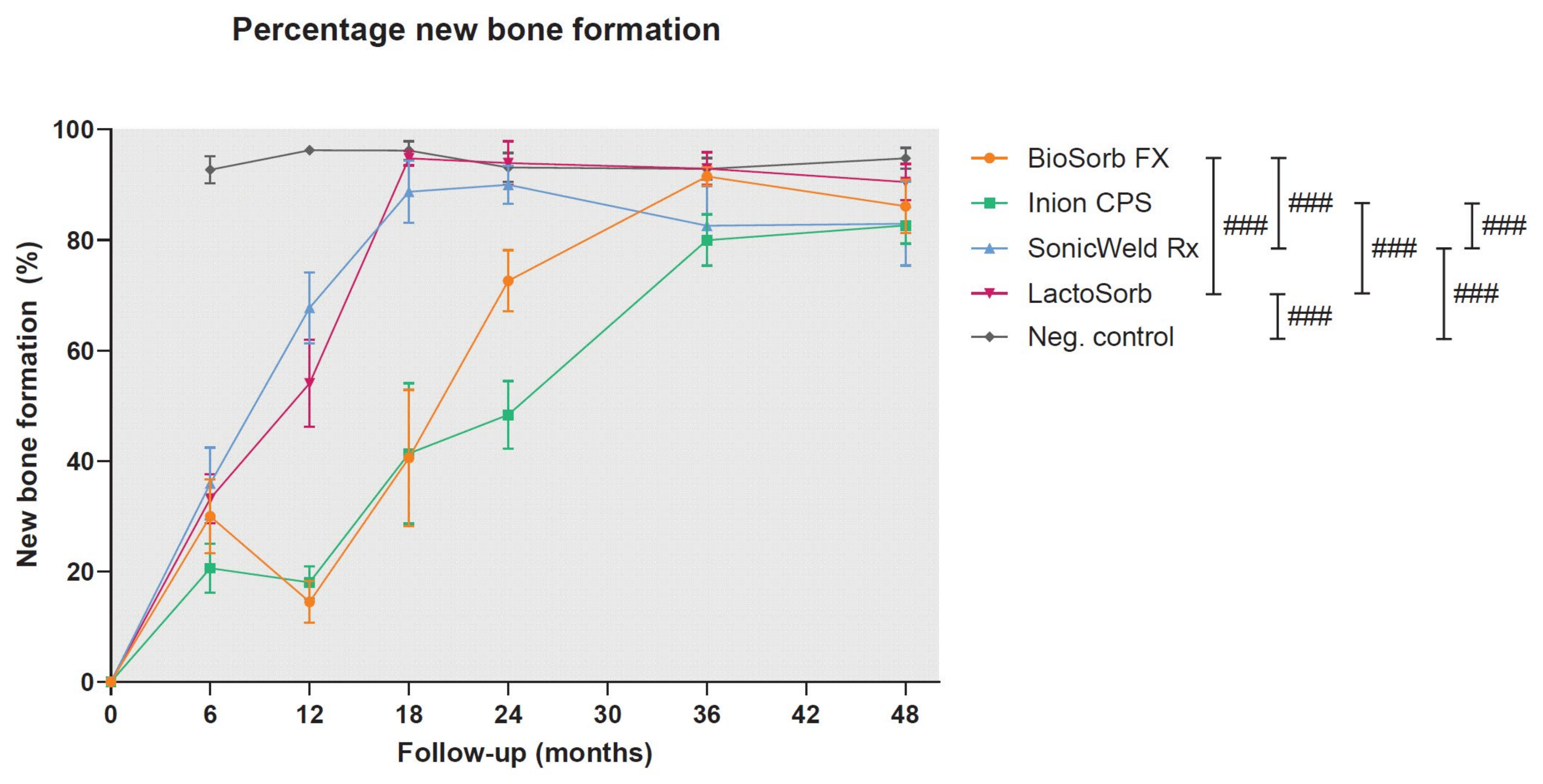
Figure 1. Percentage of total new bone formation at the implant site. Error bars: mean values ± standard error of the mean. ### represents p < 0.05, p < 0.01, and p < 0.001, respectively. The composition of each system is described in Table 1. A similar curve for titanium systems is not applicable due to the non-degradable nature of titanium systems. Error bars: mean values ± standard error of the mean. Reprinted with permission from [25].
Polymers 14 02782 g001
Figure 2. HE-sections of the Inion CPS system at 48-month follow-up under LM (a) and LM-pol (b) with observable residual polymer fragments (examples are indicated with black arrows). HE, hematoxylin and eosin; LM, light microscopy; LM-pol, polarized light microscopy. From the study of [25].
Polymers 14 02782 g002
Figure 3. HE-sections (LM and LM-pol), SEM, and EDX by element and with overlay (red: carbon, and blue: nitrogen) of birefringent polymeric residual fragments of every osteosynthesis system at 48-month follow-up. HE, hematoxylin and eosin; LM, light microscopy; LM-pol, polarized light microscopy; SEM, scanning electron microscopy; EDX, energy-dispersive X-ray analysis; C, carbon; N, nitrogen; O, oxygen. Reprinted with permission from [25].
Polymers 14 02782 g003
Figure 4. HE-section of a soft tissue biopsy surrounding commercially pure titanium plates after mandibular osteosynthesis under LM. Dust-like (1 micron) particles are indicated with a blue arrow (magnification ×400). HE, hematoxylin and eosin; LM, light microscopy. Reprinted with permission from [15].
Polymers 14 02782 g004
Figure 5. The initial curve represents the percentage survival of colony formation of HF19 cells exposed to titanium particles for 24 h. The percentage of survival of the progeny of these cells is also shown, indicating delayed reproductive death 10 generations postexposure. All percentages are expressed relative to the control expressed as 100%. Reprinted with permission from [105].
Polymers 14 02782 g005
Figure 6. (a) Schematic overview of nanogel coating with antifouling and antimicrobial properties. (b) Staphylococcus aureus adhered to glass with and without the nanogel coatings under fluorescence microscopy (scale bars 20 µm). (c) The number of colony-forming units of surviving Staphylococcus aureus after 24 h incubation on the surface of uncoated and coated glass (*** and **** indicate p < 0.001 and p < 0.0001, respectively). N-nGel, nonquaternized nanogel; Q-nGel, quaternized nanogel; TCS, triclosan; CFU, colony-forming units. Reprinted with permission from [122].
Polymers 14 02782 g006
Publisher’s Note: MDPI stays neutral with regard to jurisdictional claims in published maps and institutional affiliations.