1. Introduction
Due to their unique combinations of biological and biophysical properties, hyaluronan (HA)-based hydrogels are broadly applied for dermal tissue wound healing, engineering, and aesthetic treatments [
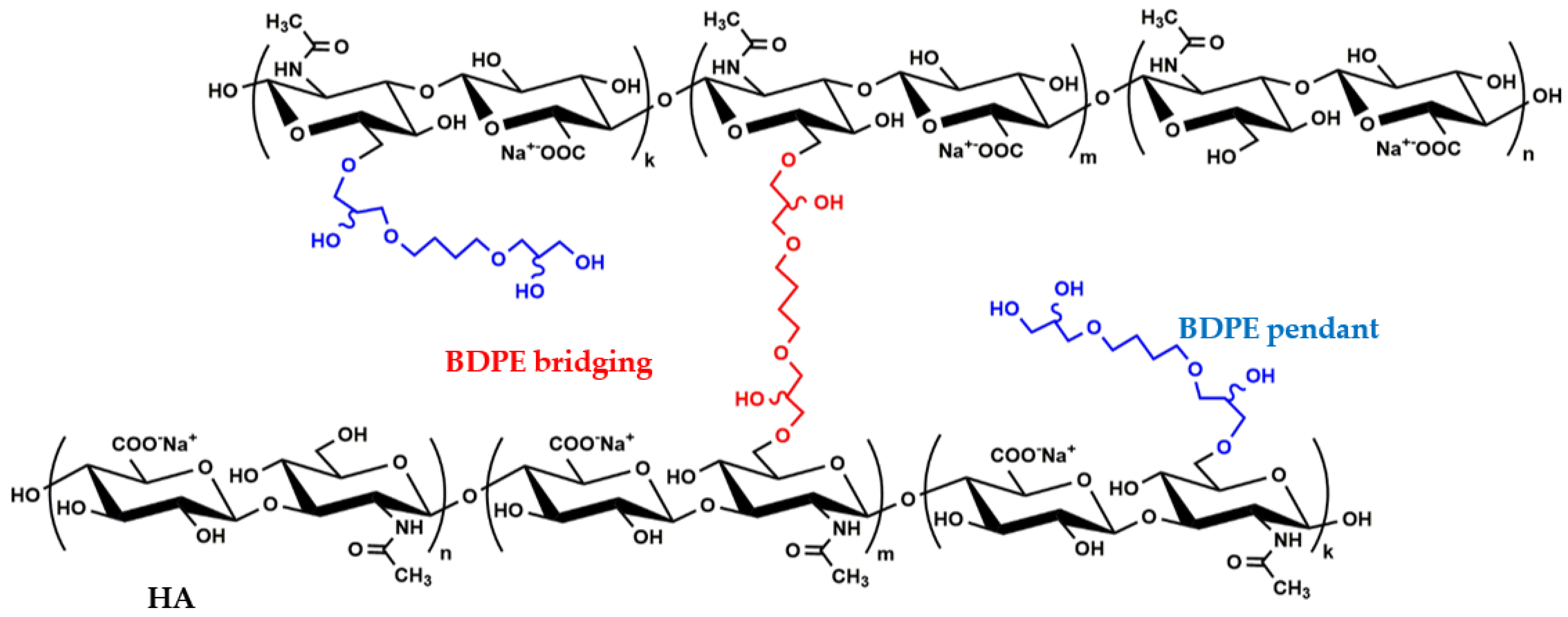
1]. Specifically, the hydrogels obtained through the reaction of HA with 1,4-butanediol-diglycidyl ether (BDDE) are widely used for dermo-aesthetic purposes. Intradermal injections for skin rejuvenation represent the most broadly used treatment option. Commercially available gels are variously concentrated suspensions of HA–BDDE networks in a physiological solution. They can differ according to the degree of HA modification by BDDE. Further, they are known to contain two different 1,4-butanediol di-(propan-2,3-diolyl) ether (BDPE) functionalizations (
Figure 1).
In fact, part of BDDE reacts with HA at both ends, effectively bridging two disaccharide units (BDPE bridging) and forming crosslinked HA (
Figure 1). Another portion of the di-epoxide reacts with water/hydroxide at one end and with the polymer at the other, thus installing only an ether-linked appendage to HA (BDPE pendant) and forming branched HA (
Figure 1) [
2,
3,
4,
5].
The modification of HA by BDDE endows it with key features for various applications. Unlike the linear polymer, the crosslinked network is able to swell in an aqueous solution without dissolving, rheologically behaves as a gel-like material (with the storage modulus exceeding the loss modulus and with a slight dependence of the moduli on frequency), shows a viscosity that decreases with the shear rate under flow conditions, and is less sensitive to degradation by hyaluronidases [
1,
6]. These features are at the basis of a gel’s overall performance, as they are responsible for its hydration, projection (filling effect), injectability, and longer permanence in tissues compared to linear HA. The rheology of the final formula is key for its clinical designation. As the storage modulus is a measure of the gel’s resistance to external forces and its ability to retain its shape, stiffer gels (high-G′ gels) are suited for restoring lost tissue volume (lifting support) and are designated for injection into deeper layers of skin; more deformable preparations (lower-G′ gels) are intended for injection into more superficial dermal layers in order to enhance face contours and for a skin-boosting effect. Studies providing rheological and other biophysical data (swelling, sensitivity to enzymatic hydrolysis, cohesivity, and so on) for clinically available HA–BDDE gels have been intensifying. By allowing the comparison of available gels in terms of certain parameters, they assist clinicians in the selection of the most appropriate products for the specific clinical needs. Fewer studies aiming at properly ascertaining the complexity of the rheological behaviors of these products have been carried out, with Ilyin et al. describing, for the first time, to the best of our knowledge, the mechanical behavior of these gels after dilution in a physiological medium. This represents an aspect that should be further explored in the science of HA-based dermal treatments to indicate gels’ behaviors when interacting with surrounding tissues [
6].
Gel rheology certainly depends on the degree of HA derivatization—with different effects expected for the two types of modification—and on the biopolymer concentration (i.e., the higher the HA modification and concentration, the stiffer the gel is expected to be).
However, to the best of our knowledge, a quantitative analysis establishing this correlation, which would be valuable for tailoring gel design towards specific performance, has not been carried out so far. In fact, the biophysical parameters provided for clinically available gels have not been related to level/type of chemical modification of HA [
3,
7,
8,
9,
10,
11,
12,
13,
14,
15,
16,
17,
18,
19,
20,
21,
22,
23,
24,
25,
26,
27,
28].
Conversely, characterization studies of the structural modifications obtained after the reaction of HA with BDDE have been performed to a lower extent [
2,
3,
4,
5,
9,
12,
19,
20,
29,
30,
31]. Only some of them have discriminated between the BDPE-bridging and BDPE-pendant moieties. Furthermore, these analyses were mostly performed for homemade gels, and the acquired chemical data have never been considered in relation to hydrogel rheology [
3,
4,
5,
12,
19,
31].
To fill this gap, four commercially available HA–BDDE hydrogels with the same clinical indications as volumetric dermal fillers were investigated here for the determination of their amounts of crosslinked and branched HA, stiffness (G′), and overall elasticity (tan δ). Their sensitivity to enzymatic degradation was also studied as another key parameter in view of their application, and it was influenced by the HA modification level. The collected data were then examined to verify whether and how gel rheology and stability are correlated with the type and degree of HA modification. The rheology was also unconventionally studied during the hydration of the gel (progressive gel dilution) in a physiological medium so that it would, thus, better resemble in vivo conditions. Information that will be useful for the improvement of the design and characterization of this type of gel is expected.
3. Results
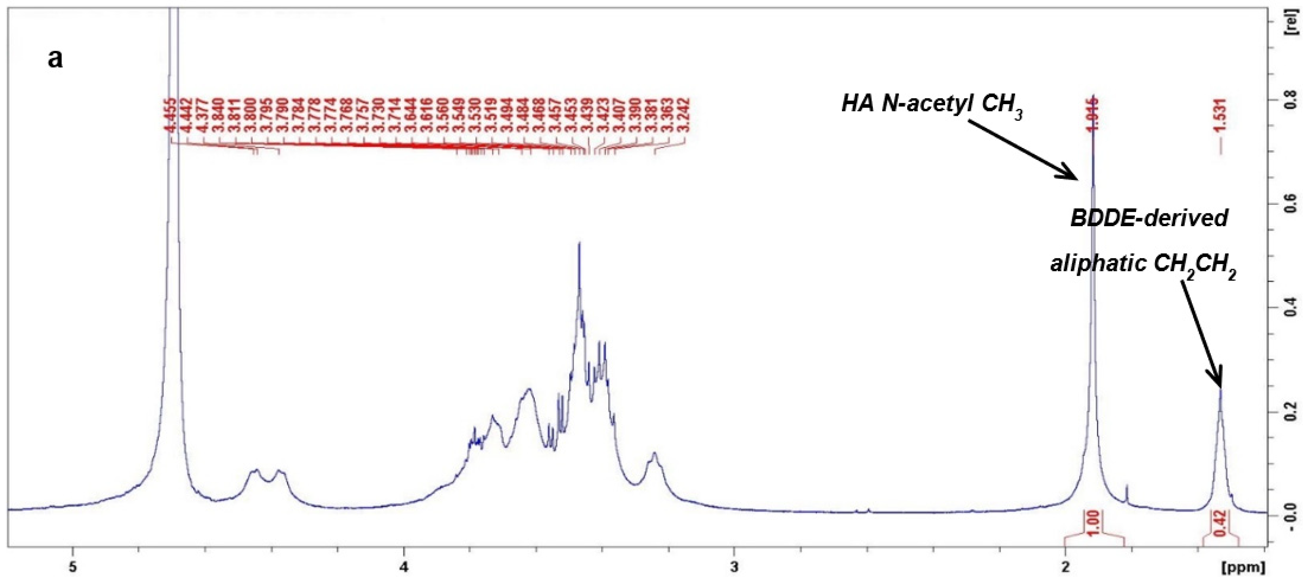
3.1. NMR Analyses
The BDPE/HAd (mol%) values calculated from the 1H-NMR analyses (A) varied over a wide range. Namely, the total BDPE/HAd(mol%) spanned from a very low amount (2.4%), which was recorded for RL, up to around 31%, which was recorded for ASV. Similar intermediate values of around 7–8% were found for both TRHA4 and JV. When considering the BDPE/HA weight ratio (E), the values ranged from 1.2 (RL) to 16.0 wt% (ASV), with TRHA4 and JV still showing similar results (3.4–3.9 wt%). The total BDPE amount (wt%) in the gel (F) was in the range of 0.02–0.4 wt%.
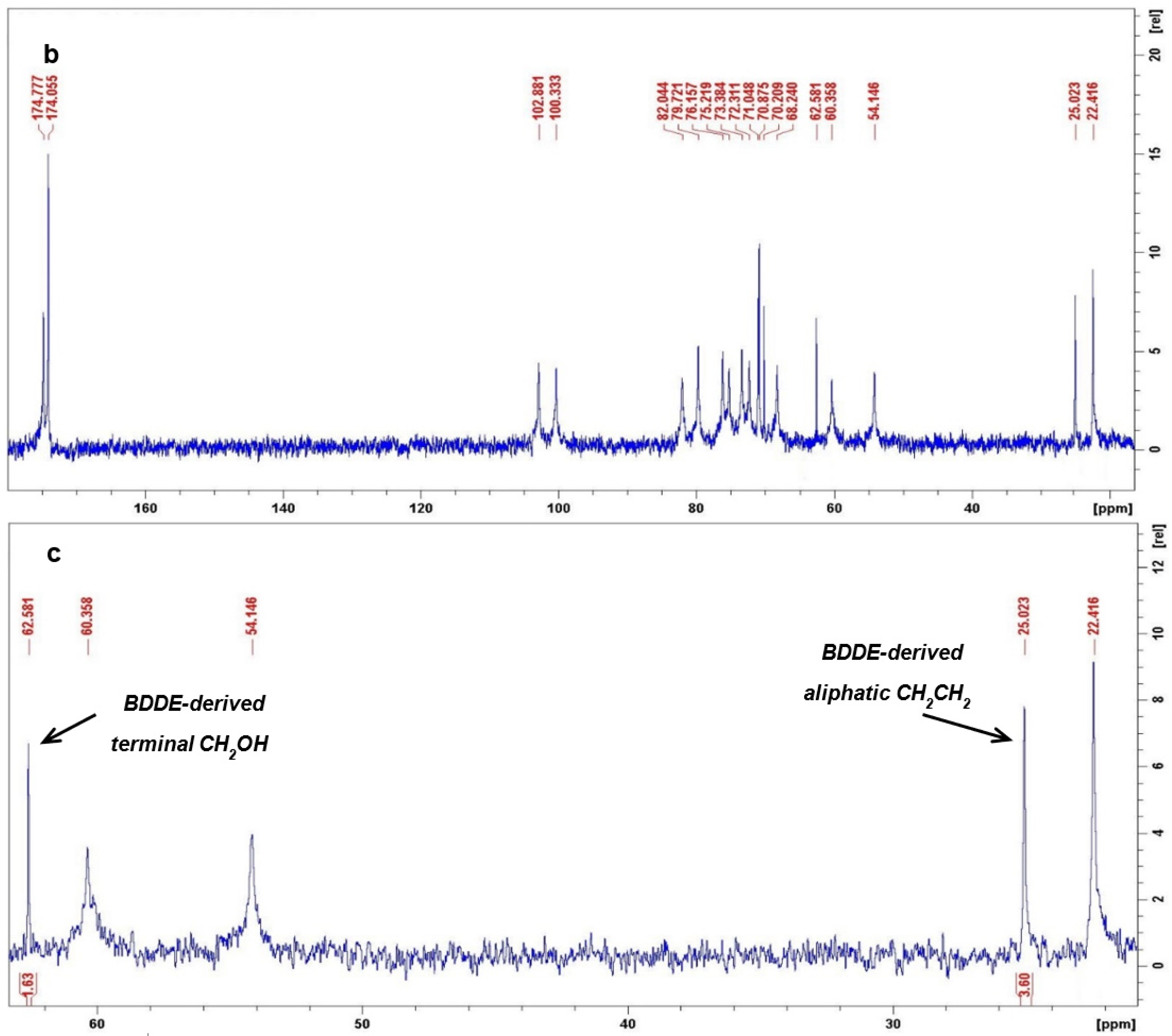
The 13C-NMR spectra allowed us to quantify the BDPE pendant/HAd molar ratio (B) and, therefore, the BDPE bridging/HAd (mol%) (C) and the BDPE bridging/BDPE (mol%) values (D). The data indicate that BDPE that effectively crosslinked two disaccharide units varied from 4.2 to 28.8% compared to the total BDPE amount, with the highest value recorded for TRHA4 (D). Therefore, most of the BDDE-derived functionalizations were present on HA chains as pendant groups, rather than as crosslinking moieties.
The amount (mol%) of crosslinked HA disaccharide units (G) and the amount of branched HAd (mol%) (H) are also reported. Finally, the amount (mol%) of HAd that was chemically modified by BDPE, which describes the total HA modification level (crosslinked + branched) (I), is indicated. ASV turned out to be the most crosslinked gel (6.0 mol% of crosslinked-HAd (G)) and the product that presented the highest percentage of branched disaccharide units (28.5 mol%) (H). It also showed the highest overall modification degree, with 34.5% of its disaccharide units being modified by BDPE (I).
RL was the least modified gel, with only 2.5% of the HAd being modified. TRHA4 and JV showed intermediate HA modification degrees that were closer to that of TRHA4, and they turned out to be slightly less modified (modified HAd equal to 8.5 and 9.0 mol%, respectively). However, the crosslinking degree was higher for TRHA4, while the amount of branched HAd was greater for JV.
3.2. Rheological Analyses and Correlations with HA Modification Parameters
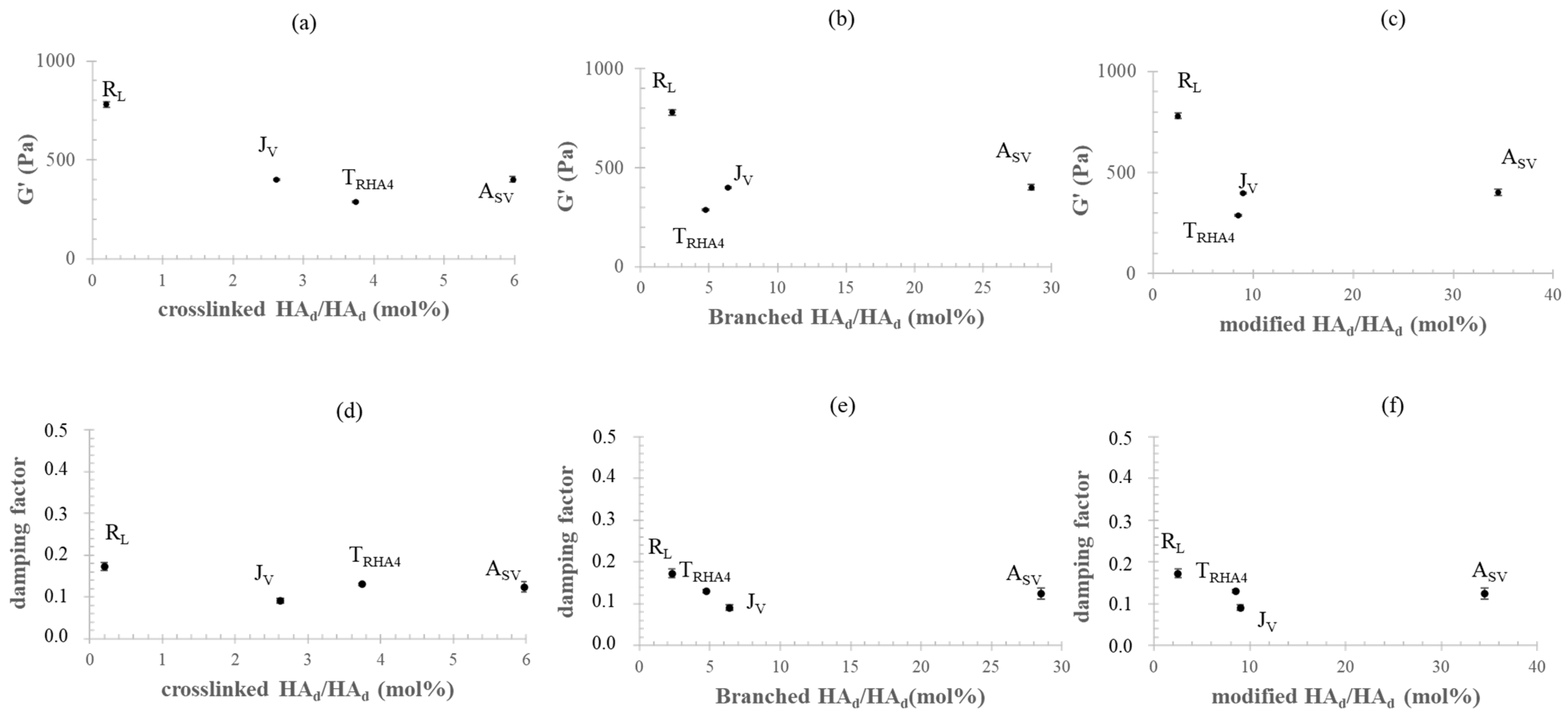
Figure 3 shows the G′ and damping factor values measured for the gels, which are reported as a function of the amounts (mol%) of crosslinked HA
d and branched HA
d and as a function of the total amount (mol%) of HA
d modified by BDPE.
Surprisingly, the rheological parameters were not correlated with the biopolymer crosslinking degree the branched HA amount, or the total degree of modification (
Figure 3a–f). The data indicated that the least crosslinked gel (R
L) behaved as the most rigid (
Figure 3a). Although turned out to be 2.3-fold more crosslinked than J
v, A
SV showed comparable G′ values. When compared to R
L, A
SV presented a 30-fold higher crosslinking, but was almost twofold less stiff. Even the loss tangent values, which were expected to decrease with increasing crosslinking, did not show any proportional variation with the HA chemical modification parameters (
Figure 3d–f).
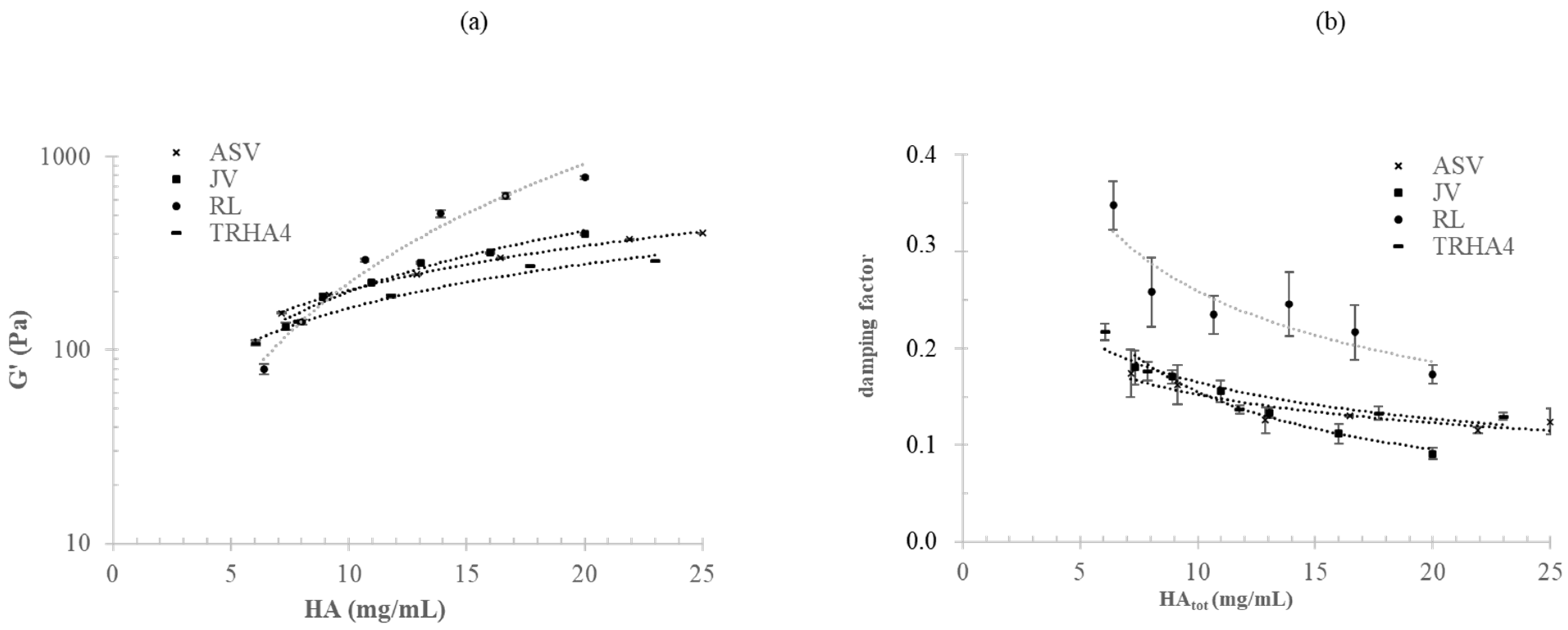
The G′ and damping factor values were then studied as a function of the progressive dilution of the gels in a physiological medium. The results are shown in
Figure 4.
G′ decreased with the decrease in HA concentration (a), while tanδ increased with progressive gel dilution (b). Both G′ and the damping factor varied as a function of HA concentration, following a power law relation. The dependence of G′ and the loss tangent on HA concentration (CHA) was differently marked for the various gels. RL showed the most marked increase in G′ with CHA (G′ = 2.01 (CHA)2.04; R2 = 0.98), followed by JV (G′ = 17.7 (CHA)1.05; R2 = 0.98), and then by ASV and TRHA4, which showed the lowest and similar dependences (G′ = 34.9 (CHA)0.77; G′ = 28.99 (CHA)0.75). Similarly, the variation of the damping factor with dilution was more marked for RL and JV compared to ASV and TRHA4. Overall, the data indicated that the rankings in stiffness and damping factor for the gels were extensively affected by hydration.
It is worth mentioning that the mechanical spectra of the preparations as they were in the syringe and those of the same gels after the various dilutions tested showed no or a very slight dependence of the moduli on frequency (with G′ being much higher than G″), thus indicating the gel-like behavior of the gels even after dilution (
Figure S2).
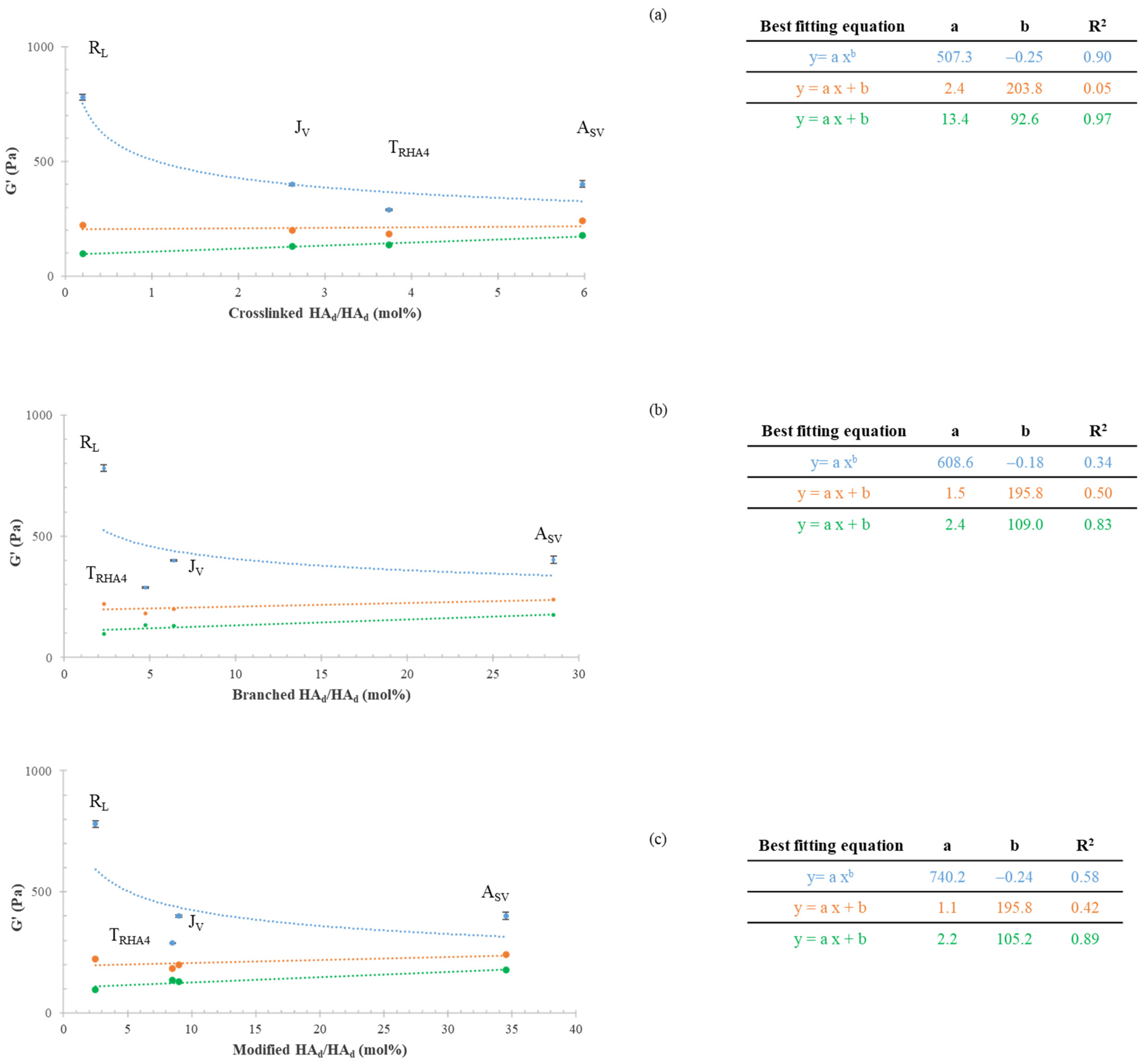
Figure 5 shows G′ in relation to the HA modification parameters during gel hydration.
Specifically, the G′ values for the gels as commercialized (blue curves) and for the same gels after twofold (orange curves) and threefold hydration (green curves) in the physiological medium are reported as a function of the amounts of crosslinked HAd (mol%) (a) and branched HAd (mol%) (b) and as a function of the total HA modification (mol%) level (c). The regression with the highest R2 value was used to fit each set of data. When considering the gels as commercialized (blue curves), the experimental data were better predicted by a power law model, and the R2 values varied from 0.34 to 0.90, with the highest value being found for the G′ vs. crosslinked HAd (mol%) relation. However, the variation of G′ with the crosslinking degree was the opposite of what was expected. Following hydration, the trend completely changed, with G′ increasing with the increase in HA modification, as expected. The goodness of the mathematical prediction of the data also varied with gel dilution. After threefold hydration (green curves), a linear regression better fit the experimental data, with the best correlation being found for G′ vs. crosslinked HA (mol%) (R2 equal to 0.97).
The correlation of G′ with the amount of branched HA
d (mol%) and with the total HA modification level (BDPE-modified HA
d mol%) also improved with hydration, but lower R
2 values (0.85–0.89) were reached (
Figure 5b,c, green curves).
The data in
Figure 5 also show that the differences in stiffness among the samples were greatly reduced after hydration. For the commercial formulations, G′ was in the range of 280–780 Pa, with the most rigid gel (R
L) being 2.7-fold stiffer than the most deformable one (T
RHA4). The threefold-hydrated gels showed G′ values in the range of 97–177 Pa, with the stiffest gel (A
SV) being only 1.8-fold more rigid than the most deformable sample.
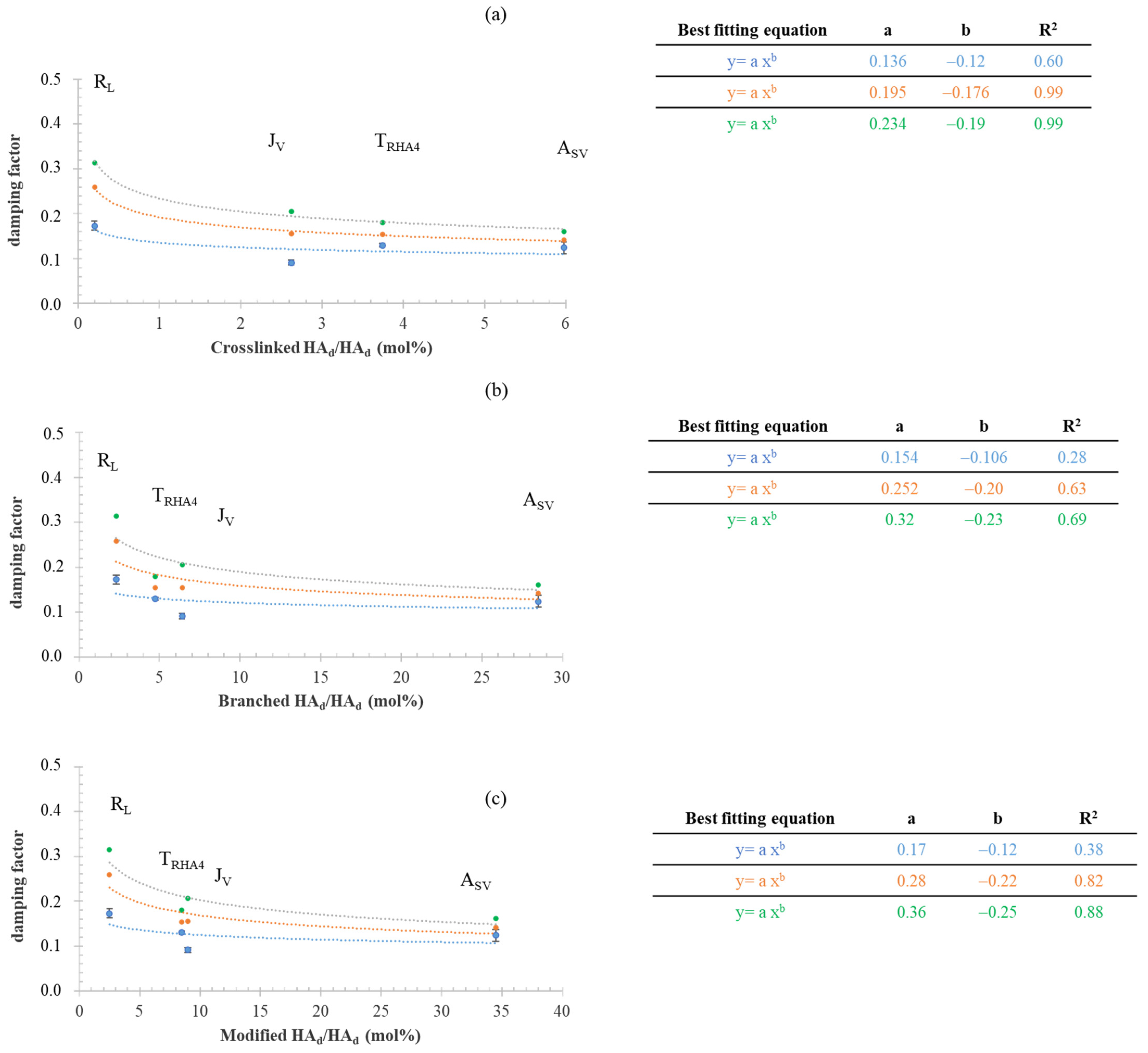
The damping factor values are reported as a function of crosslinked HA
d, branched HA
d, and modified HA
d (branched + crosslinked HA
d) in
Figure 6a–c, respectively. The blue, orange, and green curves were obtained for the commercial samples and for the samples after two- and threefold hydration, respectively (
Figure 6).
A power law regression was used to fit the data showing the highest values for the determination coefficient. The latter improved with hydration, and, for the hydrated gels, the damping factor was strongly correlated with the amount (mol%) of crosslinked HAd (R2 0.99). Even for tan δ, the correlation with the amount of branched HAd and with the total HA modification degree was worse. Unlike G′, the damping factor varied over a wider range after hydration (0.12–0.17 for the gels as commercialized and 0.16–0.31 for the hydrated gels).
3.3. Sensitivity to Enzymatic Hydrolysis and Correlation with HA Modification Parameters
The gels’ sensitivity to enzymatic action was evaluated by monitoring the amount (mg/mL) of biopolymer that was solubilized due to incubation with BTH (5 U/mL; 3 h; 37 °C). The results are reported in
Table 2. All of the samples tested were shown to have sensitivity to BTH. Different levels of solubilization extents varying from 1.1 ± 0.2 to 2.8 ± 0.5 mg/mL were recorded.
The gels presenting an amount of 2.6–6.0% crosslinked HAd (mol%) showed comparable degrees of solubilization (1.1–1.6 mg/mL, p > 0.05), while a significantly higher degree of solubilization (2.8 ± 0.5 mg/mL; p < 0.05) was recorded for the least crosslinked gel (0.2 mol% crosslinked HAd/HAd) under the same conditions. When considering solubilization due to BTH in relation to the total HA modification degree, the data indicated that variation in the amount of BDPE-modified HA (mol %) in the range of 8.5–34.5 (mol%) did not result in different levels of stability with respect to enzymatic action, while lowering the level of derivatization down to 2.5% led to worse stability.
4. Discussion
In the wide framework of studies on HA–BDDE hydrogels for dermo-aesthetic applications, we aimed to shed light on the correlation between hydrogels’ rheology and stability and their HA crosslinking/modification parameters.
Four clinically available HA–BDDE dermal fillers were investigated for this purpose. The BDPE/HA
d molar ratio values (
Table 1 (A)) derived from
1H-NMR analyses fell within the range reported in the literature for similar gels, except for those of A
SV, which presented a higher BDPE/HA
d content [
3,
9,
12,
20]. It is noteworthy that the BDPE/HA
d molar ratio varied over a wide range (2–31%) even though the same clinical indications were given for the hydrogels tested.
Interesting chemical aspects that were scarcely considered before for these hydrogels emerged from the
13C-NMR analyses. At first, the data indicated that most (more than 70%) of the BDPE bonded to HA did not lead to HA crosslinking, but to HA bearing BDPE appendages. Discrimination between the two BDPE functionalities (bridging or pendant) allowed for the calculation of the real total HA modification level (I) [
5]. The latter did not simply correspond to the BDPE/HA
d molar ratio (A), as is commonly determined for these products via
1H-NMR. In fact, each BDPE-bridging moiety was responsible for two chemically modified HA disaccharide units; therefore, a higher total HA modification (I) level was expected in comparison to the BDPE/HA
d ratio (A). However, due to the low number of BDPE-bridging functionalities, only a slight difference was recorded between the (A) and (I) parameters (
Table 1). It is noteworthy that the (I) values were in the range of 2.5–34.5%, while a total HA modification of 4–63% would be expected in the case of 100% BDPE bridging.
As very few studies in the literature have reported, our data indicated that attention must be paid when ranking the gels in terms of their HA modification parameters that are based only on the BDPE/HA
d ratio [
3,
12,
19,
31]. In fact, since the bridging BDPE/total BDPE ratio greatly varied from one product to another (
Table 1 (D)), the rankings in the HA crosslinking degree (G) can be variously marked or even completely different compared to those for the BDPE/HA ratio (A) or total HA modification level (I). For instance, for the products analyzed here, the most derivatized gel (A
SV) showed a 4.4-fold higher BDPE/HA
d ratio than that of J
V (
Table 1 (I)), but it was only 2.3-fold more crosslinked (
Table 1 (G)). Further, J
V showed a higher BDPE/HA
d amount than that of T
RHA4, but was less crosslinked.
The samples tested presented very similar concentrations of water-insoluble HA–BDDE fractions, which are mainly responsible for the typical elastic behavior of these gels. Therefore, an increase in the rheological properties with the level of HA crosslinking and/or total HA modification was expected. Hence, the absence of a correlation between the G′ and damping factor values and the biopolymer modification parameters (crosslinked HA
d; branched HA
d; modified HA
d) was surprising. The trend found for G′ and the loss tangent vs. the amount of crosslinked-HA
d was very far from what was expected (
Figure 3). These findings mainly suggested that, aside from the network’s crosslinking density, other parameters play a role in gels’ relative rheology. A certain effect related to the diversity in the water-soluble fractions and to the potential differences (limited differences considering the specific reaction conditions) in the lengths of the chains undergoing crosslinking can be expected. The extent of gel hydration was rationally considered and investigated as the other potentially highly impacting factor.
It is well known that the HA networks in the commercial formulations do not exploit their maximum hydration capabilities and that gels are further hydrated after being delivered into the dermis or subdermis. Notwithstanding this, the data on HA–BDDE gels reported so far refer only to the rheological properties of the gels as they are in the syringe, with the study by Ilyin et al. being the only one to consider the gels’ rheological parameters after fivefold dilution [
6]. Here, this latter aspect was further explored. The gels were progressively diluted in a physiological medium to simulate hydration in vivo while monitoring changes in their rheological behavior. The variation of G′ and the damping factor with decreasing HA concentration (
Figure 4) was predictable. However, it was remarkable that the dilution affected the gels to different extents due to the specific features of the network. Specifically, the data indicated a more marked variation in the rheology with dilution for less crosslinked hydrogels. As a consequence, the rankings in mechanical behavior completely changed after hydration. Finally, for the hydrated gels, the G′ and damping factor values were correlated with the crosslinking level, as expected (
Figure 5). The less significant correlation found with respect to the other HA modification parameters suggested that the tuning of HA crosslinking—more than HA branching—is key for adjusting the mechanical behavior of a gel once it is equilibrated in the tissue. Overall, the data collected here indicate that, for the gels in the syringe, the level of (partial) hydration used (HA concentration) plays a key role in their rheology, while, after hydration, their mechanical behavior is mainly driven by the extent of HA crosslinking. This is clearly represented, for instance, by the R
L data: notwithstanding that it had the lowest crosslinking/modification degree, R
L behaved as the most rigid product in its commercial form (
Figure 5). However, when allowed to hydrate, it markedly lost its rigidity, finally behaving as the most deformable sample (lowest G′), which was consistent with its low crosslinking degree (
Figure 5). The combination of the specific HA crosslinking level and the HA content in 1 mL of the commercial gel resulted, for A
SV, in an interesting performance. Based on the collected data, a higher projection/volumizing ability (highest G′ and lowest damping factor) was expected for A
SV after being equilibrated in the tissue, while the lower G′ recorded for this gel in the syringe suggested an ease of delivery that would be similar to or higher than that of the other samples. It is also worth noting that a quantification of the effect of the soluble fraction (concentration and molecular weight) on the rheology of the formula would be valuable for furthering tailoring the injectability of the final preparation.
Further, the data highlighted a key point that impacted the characterization of these hydrogels in relation to the prediction of in vivo performance. To date, the relative gel projection capacity has been predicted by comparing the rheological parameters of commercial samples [
10,
14,
15,
16,
18,
20,
24,
26]. Based on our findings, these parameters are representative of gel’s behavior only during delivery and immediately after the delivery to the dermis, but they do not characterize the gels once they have equilibrated in the tissue. Therefore, hereafter, more attention should be paid to the characterization of gels’ rheological features, which may be more predictive when they are also evaluated for hydrated gels. Comparing the rheology of the formulas to be injected only provides evidence on their relative injectability.
Useful information on the relation between gel stability with respect to hyaluronidases and HA structural modification was also provided. The derivatization of HA by BDPE (not only crosslinking, but also functionalization with pendant moieties) is expected to affect the recognition of the biopolymer by the enzyme, thus increasing gel stability and, therefore, prolonging action in vivo. The collected data confirmed what was expected only for degrees of derivatization/crosslinking up to 8-9/3-4 mol%, and they showed that there was no significant effect on BTH activity for higher levels of HA modification. This would suggest that the conformational variation in HA occurring in the lower range of modification is more significant for the recognition of the biopolymer by the enzyme. A further advance in the understanding of the dependence of HA–BDDE gels’ stability on HA modification may be achieved when investigating whether the increase in crosslinking—while keeping the BDPE/HA ratio constant—influences sensitivity to hyaluronidases or not.
5. Conclusions
For the first time, HA–BDDE gels that were intended as dermal fillers were analyzed to investigate how the formulations’ rheology and stability related to their HA chemical modification parameters. Rheological data were also provided for the gels while being hydrated in a physiological medium (to mimic the gels’ equilibration in tissue). The gels greatly differed in terms of the levels of total HA derivatization and the functionalities of branched HA and HA crosslinked by BDPE, with branching representing the main type of modification.
The rheological properties of the hydrated gels were correlated well with the level of HA crosslinking, while the mechanical behavior of the samples as commercialized was mainly controlled by the HA concentration. An unexpected and strong effect of hydration on the gels’ ranking in terms of rheology was demonstrated, with implications for the characterization of these products. The results indicated that there was no improvement in the gels’ stability with respect to enzymatic hydrolysis for derivatization and crosslinking levels higher than 9 and 3 mol%, respectively.
Overall, the collected data are of interest for the design, in vitro characterization, and prediction of the performance of these types of hydrogels.


 ,
, 


