
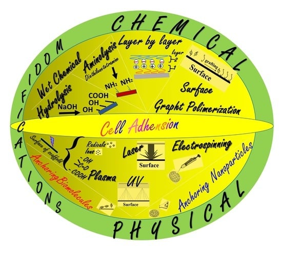
Surface Functionalities of Polymers for Biomaterial Applications


Abstract
:
1. Introduction
2. Chemical Modification Techniques
2.1. Wet Chemical Techniques
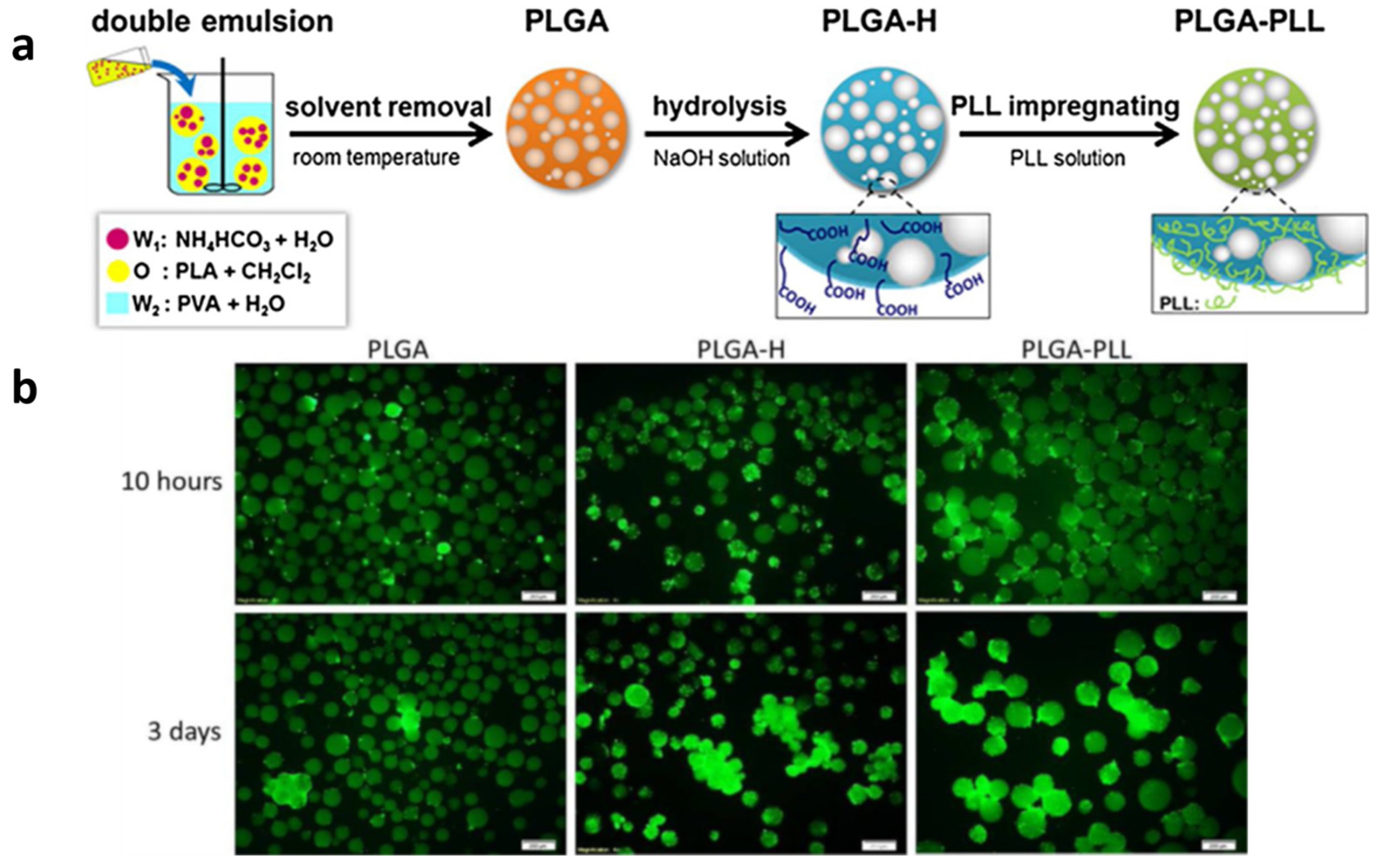
2.1.1. Hydrolysis
2.1.2. Aminolysis
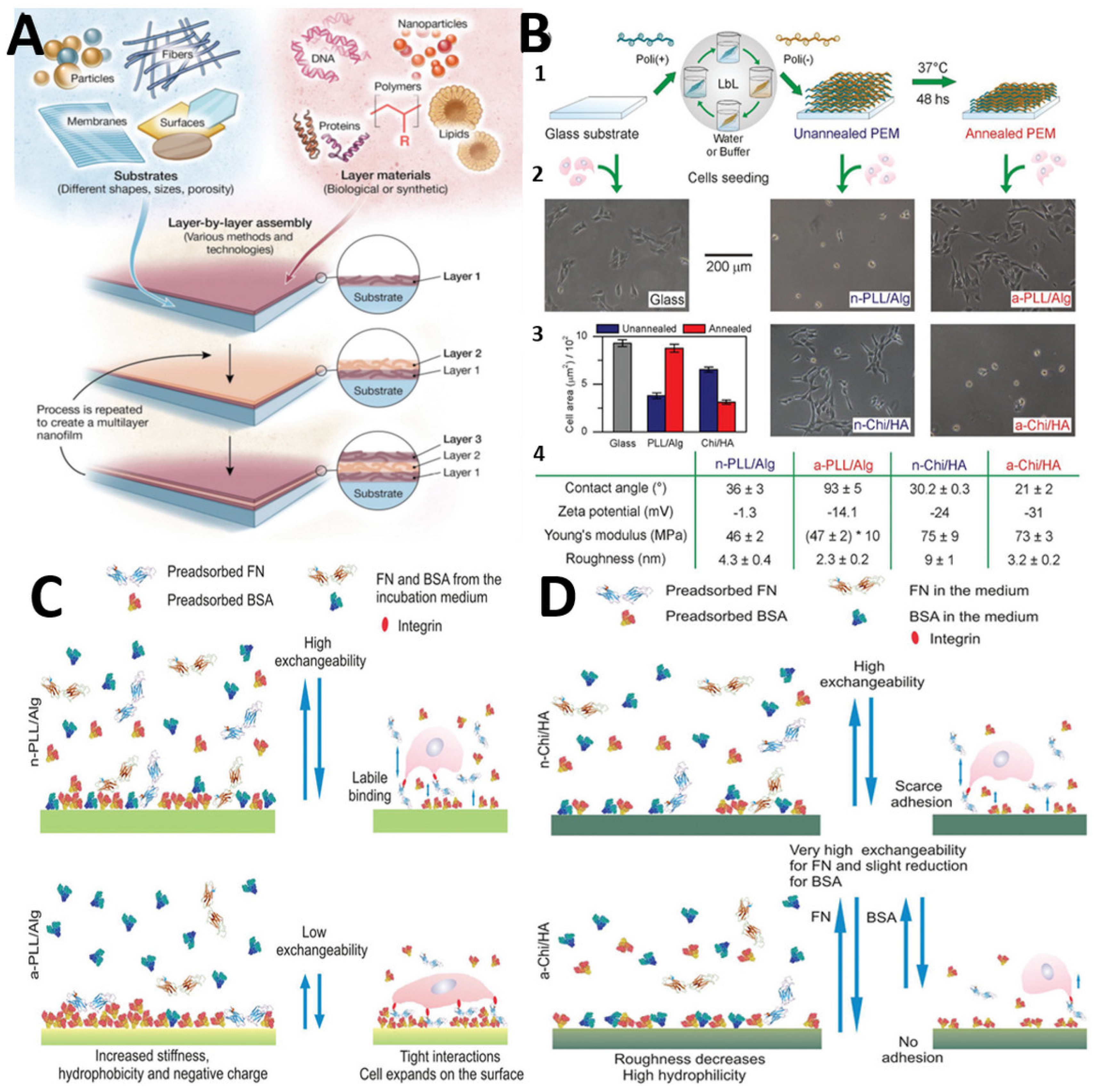
2.2. Layer-by-Layer
2.3. Surface Graft Polymerization
3. Physical Modification Techniques
3.1. Plasmas Techniques
Adsorbtion Molecules
3.2. Ultraviolet Technique
3.2.1. Principle of the Technique
3.2.2. Activation Effect
3.2.3. Curing Effect
3.2.4. UV Degradation Effect
3.3. Laser Ablation (LA)
UV Excimer Laser
3.4. Electrospinning
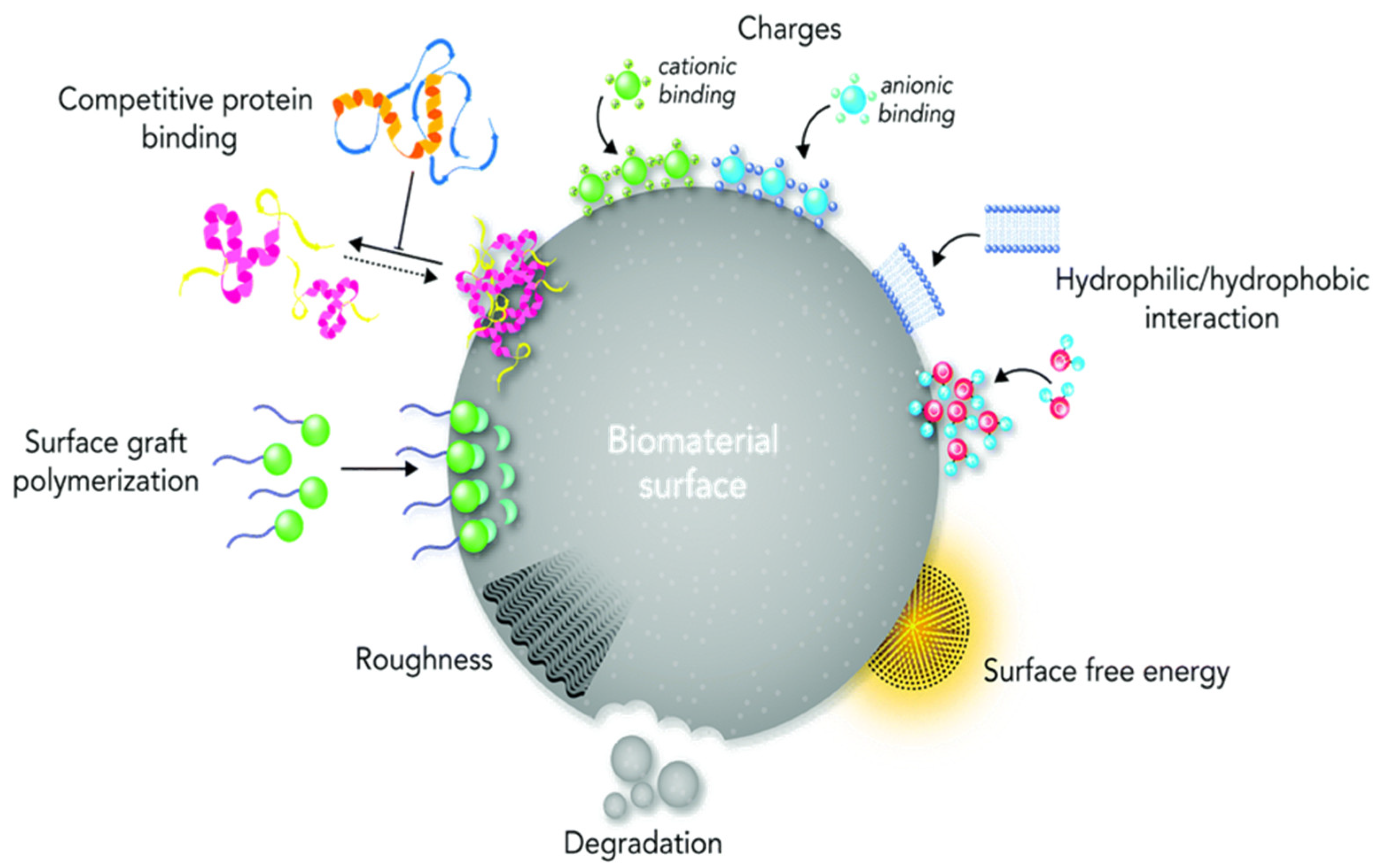
4. Effects of Surface Properties on Biological Responses of the Materials
- (a)
- the successive immersion of a single surface in different solutions biomolecules containing a mixture of biomolecules [149];
- (b)
- the single immersion of a surface in a solution containing a mixture of biomolecules [150];
- (c)
- the immobilization of synthetic and multifunctional biomolecules, as well as proteins [151].
4.1. Antibacterial Properties
4.2. Biocompatibility Properties
4.3. Cell Adhension
5. Conclusions
Author Contributions
Funding
Institutional Review Board Statement
Informed Consent Statement
Data Availability Statement
Acknowledgments
Conflicts of Interest
References
- John, A.A.; Subramanian, A.P.; Vellayappan, M.V.; Balaji, A.; Jaganathan, S.K.; Mohandas, H.; Paramalinggam, T.; Supriyanto, E.; Yusof, M. Review: Physico-chemical modification as a versatile strategy for the biocompatibility enhancement of biomaterials. RSC Adv. 2015, 5, 39232–39244. [Google Scholar] [CrossRef]
- Duan, K.; Wang, R. Surface modifications of bone implants through wet chemistry. J. Mater. Chem. 2006, 16, 2309–2321. [Google Scholar] [CrossRef]
- Curran, J.M.; Fawcett, S.; Hamilton, L.; Rhodes, N.P.; Rahman, C.V.; Alexander, M.; Shakesheff, K.; Hunt, J.A. The osteogenic response of mesenchymal stem cells to an injectable PLGA bone regeneration system. Biomaterials 2013, 34, 9352–9364. [Google Scholar] [CrossRef]
- Yuan, Y.; Shi, X.; Gan, Z.; Wang, F. Modification of porous PLGA microspheres by poly-l-lysine for use as tissue engineering scaffolds. Colloids Surf. B Biointerfaces 2018, 161, 162–168. [Google Scholar] [CrossRef] [PubMed]
- Cai, Y.; Zhang, Z.; Ding, Y.; Hu, L.; Wang, J.; Chen, T.; Yao, Y. Recent development of pillar[n]arene-based amphiphiles. Chin. Chem. Lett. 2021, 32, 1267–1279. [Google Scholar] [CrossRef]
- Amani, H.; Arzaghi, H.; Bayandori, M.; Dezfuli, A.S.; Pazoki-Toroudi, H.; Shafiee, A.; Moradi, L. Controlling Cell Behavior through the Design of Biomaterial Surfaces: A Focus on Surface Modification Techniques. Adv. Mater. Interfaces 2019, 6, 1900572. [Google Scholar] [CrossRef] [Green Version]
- Zhang, K.; Zheng, H.; Liang, S.; Gao, C. Aligned PLLA nanofibrous scaffolds coated with graphene oxide for promoting neural cell growth. Acta Biomater. 2016, 37, 131–142. [Google Scholar] [CrossRef]
- Guo, C.; Xiang, M.; Dong, Y. Surface modification of poly (lactic acid) with an improved alkali-acid hydrolysis method. Mater. Lett. 2015, 140, 144–147. [Google Scholar] [CrossRef]
- Wang, Y.; Murcia Valderrama, M.A.; van Putten, R.-J.; Davey, C.J.E.; Tietema, A.; Parsons, J.R.; Wang, B.; Gruter, G.-J.M. Biodegradation and Non-Enzymatic Hydrolysis of Poly(Lactic-co-Glycolic Acid) (PLGA12/88 and PLGA6/94). Polymers 2021, 14, 15. [Google Scholar] [CrossRef]
- Erdoğan, M.K.; Akdemir, Ö.; Hamitbeyli, A.; Karakışla, M. Preparation of hydrophilic woven fabrics: Surface modification of poly(ethylene terephthalate) by grafting of poly(vinyl alcohol) and poly(vinyl alcohol)-g-(N-vinyl-2-pyrrolidone). J. Appl. Polym. Sci. 2020, 137, 48584. [Google Scholar] [CrossRef]
- Brown, J.H.; Das, P.; DiVito, M.D.; Ivancic, D.; Tan, L.P.; Wertheim, J.A. Nanofibrous PLGA electrospun scaffolds modified with type I collagen influence hepatocyte function and support viability in vitro. Acta Biomater. 2018, 73, 217–227. [Google Scholar] [CrossRef]
- Pérez-Álvarez, L.; Lizundia, E.; del Hoyo, S.; Sagasti, A.; Rubio, L.R.; Vilas, J.L. Polysaccharide polyelectrolyte multilayer coating on poly(ethylene terephthalate). Polym. Int. 2016, 65, 915–920. [Google Scholar] [CrossRef]
- Ramachandran, B.; Chakraborty, S.; Dixit, M.; Muthuvijayan, V. A comparative study of polyethylene terephthalate surface carboxylation techniques: Characterization, in vitro haemocompatibility and endothelialization. React. Funct. Polym. 2018, 122, 22–32. [Google Scholar] [CrossRef]
- Baba Ismail, Y.M.; Ferreira, A.M.; Bretcanu, O.; Dalgarno, K.; El Haj, A.J. Polyelectrolyte multi-layers assembly of SiCHA nanopowders and collagen type I on aminolysed PLA films to enhance cell-material interactions. Colloids Surf. B Biointerfaces 2017, 159, 445–453. [Google Scholar] [CrossRef] [Green Version]
- Drobota, M.; Persin, Z.; Zemljic, L.F.; Mohan, T.; Stana-Kleinschek, K.; Doliska, A.; Bracic, M.; Ribitsch, V.; Harabagiu, V.; Coseri, S. Chemical modification and characterization of poly(ethylene terephthalate) surfaces for collagen immobilization. Cent. Eur. J. Chem. 2013, 11, 1786–1798. [Google Scholar] [CrossRef]
- Zhou, J.; Li, M.; Zhong, L.; Zhang, F.; Zhang, G. Aminolysis of polyethylene terephthalate fabric by a method involving the gradual concentration of dilute ethylenediamine. Colloids Surf. A Physicochem. Eng. Asp. 2017, 513, 146–152. [Google Scholar] [CrossRef]
- Haddad, T.; Noel, S.; Liberelle, B.; El Ayoubi, R.; Ajji, A.; De Crescenzo, G. Fabrication and surface modification of poly lactic acid (PLA) scaffolds with epidermal growth factor for neural tissue engineering. Biomatter 2016, 6, e1231276. [Google Scholar] [CrossRef] [Green Version]
- Asadpour, S.; Yeganeh, H.; Ai, J.; Ghanbari, H. A novel polyurethane modified with biomacromolecules for small-diameter vascular graft applications. J. Mater. Sci. 2018, 53, 9913–9927. [Google Scholar] [CrossRef]
- Zhu, Y.; Mao, Z.; Gao, C. Aminolysis-based surface modification of polyesters for biomedical applications. RSC Adv. 2013, 3, 2509–2519. [Google Scholar] [CrossRef]
- Monnier, A.; Al Tawil, E.; Nguyen, Q.T.; Valleton, J.-M.; Fatyeyeva, K.; Deschrevel, B. Functionalization of poly(lactic acid) scaffold surface by aminolysis and hyaluronan immobilization: How it affects mesenchymal stem cell proliferation. Eur. Polym. J. 2018, 107, 202–217. [Google Scholar] [CrossRef]
- Hoseinpour, V.; Ghaee, A.; Vatanpour, V.; Ghaemi, N. Surface modification of PES membrane via aminolysis and immobilization of carboxymethylcellulose and sulphated carboxymethylcellulose for hemodialysis. Carbohydr. Polym. 2018, 188, 37–47. [Google Scholar] [CrossRef] [PubMed]
- Nashchekina, Y.; Chabina, A.; Nashchekin, A.; Mikhailova, N. Different Conditions for the Modification of Polycaprolactone Films with L-Arginine. Int. J. Mol. Sci. 2020, 21, 6989. [Google Scholar] [CrossRef] [PubMed]
- Jeznach, O.; Kolbuk, D.; Sajkiewicz, P. Aminolysis of Various Aliphatic Polyesters in a Form of Nanofibers and Films. Polymers 2019, 11, 1669. [Google Scholar] [CrossRef] [Green Version]
- Richardson, J.J.; Björnmalm, M.; Caruso, F. Technology-driven layer-by-layer assembly of nanofilms. Science 2015, 348, aaa2491. [Google Scholar] [CrossRef] [Green Version]
- Srivastava, S.; Kotov, N.A. Composite Layer-by-Layer (LBL) Assembly with Inorganic Nanoparticles and Nanowires. Acc. Chem. Res. 2008, 41, 1831–1841. [Google Scholar] [CrossRef]
- Tanaka, T.; Nishimoto, S.; Kameshima, Y.; Miyake, M. Fabrication of layered double hydroxide/photoresponsive dendron nanocomposite multilayer film by electrostatic layer-by-layer assembly. Mater. Lett. 2011, 65, 2315–2318. [Google Scholar] [CrossRef]
- Michel, M.; Toniazzo, V.; Ruch, D.; Ball, V. Deposition Mechanisms in Layer-by-Layer or Step-by-Step Deposition Methods: From Elastic and Impermeable Films to Soft Membranes with Ion Exchange Properties. ISRN Mater. Sci. 2012, 2012, 701695. [Google Scholar] [CrossRef] [Green Version]
- Schuh, K.; Prucker, O.; Rühe, J. Tailor-Made Polymer Multilayers. Adv. Funct. Mater. 2013, 23, 6019–6023. [Google Scholar] [CrossRef]
- Casson, J.L.; Wang, H.-L.; Roberts, J.B.; Parikh, A.N.; Robinson, J.M.; Johal, M.S. Kinetics and Interpenetration of Ionically Self-Assembled Dendrimer and PAZO Multilayers. J. Phys. Chem. B 2002, 106, 1697–1702. [Google Scholar] [CrossRef]
- Kim, B.Y.; Bruening, M.L. pH-Dependent Growth and Morphology of Multilayer Dendrimer/Poly(acrylic acid) Films. Langmuir 2003, 19, 94–99. [Google Scholar] [CrossRef]
- Khopade, A.J.; Caruso, F. Investigation of the Factors Influencing the Formation of Dendrimer/Polyanion Multilayer Films. Langmuir 2002, 18, 7669–7676. [Google Scholar] [CrossRef]
- Li, C.; Mitamura, K.; Imae, T. Electrostatic Layer-by-Layer Assembly of Poly(amido amine) Dendrimer/Conducting Sulfonated Polyaniline: Structure and Properties of Multilayer Films. Macromolecules 2003, 36, 9957–9965. [Google Scholar] [CrossRef]
- Kim, D.H.; Lee, O.-J.; Barriau, E.; Li, X.; Caminade, A.-M.; Majoral, J.-P.; Frey, H.; Knoll, W. Hybrid Organic-Inorganic Nanostructures Fabricated from Layer-by-Layer Self-Assembled Multilayers of Hyperbranched Polyglycerols and Phosphorus Dendrimers. J. Nanosci. Nanotechnol. 2006, 6, 3871–3876. [Google Scholar] [CrossRef]
- Sato, K.; Anzai, J.-i. Dendrimers in layer-by-layer assemblies: Synthesis and applications. Molecules 2013, 18, 8440–8460. [Google Scholar] [CrossRef] [Green Version]
- Muzzio, N.E.; Pasquale, M.A.; Rios, X.; Azzaroni, O.; Llop, J.; Moya, S.E. Adsorption and Exchangeability of Fibronectin and Serum Albumin Protein Corona on Annealed Polyelectrolyte Multilayers and Their Consequences on Cell Adhesion. Adv. Mater. Interfaces 2019, 6, 1900008. [Google Scholar] [CrossRef]
- Surmaitis, R.L.; Arias, C.J.; Schlenoff, J.B. Stressful Surfaces: Cell Metabolism on a Poorly Adhesive Substrate. Langmuir 2018, 34, 3119–3125. [Google Scholar] [CrossRef]
- Andreeva, D.V.; Fix, D.; Möhwald, H.; Shchukin, D.G. Buffering polyelectrolyte multilayers for active corrosion protection. J. Mater. Chem. 2008, 18, 1738–1740. [Google Scholar] [CrossRef]
- Borase, T.; Heise, A. Hybrid Nanomaterials by Surface Grafting of Synthetic Polypeptides Using N-Carboxyanhydride (NCA) Polymerization. Adv. Mater. 2016, 28, 5725–5731. [Google Scholar] [CrossRef]
- Hawker, C.J.; Wooley, K.L. The Convergence of Synthetic Organic and Polymer Chemistries. Science 2005, 309, 1200. [Google Scholar] [CrossRef] [Green Version]
- Zdyrko, B.; Luzinov, I. Polymer Brushes by the “Grafting to” Method. Macromol. Rapid Commun. 2011, 32, 859–869. [Google Scholar] [CrossRef]
- Li, M.; Pester, C.W. Mixed Polymer Brushes for “Smart” Surfaces. Polymers 2020, 12, 1553. [Google Scholar] [CrossRef] [PubMed]
- Bratek-Skicki, A.; Eloy, P.; Morga, M.; Dupont-Gillain, C. Reversible Protein Adsorption on Mixed PEO/PAA Polymer Brushes: Role of Ionic Strength and PEO Content. Langmuir 2018, 34, 3037–3048. [Google Scholar] [CrossRef]
- Nebhani, L.; Schmiedl, D.; Barner, L.; Barner-Kowollik, C. Quantification of Grafting Densities Achieved via Modular “Grafting-to” Approaches onto Divinylbenzene Microspheres. Adv. Funct. Mater. 2010, 20, 2010–2020. [Google Scholar] [CrossRef]
- Dhahri, M.; Abed, A.; Lajimi, R.H.; Mansour, M.B.; Gueguen, V.; Abdesselem, S.B.; Chaubet, F.; Letourneur, D.; Meddahi-Pellé, A.; Maaroufi, R.M. Grafting of dermatan sulfate on polyethylene terephtalate to enhance biointegration. J. Biomed. Mater. Res. Part A 2011, 98, 114–121. [Google Scholar] [CrossRef]
- Hayder, J.; Chaouch, M.A.; Amira, N.; Ben Mansour, M.; Majdoub, H.; Chaubet, F.; Maaroufi, R.M. Co-immobilization of chitosan and dermatan sulfate from Raja montagui skin on polyethylene terephthalate surfaces: Characterization and antibiofilm activity. Int. J. Polym. Mater. Polym. Biomater. 2018, 67, 277–287. [Google Scholar] [CrossRef]
- Lim, C.-M.; Seo, J.; Jang, H.; Seo, J.-H. Optimizing grafting thickness of zwitterionic sulfobetaine polymer on cross-linked polyethylene surface to reduce friction coefficient. Appl. Surf. Sci. 2018, 452, 102–112. [Google Scholar] [CrossRef]
- Wang, Y.; Wang, L.-L.; He, X.-C.; Zhang, Z.-J.; Yu, H.-Y.; Gu, J.-S. Integration of RAFT polymerization and click chemistry to fabricate PAMPS modified macroporous polypropylene membrane for protein fouling mitigation. J. Colloid Interface Sci. 2014, 435, 43–50. [Google Scholar] [CrossRef]
- Wu, Z.; Chen, H.; Liu, X.; Brash, J.L. Protein-Resistant and Fibrinolytic Polyurethane Surfaces. Macromol. Biosci. 2012, 12, 126–131. [Google Scholar] [CrossRef] [PubMed]
- Liu, W.; Dong, Y.; Zhang, S.; Wu, Z.; Chen, H. A rapid one-step surface functionalization of polyvinyl chloride by combining click sulfur(vi)-fluoride exchange with benzophenone photochemistry. Chem. Commun. 2019, 55, 858–861. [Google Scholar] [CrossRef] [PubMed]
- Ping, M.; Zhang, X.; Liu, M.; Wu, Z.; Wang, Z. Surface modification of polyvinylidene fluoride membrane by atom-transfer radical-polymerization of quaternary ammonium compound for mitigating biofouling. J. Membr. Sci. 2019, 570–571, 286–293. [Google Scholar] [CrossRef]
- Özçam, A.E.; Efimenko, K.; Spontak, R.J.; Fischer, D.A.; Genzer, J. Multipurpose Polymeric Coating for Functionalizing Inert Polymer Surfaces. ACS Appl. Mater. Interfaces 2016, 8, 5694–5705. [Google Scholar] [CrossRef]
- Alauzun, J.G.; Young, S.; D’Souza, R.; Liu, L.; Brook, M.A.; Sheardown, H.D. Biocompatible, hyaluronic acid modified silicone elastomers. Biomaterials 2010, 31, 3471–3478. [Google Scholar] [CrossRef] [PubMed]
- Henze, M.; Mädge, D.; Prucker, O.; Rühe, J. “Grafting Through”: Mechanistic Aspects of Radical Polymerization Reactions with Surface-Attached Monomers. Macromolecules 2014, 47, 2929–2937. [Google Scholar] [CrossRef]
- Hedayati, M.; Neufeld, M.J.; Reynolds, M.M.; Kipper, M.J. The quest for blood-compatible materials: Recent advances and future technologies. Mater. Sci. Eng. R Rep. 2019, 138, 118–152. [Google Scholar] [CrossRef]
- Arslan, M.; Günay, K. Synthesis of modified poly(ethylene terephthalate) fibers with antibacterial properties and their characterization. Int. J. Polym. Mater. Polym. Biomater. 2019, 68, 811–818. [Google Scholar] [CrossRef]
- Mabry, J.N.; Skaug, M.J.; Schwartz, D.K. Single-Molecule Insights into Retention at a Reversed-Phase Chromatographic Interface. Anal. Chem. 2014, 86, 9451–9458. [Google Scholar] [CrossRef]
- Faulón Marruecos, D.; Kastantin, M.; Schwartz, D.K.; Kaar, J.L. Dense Poly(ethylene glycol) Brushes Reduce Adsorption and Stabilize the Unfolded Conformation of Fibronectin. Biomacromolecules 2016, 17, 1017–1025. [Google Scholar] [CrossRef]
- Matyjaszewski, K.; Tsarevsky, N.V. Macromolecular Engineering by Atom Transfer Radical Polymerization. J. Am. Chem. Soc. 2014, 136, 6513–6533. [Google Scholar] [CrossRef] [PubMed]
- Valencia, L.; Kumar, S.; Jalvo, B.; Mautner, A.; Salazar-Alvarez, G.; Mathew, A.P. Fully bio-based zwitterionic membranes with superior antifouling and antibacterial properties prepared via surface-initiated free-radical polymerization of poly(cysteine methacrylate). J. Mater. Chem. A 2018, 6, 16361–16370. [Google Scholar] [CrossRef] [Green Version]
- Matyjaszewski, K. Atom Transfer Radical Polymerization (ATRP): Current Status and Future Perspectives. Macromolecules 2012, 45, 4015–4039. [Google Scholar] [CrossRef]
- Chiefari, J.; Chong, Y.K.; Ercole, F.; Krstina, J.; Jeffery, J.; Le, T.P.T.; Mayadunne, R.T.A.; Meijs, G.F.; Moad, C.L.; Moad, G.; et al. Living Free-Radical Polymerization by Reversible Addition−Fragmentation Chain Transfer: The RAFT Process. Macromolecules 1998, 31, 5559–5562. [Google Scholar] [CrossRef]
- Kuliasha, C.A.; Fedderwitz, R.L.; Calvo, P.R.; Sumerlin, B.S.; Brennan, A.B. Engineering the Surface Properties of Poly(dimethylsiloxane) Utilizing Aqueous RAFT Photografting of Acrylate/Methacrylate Monomers. Macromolecules 2018, 51, 306–317. [Google Scholar] [CrossRef]
- Phommalysack-Lovan, J.; Chu, Y.; Boyer, C.; Xu, J. PET-RAFT polymerisation: Towards green and precision polymer manufacturing. Chem. Commun. 2018, 54, 6591–6606. [Google Scholar] [CrossRef] [PubMed]
- Domenichelli, I.; Banerjee, S.; Taddei, S.; Martinelli, E.; Passaglia, E.; Ameduri, B. Styrene and substituted styrene grafted functional polyolefins via nitroxide mediated polymerization. Polym. Chem. 2018, 9, 307–314. [Google Scholar] [CrossRef]
- Hetemi, D.; Pinson, J. Surface functionalisation of polymers. Chem. Soc. Rev. 2017, 46, 5701–5713. [Google Scholar] [CrossRef]
- Eyckens, D.J.; Jarvis, K.; Barlow, A.J.; Yin, Y.; Soulsby, L.C.; Athulya Wickramasingha, Y.; Stojcevski, F.; Andersson, G.; Francis, P.S.; Henderson, L.C. Improving the effects of plasma polymerization on carbon fiber using a surface modification pretreatment. Compos. Part A Appl. Sci. Manuf. 2021, 143, 106319. [Google Scholar] [CrossRef]
- Sundriyal, P.; Pandey, M.; Bhattacharya, S. Plasma-assisted surface alteration of industrial polymers for improved adhesive bonding. Int. J. Adhes. Adhes. 2020, 101, 102626. [Google Scholar] [CrossRef]
- Navaneetha Pandiyaraj, K.; Selvarajan, V.; Deshmukh, R.R.; Gao, C. Adhesive properties of polypropylene (PP) and polyethylene terephthalate (PET) film surfaces treated by DC glow discharge plasma. Vacuum 2008, 83, 332–339. [Google Scholar] [CrossRef]
- Aflori, M.; Drobota, M.; Dimitriu, D.G.; Stoica, I.; Simionescu, B.; Harabagiu, V. Collagen immobilization on polyethylene terephthalate surface after helium plasma treatment. Mater. Sci. Eng. B 2013, 178, 1303–1310. [Google Scholar] [CrossRef]
- López-García, J.; Bílek, F.; Lehocký, M.; Junkar, I.; Mozetič, M.; Sowe, M. Enhanced printability of polyethylene through air plasma treatment. Vacuum 2013, 95, 43–49. [Google Scholar] [CrossRef]
- Li, M.-S.; Zhao, Z.-P.; Li, N.; Zhang, Y. Controllable modification of polymer membranes by long-distance and dynamic low-temperature plasma flow: Treatment of PE hollow fiber membranes in a module scale. J. Membr. Sci. 2013, 427, 431–442. [Google Scholar] [CrossRef]
- Bitar, R.; Cools, P.; De Geyter, N.; Morent, R. Atmospheric pressure plasma activation of PP films with a localized μplasma. Surf. Coat. Technol. 2016, 307, 1074–1083. [Google Scholar] [CrossRef]
- Aflori, M.; Butnaru, M.; Tihauan, B.M.; Doroftei, F. Eco-Friendly Method for Tailoring Biocompatible and Antimicrobial Surfaces of Poly-L-Lactic Acid. Nanomaterials 2019, 9, 428. [Google Scholar] [CrossRef] [PubMed] [Green Version]
- Ma, C.; Wang, L.; Nikiforov, A.; Onyshchenko, Y.; Cools, P.; Ostrikov, K.; De Geyter, N.; Morent, R. Atmospheric-pressure plasma assisted engineering of polymer surfaces: From high hydrophobicity to superhydrophilicity. Appl. Surf. Sci. 2021, 535, 147032. [Google Scholar] [CrossRef]
- Demina, T.S.; Piskarev, M.S.; Romanova, O.A.; Gatin, A.K.; Senatulin, B.R.; Skryleva, E.A.; Zharikova, T.M.; Gilman, A.B.; Kuznetsov, A.A.; Akopova, T.A.; et al. Plasma Treatment of Poly(ethylene terephthalate) Films and Chitosan Deposition: DC- vs. AC-Discharge. Materials 2020, 13, 508. [Google Scholar] [CrossRef] [Green Version]
- Luque-Agudo, V.; Hierro-Oliva, M.; Gallardo-Moreno, A.M.; González-Martín, M.L. Effect of plasma treatment on the surface properties of polylactic acid films. Polym. Test. 2021, 96, 107097. [Google Scholar] [CrossRef]
- Morro, A.; Catalina, F.; Pablos, J.L.; Corrales, T.; Marin, I.; Abrusci, C. Surface modification of poly(ε-caprolactone) by oxygen plasma for antibacterial applications. Biocompatibility and monitoring of live cells. Eur. Polym. J. 2017, 94, 405–416. [Google Scholar] [CrossRef]
- Liu, W.; Zhan, J.; Su, Y.; Wu, T.; Wu, C.; Ramakrishna, S.; Mo, X.; Al-Deyab, S.S.; El-Newehy, M. Effects of plasma treatment to nanofibers on initial cell adhesion and cell morphology. Colloids Surf. B Biointerfaces 2014, 113, 101–106. [Google Scholar] [CrossRef]
- Jacobs, T.; Morent, R.; De Geyter, N.; Dubruel, P.; Leys, C. Plasma Surface Modification of Biomedical Polymers: Influence on Cell-Material Interaction. Plasma Chem. Plasma Process. 2012, 32, 1039–1073. [Google Scholar] [CrossRef]
- Károly, Z.; Kalácska, G.; Sukumaran, J.; Fauconnier, D.; Kalácska, Á.; Mohai, M.; Klébert, S. Effect of Atmospheric Cold Plasma Treatment on the Adhesion and Tribological Properties of Polyamide 66 and Poly(Tetrafluoroethylene). Materials 2019, 12, 658. [Google Scholar] [CrossRef] [PubMed] [Green Version]
- Primc, G. Recent Advances in Surface Activation of Polytetrafluoroethylene (PTFE) by Gaseous Plasma Treatments. Polymers 2020, 12, 2295. [Google Scholar] [CrossRef] [PubMed]
- Qiu, H.; Si, Z.; Luo, Y.; Feng, P.; Wu, X.; Hou, W.; Zhu, Y.; Chan-Park, M.B.; Xu, L.; Huang, D. The Mechanisms and the Applications of Antibacterial Polymers in Surface Modification on Medical Devices. Front. Bioeng. Biotechnol. 2020, 8, 910. [Google Scholar] [CrossRef] [PubMed]
- Nemani, S.K.; Annavarapu, R.K.; Mohammadian, B.; Raiyan, A.; Heil, J.; Haque, M.A.; Abdelaal, A.; Sojoudi, H. Surface Modification of Polymers: Methods and Applications. Adv. Mater. Interfaces 2018, 5, 1801247. [Google Scholar] [CrossRef]
- Neděla, O.; Slepička, P.; Švorčík, V. Surface Modification of Polymer Substrates for Biomedical Applications. Materials 2017, 10, 1115. [Google Scholar] [CrossRef] [PubMed]
- Vesel, A.; Mozetic, M. New developments in surface functionalization of polymers using controlled plasma treatments. J. Phys. D Appl. Phys. 2017, 50, 293001. [Google Scholar] [CrossRef]
- Zhang, H.; Ma, J.; Shen, J.; Lan, Y.; Ding, L.; Qian, S.; Xia, W.; Cheng, C.; Chu, P.K. Roles of membrane protein damage and intracellular protein damage in death of bacteria induced by atmospheric-pressure air discharge plasmas. RSC Adv. 2018, 8, 21139–21149. [Google Scholar] [CrossRef] [Green Version]
- Wei, P.; Lou, H.; Xu, X.; Xu, W.; Yang, H.; Zhang, W.; Zhang, Y. Preparation of PP non-woven fabric with good heavy metal adsorption performance via plasma modification and graft polymerization. Appl. Surf. Sci. 2021, 539, 148195. [Google Scholar] [CrossRef]
- Drobota, M.; Butnaru, M.; Vornicu, N.; Plopa, O.; Aflori, M. Facile Method for Obtaining Gold-Coated Polyester Surfaces with Antimicrobial Properties. Adv. Polym. Technol. 2020, 2020, 4504062. [Google Scholar] [CrossRef]
- Aflori, M.; Drobota, M. 2—Modification of Polyethylene Terephthalate. In Poly(Ethylene Terephthalate) Based Blends, Composites and Nanocomposites; Visakh, P.M., Liang, M., Eds.; William Andrew Publishing: Oxford, UK, 2015; pp. 15–39. [Google Scholar] [CrossRef]
- Majhi, S.M.; Mirzaei, A.; Navale, S.; Kim, H.W.; Kim, S.S. Boosting the sensing properties of resistive-based gas sensors by irradiation techniques: A review. Nanoscale 2021, 13, 4728–4757. [Google Scholar] [CrossRef]
- Silovská, T.; Matoušek, J.; Fajstavr, D.; Švorčík, V.; Kolská, Z. Antimicrobial effect of polymers grafted with cinnamaldehyde. Mater. Lett. 2020, 277, 128274. [Google Scholar] [CrossRef]
- Du, X.; Wang, M.; Welle, A.; Behboodi-Sadabad, F.; Wang, Y.; Levkin, P.A.; Gu, Z. Reparable Superhydrophobic Surface with Hidden Reactivity, Its Photofunctionalization and Photopatterning. Adv. Funct. Mater. 2018, 28, 1803765. [Google Scholar] [CrossRef]
- Raffi, A.A.; Rahman, M.A.; Salim, M.A.M.; Ismail, N.J.; Othman, M.H.D.; Ismail, A.F.; Bakhtiar, H. Surface treatment on polymeric polymethyl methacrylate (PMMA) core via dip-coating photopolymerisation curing method. Opt. Fiber Technol. 2020, 57, 102215. [Google Scholar] [CrossRef]
- Samadian, H.; Maleki, H.; Allahyari, Z.; Jaymand, M. Natural polymers-based light-induced hydrogels: Promising biomaterials for biomedical applications. Coord. Chem. Rev. 2020, 420, 213432. [Google Scholar] [CrossRef]
- Satoh, K.; Ishizuka, K.; Hamada, T.; Handa, M.; Abe, T.; Ozawa, S.; Miyajima, M.; Kamigaito, M. Construction of Sequence-Regulated Vinyl Copolymers via Iterative Single Vinyl Monomer Additions and Subsequent Metal-Catalyzed Step-Growth Radical Polymerization. Macromolecules 2019, 52, 3327–3341. [Google Scholar] [CrossRef]
- Yao, H.; Wang, J.; Mi, S. Photo Processing for Biomedical Hydrogels Design and Functionality: A Review. Polymers 2018, 10, 11. [Google Scholar] [CrossRef] [PubMed] [Green Version]
- Hu, W.; Wang, Z.; Xiao, Y.; Zhang, S.; Wang, J. Advances in crosslinking strategies of biomedical hydrogels. Biomater. Sci. 2019, 7, 843–855. [Google Scholar] [CrossRef]
- Xu, J.; Boyer, C. Visible Light Photocatalytic Thiol–Ene Reaction: An Elegant Approach for Fast Polymer Postfunctionalization and Step-Growth Polymerization. Macromolecules 2015, 48, 520–529. [Google Scholar] [CrossRef] [Green Version]
- Truong, V.X.; Li, F.; Forsythe, J.S. Versatile Bioorthogonal Hydrogel Platform by Catalyst-Free Visible Light Initiated Photodimerization of Anthracene. ACS Macro Lett. 2017, 6, 657–662. [Google Scholar] [CrossRef]
- Wu, B.; Sufi, A.; Ghosh Biswas, R.; Hisatsune, A.; Moxley-Paquette, V.; Ning, P.; Soong, R.; Dicks, A.P.; Simpson, A.J. Direct Conversion of McDonald’s Waste Cooking Oil into a Biodegradable High-Resolution 3D-Printing Resin. ACS Sustain. Chem. Eng. 2020, 8, 1171–1177. [Google Scholar] [CrossRef]
- Yu, F.; Han, X.; Zhang, K.; Dai, B.; Shen, S.; Gao, X.; Teng, H.; Wang, X.; Li, L.; Ju, H.; et al. Evaluation of a polyvinyl alcohol-alginate based hydrogel for precise 3D bioprinting. J. Biomed. Mater. Res. Part A 2018, 106, 2944–2954. [Google Scholar] [CrossRef]
- Wang, L.; Wang, Y.; Chen, D.; Yang, W. Visible light-induced thione-ene cycloaddition reaction for the surface modification of polymeric materials. Chin. Chem. Lett. 2018, 29, 157–160. [Google Scholar] [CrossRef]
- Xia, Y.; Zhang, D.; Li, Z.; Lin, H.; Chen, X.; Oliver, S.; Shi, S.; Lei, L. Toughness modification of cationic UV-cured cycloaliphatic epoxy resin by hydroxyl polymers with different structures. Eur. Polym. J. 2020, 127, 109594. [Google Scholar] [CrossRef]
- Dziemidowicz, K.; Brocchini, S.; Williams, G.R. A simple route to functionalising electrospun polymer scaffolds with surface biomolecules. Int. J. Pharm. 2021, 597, 120231. [Google Scholar] [CrossRef] [PubMed]
- Lu, T.; Solis-Ramos, E.; Yi, Y.; Kumosa, M. UV degradation model for polymers and polymer matrix composites. Polym. Degrad. Stab. 2018, 154, 203–210. [Google Scholar] [CrossRef]
- Fotopoulou, K.N.; Karapanagioti, H.K. Degradation of Various Plastics in the Environment. In Hazardous Chemicals Associated with Plastics in the Marine Environment; Takada, H., Karapanagioti, H.K., Eds.; Springer International Publishing: Cham, Switzerland, 2019; pp. 71–92. [Google Scholar] [CrossRef]
- Lamnii, H.; Nait-Abdelaziz, M.; Ayoub, G.; Gloaguen, J.-M.; Maschke, U.; Mansoor, B. Effect of UV Ageing on the fatigue life of bulk polyethylene. MATEC Web Conf. 2018, 165, 08002. [Google Scholar] [CrossRef]
- Kumar, T.A.; Sandeep, I.J.S.; Nivitha, M.R.; Chowdary, V.; Krishnan, J.M. Quantification of Aging Compounds in Evotherm-Modified Warm-Mix Asphalt Binder Using Fourier Transform Infrared Spectroscopy. Arab. J. Sci. Eng. 2019, 44, 8429–8437. [Google Scholar] [CrossRef]
- Khoironi, A.; Hadiyanto, H.; Anggoro, S.; Sudarno, S. Evaluation of polypropylene plastic degradation and microplastic identification in sediments at Tambak Lorok coastal area, Semarang, Indonesia. Mar. Pollut. Bull. 2020, 151, 110868. [Google Scholar] [CrossRef]
- Iñiguez, M.E.; Conesa, J.A.; Fullana, A. Recyclability of four types of plastics exposed to UV irradiation in a marine environment. Waste Manag. 2018, 79, 339–345. [Google Scholar] [CrossRef] [PubMed] [Green Version]
- Tang, C.-C.; Chen, H.-I.; Brimblecombe, P.; Lee, C.-L. Textural, surface and chemical properties of polyvinyl chloride particles degraded in a simulated environment. Mar. Pollut. Bull. 2018, 133, 392–401. [Google Scholar] [CrossRef]
- Fairbrother, A.; Hsueh, H.-C.; Kim, J.H.; Jacobs, D.; Perry, L.; Goodwin, D.; White, C.; Watson, S.; Sung, L.-P. Temperature and light intensity effects on photodegradation of high-density polyethylene. Polym. Degrad. Stab. 2019, 165, 153–160. [Google Scholar] [CrossRef]
- Allayarov, S.R.; Kalinin, L.A.; Tolstopyatov, E.M.; Grakovich, P.N.; Ivanov, L.F.; Dixon, D.A. Kinetic Features of the Laser Ablation of Gamma-Irradiated Polyvinylidene Fluoride. J. Russ. Laser Res. 2017, 38, 364–368. [Google Scholar] [CrossRef]
- Allayarov, S.R.; Tolstopyatov, E.M.; Dixon, D.A.; Kalinin, L.A.; Grakovich, P.N.; Ivanov, L.F.; Belov, G.P.; Golodkov, O.N. Effect of Gamma-Ray Pre-Irradiation on the Ablation of Polyethylene and Ethylene-Propylene Copolymer Under Continuous CO2 Laser Radiation. J. Russ. Laser Res. 2017, 38, 369–374. [Google Scholar] [CrossRef]
- Frolov, I.A.; Allayarov, S.R.; Kalinin, L.A.; Bogdanova, Y.G.; Tolstopyatov, E.M.; Grakovich, P.N.; Ivanov, L.F.; Dremova, N.N.; Golodkov, O.N. Infrared Laser Ablation of Gamma-Irradiated Polyamide-6. High Energy Chem. 2019, 53, 459–465. [Google Scholar] [CrossRef]
- Allayarov, S.R.; Confer, M.P.; Dixon, D.A.; Rudneva, T.N.; Kalinin, L.A.; Tolstopyatov, E.M.; Frolov, I.A.; Ivanov, L.F.; Grakovich, P.N.; Golodkov, O.N. Effect of initial γ-irradiation on infrared laser ablation of poly(vinyl alcohol) studied by infrared spectroscopy. Polym. Degrad. Stab. 2020, 181, 109331. [Google Scholar] [CrossRef]
- Žemaitis, A.; Mikšys, J.; Gaidys, M.; Gečys, P.; Gedvilas, M. High-efficiency laser fabrication of drag reducing riblet surfaces on pre-heated Teflon. Mater. Res. Express 2019, 6, 065309. [Google Scholar] [CrossRef]
- Qin, Z.; Ai, J.; Du, Q.; Liu, J.; Zeng, X. Superhydrophobic polytetrafluoroethylene surfaces with accurately and continuously tunable water adhesion fabricated by picosecond laser direct ablation. Mater. Des. 2019, 173, 107782. [Google Scholar] [CrossRef]
- Van de Wall, D. Laser Ablation for Advanced Surface Treatments. Laser Tech. J. 2017, 14, 34–37. [Google Scholar] [CrossRef]
- Sun, K.; Yang, H.; Xue, W.; He, A.; Zhu, D.; Liu, W.; Adeyemi, K.; Cao, Y. Anti-biofouling superhydrophobic surface fabricated by picosecond laser texturing of stainless steel. Appl. Surf. Sci. 2018, 436, 263–267. [Google Scholar] [CrossRef]
- Deng, Y.; Hong, W.; He, J.; Guo, Z.; Chen, Y.; Huang, Z. Micro-cracks on crosslinked Poly(dimethylsiloxane) (PDMS) surface treated by nanosecond laser irradiation. Appl. Surf. Sci. 2018, 445, 488–495. [Google Scholar] [CrossRef]
- Mohammadtaheri, S.; Jaleh, B.; Mohazzab, B.F.; Eslamipanah, M.; Nasrollahzadeh, M.; Varma, R.S. Greener hydrophilicity improvement of polypropylene membrane by ArF excimer laser treatment. Surf. Coat. Technol. 2020, 399, 126198. [Google Scholar] [CrossRef]
- Riveiro, A.; Maçon, A.L.B.; del Val, J.; Comesaña, R.; Pou, J. Laser Surface Texturing of Polymers for Biomedical Applications. Front. Phys. 2018, 6, 16. [Google Scholar] [CrossRef]
- Kraus, E.; Baudrit, B.; Heidemeyer, P.; Bastian, M.; Stoyanov, O.; Starostina, I. Surface treatment with ultraviolet laser for adhesive bonding of polymeric materials. J. Adhes. 2017, 93, 204–215. [Google Scholar] [CrossRef]
- Tiwari, A.; Sancaktar, E. Poly (N-isopropylacrylamide) grafted temperature responsive PET membranes: An ultrafast method for membrane processing using KrF excimer laser at 248 nm. J. Membr. Sci. 2018, 552, 357–366. [Google Scholar] [CrossRef]
- Tiwari, A.; Sancaktar, E. Poly(N-isopropylacrylamide) grafting solution parameters for controlling temperature responsiveness in PET membranes fabricated using 248 nm KrF excimer laser. Eur. Polym. J. 2018, 103, 220–227. [Google Scholar] [CrossRef]
- Riveiro, A.; Abalde, T.; Pou, P.; Soto, R.; del Val, J.; Comesaña, R.; Badaoui, A.; Boutinguiza, M.; Pou, J. Influence of laser texturing on the wettability of PTFE. Appl. Surf. Sci. 2020, 515, 145984. [Google Scholar] [CrossRef]
- Chuan, D.; Fan, R.; Wang, Y.; Ren, Y.; Wang, C.; Du, Y.; Zhou, L.; Yu, J.; Gu, Y.; Chen, H.; et al. Stereocomplex poly(lactic acid)-based composite nanofiber membranes with highly dispersed hydroxyapatite for potential bone tissue engineering. Compos. Sci. Technol. 2020, 192, 108107. [Google Scholar] [CrossRef]
- Michaljaničová, I.; Slepička, P.; Heitz, J.; Barb, R.A.; Sajdl, P.; Švorčík, V. Comparison of KrF and ArF excimer laser treatment of biopolymer surface. Appl. Surf. Sci. 2015, 339, 144–150. [Google Scholar] [CrossRef]
- Szustakiewicz, K.; Stępak, B.; Antończak, A.J.; Maj, M.; Gazińska, M.; Kryszak, B.; Pigłowski, J. Femtosecond laser-induced modification of PLLA/hydroxyapatite composite. Polym. Degrad. Stab. 2018, 149, 152–161. [Google Scholar] [CrossRef]
- Szustakiewicz, K.; Kryszak, B.; Gazińska, M.; Chęcmanowski, J.; Stępak, B.; Grzymajło, M.; Antończak, A. The effect of selective mineralization of PLLA in simulated body fluid induced by ArF excimer laser irradiation: Tailored composites with potential in bone tissue engineering. Compos. Sci. Technol. 2020, 197, 108279. [Google Scholar] [CrossRef]
- Ibrahim, H.M.; Klingner, A. A review on electrospun polymeric nanofibers: Production parameters and potential applications. Polym. Test. 2020, 90, 106647. [Google Scholar] [CrossRef]
- Drobota, M.; Gradinaru, L.M.; Vlad, S.; Bargan, A.; Butnaru, M.; Angheloiu, M.; Aflori, M. Preparation and Characterization of Electrospun Collagen Based Composites for Biomedical Applications. Materials 2020, 13, 3961. [Google Scholar] [CrossRef]
- Kenry; Lim, C.T. Nanofiber technology: Current status and emerging developments. Prog. Polym. Sci. 2017, 70, 1–17. [Google Scholar] [CrossRef]
- Azimi, B.; Maleki, H.; Zavagna, L.; De la Ossa, J.G.; Linari, S.; Lazzeri, A.; Danti, S. Bio-Based Electrospun Fibers for Wound Healing. J. Funct. Biomater. 2020, 11, 67. [Google Scholar] [CrossRef]
- Bi, H.; Feng, T.; Li, B.; Han, Y. In Vitro and In Vivo Comparison Study of Electrospun PLA and PLA/PVA/SA Fiber Membranes for Wound Healing. Polymers 2020, 12, 839. [Google Scholar] [CrossRef] [Green Version]
- Wu, L.; Gu, Y.; Liu, L.; Tang, J.; Mao, J.; Xi, K.; Jiang, Z.; Zhou, Y.; Xu, Y.; Deng, L.; et al. Hierarchical micro/nanofibrous membranes of sustained releasing VEGF for periosteal regeneration. Biomaterials 2020, 227, 119555. [Google Scholar] [CrossRef] [PubMed]
- Liu, W.; Bi, W.; Sun, Y.; Wang, L.; Yu, X.; Cheng, R.; Yu, Y.; Cui, W. Biomimetic organic-inorganic hybrid hydrogel electrospinning periosteum for accelerating bone regeneration. Mater. Sci. Eng. C 2020, 110, 110670. [Google Scholar] [CrossRef]
- Chen, X.; Xu, Y.; Liang, M.; Ke, Q.; Fang, Y.; Xu, H.; Jin, X.; Huang, C. Honeycomb-like polysulphone/polyurethane nanofiber filter for the removal of organic/inorganic species from air streams. J. Hazard. Mater. 2018, 347, 325–333. [Google Scholar] [CrossRef] [PubMed]
- Alberti, T.B.; Coelho, D.S.; de Prá, M.; Maraschin, M.; Veleirinho, B. Electrospun PVA nanoscaffolds associated with propolis nanoparticles with wound healing activity. J. Mater. Sci. 2020, 55, 9712–9727. [Google Scholar] [CrossRef]
- Cao, D.; Cao, Z.; Wang, G.; Dong, X.; Dong, Y.; Ye, Y.; Hu, S. Plasma induced graft co-polymerized electrospun polyethylene terephalate membranes for removal of Cu2+ from aqueous solution. Chem. Phys. 2020, 536, 110832. [Google Scholar] [CrossRef]
- Mohamed, A.; Osman, T.A.; Toprak, M.S.; Muhammed, M.; Uheida, A. Surface functionalized composite nanofibers for efficient removal of arsenic from aqueous solutions. Chemosphere 2017, 180, 108–116. [Google Scholar] [CrossRef]
- Mohamed, A.; Nasser, W.S.; Osman, T.A.; Toprak, M.S.; Muhammed, M.; Uheida, A. Removal of chromium (VI) from aqueous solutions using surface modified composite nanofibers. J. Colloid Interface Sci. 2017, 505, 682–691. [Google Scholar] [CrossRef]
- Haddad, M.Y.; Alharbi, H.F.; Karim, M.R.; Aijaz, M.O.; Alharthi, N.H. Preparation of TiO2 incorporated polyacrylonitrile electrospun nanofibers for adsorption of heavy metal ions. J. Polym. Res. 2018, 25, 218. [Google Scholar] [CrossRef]
- El-Fiqi, A.; Kim, H.-W. Nano/Micro-structured poly(ɛ-caprolactone)/gelatin nanofibers with biomimetically-grown hydroxyapatite spherules: High protein adsorption, controlled protein delivery and sustained bioactive ions release designed as a multifunctional bone regenerative membrane. Ceram. Int. 2021, 47, 19873–19885. [Google Scholar] [CrossRef]
- Cui, C.; Liu, W. Recent advances in wet adhesives: Adhesion mechanism, design principle and applications. Prog. Polym. Sci. 2021, 116, 101388. [Google Scholar] [CrossRef]
- Rodrigues, M.M.; Fontoura, C.P.; Garcia, C.S.C.; Martins, S.T.; Henriques, J.A.P.; Figueroa, C.A.; Roesch-Ely, M.; Aguzzoli, C. Investigation of plasma treatment on UHMWPE surfaces: Impact on physicochemical properties, sterilization and fibroblastic adhesion. Mater. Sci. Eng. C 2019, 102, 264–275. [Google Scholar] [CrossRef] [PubMed]
- Ahmed, M.K.; Mansour, S.F.; Al-Wafi, R.; El-dek, S.I.; Uskoković, V. Tuning the mechanical, microstructural, and cell adhesion properties of electrospun ε-polycaprolactone microfibers by doping selenium-containing carbonated hydroxyapatite as a reinforcing agent with magnesium ions. J. Mater. Sci. 2019, 54, 14524–14544. [Google Scholar] [CrossRef]
- Bilem, I.; Chevallier, P.; Plawinski, L.; Sone, E.D.; Durrieu, M.-C.; Laroche, G. Interplay of Geometric Cues and RGD/BMP-2 Crosstalk in Directing Stem Cell Fate. ACS Biomater. Sci. Eng. 2017, 3, 2514–2523. [Google Scholar] [CrossRef]
- Padiolleau, L.; Chanseau, C.; Durrieu, S.; Chevallier, P.; Laroche, G.; Durrieu, M.-C. Single or Mixed Tethered Peptides To Promote hMSC Differentiation toward Osteoblastic Lineage. ACS Appl. Bio Mater. 2018, 1, 1800–1809. [Google Scholar] [CrossRef]
- Mas-Moruno, C.; Fraioli, R.; Albericio, F.; Manero, J.M.; Gil, F.J. Novel Peptide-Based Platform for the Dual Presentation of Biologically Active Peptide Motifs on Biomaterials. ACS Appl. Mater. Interfaces 2014, 6, 6525–6536. [Google Scholar] [CrossRef] [PubMed]
- Xu, S.; Wang, Q.; Wang, N. Chemical Fabrication Strategies for Achieving Bioinspired Superhydrophobic Surfaces with Micro and Nanostructures: A Review. Adv. Eng. Mater. 2020, 23, 2001083. [Google Scholar] [CrossRef]
- Hoseinnejad, M.; Jafari, S.M.; Katouzian, I. Inorganic and metal nanoparticles and their antimicrobial activity in food packaging applications. Crit. Rev. Microbiol. 2018, 44, 161–181. [Google Scholar] [CrossRef]
- Felice, B.; Sánchez, M.A.; Socci, M.C.; Sappia, L.D.; Gómez, M.I.; Cruz, M.K.; Felice, C.J.; Martí, M.; Pividori, M.I.; Simonelli, G.; et al. Controlled degradability of PCL-ZnO nanofibrous scaffolds for bone tissue engineering and their antibacterial activity. Mater. Sci. Eng. C 2018, 93, 724–738. [Google Scholar] [CrossRef] [Green Version]
- Aldea, A.; Leote, R.J.B.; Matei, E.; Evanghelidis, A.; Enculescu, I.; Diculescu, V.C. Gold coated electrospun polymeric fibres as new electrode platform for glucose oxidase immobilization. Microchem. J. 2021, 165, 106108. [Google Scholar] [CrossRef]
- Reddy, B.S.; In, K.H.; Panigrahi, B.B.; Paturi, U.M.R.; Cho, K.K.; Reddy, N.S. Modeling tensile strength and suture retention of polycaprolactone electrospun nanofibrous scaffolds by artificial neural networks. Mater. Today Commun. 2021, 26, 102115. [Google Scholar] [CrossRef]
- Liu, X.; Aho, J.; Baldursdottir, S.; Bohr, A.; Qu, H.; Christensen, L.P.; Rantanen, J.; Yang, M. The effect of poly (lactic-co-glycolic) acid composition on the mechanical properties of electrospun fibrous mats. Int. J. Pharm. 2017, 529, 371–380. [Google Scholar] [CrossRef]
- Arrieta, M.P.; Perdiguero, M.; Fiori, S.; Kenny, J.M.; Peponi, L. Biodegradable electrospun PLA-PHB fibers plasticized with oligomeric lactic acid. Polym. Degrad. Stab. 2020, 179, 109226. [Google Scholar] [CrossRef]
- Prasad, A. Bioabsorbable polymeric materials for biofilms and other biomedical applications: Recent and future trends. Mater. Today Proc. 2021, 44, 2447–2453. [Google Scholar] [CrossRef]
- Presley, K.F.; Reinsch, B.M.; Cybyk, D.B.; Ly, J.T.; Schweller, R.M.; Dalton, M.J.; Lannutti, J.J.; Grusenmeyer, T.A. Oxygen sensing performance of biodegradable electrospun nanofibers: Influence of fiber composition and core-shell geometry. Sens. Actuators B Chem. 2021, 329, 129191. [Google Scholar] [CrossRef]
- Fahimirad, S.; Abtahi, H.; Satei, P.; Ghaznavi-Rad, E.; Moslehi, M.; Ganji, A. Wound healing performance of PCL/chitosan based electrospun nanofiber electrosprayed with curcumin loaded chitosan nanoparticles. Carbohydr. Polym. 2021, 259, 117640. [Google Scholar] [CrossRef]
- Chaparro, F.J.; Presley, K.F.; Coutinho da Silva, M.A.; Lannutti, J.J. Sintered electrospun polycaprolactone for controlled model drug delivery. Mater. Sci. Eng. C 2019, 99, 112–120. [Google Scholar] [CrossRef]
- Konai, M.M.; Bhattacharjee, B.; Ghosh, S.; Haldar, J. Recent Progress in Polymer Research to Tackle Infections and Antimicrobial Resistance. Biomacromolecules 2018, 19, 1888–1917. [Google Scholar] [CrossRef] [PubMed]
- Kim, J.-H.; Hong, W.-s.; Oh, S.-W. Effect of layer-by-layer antimicrobial edible coating of alginate and chitosan with grapefruit seed extract for shelf-life extension of shrimp (Litopenaeus vannamei) stored at 4 °C. Int. J. Biol. Macromol. 2018, 120, 1468–1473. [Google Scholar] [CrossRef]
- Sheridan, M.; Winters, C.; Zamboni, F.; Collins, M.N. Biomaterials: Antimicrobial surfaces in biomedical engineering and healthcare. Curr. Opin. Biomed. Eng. 2022, 22, 100373. [Google Scholar] [CrossRef]
- Balasubramaniam, B.; Prateek; Ranjan, S.; Saraf, M.; Kar, P.; Singh, S.P.; Thakur, V.K.; Singh, A.; Gupta, R.K. Antibacterial and Antiviral Functional Materials: Chemistry and Biological Activity toward Tackling COVID-19-like Pandemics. ACS Pharm. Transl. Sci. 2020, 4, 8–54. [Google Scholar] [CrossRef] [PubMed]
- Kwon, K.Y.; Cheeseman, S.; Frias-De-Diego, A.; Hong, H.; Yang, J.; Jung, W.; Yin, H.; Murdoch, B.J.; Scholle, F.; Crook, N.; et al. A Liquid Metal Mediated Metallic Coating for Antimicrobial and Antiviral Fabrics. Adv. Mater. 2021, 33, 2104298. [Google Scholar] [CrossRef]
- Yang, K.; Han, Q.; Chen, B.; Zheng, Y.; Zhang, K.; Li, Q.; Wang, J. Antimicrobial hydrogels: Promising materials for medical application. Int. J. Nanomed. 2018, 13, 2217–2263. [Google Scholar] [CrossRef] [Green Version]
- Schwibbert, K.; Menzel, F.; Epperlein, N.; Bonse, J.; Krüger, J. Bacterial Adhesion on Femtosecond Laser-Modified Polyethylene. Materials 2019, 12, 3107. [Google Scholar] [CrossRef] [Green Version]
- Shafagh, N.; Sabzi, M.; Afshari, M.J. Development of pH-sensitive and antibacterial gelatin/citric acid/Ag nanocomposite hydrogels with potential for biomedical applications. J. Polym. Res. 2018, 25, 259. [Google Scholar] [CrossRef]
- Khan, M.I.; Paul, P.; Behera, S.K.; Jena, B.; Tripathy, S.K.; Stålsby Lundborg, C.; Mishra, A. To decipher the antibacterial mechanism and promotion of wound healing activity by hydrogels embedded with biogenic Ag@ZnO core-shell nanocomposites. Chem. Eng. J. 2020, 417, 128025. [Google Scholar] [CrossRef]
- Mallakpour, S.; Sirous, F.; Hussain, C.M. A journey to the world of fascinating ZnO nanocomposites made of chitosan, starch, cellulose, and other biopolymers: Progress in recent achievements in eco-friendly food packaging, biomedical, and water remediation technologies. Int. J. Biol. Macromol. 2021, 170, 701–716. [Google Scholar] [CrossRef] [PubMed]
- Wahid, F.; Wang, H.-S.; Lu, Y.-S.; Zhong, C.; Chu, L.-Q. Preparation, characterization and antibacterial applications of carboxymethyl chitosan/CuO nanocomposite hydrogels. Int. J. Biol. Macromol. 2017, 101, 690–695. [Google Scholar] [CrossRef]
- Kouhi, M.; Fathi, M.; Jayarama Reddy, V.; Ramakrishna, S. Bredigite Reinforced Electrospun Nanofibers for Bone Tissue Engineering. Mater. Today Proc. 2019, 7, 449–454. [Google Scholar] [CrossRef]
- Yue, T.-T.; Li, X.; Wang, X.-X.; Yan, X.; Yu, M.; Ma, J.-W.; Zhou, Y.; Ramakrishna, S.; Long, Y.-Z. Electrospinning of Carboxymethyl Chitosan/Polyoxyethylene Oxide Nanofibers for Fruit Fresh-Keeping. Nanoscale Res. Lett. 2018, 13, 239. [Google Scholar] [CrossRef]
- Rezaei, S.; Valipouri, A.; Hosseini Ravandi, S.A.; Kouhi, M.; Ghasemi Mobarakeh, L. Fabrication, characterization, and drug release study of vitamin C–loaded alginate/polyethylene oxide nanofibers for the treatment of a skin disorder. Polym. Adv. Technol. 2019, 30, 2447–2457. [Google Scholar] [CrossRef]
- Tang, Y.; Zhou, Y.; Lan, X.; Huang, D.; Luo, T.; Ji, J.; Mafang, Z.; Miao, X.; Wang, H.; Wang, W. Electrospun Gelatin Nanofibers Encapsulated with Peppermint and Chamomile Essential Oils as Potential Edible Packaging. J. Agric. Food Chem. 2019, 67, 2227–2234. [Google Scholar] [CrossRef]
- Kouhi, M.; Jayarama Reddy, V.; Ramakrishna, S. GPTMS-Modified Bredigite/PHBV Nanofibrous Bone Scaffolds with Enhanced Mechanical and Biological Properties. Appl. Biochem. Biotechnol. 2019, 188, 357–368. [Google Scholar] [CrossRef] [PubMed]
- Kouhi, M.; Prabhakaran, M.P.; Ramakrishna, S. Edible polymers: An insight into its application in food, biomedicine and cosmetics. Trends Food Sci. Technol. 2020, 103, 248–263. [Google Scholar] [CrossRef]
- Sahariah, P.; Másson, M. Antimicrobial Chitosan and Chitosan Derivatives: A Review of the Structure–Activity Relationship. Biomacromolecules 2017, 18, 3846–3868. [Google Scholar] [CrossRef] [PubMed]
- Yang, M.; Jiang, P.; Ge, Y.; Lan, F.; Zhou, X.; He, J.; Wu, Y. Dopamine self-polymerized along with hydroxyapatite onto the preactivated titanium percutaneous implants surface to promote human gingival fibroblast behavior and antimicrobial activity for biological sealing. J. Biomater. Appl. 2018, 32, 1071–1082. [Google Scholar] [CrossRef]
- Soundararajan, A.; Muralidhar R, J.; Dhandapani, R.; Radhakrishnan, J.; Manigandan, A.; Kalyanasundaram, S.; Sethuraman, S.; Subramanian, A. Surface topography of polylactic acid nanofibrous mats: Influence on blood compatibility. J. Mater. Sci. Mater. Med. 2018, 29, 145. [Google Scholar] [CrossRef] [PubMed]
- Ji, H.; Xu, H.; Jin, L.; Song, X.; He, C.; Liu, X.; Xiong, L.; Zhao, W.; Zhao, C. Surface engineering of low-fouling and hemocompatible polyethersulfone membranes via in-situ ring-opening reaction. J. Membr. Sci. 2019, 581, 373–382. [Google Scholar] [CrossRef]
- Sulaiman, G.M.; Jabir, M.S.; Hameed, A.H. Nanoscale modification of chrysin for improved of therapeutic efficiency and cytotoxicity. Artif. Cells Nanomed. Biotechnol. 2018, 46, 708–720. [Google Scholar] [CrossRef] [PubMed]
- Nday, C.M.; Eleftheriadou, D.; Jackson, G. Magnetic chrysin silica nanomaterials behavior in an amyloidogenic environment. Hell. J. Nucl. Med. 2019, 22, 42–50. [Google Scholar] [PubMed]
- Firouzi-Amandi, A.; Dadashpour, M.; Nouri, M.; Zarghami, N.; Serati-Nouri, H.; Jafari-Gharabaghlou, D.; Karzar, B.H.; Mellatyar, H.; Aghebati-Maleki, L.; Babaloo, Z.; et al. Chrysin-nanoencapsulated PLGA-PEG for macrophage repolarization: Possible application in tissue regeneration. Biomed. Pharm. 2018, 105, 773–780. [Google Scholar] [CrossRef] [PubMed]
- Zeinali, M.; Rezaee, S.A.; Hosseinzadeh, H. An overview on immunoregulatory and anti-inflammatory properties of chrysin and flavonoids substances. Biomed. Pharmacother. 2017, 92, 998–1009. [Google Scholar] [CrossRef]
- Halevas, E.; Kokotidou, C.; Zaimai, E.; Moschona, A.; Lialiaris, E.; Mitraki, A.; Lialiaris, T.; Pantazaki, A. Evaluation of the Hemocompatibility and Anticancer Potential of Poly(ε-Caprolactone) and Poly(3-Hydroxybutyrate) Microcarriers with Encapsulated Chrysin. Pharmaceutics 2021, 13, 109. [Google Scholar] [CrossRef]
- Elliott, M.B.; Ginn, B.; Fukunishi, T.; Bedja, D.; Suresh, A.; Chen, T.; Inoue, T.; Dietz, H.C.; Santhanam, L.; Mao, H.-Q.; et al. Regenerative and durable small-diameter graft as an arterial conduit. Proc. Natl. Acad. Sci. USA 2019, 116, 12710. [Google Scholar] [CrossRef] [Green Version]
- Rahmati, M.; Silva, E.A.; Reseland, J.E.; Heyward, C.A.; Haugen, H.J. Biological responses to physicochemical properties of biomaterial surface. Chem. Soc. Rev. 2020, 49, 5178–5224. [Google Scholar] [CrossRef]
- Zhou, S.; Wen, L.; Tian, Z.; Chang Yan, K.; Cheng, J.; Xia, L.; Wang, H.; Chu, J.; Zou, G. Parameter optimization of O2/He atmospheric pressure plasma for surface modification of poly (L-lactic) acid oriented fiber membranes: Improving cell adhesion and proliferation. Vacuum 2020, 182, 109763. [Google Scholar] [CrossRef]


| No | Methods | Properties | Applications | Reference |
|---|---|---|---|---|
| 1 | Chemically hydrolyzed | Increased surface roughness and hydrophilicity antimicrobial wettability | endothelial cell adhesion, hemocompatibility mesenchymal stem cells, osteoblast cell S. aureus, E. coli human gingival fibroblasts | [11] |
| [148,149,168] | ||||
| [176] | ||||
| 2 | Aminolysis | highest wettability high surface roughness increased hydrophilicity highest sponge structure, surface hydrophilicity increase surface roughness | immobilize bioactive agents such as collagen neural stem-like cells endothelialization cells—vascular grafts, mesenchymal stem cells—regenerative medicine protein adsorption, platelet adhesion—hemodialysis mesenchymal stem cell (MSC) proliferation | [13,14] |
| [15] | ||||
| [16,19] | ||||
| [17] | ||||
| [18] | ||||
| 3. | Layer by layer | surface roughness, porosity antimicrobial, biocompatibility | human osteoblasts | [22] |
| [42] | ||||
| [162] | ||||
| 4. | Surface graft polymerization | hydrophilicity friction coefficient modified topography antibacterial antibacterial, hydrophilicity biocompatibility surface roughness, antimicrobial activity, hemolysis, hemocompatibility | endothelial cell, corneal epithelial cell, MRI contrast imaging artificial hip-joint—osteolysis adsorbtion fibrinogen, human serum albumin, lysozyme and human fibrinogen S. aureus, K. pneumoniae, P. aeruginosa, and C. albicans S. aureus, E. coli fibronectin BSA adsorbtion erythrocyte plasma | [35,41,49,51] |
| [43] | ||||
| [37,38,39] | ||||
| [42] | ||||
| [52] | ||||
| [54] | ||||
| [56] | ||||
| [178] | ||||
| 5. | Plasma | wettability, hydrophilicity, topography, morphology, wettability disinfection mechanism, antimicrobial surface wettability | collagen adsorbtion fibroblasts cells, human mesenchymal stem cells, porcine mesenchymal stem cells mouse NIH 3T3 fibroblasts, osteoblast-cells induced bacterial death fibroblast cell fibroblast adhesion L929 cells mesenchymal stem cells | [63,65,66] |
| [72] | ||||
| [71,73,74] | ||||
| [75] | ||||
| [82] | ||||
| [84] | ||||
| [145] | ||||
| 6. | UV | polarity wettability, antimicrobial morphology antimicrobial | D. quadricauda, E. coli, S. epidermidis fibroblasts (3T3), myoblasts (C2C12), endothelial cells human epithelial cell line, skin regenerative osteoblastic cells P. aeruginosa (ATCC 27853), S. epidermidis (MTCC 435) | [87,122] |
| [92,100] | ||||
| [147,151,161] | ||||
| 7. | Electrospinning | morphology polarity, antimicrobial biocompatibility porosity, roughness, wettability, higher mechanical properties, hemolysis | human epithelial cell line, skin regenerative human keratinocytes anti-inflammatory activity growth of rat fibroblasts L929 cells stem cells osteoblasts MC3T3-E1 cell fibroblast human osteoblast cell line biological fluids human body plasma red blood cell | [100] |
| [123] | ||||
| [132] | ||||
| [133] | ||||
| [134] | ||||
| [135] | ||||
| [142] | ||||
| [146,152,153] | ||||
| [169] | ||||
| [177] | ||||
| 8. | Laser | roughness, wettability morphology reduction contact angle, higher hydrophilicity antibacterial | human mesenchymal cell differentiation amino acids, simulated body fluid reduced inflammation S. aureus and E. coli | [121,122,127] |
| [129] | ||||
| [82] |
Publisher’s Note: MDPI stays neutral with regard to jurisdictional claims in published maps and institutional affiliations. |
© 2022 by the authors. Licensee MDPI, Basel, Switzerland. This article is an open access article distributed under the terms and conditions of the Creative Commons Attribution (CC BY) license (https://creativecommons.org/licenses/by/4.0/).
Share and Cite
Drobota, M.; Ursache, S.; Aflori, M. Surface Functionalities of Polymers for Biomaterial Applications. Polymers 2022, 14, 2307. https://doi.org/10.3390/polym14122307
Drobota M, Ursache S, Aflori M. Surface Functionalities of Polymers for Biomaterial Applications. Polymers. 2022; 14(12):2307. https://doi.org/10.3390/polym14122307
Chicago/Turabian StyleDrobota, Mioara, Stefan Ursache, and Magdalena Aflori. 2022. "Surface Functionalities of Polymers for Biomaterial Applications" Polymers 14, no. 12: 2307. https://doi.org/10.3390/polym14122307
APA StyleDrobota, M., Ursache, S., & Aflori, M. (2022). Surface Functionalities of Polymers for Biomaterial Applications. Polymers, 14(12), 2307. https://doi.org/10.3390/polym14122307