Article

Elastic Nanofibrous Membranes for Medical and Personal Protection Applications: Manufacturing, Anti-COVID-19, and Anti-Colistin Resistant Bacteria Evaluation

1
Chemistry Department, College of Science, Princess Nourah Bint Abdulrahman University, Riyadh 11671, Saudi Arabia
2
Science and Technology Institute, Wuhan Textile University, Wuhan 430073, China
3
Centre of Smart Nanotechnology and Photonics (CSNP), SmartCI Research Centre, Alexandria University, Alexandria 21544, Egypt
4
Department of Medical Laboratory Technology, Faculty of Applied Health Sciences Technology, Pharos University in Alexandria, Alexandria 21321, Egypt
5
Industrial Microbiology and Applied Chemistry Program, Department of Botany & Microbiology, Faculty of Science, Alexandria University, Alexandria 21568, Egypt
6
Materials Science & Engineering Department, School of Innovative Design Engineering, Egypt-Japan University of Science and Technology (E-JUST), Alexandria 21934, Egypt
7
Key Laboratory of Textile Fiber and Products (Ministry of Education), Wuhan Textile University, Wuhan 430200, China
8
Hubei Key Laboratory of Advanced Textile Materials & Application, Hubei International Scientific and Technological Cooperation Base of Intelligent Textile Materials & Application, Wuhan Textile University, Wuhan 430200, China
9
Department of Engineering Mathematics and Physics, Faculty of Engineering, Alexandria University, Alexandria 21544, Egypt
10
USTAR Bioinnovations Centre, Faculty of Science, Utah State University, Logan, UT 84341, USA
11
Department of Physics, School of Engineering, Kuwait College of Science and Technology (KCST), Doha Superior Rd., Jahraa 13133, Kuwait
12
Materials Science & Engineering Department, School of Innovative Design Engineering, Egypt-Japan University of Science and Technology (E-JUST), Alexandria 21934, Egypt
13
Department of Textile Engineering, Faculty of Engineering, Alexandria University, Alexandria 21544, Egypt
14
Chemistry Department, Faculty of Science, Alexandria University, Alexandria 21321, Egypt
15
Chemistry Department, College of Sciences, Taibah University, Yanbu 30799, Saudi Arabia
*
Author to whom correspondence should be addressed.
Authors contributed equally to this work.
Polymers 2021, 13(22), 3987; https://doi.org/10.3390/polym13223987
Submission received: 18 October 2021 / Revised: 6 November 2021 / Accepted: 10 November 2021 / Published: 18 November 2021
(This article belongs to the Special Issue Biobased Materials for Emerging Applications)

Abstract

:
Herein, in the present work two series of thermoplastic polyurethane (TPU) nanofibers were manufactured using the electrospinning techniques with ZnO and CuO nanoparticles for a potential use as an elastic functional layer in antimicrobial applications. Percentages of 0%, 2 wt%, and 4 wt% of the nanoparticles were used. The morphological characterization of the electrospun TPU and TPU/NPs composites nanofibers were observed by using scanning electron microscopy to show the average fiber diameter and it was in the range of 90–150 nm with a significant impact of the nanoparticle type. Mechanical characterization showed that TPU nanofiber membranes exhibit excellent mechanical properties with ultra-high elastic properties. Elongation at break reached up to 92.5%. The assessment of the developed nanofiber membranes for medical and personal protection applications was done against various colistin resistant bacterial strains and the results showed an increment activity by increasing the metal oxide concentration up to 83% reduction rate by using TPU/ZnO 4% nanofibers against K. pneumoniae strain 10. The bacterial growth was completely eradicated after 8 and 16 h incubation with TPU/ZnO and TPU/CuO nanofibers, respectively. The nanofibers SEM study reveals the adsorption of the bacterial cells on the metal oxides nanofibers surface which led to cell lysis and releasing of their content. Finally, in vitro study against Spike S-protein from SARS-CoV-2 was also evaluated to investigate the potent effectiveness of the proposed nanofibers in the virus deactivation. The results showed that the metal oxide concentration is an effective factor in the antiviral activity due to the observed pattern of increasing the antibacterial and antiviral activity by increasing the metal oxide concentration; however, TPU/ZnO nanofibers showed a potent antiviral activity in relation to TPU/CuO.

1. Introduction

The global health system is struggling to provide enough protection for the public. Alongside the health care systems that are seeking all the possible ways for suitable testing and care for the potentially infectious patients [1,2], the COVID-19 pandemic has caused many countries to enter a crisis mode [3]. The sudden rapid increase in the personal protective equipment (PPE) demand caused the supply chains to become dysfunctional [4] and led to a shortage in many local hospitals [3,5]. Viral infections spread, microorganisms, and other occupational diseases are everywhere especially in workplaces related to health care facilities, which can cause a severe physical, chemical, and microbial infections [6].
Multidrug-resistant bacterial infections are becoming a severe problem in both healthcare facilities and communities. Klebsiella pneumoniae is an opportunistic Gram-negative bacteria that can cause bacteremia, pneumonia, urinary tract infection, and wound infection in hospitalized patients [7]. Carbapenemase enzyme has become a major problem since the introduction of multidrug resistance and extended spectrum beta lactamases [8]. The discovery of carbapenemases in K. pneumoniae emphasizes the critical therapeutic significance of polymyxins like colistin, a tiny lipopeptide antibiotic with bactericidal activity against Gram-negative bacteria [9,10]. Colistin has been utilized as a first-line treatment for acute multidrug-resistant bacterial infections in intensive care units [11]. The usage of polymyxins, on the other hand, has been linked to the formation of resistance to them [12].
Global communities are seeking to solve this shortage through unconventional solutions. Many ideas were proposed from different parties [13] including using new materials such as copper, sodium chloride, and metal oxides. New techniques such as applying an antimicrobial coating, impregnation, nanoparticles, and using filter materials. Recycling of the used PPE [14,15] and freeing up supplies by suspending some practices such as contact precautions for some infectious diseases [16,17].
Recently, researchers have been focusing on the novel and insightful ideas that can be more applicable in industry to precisely solve this problem [1,18,19,20,21,22]. Textile engineering and nanotechnology are playing an important role in the fight against this pandemic [23]. Nanofibers can provide a wide range of safe, low cost, and biodegradable composites as antiviral and antimicrobial materials. Furthermore, choosing different polymers provide control over the needed properties for adding better characteristics on the end product. Latest reported investigations stated various metallic and oxide nanomaterials as the base for effective antibacterial applications [24,25].
Choosing the appropriate material and the suitable concentration of the formulation is a key element in the manufacturing of an efficient antimicrobial PPE. The suitable material will adjust the interaction with the cell wall and start destabilizing the metabolic process, alongside the prevention of the reproduction and growth [26,27].
The excellent properties of Thermoplastic polyurethane (TPU) nanofibers have a significant influence on the biomedical applications [28,29]. High mechanical strength, high toughness, oil resistance, and durable wear resistance alongside their biocompatibility and biodegradability made them an efficient choice to be used in textiles, transportation, food industries, construction, national defense, and commodity production [30,31,32,33,34,35,36,37,38].
Electrospinning can be used in a variety of ways to create new functional hybrids. For example, single-fluid processes [39], coaxial [40], tri-axial, modified triaxial, side-by-side [41], multiple-fluid [42], solid needle processes, and needleless processes are all about guiding polymer solutions into electrical fields in intricate ways to create polymer-based nanohybrids. Electrospinning potential to generate new functional hybrids can be extended by nano suspensions of filament-forming polymers including inorganic nanoparticles.
Recently, the biodegradable materials have been extensively used as antimicrobial/antiviral PPE including face masks, filters, and gowns. Some examples of initial precursors of PPE biodegradable/antimicrobial materials using the electrospun nanofiber mats that use non-woven cellulose, gluten, PLA/chitosan, licorice extract, and silk fibroin. Although more challenges have been raised especially within facemask fabrication related to fabrication feasibility, scaling-up, elastic performance, and breathability, but such raw materials are still promising for optimum protective layers against bacteria/virus for current and possible future pandemics [43]. Among the recent trials to fabricate elastic nanofibers mats, Abdoluosef et al. [44] studied the electrospinning process and the effect of changing different parameters such as polymer concentration, applied voltage, flow rate, and solution temperature on TPU nanofibers. The results showed a potential decrease in nanofibers diameter at 10 wt% from 156 to 139 nm when changing the applied voltage from 5 kV to 15 kV. While the smallest average fibers diameter of 115 nm was obtained at an applied voltage 20 kV and polymer concentration 8 wt%.
Pedicini et al. [45] observed the difference in the mechanical behavior between electrospun TPU and the bulk material. The uniaxial tensile tests showed distinctly different behavior for the tested samples. While both bulk and electrospun TPU materials are elastomeric in nature, the characteristic response varied. The resulting stress–strain curve showed sigmoidal shape for the bulk material with strain hardening due to molecular orientation at high degree of strain, whilst the electrospun mat curve was monotonic without experiencing any inflection in the slope and underwent an upturn in stress at high strain.
Lee et al. [46] attributed the mechanical behavior of TPU electrospun NF to the geometrical arrangement and the point bonded structure represented in the size and distribution of the fibers formed during the process. The results in cyclic loading experiments showed elastomeric hysteresis and stress-softening behavior due to the breakage and slipping apart of the relatively weak non-bonded structures.
In addition, copper which has excellent chemical stability [47], besides being an essential trace element in the human body and participate in the metabolism and take part in wound healing process [48], can enhance the antimicrobial activity in the biomedical applications. Copper is much cheaper than silver and has been reported that the lower content in biomedical materials do not have any cytotoxicity effect [49].
Malwal et al. [50] fabricated Copper oxide and zinc oxide (CuO/ZnO) composite nanofibers and examined the antibacterial activity against antibiotic resistant GFP-E. coli and S. aureus. The growth of bacteria was completely inhibited in the presence of CuO/ZnO NF at 300 µg/mL and 450 µg/mL concentrations as was shown by visual turbidity analysis. The lower concentration was more efficient in the removal of S. aureus, on the contrary E. coli needed a higher concentration of CuO/ZnO due to a special cell membrane structure that has a resistance against antimicrobial agents.
Recent investigations are currently working on examining the antibacterial effect of ZnO in both microscale and nanoscale formulations. As it is shown that the smaller the particle size, the more significant the antimicrobial activities [51] several studies have reported that ZnO nanoparticles are non-toxic to human cells [52,53], which makes it a suitable material for medical applications.
Bužarovska et al. [54] produced TPU/ZnO nanocomposite foams to test their potential use as wound dressing materials. The results displayed significant activity against formed biofilms by different Gram-positive and Gram-negative bacteria. Besides low cytotoxicity potential proportional to ZnO wt%.
Herein, the aim of this study is to report the fabrication process of TPU polymer solution via electrospinning process, and to introduce TPU nanofibers with different additives comparing the net structure and evaluating the antimicrobial effect of electrospun TPU when blended with ZnO and CuO for enhancing the antimicrobial effect and restraining the biological activity of K. pneumoniae to be applied as a function layer in antimicrobial applications and for usage in the prevention of spreading the most threatening infectious diseases including personal protective equipment.

2. Materials and Methods

2.1. Materials

Thermoplastic polyurethane (TPU) with Polydispersity Index (PDI) of 1.83 and 107,020 g mol−1 molecular weight was supplied by (BASF Co., Ltd., Berlin, Germany). Dimethylformamide (DMF 98%, Sigma Aldrich, Taufkirchen, Germany). ZnO nanoparticles (<100 nm particle size) were purchased from (Sigma Aldrich, St. Louis, MO, USA). Copper (II) sulfate pentahydrate (CuSO4·5H2O) was purchased from (Sigma Aldrich, St. Louis, MO, USA). Sodium Hydroxide (NaOH) was supplied by (El Nasr Pharmaceutical Chemicals Co., Alexandria, Egypt).

2.1.1. Synthesis of CuO Nanoparticles

The CuO nanoparticles were prepared experimentally according to Suleiman et al. method [53]. A total of 0.04 M of copper sulfate aqueous solution was heated to 85 °C and kept under constant stirring till complete dissolving. While the NaOH aqueous solution was added as a reducing agent. A black precipitate of [Cu(OH)2] was formed and filtered out followed by washing and drying at 200 °C for 3 h. The characterization of the prepared CuO nanoparticles and ZnO one was achieved using different spectroscopic tools (see supplementary information Figures S1–S8).

2.1.2. Manufacturing of Nanofibrous Membranes

TPU polymer solution 10 wt% was attained by dispersing 2 mg of TPU pellets into 20 mL of DMF. Different concentrations of ZnO nanoparticles (2 wt%, 4 wt%) were added to 5 mL of TPU solution and stirred overnight.
Similar concentrations of CuO (2 wt%, 4 wt%) were added to 5 mL of TPU solution and prepared for electrospinning through continuous stirring overnight and sonication.
The prepared solutions were electrospun by adding each concentration into a plastic syringe with 18-gauge stainless-steel needle. High voltage power supply CZE1000R (purchased from Spellman, Hauppauge, NY, USA) was connected and provided positive volt of 25 kV to the needle. Feed rate of 1 mL/h was fixed by using a syringe pump NE1000 (New Era Pump Systems, Suffolk County, NY, USA). Distance between needle tip and grounded rotating collector was adjusted to 10 cm.

2.1.3. Fiber Morphological and Physical Characterizations

The morphology of the electrospun TPU and TPU composites nanofibers were observed by using scanning electron microscopy (JEOL, JSM-6010LV-SEM, Tokyo, Japan). One sample of each concentration was cut and sputtered with gold. The average fiber diameter and fiber diameter distribution were measured by using Image-J software (Madison, WI, USA)
A Fourier transform infrared spectrometer (FT-IR) (Vertex 70 FT-IR, Bruker, Billerica, MA, USA) was operated in Attenuated Total Reflection (ATR) mode. Samples (TPU; TPU/ZnO 2 wt%; TPU/ZnO 4 wt%; TPU/CuO 2 wt%; TPU/CuO 4 wt% were scanned 120 times at a resolution of 5 cm−1 over a range of 4000–400 cm−1.

2.1.4. Mechanical Characterization

TPU nanofibers membranes tensile properties were tested to study the effect of adding different nanoparticles to the polymer solution. The samples were cut into a rectangular strip with dimensions of (1 cm × 6 cm). Each sample were held and fixed between two layers of cardboard frames with 4 cm gauge length. A universal testing machine was used (TENSO LAB 5000, Mesdan, Italy) to perform the test. The test was performed at a strain rate of 10 mm/min and zero initial load. The load cell used was equal to 100 N.

2.2. Antibacterial Activity

2.2.1. Microorganisms

Colistin resistant strains (K. pneumoniae) under test were identified and provided by the Surveillance Microbiology Department Strain Bank, at Al-Shatby Pediatric Hospital, Alexandria, Egypt.

2.2.2. Antibacterial Activity of the Prepared Nanofibers against Colistin Resistant Bacteria

Antibacterial activity of the prepared nanofibers was investigated using different techniques. Bacterial suspensions (106 CFU/mL) were inoculated on Muller Hinton agar (MHA) plates and then different nanofibers with standard dimensions (1 cm × 1 cm) overlaid the inoculated agar. Each plate was incubated at 37 ± 2 °C for 24 h. The antibacterial activity was observed as inhibition zone halos around the nanofibers [55]. Further investigations were evaluated according to ASTM E 2149-01 (Standard Test Method for Determining the Antimicrobial Activity of Immobilized Antimicrobial Agents under Dynamic Contact Conditions). The antimicrobial activities of the prepared nanofibers were expressed as the reduction percent of the test organisms after 24 h incubation with the nanofibers according to the following formula:
R   % = B A B × 100
R is the reduction rate of the colonies number, A is the number of bacterial colonies in the flask containing the nanofibers after 24 h of contact time, and B is the number of the initial bacterial colonies in the flask prior to the addition of the nanofibers [56].
Another antibacterial test was done through the assessment of the bacterial lethality curve. Different nanofibers with standard dimensions (1 cm × 1 cm) were placed inside inoculated bacterial suspension tubes (102 CFU/mL). A total of 100 µL of the bacterial solution was extracted at different time intervals, plated on agar plates, and incubated to calculate the number of colonies formed [57]. Each experiment was done in triplicate.

2.2.3. Metal Oxides Release from the Nanofiber and the Corresponding Antibacterial Activity

The most promising nanofibers were tested for their metal oxides leaching and the effect of this phenomenon on the antibacterial activity was evaluated. A total of 0.02 g (1 cm × 1 cm) nanofiber was placed in sterilized distilled water and 1 mL of the solution was extracted every 180 s for 1 h and measured the concentration of the metal oxides by aspiration Flame atomic absorption [58].

2.2.4. The Mechanism of Action of the Antibacterial Activity

TPU/ZnO and TPU/CuO nanofibers antibacterial activities were assessed using electron microscope study. TPU/ZnO and TPU/CuO nanofibers (1 cm × 1 cm) were inoculated in 50 mL of the tested bacterial suspension (102 CFU/mL). After 24 h contact time in the bacterial suspension, the nanofibers were examined using scanning electron microscope (SEM) [55].

2.3. Antiviral Activity of the Prepared Nanofibers

COVID-19 Coronavirus Assay Kit (Biosource, Camarillo, CA, USA) was used to screen the SARS-CoV-2 inhibition of the synthesized nanofibers. SARS-CoV-2 inhibitor screening assay is based on a colorimetric ELISA kit, which measures the binding of the Spike S protein (from SARS-CoV-2) to its human receptor ACE2. Hence, it has been used to identify the inhibitory effect of between the viral protein and its human cell receptor using different compounds. Incubated between the tested materials and Spike S was needed according to the kit manual (one hour at 37 °C) then the OD was measured at 450 nm using ELISA reader [59].

3. Results

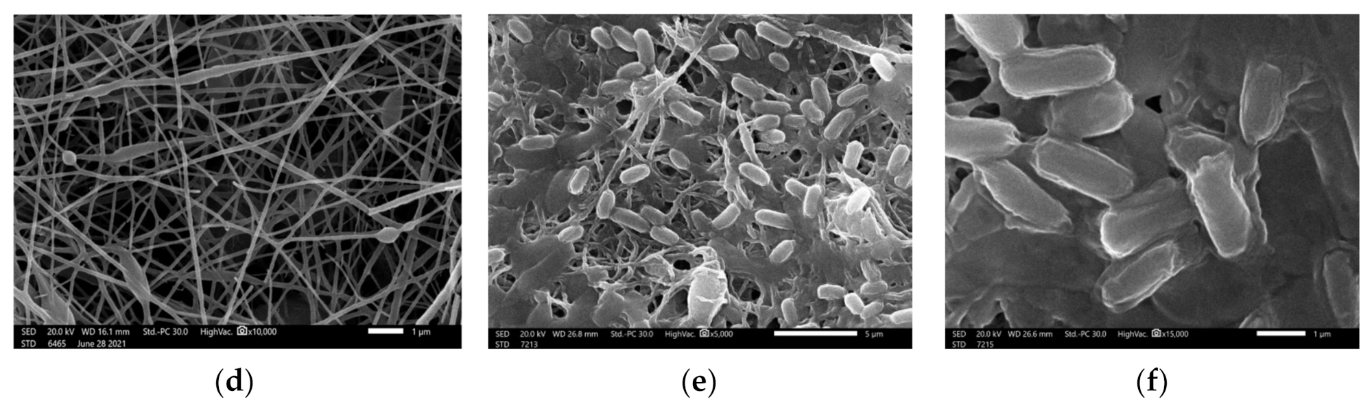
3.1. Morphological Characterization of Nanofibers

The morphology of TPU and TPU composites nanofibrous membranes revealed that the electrospun nanofibers exhibited smooth surface and high homogeneity. The diameter of the TPU nanofibers were in the range of 90–150 nm with average fiber diameter of 116 nm along their length as shown in Figure 1A. Significant morphological differences can be observed after the addition of conductive nanoparticles as shown in Table 1.
Adding ZnO nanoparticles with concentration of 2 wt% and 4 wt% affected the average fiber diameter significantly to be reduced to 102 and 89 nm, respectively, as shown in Figure 1B,C. The presence of CuO nanoparticles in electrospun TPU nanofibers with concentration of 2 wt% and 4 wt% in Figure 1D and Figure 1E shows the decrease in fiber diameter to 66 and 68 nm, respectively.
This decrease in fiber diameter at the concentration of 2 wt% and 4 wt% could be attributed to the excellent preparation conditions and the homogeneity of the polymer solution even after adding the nanoparticles. The nanoparticles concentration was not high enough to disturb the spinning process.

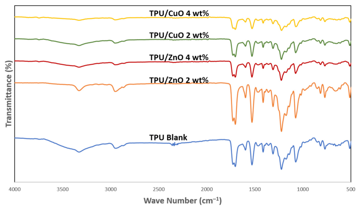
3.2. Physicochemical Characterization of Nanofibers

The manufactured nanofibers were characterized using FTIR to confirm the presence of ZnO and CuO nanoparticles into the TPU nanofibers matrix. FTIR spectra of pure TPU, TPU/ZnO and TPU/CuO nanofibers composites are shown in Figure 2. It emphasizes from Figure 2 the presence of three primary bands at 1703 cm−1 attributed to C=O stretching, 1529 cm−1 for N–H in-plane bending, and 1232 cm−1 that is refer to C–N stretching for the pure TPU nanofibers [60]. Furthermore, a broad peak at 3329 cm−1 was assigned to O–H stretching; however, an asymmetric vibration of CH2 stretching was appeared at 2945 cm−1 Furthermore, TPU/ZnO 2 wt% and 4wt% composites showed intense peak at 1727 cm−1 with more enhancement of the wavenumber of the stare aching vibration of the C=O bond, and this could be attributed to the interaction between the C=O functional group with the metal that could increase the strength of the C=O bond [61]. At the same time, new peak was observed at 3744 with the addition of CuO nanoparticles attributed to the stretching vibration frequency of the H2O molecules associated with CuO nanoparticles [62].

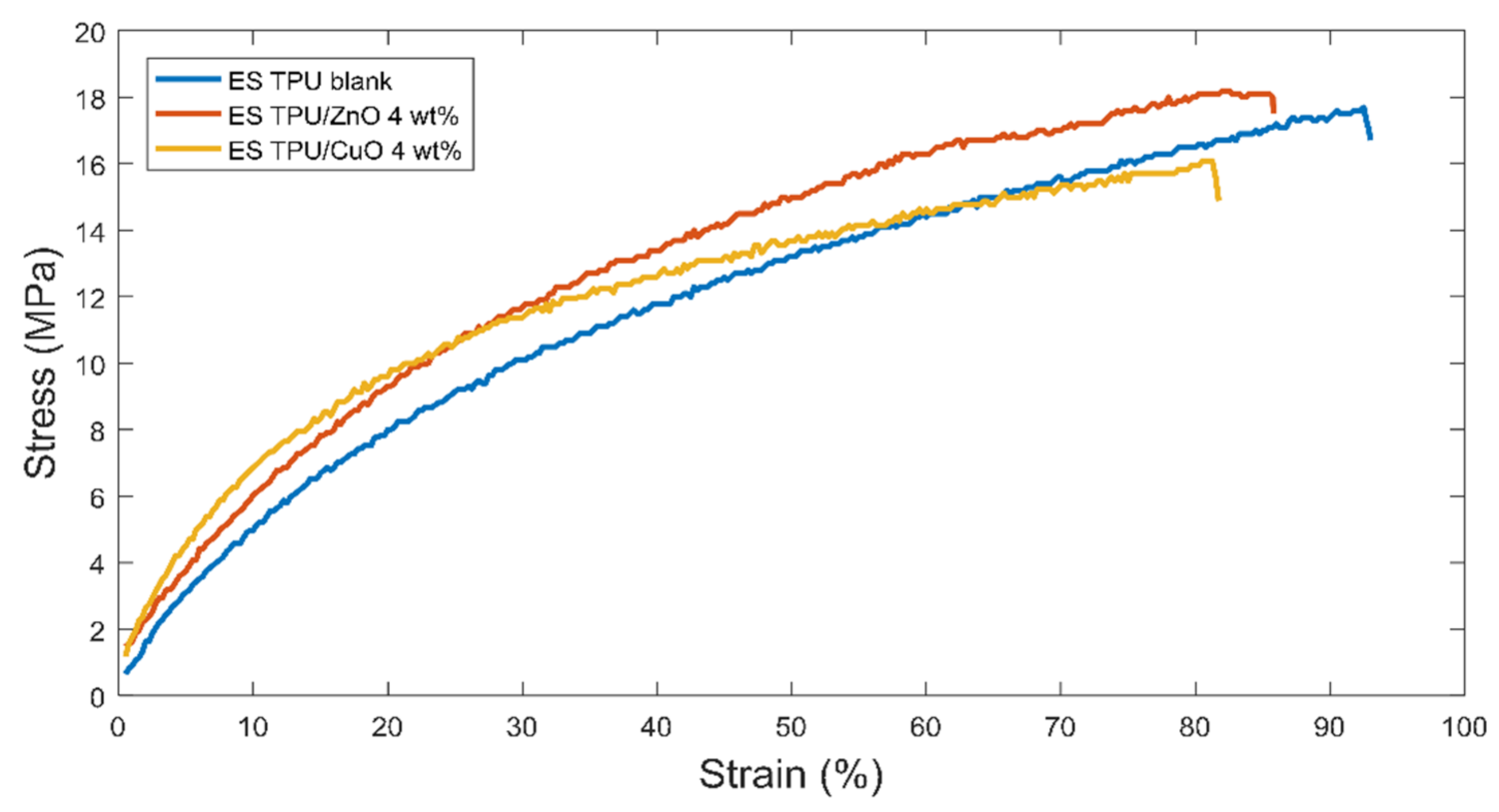
3.3. Mechanical Analysis

The mechanical properties of the electrospun TPU, TPU/ZnO, and TPU/CuO nanofiber mats were examined through the tensile testing. Figure 3 shows the stress–strain curve obtained for each sample.
We noticed that TPU nanofibers exhibited a maximum strength of 17.7 MPa with strain percentage of 92.5%, which is an indication for the excellent mechanical properties for TPU polymer that can withstand high stress and ultra-high elasticity.
In a comparative study [63] done to explore the mechanical behavior of TPU and TPU/PVDF composite nanofibers, a maximum strength of 14.98 MPa was recorded for pure TPU nanofibers, with 97.25% elongation at breakage.
In another study done on TPU microfibers fabricated through laser melt electrospinning [64] as a potential matrix for tissue engineering scaffolds, the TPU microfibers membranes recorded 134% elongation at breakage, which is attributed to the high elasticity of this polymer.
As can be seen in Table 2, the maximum strength for electrospun TPU/ZnO 4 wt% and TPU/CuO 4 wt% nanofibers reached 18.1 MPa and 16.08 MPa, while elongation at breakage reached 85.5% and 81.25%, respectively. The result showed that adding nanoparticle has affected the mechanical properties of the nanofiber membrane by decreasing the elongation at break. This could be due to acting the nanoparticle as nucleating agents, which increase the possibility of forming more crystal regions with high crystallinity, consequently decrease the elasticity and toughness properties of the nanofiber membranes.

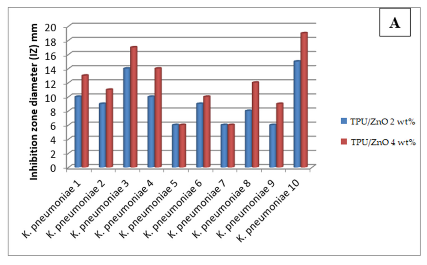
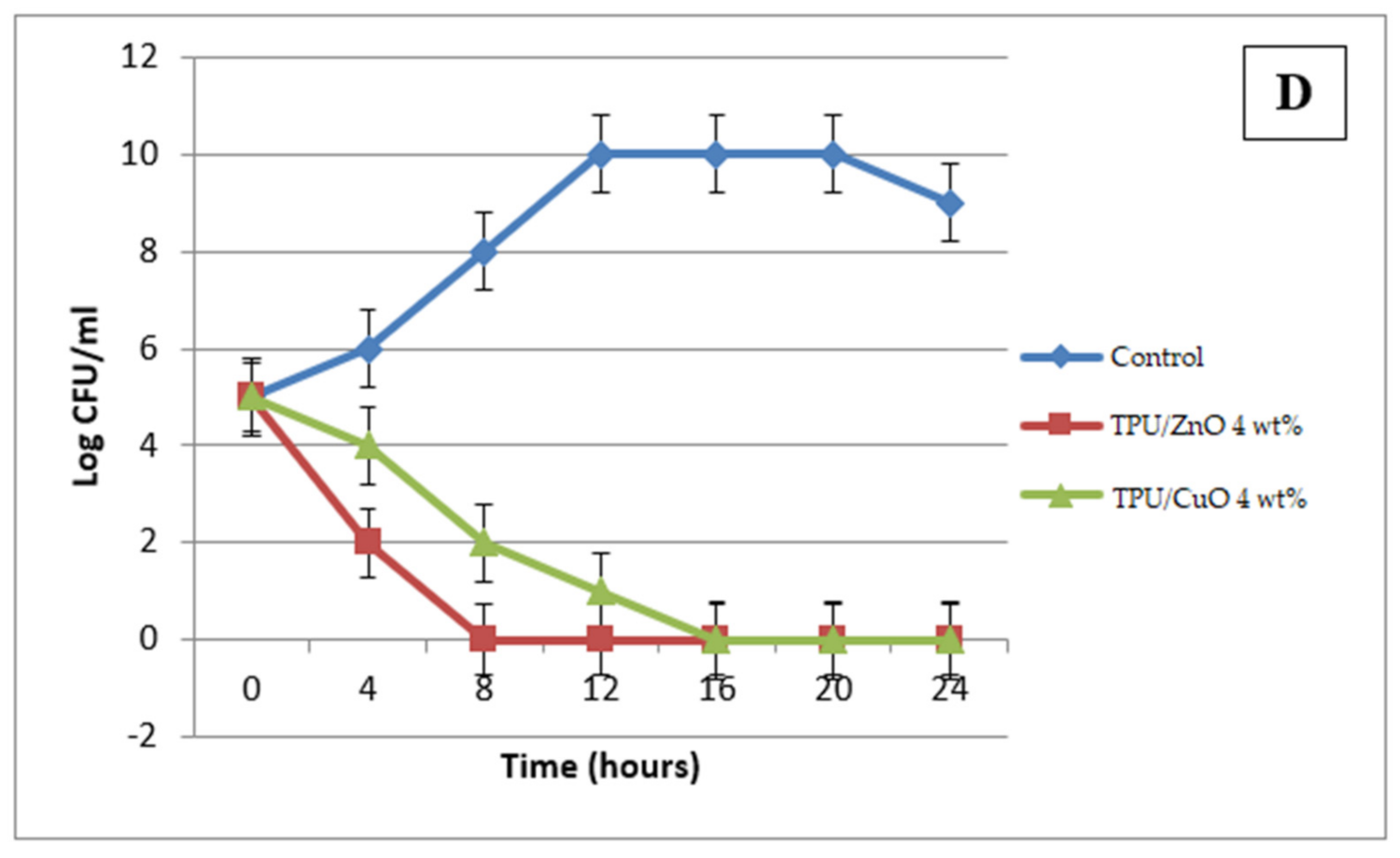
3.4. Antibacterial Activity

The antibacterial activity of the prepared nanofibers was evaluated using various techniques due to the significant fear of the colistin resistant bacterial strains. Promising activity was noticed upon using TPU/ZnO nanofibers (Table 3 and Figure 4). The observed activity increased by increasing the metal oxide concentration to reach 83% reduction rate upon using TPU/ZnO 4 wt% nanofibers against K. pneumoniae strain 10. The most susceptible strain (K. pneumoniae strain 10) was chosen for further antibacterial analyses. The bacterial lethality curve indicated that the bacterial growth was completely eradicated after 8 and 16 h incubation with TPU/ZnO and TPU/CuO, respectively.
This may be explained by the release of the metal ions into the solution. Hence, the metal oxides release from the nanofiber was assessed. Data revealed the Cu and Zn ions releasing into the solution were burst in the first 360 s till reaching stability state after 540 s and 720 s, respectively (Figure 5). This burst releasing could be explained by the porous structure of the nanofibers that assisted the water absorption and in turn releasing the metal oxide nanoparticles into the solution [58].
To determine the mechanism of action of the prepared nanofibers SEM study was used. It was found that the tested bacteria were adsorbed on the metal oxides nanofibers surface which led to bacterial cells lysis and releasing of the cell content (Figure 6).

3.5. Antiviral Activity

Few researchers started to prevent the viral infectivity through using textile materials, which are usually fibrous structures. Hence, in the present manuscript we aimed to start the journey by taking one step further using in vitro evaluation against Spike S-protein from SARS-CoV-2. Data in Figure 7 revealed that, the potent effectiveness of the proposed nanofibers in deactivation the adherence of the SARS-CoV-2 spike protein with the human cell receptor (ACE2). It was revealed that the metal oxide concentration is the most effective factor in the antiviral activity due to the observed pattern of increasing the antibacterial and antiviral activity by increasing the metal oxide concentration. TPU/ZnO showed a potent antibacterial and antiviral activity in relation to TPU/CuO.

4. Conclusions

The manufacture of two concentrations of ZnO and CuO nanoparticles with TPU using electrospinning technique has been accomplished. The SEM morphological investigation showed the significant impact of the nanoparticle type average nanofiber diameter. Mechanical investigation showed the effect of adding nanoparticle on the mechanical properties of nanofibers membranes. The elastic properties have been affected clearly by adding the nanoparticle, where the elongation at break reduced from 92.5% in case of TPU nanofiber membrane to 81.25% in case of TPU/CuO 4 wt%. The evaluation of the manufactured nanofibers as anti-multidrug-resistant bacteria and antiviral has been achieved through assessment using various colistin resistant bacterial strains and Spike S-protein from SARS-CoV-2 deactivation of the viruses, respectively. Based on the observed trend of rising antibacterial and antiviral activity by raising the metal oxide concentration, the results revealed that the metal oxide concentration is an effective factor in antiviral activity, and TPU/ZnO displayed a potent antiviral activity in comparison to TPU/CuO. The developed nanofiber membrane showed promising potential to be used in medical and personal protection applications, especially in applications that need good mechanical and hygienic performance.

Supplementary Materials

The following are available online at https://www.mdpi.com/article/10.3390/polym13223987/s1. Figure S1: TEM micrographs of ZnO NPs, Figure S2: TEM micrographs of CuO NPs, Figure S3: SEM micrographs of ZnO NPs, Figure S4: SEM micrographs of CuO NPs, Figure S5: XRD pattern of ZnO NPs, Figure S6: XRD pattern of CuO NPs, Figure S7: FTIR spectra of the ZnO NPs, Figure S8: FTIR spectra of the CuO NPs.

Author Contributions

Conceptualization, M.H., N.O., B.H.E. and A.H.H.; Data curation, N.O., B.H.E., M.T.H., S.M.A., N.O., N.S., A.H.H., D.W., Q.L. and M.H.; Formal analysis, N.O., M.T.H., S.M.A., N.S., A.H.H. and D.W.; Funding acquisition, N.O. and M.H.; Investigation, N.O. and A.H.H.; Methodology, L.A.A., L.A.A.-M., N.O., B.H.E., M.T.H., N.S., A.H.H. and M.H.; Resources, B.H.E.; Supervision, A.H.H., D.W. and Q.L.; Writing—original draft, N.O., B.H.E. and M.H.; Writing—review and editing, N.O., B.H.E., A.H.H. and M.H. All authors have read and agreed to the published version of the manuscript.

Funding

This research work was funded by the Deanship of Scientific Research at Princess Nourah Bint Abdulrahman University through the Fast-track Research Funding Program.

Institutional Review Board Statement

Not applicable.

Informed Consent Statement

Not applicable.

Data Availability Statement

Not applicable.

Acknowledgments

We would appreciate the Deanship of Scientific Research at Princess Nourah Bint Abdulrahman University for funding of this work though the Fast-track Research Funding Program.

Conflicts of Interest

The authors declare no conflict of interest.

References

  1. Livingston, E.; Desai, A.; Berkwits, M.J.J. Sourcing personal protective equipment during the COVID-19 pandemic. JAMA 2020, 323, 1912–1914. [Google Scholar] [CrossRef] [Green Version]
  2. Bauchner, H.; Fontanarosa, P.B.; Livingston, E.H. Conserving supply of personal protective equipment—A call for ideas. JAMA 2020, 323, 1911. [Google Scholar] [CrossRef] [PubMed] [Green Version]
  3. Bowden, E.; Campanile, C.; Golding, B. Worker at NYC Hospital Where Nurses Wear Trash Bags as Protection Dies from Coronavirus. N. Y. Post 2020, 3, 25. Available online: https://nypost.com/2020/03/25/worker-at-nyc-hospital-where-nurses-wear-trash-bags-as-protection-dies-from-coronavirus/ (accessed on 25 August 2021).
  4. Chris, M. Some Amazon Prime shipments won’t arrive for a month due to coronavirus. Fortune, 23 March 2020. Available online: https://fortune.com/2020/03/23/amazon-prime-delays-coronavirus/(accessed on 25 August 2021).
  5. Trexler, P.; Tiffany, T. Strategic National Stockpile fails to quench Ohio’s need for medical supplies. In MSN, 2020th ed.; 26 March 2020; Available online: ttps://www.wkyc.com/article/news/health/coronavirus/strategic-nation-stockpile-fails-to-quench-ohio-need-for-medical-supplies/95-9c05ef80-5152-4951-b8f0-210786b23d33 (accessed on 25 August 2021).
  6. Harris, A.D.; Pineles, L.; Belton, B.; Johnson, J.K.; Shardell, M.; Loeb, M.; Newhouse, R.; Dembry, L.; Braun, B.; Perencevich, E.N.; et al. Universal Glove and Gown Use and Acquisition of Antibi-otic-Resistant Bacteria in the ICU: A Randomized Trial. JAMA 2013, 310, 1571–1580. [Google Scholar] [CrossRef] [PubMed]
  7. Martin, R.M.; Bachman, M.A.; Microbiology, I. Colonization, infection, and the accessory genome of Klebsiella pneumoniae. Front. Cell Infect. Microbiol. 2018, 8, 4. [Google Scholar] [CrossRef] [PubMed] [Green Version]
  8. Gupta, K.; Gupta, A.; Shrivastava, D. The last resort antibiotics: Carbapenems. Int. J. Adv. Res. 2017, 5, 1410–1413. [Google Scholar] [CrossRef] [Green Version]
  9. Tsuji, B.T.; Pogue, J.M.; Zavascki, A.P.; Paul, M.; Daikos, G.L.; Forrest, A.; Giacobbe, D.R.; Viscoli, C.; Giamarellou, H.; Karaiskos, I.; et al. International consensus guidelines for the optimal use of the polymyxins: Endorsed by the American college of clinical pharmacy (ACCP), European society of clinical microbiology and infectious diseases (ESCMID), infectious diseases society of America (IDSA), international society for anti-infective Pharmacology (ISAP), society of critical care medicine (SCCM), and society of infectious diseases pharmacists (SIDP). Pharmacother. J. Hum. Pharmacol. Drug Ther. 2019, 39, 10–39. [Google Scholar]
  10. Brown, P.; Dawson, M.J. Development of new polymyxin derivatives for multi-drug resistant Gram-negative infections. J. Antibiot. 2017, 70, 386–394. [Google Scholar] [CrossRef] [PubMed] [Green Version]
  11. Spapen, H.; Jacobs, R.; Van Gorp, V.; Troubleyn, J.; Honoré, P.M. Renal and neurological side effects of colistin in critically ill patients. Ann. Intensiv. Care 2011, 1, 14. [Google Scholar] [CrossRef] [PubMed] [Green Version]
  12. MacGowan, A.; Macnaughton, E. Antimicrobial therapy: Principles of use. Medcine 2017, 45, 614–621. [Google Scholar] [CrossRef]
  13. Fratini, S.; Nikolka, M.; Salleo, A.; Schweicher, G.; Sirringhaus, H. Charge transport in high-mobility conjugated polymers and molecular semiconductors. Nat. Mater. 2020, 19, 491–502. [Google Scholar] [CrossRef] [Green Version]
  14. Ogunseitan, O.A. The Materials Genome and COVID-19 Pandemic. Jom 2020, 72, 1–3. [Google Scholar]
  15. Vozzola, E.; Overcash, M.; Griffing, E. Environmental considerations in the selection of isolation gowns: A life cycle assessment of reusable and disposable alternatives. Am. J. Infect. Control. 2018, 46, 881–886. [Google Scholar] [CrossRef] [PubMed] [Green Version]
  16. Wang, D.; Hu, B.; Hu, C.; Zhu, F.; Liu, X.; Zhang, J.; Wang, B.; Xiang, H.; Cheng, Z.; Xiong, Y. Clinical characteristics of 138 hospitalized patients with 2019 novel coronavirus–infected pneumonia in Wuhan, China. JAMA 2020, 323, 1061–1069. [Google Scholar] [CrossRef] [PubMed]
  17. Rubin, M.A.; Samore, M.H.; Harris, A.D. The importance of contact precautions for endemic methicillin-resistant Staphylococcus aureus and vancomycin-resistant enterococci. JAMA 2018, 319, 863–864. [Google Scholar] [CrossRef]
  18. Gutarowska, B.; Skóra, J.; Nowak, E.; Łysiak, I.; Wdówka, M. Antimicrobial Activity and Filtration Effectiveness of Nonwovens with Sanitized for Respiratory Protective Equipment. 2014. Available online: http://www.fibtex.lodz.pl/pliki/Fibtex_(lkzi8pi6xun6202m).pdf (accessed on 27 August 2021).
  19. Longano, D.; Ditaranto, N.; Sabbatini, L.; Torsi, L.; Cioffi, N. Synthesis and antimicrobial activity of copper nanomaterials. In Nano-Antimicrobials; Springer: Berlin/Heidelberg, Germany, 2012; pp. 85–117. [Google Scholar]
  20. Karim, N.; Afroj, S.; Lloyd, K.; Oaten, L.C.; Andreeva, D.V.; Carr, C.; Farmery, A.D.; Kim, I.-D.; Novoselov, K.S. Sustainable personal protective clothing for healthcare applications: A review. ACS Nano 2020, 14, 12313–12340. [Google Scholar] [CrossRef] [PubMed]
  21. Alshabanah, L.A.; Hagar, M.; Al-Mutabagani, L.A.; Abozaid, G.M.; Abdallah, S.M.; Ahmed, H.; Hassanin, A.H.; Shehata, N. Biodegradable Nanofibrous Membranes for Medical and Personal Protection Applications: Manufacturing, Anti-COVID-19 and Anti-Multidrug Resistant Bacteria Evaluation. Materials 2021, 14, 3862. [Google Scholar] [CrossRef] [PubMed]
  22. Alshabanah, L.A.; Hagar, M.; Al-Mutabagani, L.A.; Abozaid, G.M.; Abdallah, S.M.; Shehata, N.; Ahmed, H.; Hassanin, A.H. Hybrid Nanofibrous Membranes as a Promising Functional Layer for Personal Protection Equipment: Manufacturing and Antiviral/Antibacterial Assessments. Polymers 2021, 13, 1776. [Google Scholar] [CrossRef] [PubMed]
  23. Sportelli, M.C.; Izzi, M.; Kukushkina, E.A.; Hossain, S.I.; Picca, R.A.; Ditaranto, N.; Cioffi, N. Can nanotechnology and materials science help the fight against SARS-CoV-2? Nanomaterials 2020, 10, 802. [Google Scholar] [CrossRef] [PubMed] [Green Version]
  24. Patel, M. Nanoparticle-based Antimicrobial Paper as Spread-breaker for Coronavirus. Pap. Technol. Int. 2020, 62, 20–25. [Google Scholar]
  25. Magill, S.S.; Edwards, J.R.; Bamberg, W.; Beldavs, Z.G.; Dumyati, G.; Kainer, M.A.; Lynfield, R.; Maloney, M.; McAllister-Hollod, L.; Nadle, J. Multistate point-prevalence survey of health care–associated infections. N. Engl. J. Med. 2014, 370, 1198–1208. [Google Scholar] [CrossRef] [Green Version]
  26. Żakowska, Z. Microbial biodegradation and biodeterioration of technical materials. In Proceedings of the IV International Scientific Conference, Conference Materials, Łódź, Poland, 2006; pp. 75–84. [Google Scholar]
  27. Abo-Shosha, M.; Hashem, A.; El-Hosamy, M.; El-Nagar, A. Easy care finishing of knitted cotton fabric in presence of a reactive-type antibacterial agent. J. Ind. Text. 2008, 38, 103–126. [Google Scholar] [CrossRef]
  28. Chen, R.; Morsi, Y.; Patel, S.; Ke, Q.-F.; Mo, X.-M. A novel approach via combination of electrospinning and FDM for tri-leaflet heart valve scaffold fabrication. Front. Mater. Sci. China 2009, 3, 359–366. [Google Scholar] [CrossRef]
  29. Fallahiarezoudar, E.; Ahmadipourroudposht, M.; Yusof, N.M.; Idris, A.; Ngadiman, N. 3D biofabrication of thermoplastic polyurethane (TPU)/poly-L-lactic acid (PLLA) electrospun nanofibers containing maghemite (γ-Fe2O3) for tissue engineering aortic heart valve. Polymer 2017, 9, 584. [Google Scholar] [CrossRef] [PubMed] [Green Version]
  30. Choi, J.; Jang, J.U.; Yin, W.B.; Lee, B.; Lee, K. Synthesis of highly functionalized thermoplastic polyurethanes and their potential applications. Polymer 2017, 116, 287–294. [Google Scholar] [CrossRef]
  31. Hattig, J.; Drube, W. Wide area of applications: Upward trend for thermoplastic polyurethanes. Kunstst.-Plast Eur. 2003, 93, BA28. [Google Scholar]
  32. Kharbas, H.A.; Ellingham, T.; Manitiu, M.; Scholz, G.; Turng, L.-S. Effect of a cross-linking agent on the foamability of microcellular injection molded thermoplastic polyurethane. J. Cell. Plast. 2017, 53, 407–423. [Google Scholar] [CrossRef]
  33. Bazmara, B.; Tahersima, M.; Behravan, A.J.C.; Materials, B. Influence of thermoplastic polyurethane and synthesized polyurethane additive in performance of asphalt pavements. Constr. Build. Mater. 2018, 166, 1–11. [Google Scholar] [CrossRef]
  34. Dong, M.; Li, Q.; Liu, H.; Liu, C.; Wujcik, E.K.; Shao, Q.; Ding, T.; Mai, X.; Shen, C.; Guo, Z. Thermoplastic polyurethane-carbon black nanocomposite coating: Fabrication and solid particle erosion resistance. Polymer 2018, 158, 381–390. [Google Scholar] [CrossRef]
  35. Li, Y.; Zhou, B.; Zheng, G.; Liu, X.; Li, T.; Yan, C.; Cheng, C.; Dai, K.; Liu, C.; Shen, C. Continuously prepared highly conductive and stretchable SWNT/MWNT synergistically composited electrospun thermoplastic polyurethane yarns for wearable sensing. J. Mater. Chem. C 2018, 6, 2258–2269. [Google Scholar] [CrossRef]
  36. Ke, K.; Bonab, V.S.; Yuan, D.; Manas-Zloczower, I. Piezoresistive thermoplastic polyurethane nanocomposites with carbon nanostructures. Carbon 2018, 139, 52–58. [Google Scholar] [CrossRef]
  37. Chen, R.; Zhang, X.; Wang, P.; Xie, K.; Jian, J.; Zhang, Y.; Zhang, J.; Yuan, Y.; Na, P.; Yi, M. Transparent thermoplastic polyurethane air filters for efficient electrostatic capture of particulate matter pollutants. Nanotechnology 2018, 30, 015703. [Google Scholar] [CrossRef] [PubMed]
  38. Ren, M.; Zhou, Y.; Wang, Y.; Zheng, G.; Dai, K.; Liu, C.; Shen, C. Highly stretchable and durable strain sensor based on carbon nanotubes decorated thermoplastic polyurethane fibrous network with aligned wave-like structure. Chem. Eng. J. 2019, 360, 762–777. [Google Scholar] [CrossRef]
  39. Rubin Pedrazzo, A.; Cecone, C.; Morandi, S.; Manzoli, M.; Bracco, P.; Zanetti, M. Nanosized SnO2 Prepared by Electrospinning: Influence of the Polymer on Both Morphology and Microstructure. Polymers 2021, 13, 977. [Google Scholar] [CrossRef] [PubMed]
  40. Thamer, B.M.; Aldalbahi, A.; Moydeen, A.M.; Rahaman, M.; El-Newehy, M.H. Modified Electrospun Polymeric Nanofibers and Their Nanocomposites as Nanoadsorbents for Toxic Dye Removal from Contaminated Waters: A Review. Polymers 2021, 13, 20. [Google Scholar] [CrossRef] [PubMed]
  41. Wang, M.; Li, D.; Li, J.; Li, S.; Chen, Z.; Yu, D.-G.; Liu, Z.; Guo, J.Z. Electrospun Janus zein–PVP nanofibers provide a two-stage controlled release of poorly water-soluble drugs. Mater. Des. 2020, 196, 109075. [Google Scholar] [CrossRef]
  42. Zhao, K.; Kang, S.-X.; Yang, Y.-Y.; Yu, D.-G. Electrospun Functional Nanofiber Membrane for Antibiotic Removal in Water. Polymers 2021, 13, 226. [Google Scholar] [CrossRef]
  43. Deng, C.; Seidi, F.; Yong, Q.; Jin, X.; Li, C.; Zhang, X.; Han, J.; Liu, Y.; Huang, Y.; Wang, Y.; et al. Antiviral/antibacterial biodegradable cellulose nonwovens as environmentally friendly and bioprotective materials with potential to minimize microplastic pollution. J. Hazard. Mater. 2022, 424, 127391. [Google Scholar] [CrossRef]
  44. Abdoluosefi, H.E.; Honarasa, G.; Abdoluosefi, H.E. Fabrication of polyurethane and thermoplastic polyurethane nanofiber by controlling the electrospinning parameters. Mater. Res. Express 2017, 4, 105308. [Google Scholar] [CrossRef]
  45. Pedicini, A.; Farris, R.J. Mechanical behavior of electrospun polyurethane. Polymer 2003, 44, 6857–6862. [Google Scholar] [CrossRef]
  46. Lee, K.; Lee, B.; Kim, C.; Kim, H.; Kim, K.; Nah, C. Stress-strain behavior of the electrospun thermoplastic polyurethane elastomer fiber mats. Macromol. Res. 2005, 13, 441–445. [Google Scholar] [CrossRef]
  47. Chen, Y.; Zhang, Y.; Liu, J.; Zhang, H.; Wang, K. Preparation and antibacterial property of polyethersulfone ultrafiltration hybrid membrane containing halloysite nanotubes loaded with copper ions. Chem. Eng. J. 2012, 210, 298–308. [Google Scholar] [CrossRef]
  48. Stafford, S.L.; Bokil, N.J.; Achard, M.E.; Kapetanovic, R.; Schembri, M.A.; McEwan, A.G.; Sweet, M.J. Metal ions in macrophage antimicrobial pathways: Emerging roles for zinc and copper. Biosci. Rep. 2013, 33, e00049. [Google Scholar] [CrossRef] [PubMed]
  49. Radovanović, Ž.; Jokić, B.; Veljović, D.; Dimitrijević, S.; Kojić, V.; Petrović, R.; Janaćković, D. Antimicrobial activity and biocompatibility of Ag+-and Cu2+-doped biphasic hydroxyapatite/α-tricalcium phosphate obtained from hydrothermally synthesized Ag+-and Cu2+-doped hydroxyapatite. Appl. Surf. Sci. 2014, 307, 513–519. [Google Scholar] [CrossRef]
  50. Malwal, D.; Gopinath, P. Efficient adsorption and antibacterial properties of electrospun CuO-ZnO composite nanofibers for water remediation. J. Hazard. Mater. 2017, 321, 611–621. [Google Scholar] [CrossRef] [PubMed]
  51. Sirelkhatim, A.; Mahmud, S.; Seeni, A.; Kaus, N.H.M.; Ann, L.C.; Bakhori, S.K.M.; Hasan, H.; Mohamad, D. Review on zinc oxide nanoparticles: Antibacterial activity and toxicity mechanism. Nano-Micro Lett. 2015, 7, 219–242. [Google Scholar] [CrossRef] [PubMed] [Green Version]
  52. Colon, G.; Ward, B.C.; Webster, T.J. Increased osteoblast and decreased Staphylococcus epidermidis functions on nanophase ZnO and TiO2. J. Biomed. Mater. Res. Part A 2006, 78, 595–604. [Google Scholar] [CrossRef] [PubMed]
  53. Bužarovska, A.; Dinescu, S.; Lazar, A.D.; Serban, M.; Pircalabioru, G.G.; Costache, M.; Gualandi, C.; Avérous, L. Nanocomposite foams based on flexible biobased thermoplastic polyurethane and ZnO nanoparticles as potential wound dressing materials. Mater. Sci. Eng. C 2019, 104, 109893. [Google Scholar] [CrossRef] [PubMed]
  54. Mousa, M.K.H. Wastewater Disinfection by Synthesized Copper Oxide Nanoparticles Stabilized with Surfactant. 2013. Available online: https://repository.najah.edu/bitstream/handle/20.500.11888/8493/muath%20mousa.pdf?sequence=1&isAllowed=y (accessed on 28 August 2021).
  55. Cheng, Z.; Zhang, F.; Liu, W.; Cui, L.; Kang, L. A novel preparation for a PVA/L-histidine/AgNPs membrane and its antibacterial property. RSC Adv. 2015, 5, 54182–54187. [Google Scholar] [CrossRef]
  56. Santiago-Morales, J.; Amariei, G.; Letón, P.; Rosal, R.J.C.; Biointerfaces, S.B. Antimicrobial activity of poly (vinyl alcohol)-poly (acrylic acid) electrospun nanofibers. Colloids Surf. B Biointerfaces 2016, 146, 144–151. [Google Scholar] [CrossRef]
  57. Elnaggar, Y.S.; Elwakil, B.H.; Elshewemi, S.S.; El-Naggar, M.Y.; Bekhit, A.A.; Olama, Z.A. Novel Siwa propolis and colistin-integrated chitosan nanoparticles: Elaboration; in vitro and in vivo appraisal. Nanomedicine 2020, 15, 1269–1284. [Google Scholar] [CrossRef] [PubMed]
  58. Mokhena, T.C.; Luyt, A.S. Electrospun alginate nanofibres impregnated with silver nanoparticles: Preparation, morphology and antibacterial properties. Carbohydr. Polym. 2017, 165, 304–312. [Google Scholar] [CrossRef] [PubMed]
  59. Chua, M.H.; Cheng, W.; Goh, S.S.; Kong, J.; Li, B.; Lim, J.Y.; Mao, L.; Wang, S.; Xue, K.; Yang, L.J. Face masks in the new COVID-19 normal: Materials, testing, and perspectives. Research 2020, 2020, 7286735. [Google Scholar] [CrossRef] [PubMed]
  60. Liang, W.; Xu, Y.; Li, X.; Wang, X.-X.; Zhang, H.-D.; Yu, M.; Ramakrishna, S.; Long, Y.-Z. Transparent polyurethane nanofiber air filter for high-efficiency PM2.5 capture. Nanoscale Res. Lett. 2019, 14, 361. [Google Scholar] [CrossRef] [PubMed] [Green Version]
  61. Velayutham, T.; Abd Majid, W.H.; Gan, W.; Khorsand Zak, A.; Gan, S.N. Theoretical and experimental approach on dielectric properties of ZnO nanoparticles and polyurethane/ZnO nanocomposites. J. Appl. Phys. 2012, 112, 054106. [Google Scholar] [CrossRef] [Green Version]
  62. Nirmala, R.; Jeon, K.S.; Lim, B.H.; Navamathavan, R.; Kim, H.Y. Preparation and characterization of copper oxide particles incorporated polyurethane composite nanofibers by electrospinning. Ceram. Int. 2013, 39, 9651–9658. [Google Scholar] [CrossRef]
  63. Elnabawy, E.; Hassanain, A.H.; Shehata, N.; Popelka, A.; Nair, R.; Yousef, S.; Kandas, I. Piezoelastic PVDF/TPU nanofibrous composite membrane: Fabrication and characterization. Polymers 2019, 11, 1634. [Google Scholar] [CrossRef] [PubMed] [Green Version]
  64. Li, X.; Wang, Z.; Wang, J.; Liu, J.; Li, C. Preparation and properties of TPU micro/nanofibers by a laser melt-electrospinning system. Polym. Eng. Sci. 2014, 54, 1412–1417. [Google Scholar] [CrossRef]
Figure 1. Scanning electron microscopy (SEM) images of electrospun of TPU, TPU/ZnO, TPU/CuO and nanofiber diameter distribution histograms. (A) TPU; (B) TPU/ZnO 2 wt%; (C) TPU/ZnO 4 wt%; (D) TPU/CuO 2 wt%; (E) TPU/CuO 4 wt%.
Polymers 13 03987 g001aPolymers 13 03987 g001b
Figure 2. FTIR spectra of electrospun TPU, TPU/ZnO 2 wt%; TPU/ZnO 4 wt%, TPU/CuO 2 wt%; TPU/CuO 4 wt%.
Polymers 13 03987 g002
Figure 3. Stress–strain curve of electrospun nanofibers membrane.
Polymers 13 03987 g003
Figure 4. Antibacterial activity evaluation of the prepared nanofibers against colistin resistant strains where (A): TPU/ZnO activity, (B): TPU/CuO activity, (C): the reduction rate of TPU/ZnO and TPU/CuO against colistin resistant strains, and (D): the bacterial lethality curve of TPU/ZnO and TPU/CuO against K. pneumoniae 10.
Polymers 13 03987 g004aPolymers 13 03987 g004bPolymers 13 03987 g004c
Figure 5. Metal oxides release from the nanofiber in solution.
Polymers 13 03987 g005
Figure 6. Scanning electron microscopic (SEM) study to assess the antibacterial activity of TPU/ZnO and TPU/CuO nanofibers. (a): Control uninoculated TPU/ZnO (4 wt%); (b): K. pneumoniae strain 10 treated with TPU/ZnO (4 wt%) at 5 µ magnification; (c): K. pneumoniae strain 10 treated with TPU/ZnO (4 wt%) at 1 µ magnification. While (d): Control uninoculated TPU/CuO (4 wt%); (e) K. pneumoniae strain 10 treated with TPU/CuO (4 wt%) at 5 µ magnification and (f) K. pneumoniae strain 10 treated with TPU/CuO (4 wt%)/TPU 4 wt% at 1 µ magnification.
Polymers 13 03987 g006aPolymers 13 03987 g006b
Figure 7. Remaining activity (percentage) of the SARS-CoV-2 spike protein to adhere with ACE2.
Polymers 13 03987 g007
Table 1. Average fiber diameter for electrospun nanofibers.
Nanofiber MembraneNanoparticles Concentration (%)Average Fiber Diameter (nm)
TPU0 wt%116 ± 39
TPU/ZnO2 wt%102 ± 36
4 wt%89 ± 30
TPU/CuO2 wt%66 ± 21
4 wt%68 ± 20
Table 2. Mechanical properties of electrospun nanofibers.
SampleMax. Strength (MPa)Elongation at Break (%)
TPU Blank17.7 ± 1.292.5 ± 20.4
TPU/ZnO 4 wt%18.1 ± 0.985.5 ± 10.2
TPU/CuO 4 wt%16.08 ± 2.381.25 ± 18.9
Table 3. The antibacterial activity of TPU/ZnO and TPU/CuO nanofibers against colistin resistant K. pneumoniae strains.
Tested StrainsTPU/ZnO 2 wt%TPU/ZnO 4 wt%TPU/CuO 2 wt%TPU/CuO 4 wt%
IZ (mm)Reduction Rate (%)IZ (mm)Reduction Rate (%)IZ (mm)Reduction Rate (%)IZ (mm)Reduction Rate (%)
K. pneumoniae 1105013729241216
K. pneumoniae 29441170621632
K. pneumoniae 31470177611291641
K. pneumoniae 4105414756201030
K. pneumoniae 5632653620630
K. pneumoniae 69381054620933
K. pneumoniae 7637650620631
K. pneumoniae 883912736201036
K. pneumoniae 9631957620632
K. pneumoniae 101574198312291358
Publisher’s Note: MDPI stays neutral with regard to jurisdictional claims in published maps and institutional affiliations.

