
Biomechanical Aspects of Various Attachments for Implant Overdentures: A Review

 ,
,  ,
,
Abstract
:
1. Introduction
2. Polymeric Materials Used for Overdenture
3. Effects of Attachment Types on Implant Overdenture
4. Retentive Force and Stress Distribution of Various Attachments
5. Effects of Attachments in Peri-Implant Health
6. Effects of Attachments on Patient’s Satisfaction and Quality of Life
7. Recent Advances and Future Perspectives
8. Conclusions
Author Contributions
Funding
Institutional Review Board Statement
Informed Consent Statement
Data Availability Statement
Acknowledgments
Conflicts of Interest
References
- Vi, S.; Pham, D.; Du, Y.Y.M.; Arora, H.; Tadakamadla, S.K. Mini-implant-retained overdentures for the rehabilitation of completely edentulous maxillae: A systematic review and meta-analysis. Int. J. Environ. Res. Public Health 2021, 18, 4377. [Google Scholar] [CrossRef] [PubMed]
- Aldhohrah, T.; Mashrah, M.A.; Wang, Y. Effect of 2-implant mandibular overdenture with different attachments and loading protocols on peri-implant health and prosthetic complications: A systematic review and network meta-analysis. J. Prosthet. Dent. 2021, in press. [Google Scholar] [CrossRef] [PubMed]
- Ceraulo, S.; Leonida, A.; Lauritano, D.; Baldoni, A.; Longoni, S.; Baldoni, M.; Caccianiga, G. Proposal for a clinical approach to geriatric patients with anchor need on implant for removable denture: New technique. Prosthesis 2020, 2, 185–195. [Google Scholar] [CrossRef]
- Amornvit, P.; Rokaya, D.; Bajracharya, S.; Keawcharoen, K.; Supavanich, W. Management of obstructive sleep apnea with implant retained mandibular advancement device. World J. Dent. 2014, 5, 184–189. [Google Scholar] [CrossRef]
- Daou, E.E. Stud attachments for the mandibular implant-retained overdentures: Prosthetic complications. A literature review. Saudi Dent. J. 2013, 25, 53–60. [Google Scholar] [CrossRef] [Green Version]
- Li, P.; Hasselbeck, D.; Unkovskiy, A.; Sharghi, F.; Spintzyk, S. Retentive characteristics of a polyetheretherketone post-core restoration with polyvinylsiloxane attachments. Polymers 2020, 12, 2005. [Google Scholar] [CrossRef]
- Alzahrani, A.H. Mechanical Properties of Selected Cad/Cam and Conventional Interim Fixed Restorative Materials University at Buffalo. Master’s Thesis, State University of New York, New York, NY, USA, 1 September 2018. [Google Scholar]
- Alsabeeha, N.H.; Swain, M.V.; Payne, A.G. Clinical performance and material properties of single-implant overdenture attachment systems. Int. J. Prosthodont. 2011, 24, 247–254. [Google Scholar]
- Rokaya, D.; Srimaneepong, V.; Sapkota, J.; Qin, J.; Siraleartmukul, K.; Siriwongrungson, V. Polymeric materials and films in dentistry: An overview. J. Adv. Res. 2018, 14, 25–34. [Google Scholar] [CrossRef]
- Winkler, S.; Piermatti, J.; Rothman, A.; Siamos, G. An overview of the o-ring implant overdenture attachment: Clinical reports. Oral Implant. 2002, 28, 82–86. [Google Scholar] [CrossRef] [Green Version]
- Sadowsky, S.J. Mandibular implant-retained overdentures: A literature review. J. Prosthet. Dent. 2001, 86, 468–473. [Google Scholar] [CrossRef]
- Cakarer, S.; Can, T.; Yaltirik, M.; Keskin, C. Complications associated with the ball, bar and locator attachments for implant-supported overdentures. Med. Oral Patol. Oral Cir. Bucal 2011, 16, e953–e959. [Google Scholar] [CrossRef] [PubMed] [Green Version]
- Payne, A.G.; Solomons, Y.F.; Tawse-Smith, A.; Lownie, J.F. Inter-abutment and peri-abutment mucosal enlargement with mandibular implant overdentures. Clin. Oral Implant. Res. 2001, 12, 179–187. [Google Scholar] [CrossRef] [PubMed]
- Alsabeeha, N.H.; Payne, A.G.; De Silva, R.K.; Thomson, W.M. Mandibular single-implant overdentures: Preliminary results of a randomised-control trial on early loading with different implant diameters and attachment systems. Clin. Oral Implant. Res. 2011, 22, 330–337. [Google Scholar] [CrossRef] [PubMed]
- Kleis, W.K.; Kämmerer, P.W.; Hartmann, S.; Al-Nawas, B.; Wagner, W. A comparison of three different attachment systems for mandibular two-implant overdentures: One-year report. Clin. Implant Dent. Relat. Res. 2010, 12, 209–218. [Google Scholar] [CrossRef]
- Vasant, R.; Vasant, M.K. Retention systems for implant-retained overdentures. Dent. Update 2013, 40, 28–31. [Google Scholar] [CrossRef]
- ELsyad, M.A.; Fathe Mahanna, F.; Samir Khirallah, A.; Ali Habib, A. Clinical denture base deformation with different attachments used to stabilize implant overdentures: A crossover study. Clin. Oral Implant. Res. 2020, 31, 162–172. [Google Scholar] [CrossRef]
- Chung, K.H.; Chung, C.Y.; Cagna, D.R.; Cronin, R.J., Jr. Retention characteristics of attachment systems for implant overdentures. J. Prosthodont.: Off. J. Am. Coll. Prosthodont. 2004, 13, 221–226. [Google Scholar] [CrossRef]
- Naert, I.; Gizani, S.; Vuylsteke, M.; Van Steenberghe, D. A 5-year prospective randomized clinical trial on the influence of splinted and unsplinted oral implants retaining a mandibular overdenture: Prosthetic aspects and patient satisfaction. J. Oral Rehabil. 1999, 26, 195–202. [Google Scholar] [CrossRef]
- Boeckler, A.F.; Ehring, C.; Morton, D.; Geis-Gerstorfer, J.; Setz, J.M. Corrosion of dental magnet attachments for removable prostheses on teeth and implants. J. Prosthodont.: Off. J. Am. Coll. Prosthodont. 2009, 18, 301–308. [Google Scholar] [CrossRef]
- Laverty, D.P.; Green, D.; Marrison, D.; Addy, L.; Thomas, M.B. Implant retention systems for implant-retained overdentures. Br. Dent. J. 2017, 222, 347–359. [Google Scholar] [CrossRef]
- Tallarico, M.; Cervino, G.; Scrascia, R.; Uccioli, U.; Lumbau, A.; Meloni, S.M. Minimally invasive treatment of edentulous maxillae with overdenture fully supported by a cad/cam titanium bar with a low-profile attachment screwed on four or six implants: A case series. Prosthesis 2020, 2, 53–64. [Google Scholar] [CrossRef]
- Karabuda, C.; Tosun, T.; Ermis, E.; Ozdemir, T. Comparison of 2 retentive systems for implant-supported overdentures: Soft tissue management and evaluation of patient satisfaction. J. Periodontol. 2002, 73, 1067–1070. [Google Scholar] [CrossRef] [PubMed]
- Chaware, S.H.; Thakkar, S.T. A systematic review and meta-analysis of the attachments used in implant-supported overdentures. J. Indian Prosthodont. Soc. 2020, 20, 255–268. [Google Scholar] [CrossRef] [PubMed]
- Langer, A. Telescope retainers for removable partial dentures. J. Prosthet. Dent. 1981, 45, 37–43. [Google Scholar] [CrossRef]
- Langer, A. Telescope retainers and their clinical application. J. Prosthet. Dent. 1980, 44, 516–522. [Google Scholar] [CrossRef]
- Gonda, T.; Maeda, Y. Why are magnetic attachments popular in japan and other asian countries? Jpn. Dent. Sci. Rev. 2011, 47, 124–130. [Google Scholar] [CrossRef] [Green Version]
- Mericske-Stern, R.; Oetterli, M.; Kiener, P.; Mericske, E. A follow-up study of maxillary implants supporting an overdenture: Clinical and radiographic results. Int. J. Oral Maxillofac. Implant. 2002, 17, 678–686. [Google Scholar]
- Kappel, S.; Giannakopoulos, N.N.; Eberhard, L.; Rammelsberg, P.; Eiffler, C. Immediate loading of dental implants in edentulous mandibles by use of locator ® attachments or dolder ® bars: Two-year results from a prospective randomized clinical study. Clin. Implant. Dent. Relat. Res. 2016, 18, 752–761. [Google Scholar] [CrossRef]
- Boven, G.C.; Meijer, H.J.A.; Vissink, A.; Raghoebar, G.M. Maxillary implant overdentures retained by use of bars or locator attachments: 1-year findings from a randomized controlled trial. J. Prosthodont. Res. 2020, 64, 26–33. [Google Scholar] [CrossRef]
- Petropoulos, V.C.; Mante, F.K. Comparison of retention and strain energies of stud attachments for implant overdentures. J. Prosthodont.: Off. J. Am. Coll. Prosthodont. 2011, 20, 286–293. [Google Scholar] [CrossRef]
- Daou, E.E. Biomaterial aspects: A key factor in the longevity of implant overdenture attachment systems. J. Int. Soc. Prev. Community Dent. 2015, 5, 255–262. [Google Scholar] [CrossRef] [PubMed] [Green Version]
- de Albuquerque, R.F., Jr.; Fromentin, O.; Lassauzay, C.; Conceição Pereira Saraiva, M.D. Patient satisfaction versus retention of implant overdentures with two attachment systems: A randomized trial. Clin. Implant. Dent. Relat. Res. 2019, 21, 21–31. [Google Scholar] [CrossRef] [PubMed] [Green Version]
- Amornvit, P.; Rokaya, D.; Keawcharoen, K.; Thongpulsawasdi, N. Stress distribution in implant retained finger prosthesis: A finite element study. J. Clin. Diagn. Res. 2013, 7, 2851–2854. [Google Scholar] [CrossRef]
- Quaresma, S.E.; Cury, P.R.; Sendyk, W.R.; Sendyk, C. A finite element analysis of two different dental implants: Stress distribution in the prosthesis, abutment, implant, and supporting bone. J. Oral Implantol. 2008, 34, 1–6. [Google Scholar] [CrossRef]
- Syed, A.U.Y.; Rokaya, D.; Shahrbaf, S.; Martin, N. Three-dimensional finite element analysis of stress distribution in a tooth restored with full coverage machined polymer crown. Appl. Sci. 2021, 11, 1220. [Google Scholar] [CrossRef]
- Idzior-Haufa, M.; Pilarska, A.A.; Hędzelek, W.; Boniecki, P.; Pilarski, K.; Dorocka-Bobkowska, B. A comparison of biomechanical properties of implant-retained overdenture based on precision attachment type. Materials 2021, 14, 2598. [Google Scholar] [CrossRef]
- Yang, T.C.; Maeda, Y.; Gonda, T.; Kotecha, S. Attachment systems for implant overdenture: Influence of implant inclination on retentive and lateral forces. Clin. Oral Implant. Res. 2011, 22, 1315–1319. [Google Scholar] [CrossRef] [PubMed]
- Elkerdawy, M.W.; Radi, I.A. Effect of dislodging forces on mandibular implant attachment-retained overdenture. Implant Dent. 2011, 20, 246–254. [Google Scholar] [CrossRef]
- Aroso, C.; Silva, A.S.; Ustrell, R.; Mendes, J.M.; Braga, A.C.; Berastegui, E.; Escuin, T. Effect of abutment angulation in the retention and durability of three overdenture attachment systems: An in vitro study. J. Adv. Prosthodont. 2016, 8, 21–29. [Google Scholar] [CrossRef] [Green Version]
- Porter, J.A., Jr.; Petropoulos, V.C.; Brunski, J.B. Comparison of load distribution for implant overdenture attachments. Int. J. Oral Maxillofac. Implant. 2002, 17, 651–662. [Google Scholar]
- Rokaya, D.; Srimaneepong, V.; Wisitrasameewon, W.; Humagain, M.; Thunyakitpisal, P. Peri-implantitis update: Risk indicators, diagnosis, and treatment. Eur. J. Dent. 2020, 14, 672–682. [Google Scholar] [CrossRef]
- Cune, M.; Burgers, M.; van Kampen, F.; de Putter, C.; van der Bilt, A. Mandibular overdentures retained by two implants: 10-year results from a crossover clinical trial comparing ball-socket and bar-clip attachments. Int. J. Prosthodont. 2010, 23, 310–317. [Google Scholar] [PubMed]
- Närhi, T.O.; Hevinga, M.; Voorsmit, R.A.; Kalk, W. Maxillary overdentures retained by splinted and unsplinted implants: A retrospective study. Int. J. Oral Maxillofac. Implant. 2001, 16, 259–266. [Google Scholar]
- Assad, A.S.; Abd El-Dayem, M.A.; Badawy, M.M. Comparison between mainly mucosa-supported and combined mucosa-implant-supported mandibular overdentures. Implant Dent. 2004, 13, 386–394. [Google Scholar] [CrossRef] [PubMed]
- Elsyad, M.A.; Khirallah, A.S. Circumferential bone loss around splinted and nonsplinted immediately loaded implants retaining mandibular overdentures: A randomized controlled clinical trial using cone beam computed tomography. J. Prosthet. Dent. 2016, 116, 741–748. [Google Scholar] [CrossRef] [PubMed]
- Keshk, A.M.; Alqutaibi, A.Y.; Algabri, R.S.; Swedan, M.S.; Kaddah, A. Prosthodontic maintenance and peri-implant tissue conditions for telescopic attachment-retained mandibular implant overdenture: Systematic review and meta-analysis of randomized clinical trials. Eur. J. Dent. 2017, 11, 559–568. [Google Scholar] [CrossRef] [PubMed]
- Rosa, C.D.D.R.D.; de Souza Leão, R.; Guerra, C.M.F.; Pellizzer, E.P.; Silva Casado, B.G.d.; Moraes, S.L.D.d. Do ball-type attachment systems for overdenture result in better patient-satisfaction? A systematic review of randomized crossover clinical trial. Saudi Dent. J. 2021, 33, 299–307. [Google Scholar] [CrossRef] [PubMed]
- Cepa, S.; Koller, B.; Spies, B.C.; Stampf, S.; Kohal, R.J. Implant-retained prostheses: Ball vs. Conus attachments-a randomized controlled clinical trial. Clin. Oral Implant. Res. 2017, 28, 177–185. [Google Scholar] [CrossRef] [PubMed]
- Brandt, S.; Lauer, H.-C.; Fehrenz, M.; Güth, J.-F.; Romanos, G.; Winter, A. Ball versus locator(®) attachments: A retrospective study on prosthetic maintenance and effect on oral-health-related quality of life. Materials 2021, 14, 1051. [Google Scholar] [CrossRef]
- Alauddin, M.S.; Baharuddin, A.S.; Mohd Ghazali, M.I. The modern and digital transformation of oral health care: A mini review. Healthcare 2021, 9, 118. [Google Scholar] [CrossRef]
- Humagain, M.; Rokaya, D. Integrating digital technologies in dentistry to enhance the clinical success. Kathmandu Univ. Med. J. (KUMJ) 2019, 17, 256–257. [Google Scholar]
- Schwendicke, F. Digital dentistry: Advances and challenges. J. Clin. Med. 2020, 9, 4005. [Google Scholar] [CrossRef] [PubMed]
- Zafar, M.S.; Amin, F.; Fareed, M.A.; Ghabbani, H.; Riaz, S.; Khurshid, Z.; Kumar, N. Biomimetic aspects of restorative dentistry biomaterials. Biomimetics 2020, 5, 34. [Google Scholar] [CrossRef]
- Amornvit, P.; Rokaya, D.; Peampring, C.; Sanohkan, S. Confocal 3d optical intraoral scanners and comparison of image capturing accuracy. Comput. Mater. Contin. 2021, 66, 303–314. [Google Scholar] [CrossRef]
- Najeeb, S.; Zafar, M.S.; Khurshid, Z.; Siddiqui, F. Applications of polyetheretherketone (peek) in oral implantology and prosthodontics. J. Prosthodont. Res. 2016, 60, 12–19. [Google Scholar] [CrossRef]
- Alqurashi, H.; Khurshid, Z.; Syed, A.U.Y.; Rashid Habib, S.; Rokaya, D.; Zafar, M.S. Polyetherketoneketone (pekk): An emerging bio-material for oral implants and dental prostheses. J. Adv. Res. 2021, 28, 87–95. [Google Scholar] [CrossRef] [PubMed]


| Polymeric Materials | Structure | Advantages | Disadvantages | Reference |
|---|---|---|---|---|
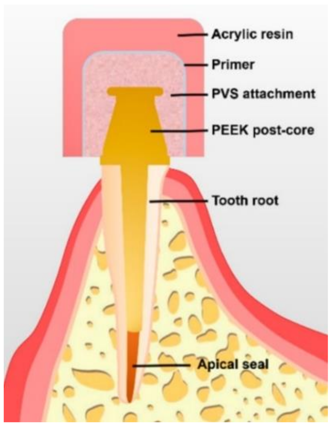
| Polyvinylsiloxane (PVS) | 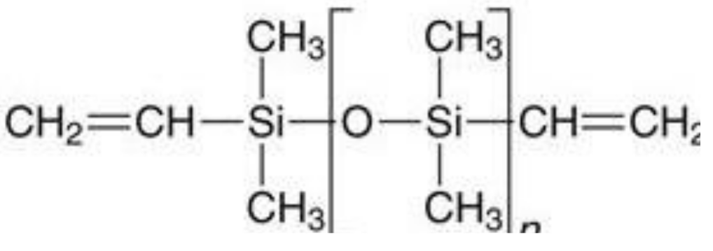 | Presents stable retention and is economical. | PVS attachments result in distortion from repeated wear and artificial saliva. | [6] |
| Polymethyl-methacrylate (PMMA) | 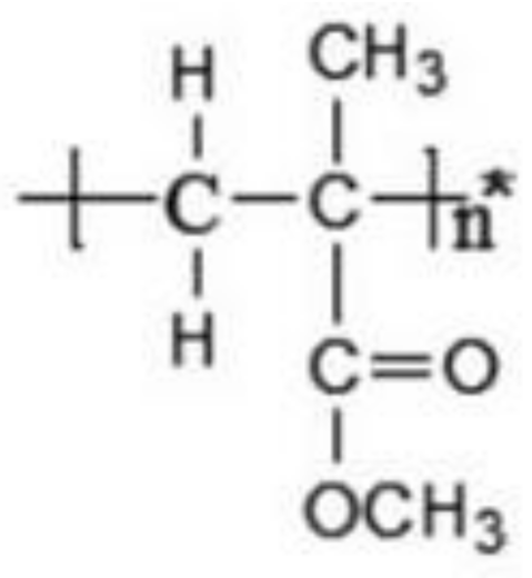 | Durable, strong, aesthetic, presents good marginal adaptation, and is economical. | Exothermic polymerization, polymerization shrinkage, poor wear resistance, and pulp irritation associated with excess monomers. | [7,9] |
| Plastic resin and Hostaform (polyoxymethylene copolymer) (POM) | 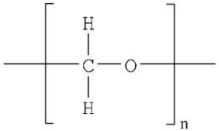 | Elastic, aesthetic, presents good marginal adaptation, and is economical. | Minimal wear is seen in plastic matrices with minimal maintenance. | [8] |
| Nylon resin and DuPont Zytel | 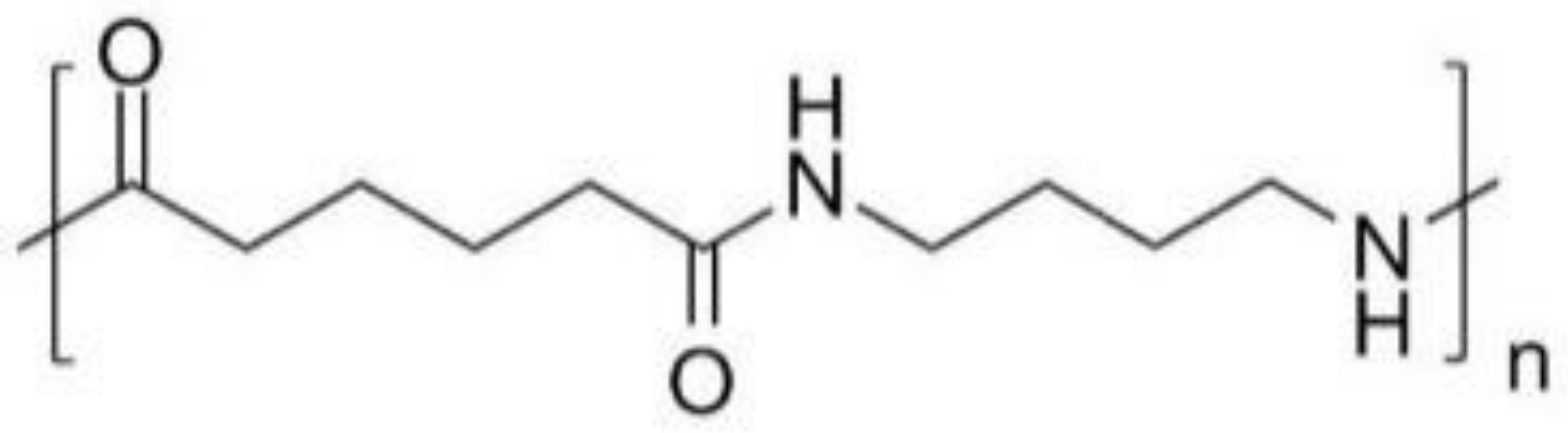 | Elastic, aesthetic, presents good marginal adaptation, and is economical. | Locator nylon matrices show extensive deformation and require considerable maintenance. | [8] |
| Attachment Type | Advantages | Disadvantages | Reference |
|---|---|---|---|
| Ball attachment (O-ring attachment) | Simple manufacturing process, the provision of a wide range of movement, cost-effectiveness, ease of use, good retention, easy hygiene maintenance, and high patient satisfaction. | The abutment requires implants to be parallelly placed, and the loss of parallelism may cause difficulty while inserting and removing the prosthesis or during the fracturing of the abutment. The O-ring needs to be regularly changed because it is subject to wear. | [10,11] |
| Bar attachment | Provides retention, implant splinting, and wide-ranging load distribution that results in a movement reduction of the implants. | Technique-sensitive, expensive, and present difficult hygiene maintenance under the bars, leading to mucosal swelling or gingival hyperplasia. Bars are not indicated in a V-shaped ridge because this leads to the infringement of tongue space. | [12,13] |
| Locators | Locators are popular attachments for implant-retained or implant-supported overdenture because of their low level of thickness (2.5 mm height) and ability to self-align, which can correct up to 40° of implant angulations. They can be used in narrow inter-arch space. Locators offer excellent retention and stability, and they allow for easy hygiene maintenance. The telescopic attachment, which offers a self-seating mechanism, is suitable for patients with reduced manual dexterity, such as those with Parkinson’s disease. | Periodic replacement of the male nylon component is required. Some prosthetic complications such as locator attachments, periodic repair, and higher maintenance double-crown locator attachments require sufficient inter-arch space and the metal display of attachments. | [8,14,15,16,17] |
| Magnetic attachments | Magnetic attachments reduce the transfer of horizontal stress to the implants and bone during the insertion and removal of the denture. | These are low-profile attachments; however, corrosion and loss of magnetism are significant complications associated with their usage. | [18,19,20,21] |
Publisher’s Note: MDPI stays neutral with regard to jurisdictional claims in published maps and institutional affiliations. |
© 2021 by the authors. Licensee MDPI, Basel, Switzerland. This article is an open access article distributed under the terms and conditions of the Creative Commons Attribution (CC BY) license (https://creativecommons.org/licenses/by/4.0/).
Share and Cite
Mirchandani, B.; Zhou, T.; Heboyan, A.; Yodmongkol, S.; Buranawat, B. Biomechanical Aspects of Various Attachments for Implant Overdentures: A Review. Polymers 2021, 13, 3248. https://doi.org/10.3390/polym13193248
Mirchandani B, Zhou T, Heboyan A, Yodmongkol S, Buranawat B. Biomechanical Aspects of Various Attachments for Implant Overdentures: A Review. Polymers. 2021; 13(19):3248. https://doi.org/10.3390/polym13193248
Chicago/Turabian StyleMirchandani, Bharat, Ting Zhou, Artak Heboyan, Sirasa Yodmongkol, and Borvornwut Buranawat. 2021. "Biomechanical Aspects of Various Attachments for Implant Overdentures: A Review" Polymers 13, no. 19: 3248. https://doi.org/10.3390/polym13193248
APA StyleMirchandani, B., Zhou, T., Heboyan, A., Yodmongkol, S., & Buranawat, B. (2021). Biomechanical Aspects of Various Attachments for Implant Overdentures: A Review. Polymers, 13(19), 3248. https://doi.org/10.3390/polym13193248