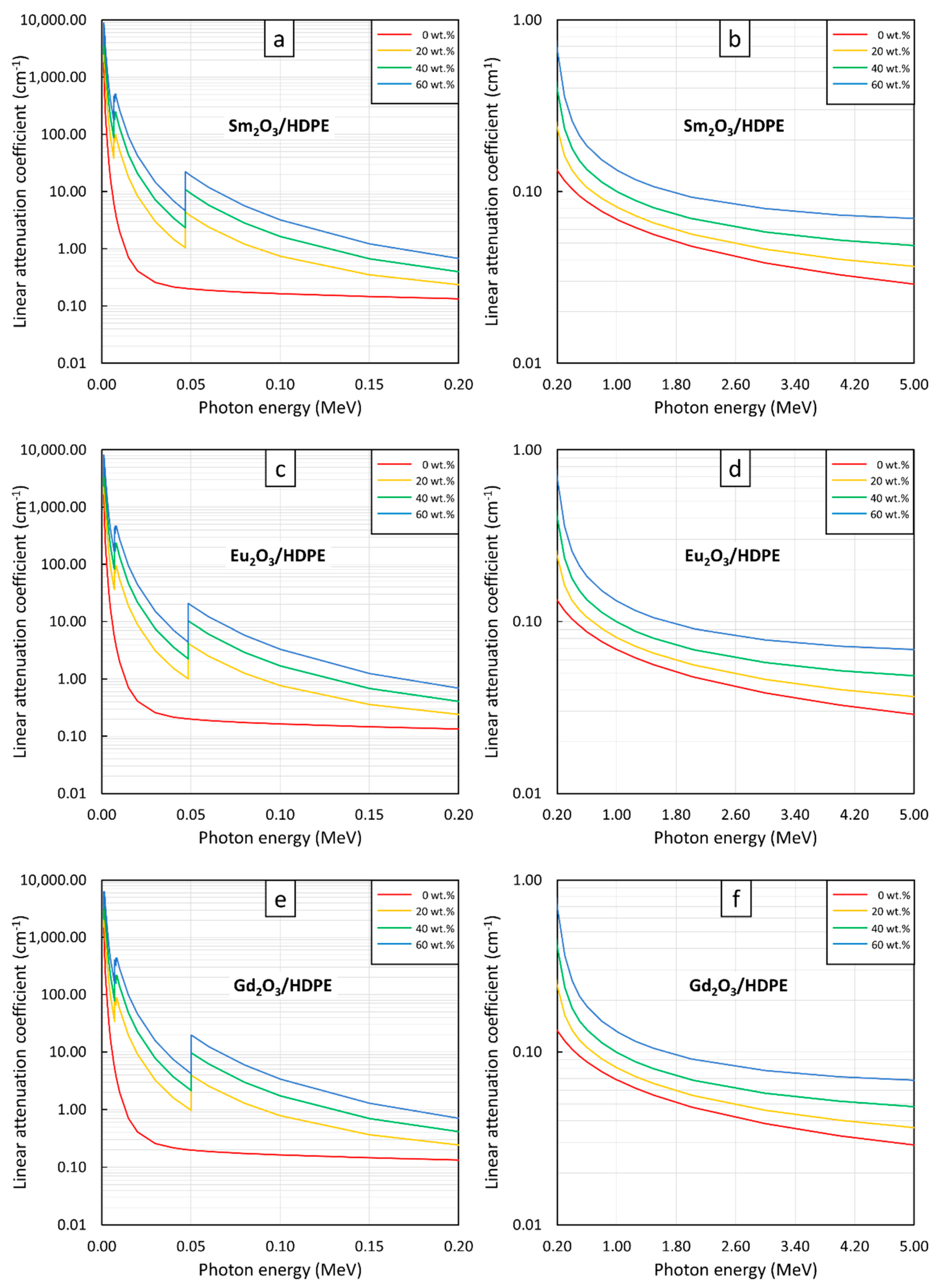
Figure 1.
µm values of (a,b) Sm2O3/HDPE, (c,d) Eu2O3/HDPE, and (e,f) Gd2O3/HDPE composites with filler contents of 0, 20, 40, and 60 wt.%, determined at photon energies of (a,c,e) 0.001–0.2 MeV and (b,d,f) 0.2–5 MeV using XCOM.
Figure 1.
µm values of (a,b) Sm2O3/HDPE, (c,d) Eu2O3/HDPE, and (e,f) Gd2O3/HDPE composites with filler contents of 0, 20, 40, and 60 wt.%, determined at photon energies of (a,c,e) 0.001–0.2 MeV and (b,d,f) 0.2–5 MeV using XCOM.
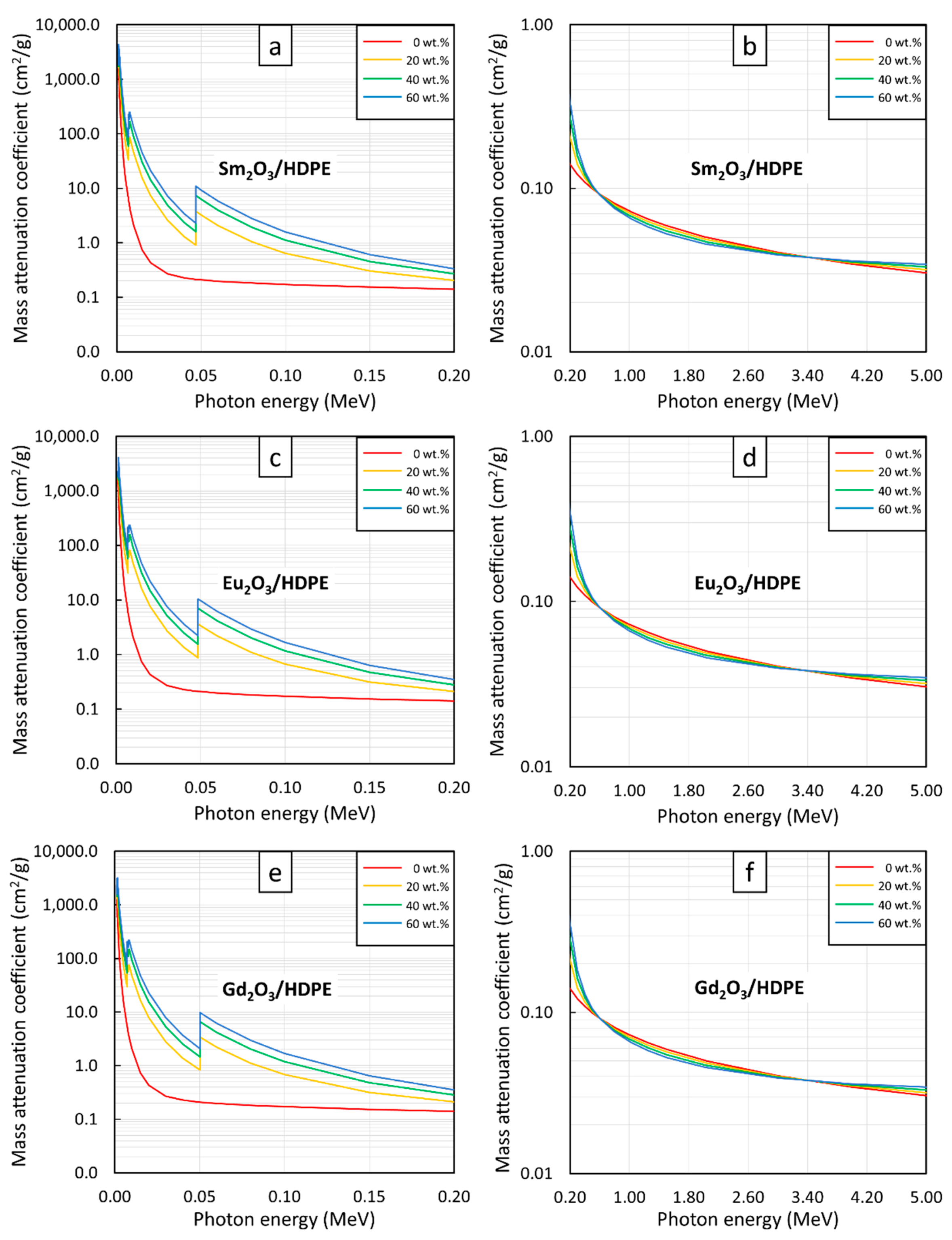
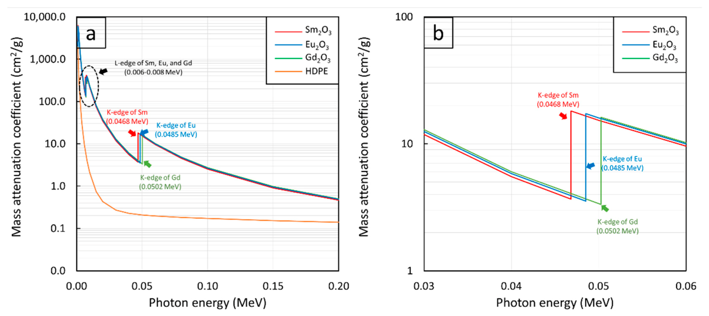
Figure 2.
µm values of Sm2O3, Eu2O3, Gd2O3, and HDPE showing K-edge and L-edge behaviors of Sm, Eu, and Gd at photon energies of (a) 0.001–0.2 MeV and (b) 0.03–0.06 MeV.
Figure 2.
µm values of Sm2O3, Eu2O3, Gd2O3, and HDPE showing K-edge and L-edge behaviors of Sm, Eu, and Gd at photon energies of (a) 0.001–0.2 MeV and (b) 0.03–0.06 MeV.
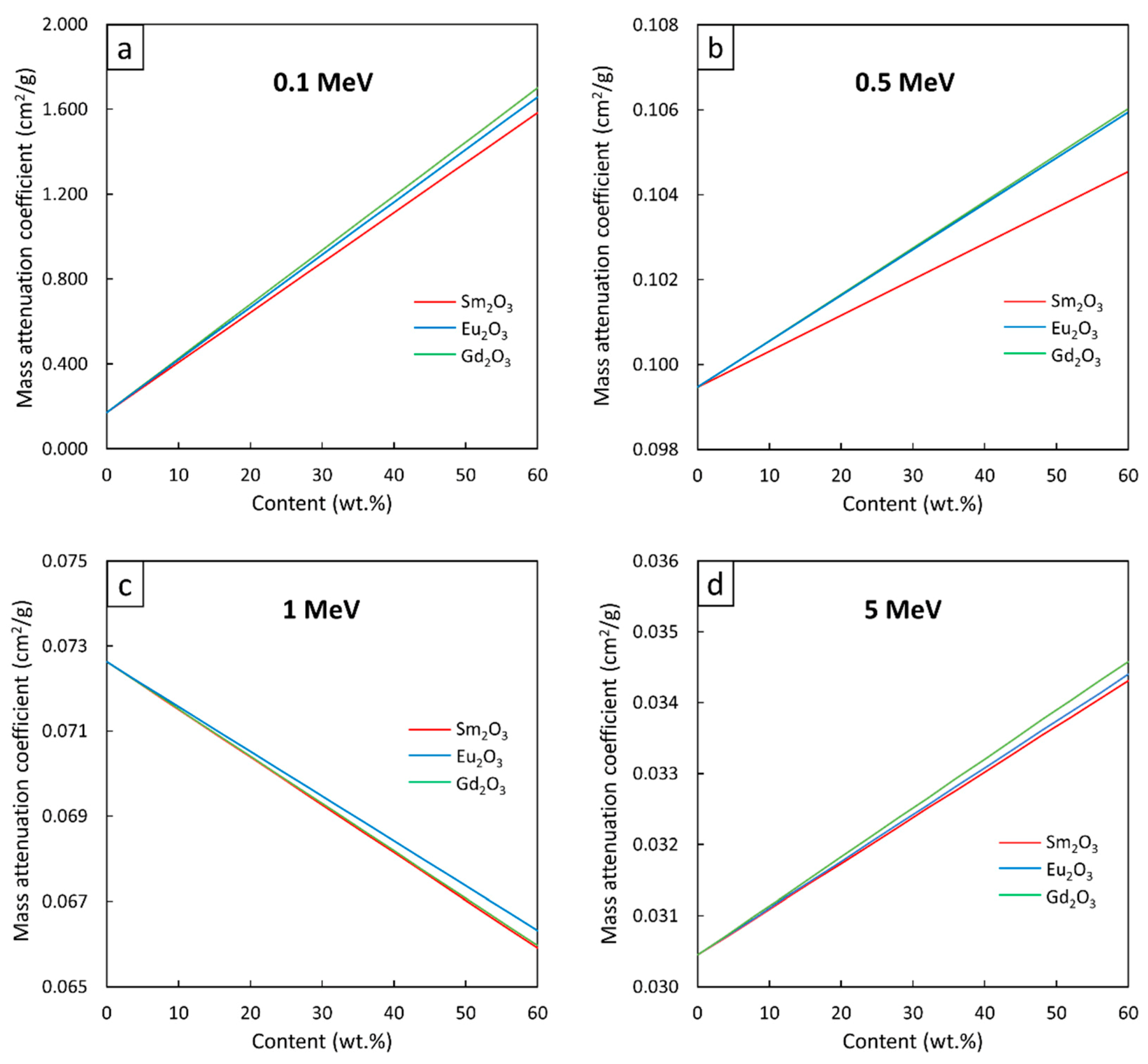
Figure 3.
µm values of Sm2O3/HDPE, Eu2O3/HDPE, and Gd2O3/HDPE composites with filler contents varied from 0–60 wt.%, determined at photon energies of (a) 0.1 MeV, (b) 0.5 MeV, (c) 1 MeV, and (d) 5 MeV using XCOM.
Figure 3.
µm values of Sm2O3/HDPE, Eu2O3/HDPE, and Gd2O3/HDPE composites with filler contents varied from 0–60 wt.%, determined at photon energies of (a) 0.1 MeV, (b) 0.5 MeV, (c) 1 MeV, and (d) 5 MeV using XCOM.
Figure 4.
µ values of (a,b) Sm2O3/HDPE, (c,d) Eu2O3/HDPE, and (e,f) Gd2O3/HDPE composites with filler contents of 0, 20, 40, and 60 wt.%, determined at photon energies of (a,c,e) 0.001–0.2 MeV and (b,d,f) 0.2–5 MeV using XCOM.
Figure 4.
µ values of (a,b) Sm2O3/HDPE, (c,d) Eu2O3/HDPE, and (e,f) Gd2O3/HDPE composites with filler contents of 0, 20, 40, and 60 wt.%, determined at photon energies of (a,c,e) 0.001–0.2 MeV and (b,d,f) 0.2–5 MeV using XCOM.
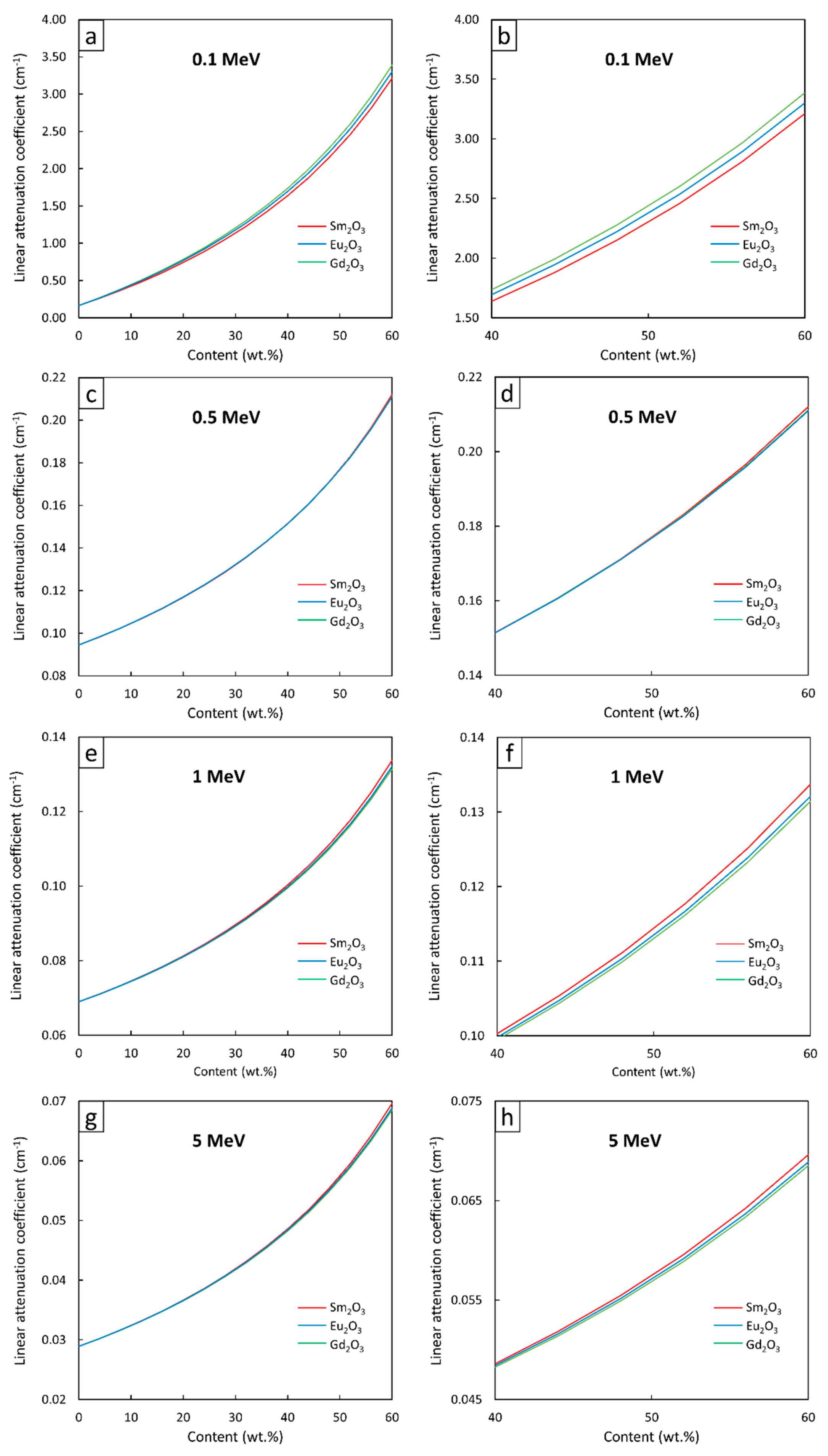
Figure 5.
µ values of Sm2O3/HDPE, Eu2O3/HDPE, and Gd2O3/HDPE composites using XCOM, with filler contents varied from (a,c,e,g) 0–60 wt.%, and (b,d,f,h) 40–60 wt.%, determined at photon energies of (a,b) 0.1 MeV, (c,d) 0.5 MeV, (e,f) 1 MeV, and (g,h) 5 MeV.
Figure 5.
µ values of Sm2O3/HDPE, Eu2O3/HDPE, and Gd2O3/HDPE composites using XCOM, with filler contents varied from (a,c,e,g) 0–60 wt.%, and (b,d,f,h) 40–60 wt.%, determined at photon energies of (a,b) 0.1 MeV, (c,d) 0.5 MeV, (e,f) 1 MeV, and (g,h) 5 MeV.
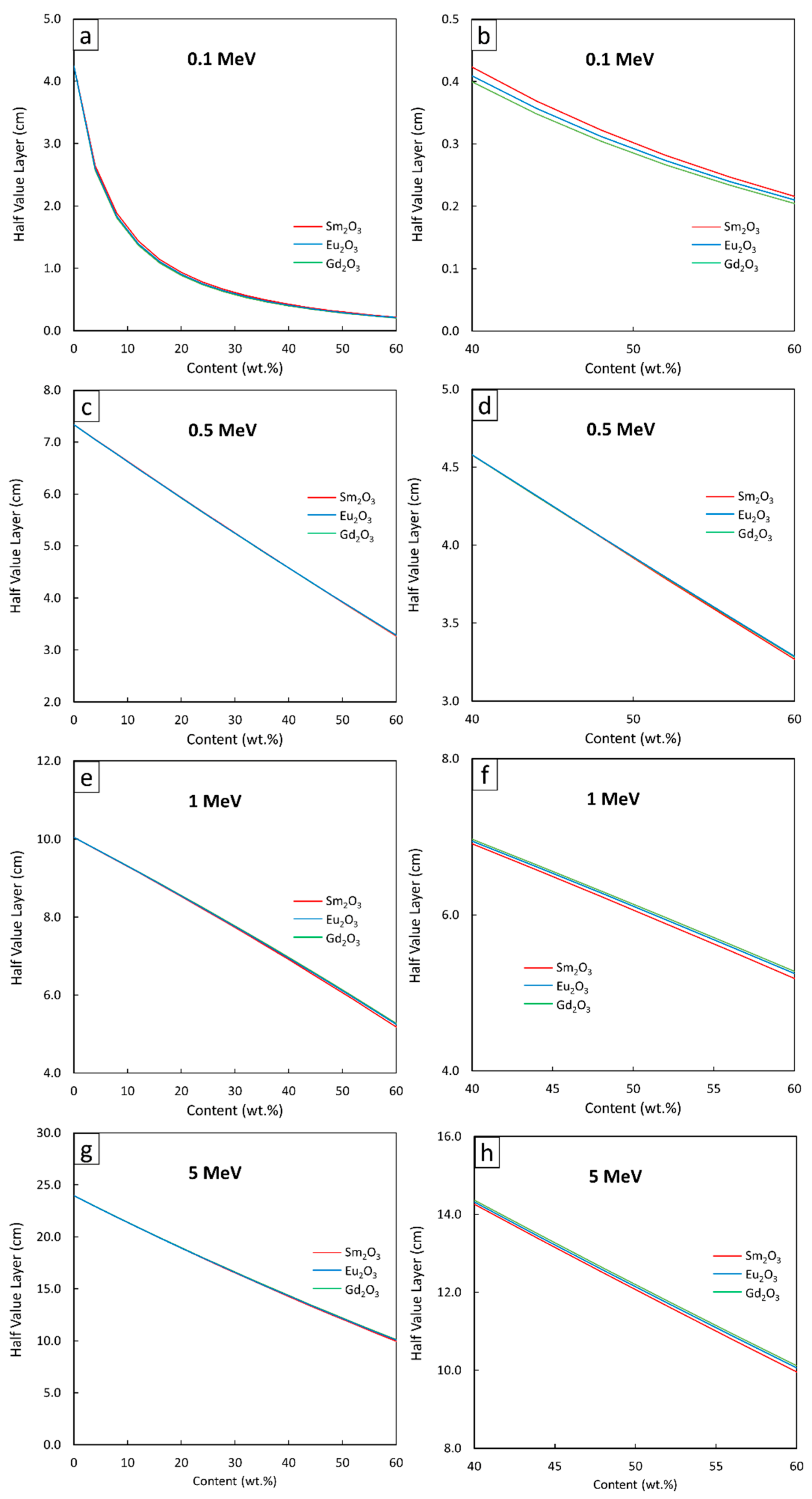
Figure 6.
HVL values of Sm2O3/HDPE, Eu2O3/HDPE, and Gd2O3/HDPE composites using XCOM, with filler contents varied from (a,c,e,g) 0–60 wt.%, and (b,d,f,h) 40–60 wt.%, determined at photon energies of (a,b) 0.1 MeV, (c,d) 0.5 MeV, (e,f) 1 MeV, and (g,h) 5 MeV.
Figure 6.
HVL values of Sm2O3/HDPE, Eu2O3/HDPE, and Gd2O3/HDPE composites using XCOM, with filler contents varied from (a,c,e,g) 0–60 wt.%, and (b,d,f,h) 40–60 wt.%, determined at photon energies of (a,b) 0.1 MeV, (c,d) 0.5 MeV, (e,f) 1 MeV, and (g,h) 5 MeV.
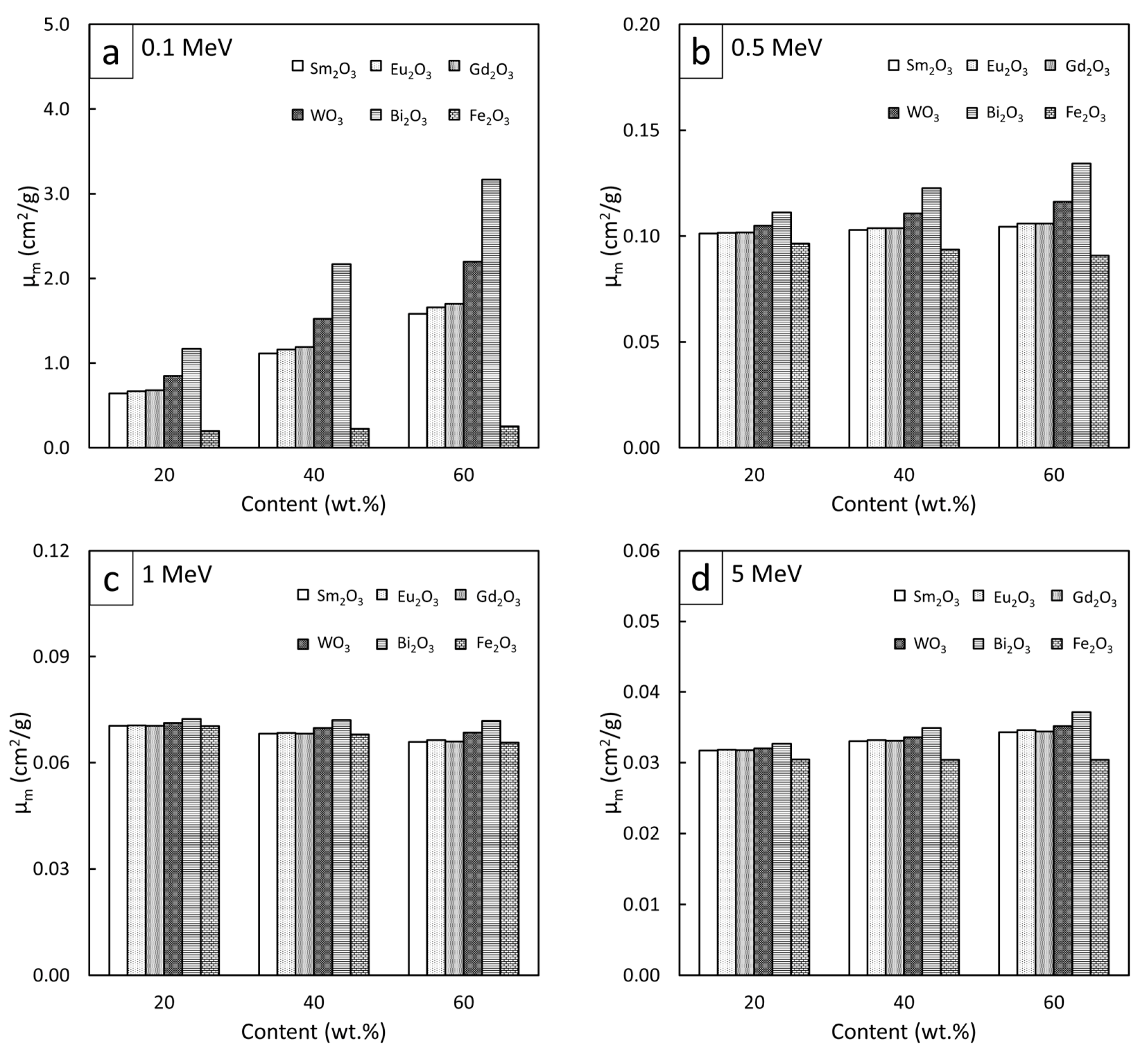
Figure 7.
Comparative µm values of Sm2O3/HDPE, Eu2O3/HDPE, and Gd2O3/HDPE composites with other common Pb-free HDPE composites (Bi2O3/HDPE, WO3/HDPE, and Fe2O3/HDPE) at filler contents of 20, 40, and 60 wt.% and photon energies of (a) 0.1 MeV, (b) 0.5 MeV, (c) 1 MeV, and (d) 5 MeV.
Figure 7.
Comparative µm values of Sm2O3/HDPE, Eu2O3/HDPE, and Gd2O3/HDPE composites with other common Pb-free HDPE composites (Bi2O3/HDPE, WO3/HDPE, and Fe2O3/HDPE) at filler contents of 20, 40, and 60 wt.% and photon energies of (a) 0.1 MeV, (b) 0.5 MeV, (c) 1 MeV, and (d) 5 MeV.
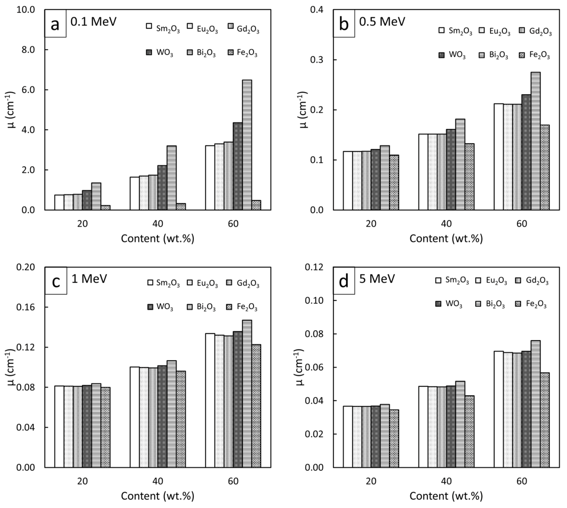
Figure 8.
Comparative µ values of Sm2O3/HDPE, Eu2O3/HDPE, and Gd2O3/HDPE composites with other common Pb-free HDPE composites (Bi2O3/HDPE, WO3/HDPE, and Fe2O3/HDPE) at filler contents of 20, 40, and 60 wt.% and photon energies of (a) 0.1 MeV, (b) 0.5 MeV, (c) 1 MeV, and (d) 5 MeV.
Figure 8.
Comparative µ values of Sm2O3/HDPE, Eu2O3/HDPE, and Gd2O3/HDPE composites with other common Pb-free HDPE composites (Bi2O3/HDPE, WO3/HDPE, and Fe2O3/HDPE) at filler contents of 20, 40, and 60 wt.% and photon energies of (a) 0.1 MeV, (b) 0.5 MeV, (c) 1 MeV, and (d) 5 MeV.
Figure 9.
Comparative HVL values of Sm2O3/HDPE, Eu2O3/HDPE, and Gd2O3/HDPE composites with other common Pb-free HDPE composites (Bi2O3/HDPE, WO3/HDPE, and Fe2O3/HDPE) at filler contents of 20, 40, and 60 wt.% and photon energies of (a) 0.1 MeV, (b) 0.5 MeV, (c) 1 MeV, and (d) 5 MeV.
Figure 9.
Comparative HVL values of Sm2O3/HDPE, Eu2O3/HDPE, and Gd2O3/HDPE composites with other common Pb-free HDPE composites (Bi2O3/HDPE, WO3/HDPE, and Fe2O3/HDPE) at filler contents of 20, 40, and 60 wt.% and photon energies of (a) 0.1 MeV, (b) 0.5 MeV, (c) 1 MeV, and (d) 5 MeV.
Table 1.
Individual density of a pristine HDPE, rare-earth oxides, metal oxides, and Pb used for determination of densities for HDPE composites [
22,
35].
Table 1.
Individual density of a pristine HDPE, rare-earth oxides, metal oxides, and Pb used for determination of densities for HDPE composites [
22,
35].
| Matrix/Compound | Density (g/cm3) |
|---|
| HDPE | 0.95 |
| Sm2O3 | 8.35 |
| Eu2O3 | 7.41 |
| Gd2O3 | 7.40 |
| WO3 | 7.16 |
| Bi2O3 | 8.90 |
| Fe2O3 | 5.24 |
| Pb | 11.35 |
Table 2.
Mathematical coefficients (A and B) for µ
m in form µ
m = Ax + B (Equation (1)) from
Figure 3.
Table 2.
Mathematical coefficients (A and B) for µ
m in form µ
m = Ax + B (Equation (1)) from
Figure 3.
| Photon Energy (MeV) | Sm2O3 | Eu2O3 | Gd2O3 |
|---|
| A | B | A | B | A | B |
|---|
| 0.1 | 0.0235 | 0.172 | 0.0248 | 0.172 | 0.0255 | 0.172 |
| 0.5 | 0.85 × 10⁻⁴ | 0.099 | 1.08 × 10⁻⁴ | 0.099 | 1.09 × 10⁻⁴ | 0.099 |
| 1.0 | −1.12 × 10⁻⁴ | 0.073 | −1.05 × 10⁻⁴ | 0.073 | −1.11 × 10⁻⁴ | 0.073 |
| 5.0 | 6.44 × 10⁻⁵ | 0.030 | 6.58 × 10⁻⁵ | 0.030 | 6.89 × 10⁻⁵ | 0.030 |
Table 3.
Comparative µm values obtained from XCOM and other programs (Phy-X/PSD and PHITS) for HDPE composites containing rare-earth oxides at various photon energies.
Table 3.
Comparative µm values obtained from XCOM and other programs (Phy-X/PSD and PHITS) for HDPE composites containing rare-earth oxides at various photon energies.
| Filler | Photon Energy (MeV) | Content (wt.%) | µm (cm2/g) | Difference (%) |
|---|
| XCOM | Phys-X (PSD) | PHITS | XCOM vs. Phys-X (PSD) | XCOM vs. PHITS |
|---|
| Sm2O3 | 0.1 | 20 | 0.64220 | 0.64223 | 0.64905 | 0.01 | 1.06 |
| 40 | 1.11300 | 1.11255 | 1.11783 | 0.04 | 0.43 |
| 60 | 1.58300 | 1.58287 | 1.58001 | 0.01 | 0.18 |
| 0.5 | 20 | 0.10120 | 0.10116 | 0.10127 | 0.04 | 0.06 |
| 40 | 0.10290 | 0.10285 | 0.10331 | 0.05 | 0.39 |
| 60 | 0.10450 | 0.10454 | 0.10458 | 0.04 | 0.07 |
| 1.0 | 20 | 0.07039 | 0.07039 | 0.07104 | 0.01 | 0.92 |
| 40 | 0.06815 | 0.06815 | 0.06841 | 0.01 | 0.38 |
| 60 | 0.06591 | 0.06591 | 0.06596 | 0.01 | 0.07 |
| 5.0 | 20 | 0.03173 | 0.03173 | 0.03194 | 0.01 | 0.66 |
| 40 | 0.03302 | 0.03302 | 0.03338 | 0.00 | 1.09 |
| 60 | 0.03431 | 0.03431 | 0.03466 | 0.01 | 1.02 |
| Eu2O3 | 0.1 | 20 | 0.66690 | 0.66691 | 0.67318 | 0.00 | 0.94 |
| 40 | 1.16200 | 1.16191 | 1.16680 | 0.01 | 0.41 |
| 60 | 1.65700 | 1.65691 | 1.65340 | 0.01 | 0.21 |
| 0.5 | 20 | 0.10160 | 0.10163 | 0.10176 | 0.03 | 0.15 |
| 40 | 0.10380 | 0.10379 | 0.10331 | 0.01 | 0.47 |
| 60 | 0.10590 | 0.10595 | 0.10458 | 0.05 | 1.24 |
| 1.0 | 20 | 0.07052 | 0.07052 | 0.07121 | 0.00 | 0.97 |
| 40 | 0.06842 | 0.06842 | 0.06868 | 0.00 | 0.38 |
| 60 | 0.06632 | 0.06632 | 0.06639 | 0.01 | 0.10 |
| 5.0 | 20 | 0.03183 | 0.03183 | 0.03204 | 0.01 | 0.65 |
| 40 | 0.03320 | 0.03320 | 0.03358 | 0.01 | 1.14 |
| 60 | 0.03458 | 0.03458 | 0.03491 | 0.01 | 0.95 |
| Gd2O3 | 0.1 | 20 | 0.68120 | 0.68118 | 0.68734 | 0.00 | 0.90 |
| 40 | 1.19000 | 1.19045 | 1.19608 | 0.04 | 0.51 |
| 60 | 1.70000 | 1.69971 | 1.69741 | 0.02 | 0.15 |
| 0.5 | 20 | 0.10170 | 0.10166 | 0.10178 | 0.04 | 0.07 |
| 40 | 0.10380 | 0.10384 | 0.10427 | 0.04 | 0.45 |
| 60 | 0.10600 | 0.10603 | 0.10603 | 0.03 | 0.02 |
| 1.0 | 20 | 0.07041 | 0.07041 | 0.07107 | 0.00 | 0.93 |
| 40 | 0.06819 | 0.06819 | 0.06844 | 0.00 | 0.36 |
| 60 | 0.06597 | 0.06597 | 0.06601 | 0.00 | 0.06 |
| 5.0 | 20 | 0.03176 | 0.03176 | 0.03197 | 0.01 | 0.66 |
| 40 | 0.03308 | 0.03308 | 0.03344 | 0.00 | 1.08 |
| 60 | 0.03440 | 0.03440 | 0.03472 | 0.01 | 0.93 |
Table 4.
Densities of Sm2O3/HDPE, Eu2O3/HDPE, and Gd2O3/HDPE composites with filler contents varying from 0 to 60 wt.% (in 4 wt.% increments), calculated using Equation (5).
Table 4.
Densities of Sm2O3/HDPE, Eu2O3/HDPE, and Gd2O3/HDPE composites with filler contents varying from 0 to 60 wt.% (in 4 wt.% increments), calculated using Equation (5).
| Content (wt.%) | Density (g/cm3) |
|---|
| Sm2O3 | Eu2O3 | Gd2O3 |
|---|
| 0 | 0.95 | 0.95 | 0.95 |
| 4 | 0.98 | 0.98 | 0.98 |
| 8 | 1.02 | 1.02 | 1.02 |
| 12 | 1.06 | 1.06 | 1.06 |
| 16 | 1.11 | 1.10 | 1.10 |
| 20 | 1.15 | 1.15 | 1.15 |
| 24 | 1.21 | 1.20 | 1.20 |
| 28 | 1.26 | 1.26 | 1.26 |
| 32 | 1.33 | 1.32 | 1.32 |
| 36 | 1.40 | 1.38 | 1.38 |
| 40 | 1.47 | 1.46 | 1.46 |
| 44 | 1.56 | 1.54 | 1.54 |
| 48 | 1.65 | 1.63 | 1.63 |
| 52 | 1.76 | 1.74 | 1.74 |
| 56 | 1.89 | 1.86 | 1.86 |
| 60 | 2.03 | 1.99 | 1.99 |
Table 5.
Mathematical constants (A and B) of µ in the form µ = Ae
Bx (Equation (6)) from
Figure 5.
Table 5.
Mathematical constants (A and B) of µ in the form µ = Ae
Bx (Equation (6)) from
Figure 5.
| Photon Energy (MeV) | Sm2O3 | Eu2O3 | Gd2O3 |
|---|
| A | B | A | B | A | B |
|---|
| 0.1 | 0.2534 | 0.0453 | 0.2591 | 0.0456 | 0.2622 | 0.0459 |
| 0.5 | 0.0909 | 0.0133 | 0.0911 | 0.0132 | 0.0911 | 0.0132 |
| 1 | 0.0665 | 0.0105 | 0.0664 | 0.0108 | 0.0665 | 0.0106 |
| 5 | 0.0279 | 0.0144 | 0.0279 | 0.0143 | 0.0279 | 0.0142 |
Table 6.
Mathematical constants (A and B) of HVL in the form HVL = Ae
Bx (Equation (6)) from
Figure 6.
Table 6.
Mathematical constants (A and B) of HVL in the form HVL = Ae
Bx (Equation (6)) from
Figure 6.
| Photon Energy (MeV) | Sm2O3 | Eu2O3 | Gd2O3 |
|---|
| A | B | A | B | A | B |
|---|
| 0.1 | 2.735 | −0.0453 | 2.675 | −0.0456 | 2.644 | −0.0459 |
| 0.5 | 7.622 | −0.0133 | 7.608 | −0.0132 | 7.609 | −0.0132 |
| 1 | 10.42 | −0.0108 | 10.42 | −0.0106 | 10.42 | −0.0106 |
| 5 | 24.88 | −0.0144 | 24.83 | −0.0143 | 24.83 | −0.0142 |
Table 7.
Pbeq of Sm2O3/HDPE, Eu2O3/HDPE, and Gd2O3/HDPE composites with filler contents of 0, 20, 40, and 60 wt.%, determined at photon energies of 0.06, 0.08, and 0.1 MeV using XCOM.
Table 7.
Pbeq of Sm2O3/HDPE, Eu2O3/HDPE, and Gd2O3/HDPE composites with filler contents of 0, 20, 40, and 60 wt.%, determined at photon energies of 0.06, 0.08, and 0.1 MeV using XCOM.
| Photon Energy (MeV) | Filler | Lead Equivalence (mmPb) |
|---|
| 20 wt.% | 40 wt.% | 60 wt.% |
|---|
| 0.06 | Sm2O3 | 0.42 | 1.02 | 2.07 |
| Eu2O3 | 0.44 | 1.05 | 2.13 |
| Gd2O3 | 0.45 | 1.08 | 2.17 |
| 0.08 | Sm2O3 | 0.44 | 1.03 | 2.06 |
| Eu2O3 | 0.46 | 1.06 | 2.11 |
| Gd2O3 | 0.47 | 1.09 | 2.17 |
| 0.1 | Sm2O3 | 0.11 | 0.26 | 0.51 |
| Eu2O3 | 0.12 | 0.27 | 0.52 |
| Gd2O3 | 0.12 | 0.28 | 0.54 |
Table 8.
Comparative µm, µ, and HVL values of Sm2O3/HDPE, Eu2O3/HDPE, and Gd2O3/HDPE composites with glassy alloys containing different types and contents of rare-earth elements (Gd, Tb, Dy, Ho, Er, and Tm) at photon energies of 0.1 and 5 MeV.
Table 8.
Comparative µm, µ, and HVL values of Sm2O3/HDPE, Eu2O3/HDPE, and Gd2O3/HDPE composites with glassy alloys containing different types and contents of rare-earth elements (Gd, Tb, Dy, Ho, Er, and Tm) at photon energies of 0.1 and 5 MeV.
| Photon Energy (MeV) | Material | Rare-Earth Element (Weight Fraction in the Material) | µm (cm2/g) | µ (cm−1) | HVL (cm) | Reference |
|---|
| 0.1 | HDPE | Sm (0.5174) | 1.583 | 3.213 | 0.216 | This work |
| HDPE | Eu (0.5182) | 1.657 | 3.297 | 0.210 |
| HDPE | Gd (0.5206) | 1.700 | 3.383 | 0.205 |
| Glassy alloys | Gd (0.3911), Tb (0.3952) | 2.451 | 16.906 | 0.041 | [30] |
| Glassy alloys | Gd (0.3877), Dy (0.4006) | 2.498 | 17.429 | 0.039 |
| Glassy alloys | Gd (0.3853), Ho (0.4041) | 2.554 | 17.980 | 0.039 |
| Glassy alloys | Tb (0.2766), Dy (0.2828), Er (0.2911) | 2.821 | 21.050 | 0.033 |
| Glassy alloys | Dy (0.2780), Er (0.2861), Tm (0.2890) | 2.973 | 22.833 | 0.030 |
| Glassy alloys | Gd (0.2786), Tb (0.2816), Dy (0.2879) | 2.675 | 19.982 | 0.035 |
| Glassy alloys | Gd (0.2785), Tb (0.2815), Dy (0.2878) | 2.669 | 19.916 | 0.035 |
| 5.0 | HDPE | Sm (0.5174) | 0.034 | 0.069 | 10.046 | This work |
| HDPE | Eu (0.5182) | 0.035 | 0.070 | 9.902 |
| HDPE | Gd (0.5206) | 0.034 | 0.068 | 10.193 |
| Glassy alloys | Gd (0.3911), Tb (0.3952) | 0.037 | 0.255 | 2.718 | [30] |
| Glassy alloys | Gd (0.3877), Dy (0.4006) | 0.037 | 0.258 | 2.687 |
| Glassy alloys | Gd (0.3853), Ho (0.4041) | 0.037 | 0.260 | 2.666 |
| Glassy alloys | Tb (0.2766), Dy (0.2828), Er (0.2911) | 0.038 | 0.284 | 2.441 |
| Glassy alloys | Dy (0.2780), Er (0.2861), Tm (0.2890) | 0.038 | 0.292 | 2.374 |
| Glassy alloys | Gd (0.2786), Tb (0.2816), Dy (0.2879) | 0.038 | 0.284 | 2.441 |
| Glassy alloys | Gd (0.2785), Tb (0.2815), Dy (0.2878) | 0.038 | 0.284 | 2.441 |


 ,
, 


