Natural 3D-Printed Bioinks for Skin Regeneration and Wound Healing: A Systematic Review

 ,
,  ,
, 
 ,
, 
Abstract
1. Introduction
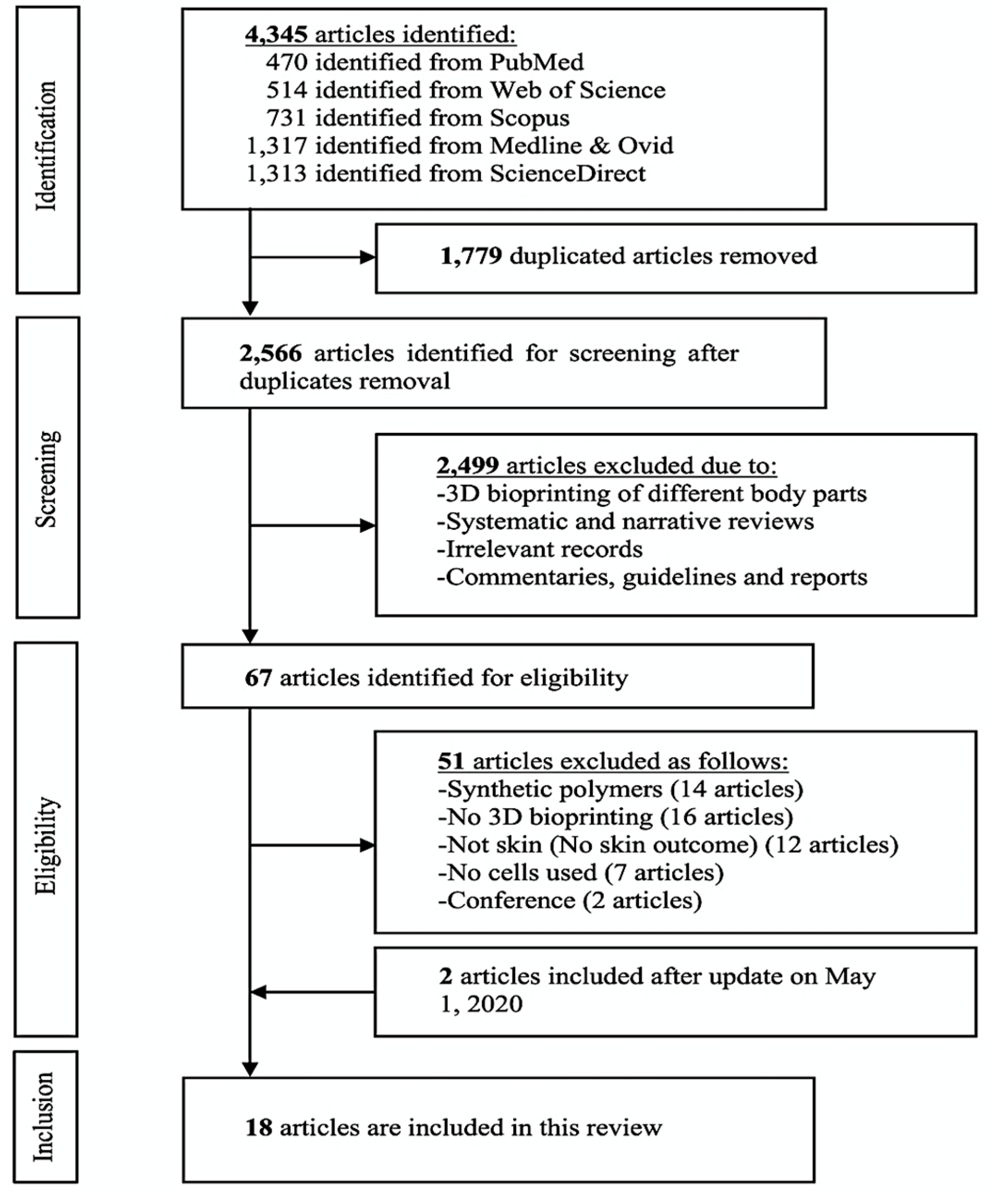
2. Methods
2.1. Search Strategy
2.2. Search Terms
2.3. Study Selection
2.4. Quality Evaluation
3. Results
3.1. Included Studies Design
3.2. Cell and Animal Models
3.3. Skin Bioinks
3.4. Bioprinting and Crosslinking Techniques
3.5. Biocompatibility Measures
3.6. Quality Evaluation
4. Discussion
4.1. Overview of the Included Studies
4.2. Bioinks Materials & Combinations
4.2.1. Collagen
4.2.2. Gelatin
4.2.3. Alginate
4.2.4. Skin-decellularized Extracellular Matrix (S-dECM)
4.3. Bioink Biocompatibility & Cellular Behavior
4.4. Structural Design & Mechanical Properties
4.5. Animal Models & Wound Healing
5. Limitations of the Present Review
6. Conclusions and Future Perspectives
Author Contributions
Funding
Acknowledgments
Conflicts of Interest
References
- Sen, C.K. Human Wounds and Its Burden: An Updated Compendium of Estimates. Adv. Wound Care 2019, 8, 39–48. [Google Scholar] [CrossRef] [PubMed]
- Beldon, P. Basic science of wound healing. Surgery 2010, 28, 409–412. [Google Scholar] [CrossRef]
- Cooper, E.H.; Anderson, C.K.; Steele, L.; O’Boyle, P. Assessment of bladder cancer. Cancer 1973, 32, 1263–1266. [Google Scholar] [CrossRef][Green Version]
- Dhivya, S.; Padma, V.V.; Santhini, E. Wound dressings—A review. BioMedicine 2015, 5, 22. [Google Scholar] [CrossRef]
- Chouhan, D.; Dey, N.; Bhardwaj, N.; Mandal, B.B. Emerging and innovative approaches for wound healing and skin regeneration: Current status and advances. Biomaterials 2019, 216, 119267. [Google Scholar] [CrossRef]
- Melchels, F.P.W.; Domingos, M.A.N.; Klein, T.J.; Malda, J.; Bartolo, P.J.; Hutmacher, D.W. Additive manufacturing of tissues and organs. Prog. Polym. Sci. 2012, 37, 1079–1104. [Google Scholar] [CrossRef]
- He, P.; Zhao, J.; Zhang, J.; Li, B.; Gou, Z.; Gou, M.; Li, X. Bioprinting of skin constructs for wound healing. Burn. Trauma 2018, 6, 1–10. [Google Scholar] [CrossRef]
- Groll, J.; Burdick, J.A.; Cho, D.W.; Derby, B.; Gelinsky, M.; Heilshorn, S.C.; Jüngst, T.; Malda, J.; Mironov, V.A.; Nakayama, K.; et al. A definition of bioinks and their distinction from biomaterial inks. Biofabrication 2019, 11, 013001. [Google Scholar] [CrossRef]
- Gopinathan, J.; Noh, I. Recent trends in bioinks for 3D printing. Biomater. Res. 2018, 22, 1–15. [Google Scholar] [CrossRef]
- Panwar, A.; Tan, L.P. Current status of bioinks for micro-extrusion-based 3D bioprinting. Molecules 2016, 21, 685. [Google Scholar] [CrossRef]
- Xia, Z.; Jin, S.; Ye, K. Tissue and Organ 3D Bioprinting. SLAS Technol. 2018, 23, 301–314. [Google Scholar] [CrossRef]
- Ng, W.L.; Wang, S.; Yeong, W.Y.; Naing, M.W. Skin Bioprinting: Impending Reality or Fantasy? Trends Biotechnol. 2016, 34, 689–699. [Google Scholar] [CrossRef] [PubMed]
- Kumar, A.; Starly, B. Large scale industrialized cell expansion: Producing the critical raw material for biofabrication processes. Biofabrication 2015, 7, 44103. [Google Scholar] [CrossRef]
- Montero, F.E.; Rezende, R.A.; da Silva, J.V.L.; Sabino, M.A. Development of a Smart Bioink for Bioprinting Applications. Front. Mech. Eng. 2019, 5, 1–12. [Google Scholar] [CrossRef]
- Valot, L.; Martinez, J.; Mehdi, A.; Subra, G. Chemical insights into bioinks for 3D printing. Chem. Soc. Rev. 2019, 48, 4049–4086. [Google Scholar] [CrossRef] [PubMed]
- Kim, J.E.; Kim, S.H.; Jung, Y. Current status of three-dimensional printing inks for soft tissue regeneration. Tissue Eng. Regen. Med. 2016, 13, 636–646. [Google Scholar] [CrossRef] [PubMed]
- Liberati, A.; Altman, D.G.; Tetzlaff, J.; Mulrow, C.; Gøtzsche, P.C.; Ioannidis, J.P.A.; Clarke, M.; Devereaux, P.J.; Kleijnen, J.; Moher, D. The PRISMA statement for reporting systematic reviews and meta-analyses of studies that evaluate health care interventions: Explanation and elaboration. J. Clin. Epidemiol. 2009, 62, e1–e34. [Google Scholar] [CrossRef]
- National Toxicology Program(NTP). OHAT Risk of Bias Rating Tool for Human and Animal Studies. Available online: https://ntp.niehs.nih.gov/whatwestudy/assessments/noncancer/riskbias/index.html (accessed on 10 January 2020).
- Heidenreich, A.C.; Pérez-Recalde, M.; González Wusener, A.; Hermida, É.B. Collagen and chitosan blends for 3D bioprinting: A rheological and printability approach. Polym. Test. 2020, 82, 106297. [Google Scholar] [CrossRef]
- Chen, X.; Yue, Z.; Winberg, P.C.; Dinoro, J.N.; Hayes, P.; Beirne, S.; Wallace, G.G. Development of rhamnose-rich hydrogels based on sulfated xylorhamno-uronic acid toward wound healing applications. Biomater. Sci. 2019, 7, 3497–3509. [Google Scholar] [CrossRef]
- Choi, D.J.; Park, S.J.; Gu, B.K.; Kim, Y.J.; Chung, S.; Kim, C.H. Effect of the pore size in a 3D bioprinted gelatin scaffold on fibroblast proliferation. J. Ind. Eng. Chem. 2018, 67, 388–395. [Google Scholar] [CrossRef]
- Lee, V.; Singh, G.; Trasatti, J.P.; Bjornsson, C.; Xu, X.; Tran, T.N.; Yoo, S.S.; Dai, G.; Karande, P. Design and fabrication of human skin by three-dimensional bioprinting. Tissue Eng. Part C Methods 2014, 20, 473–484. [Google Scholar] [CrossRef] [PubMed]
- Xu, W.; Molino, B.Z.; Cheng, F.; Molino, P.J.; Yue, Z.; Su, D.; Wang, X.; Willför, S.; Xu, C.; Wallace, G.G. On Low-Concentration Inks Formulated by Nanocellulose Assisted with Gelatin Methacrylate (GelMA) for 3D Printing toward Wound Healing Application. ACS Appl. Mater. Interfaces 2019, 11, 8838–8848. [Google Scholar] [CrossRef] [PubMed]
- Shi, L.; Hu, Y.; Ullah, M.W.; Ullah, I.; Ou, H.; Zhang, W.; Xiong, L.; Zhang, X. Cryogenic free-form extrusion bioprinting of decellularized small intestinal submucosa for potential applications in skin tissue engineering. Biofabrication 2019, 11, 035023. [Google Scholar] [CrossRef] [PubMed]
- Osidak, E.O.; Karalkin, P.A.; Osidak, M.S.; Parfenov, V.A.; Sivogrivov, D.E.; Pereira, F.D.A.S.; Gryadunova, A.A.; Koudan, E.V.; Khesuani, Y.D.; Kasyanov, V.A.; et al. Viscoll collagen solution as a novel bioink for direct 3D bioprinting. J. Mater. Sci. Mater. Med. 2019, 30, 31. Available online: https://pubmed.ncbi.nlm.nih.gov/30830351/ (accessed on 21 July 2020). [CrossRef]
- Liu, P.; Shen, H.; Zhi, Y.; Si, J.; Shi, J.; Guo, L.; Shen, S.G. 3D bioprinting and in vitro study of bilayered membranous construct with human cells-laden alginate/gelatin composite hydrogels. Colloids Surfaces B Biointerfaces 2019, 181, 1026–1034. [Google Scholar] [CrossRef]
- Xu, C.; Zhang Molino, B.; Wang, X.; Cheng, F.; Xu, W.; Molino, P.; Bacher, M.; Su, D.; Rosenau, T.; Willför, S.; et al. 3D printing of nanocellulose hydrogel scaffolds with tunable mechanical strength towards wound healing application. J. Mater. Chem. B 2018, 6, 7066–7075. [Google Scholar] [CrossRef]
- Shi, L.; Xiong, L.; Hu, Y.; Li, W.; Chen, Z.C.; Liu, K.; Zhang, X. Three-dimensional printing alginate/gelatin scaffolds as dermal substitutes for skin tissue engineering. Polym. Eng. Sci. 2018, 58, 1782–1790. [Google Scholar] [CrossRef]
- Nocera, A.D.; Comín, R.; Salvatierra, N.A.; Cid, M.P. Development of 3D printed fibrillar collagen scaffold for tissue engineering. Biomed. Microdevices 2018, 20, 1–13. [Google Scholar] [CrossRef]
- Datta, S.; Sarkar, R.; Vyas, V.; Bhutoria, S.; Barui, A.; Roy Chowdhury, A.; Datta, P. Alginate-honey bioinks with improved cell responses for applications as bioprinted tissue engineered constructs. J. Mater. Res. 2018, 33, 2029–2039. [Google Scholar] [CrossRef]
- Albanna, M.; Binder, K.W.; Murphy, S.V.; Kim, J.; Qasem, S.A.; Zhao, W.; Tan, J.; El-Amin, I.B.; Dice, D.D.; Marco, J.; et al. In Situ Bioprinting of Autologous Skin Cells Accelerates Wound Healing of Extensive Excisional Full-Thickness Wounds. Sci. Rep. 2019, 9, 1–15. [Google Scholar] [CrossRef]
- Liu, J.; Chi, J.; Wang, K.; Liu, X.; Liu, J.; Gu, F. Full-thickness wound healing using 3D bioprinted gelatin-alginate scaffolds in mice: A histopathological study. Int. J. Clin. Exp. Pathol. 2016, 9, 11197–11205. [Google Scholar]
- Huang, L.; Du, X.; Fan, S.; Yang, G.; Shao, H.; Li, D.; Cao, C.; Zhu, Y.; Zhu, M.; Zhang, Y. Bacterial cellulose nanofibers promote stress and fidelity of 3D-printed silk based hydrogel scaffold with hierarchical pores. Carbohydr. Polym. 2019, 221, 146–156. [Google Scholar] [CrossRef] [PubMed]
- Kim, B.S.; Kwon, Y.W.; Kong, J.S.; Park, G.T.; Gao, G.; Han, W.; Kim, M.B.; Lee, H.; Kim, J.H.; Cho, D.W. 3D cell printing of in vitro stabilized skin model and in vivo pre-vascularized skin patch using tissue-specific extracellular matrix bioink: A step towards advanced skin tissue engineering. Biomaterials 2018, 168, 38–53. [Google Scholar] [CrossRef] [PubMed]
- Chen, C.S.; Zeng, F.; Xiao, X.; Wang, Z.; Li, X.L.; Tan, R.W.; Liu, W.Q.; Zhang, Y.S.; She, Z.D.; Li, S.J. Three-Dimensionally Printed Silk-Sericin-Based Hydrogel Scaffold: A Promising Visualized Dressing Material for Real-Time Monitoring of Wounds. ACS Appl. Mater. Interfaces 2018, 10, 33879–33890. [Google Scholar] [CrossRef] [PubMed]
- Xiong, S.; Zhang, X.; Lu, P.; Wu, Y.; Wang, Q.; Sun, H.; Heng, B.C.; Bunpetch, V.; Zhang, S.; Ouyang, H. A Gelatin-sulfonated Silk Composite Scaffold based on 3D Printing Technology Enhances Skin Regeneration by Stimulating Epidermal Growth and Dermal Neovascularization. Sci. Rep. 2017, 7, 1–12. [Google Scholar] [CrossRef] [PubMed]
- Dzobo, K.; Motaung, K.S.C.M.; Adesida, A. Recent trends in decellularized extracellular matrix bioinks for 3D printing: An updated review. Int. J. Mol. Sci. 2019, 20, 4628. [Google Scholar] [CrossRef]
- Mitchell, A.C.; Briquez, P.S.; Hubbell, J.A.; Cochran, J.R. Engineering growth factors for regenerative medicine applications. Acta Biomater. 2016, 30, 1–12. [Google Scholar] [CrossRef]


| Bioinks | Objectives | Study Design | Experimental Design | Cross-linking Method/Materials | Ref. |
|---|---|---|---|---|---|
| Collagen-Chitosan blends | Evaluating the rheological and printability of collagen-chitosan composite as a potential bioink. | In vitro | NIH 3T3 cells | NHS/EDC | [19] |
| CNF/GelMA | Utilizing the use of deficient GelMA concentrations as supporting materials to CNF-based bioink | In vitro | Mouse 3T3 fibroblasts | Ca+2 to crosslink CNF UV light to crosslink GelMA | [23] |
| Sulfated and Rhamnose-rich XRU | Developing polysaccharide modification of 3D bioprinted XRU extract and evaluate its validity. | In vitro | Human dermal fibroblasts (HDFs) | Photo-crosslinking by UV light | [20] |
| dSIS slurry | Studying the physicochemical and biological properties of dSIS bioink. | In vitro | Normal skin fibroblasts (NSFs) | EDC | [24] |
| Viscoll Collagen | Evaluating the impact of different collagen concentrations on viscoll to produce high fidelity constructs | In vitro | NIH 3T3 | No crosslinking applied | [25] |
| Alginate/Gelatin | Investigating the rheological behavior of alginate/gelatin as a complex construct. | In vitro | AECs and WJMSCs | Two-steps gelation: a) Gelatin crosslinked by low temperature; b) Alginate crosslinked by Ca+2 | [26] |
| BCNFs+ SF/Gelatin | Enhancing the resolution and the mechanical performance of SF/gelatin scaffolds. | In vitro & in vivo | L929 cells & 12 mice | BCNFs work as a crosslinking agent | [33] |
| Fibrinogen and thrombin/Collagen I | Validating a mobile skin bioprinting system for rapid direct wound management | In vivo | Autologous fibroblasts and keratinocytes& 36 female nude mice + 6 porcine | No crosslinking applied | [31] |
| CNF | Developing an approach of double cross-linked CNF | In vitro | HDFs | Two-steps gelation: (a) During printing crosslinking with Ca+2; (b) Post-printing chemical crosslinking with BDDE | [27] |
| Sodium Alginate/ Gelatin | Developing dermal skin substitute with controlled structure and adjustable physicochemical properties | In vitro | Human skin fibroblasts (HSFs) | Three-steps gelation: (a) Immediate crosslinking at 4 °C for 30 min; (b) alginate crosslinking by CaCl2 for 1 h; (c) crosslinking by EDC-NHS | [28] |
| Collagen | Developing 3D bioprinted scaffold for tissue engineering application | In vitro | Fibroblastic NIH 3T3, and epithelial Vero cell | No crosslinking applied | [29] |
| S-dECM | Investigating the ability of printing S-dECM for skin tissue regeneration | In vitro & in vivo | HDFs and HEKs & 8 weeks old male BALB/ cA-nu/nu mice | No crosslinking agent was applied. | [34] |
| Alginate/Honey | Evaluating the shape fidelity of honey-alginate | In vitro | 3T3 fibroblast | No crosslinking applied | [30] |
| Gelatin | Evaluating the impact of pore size of gelatin scaffold on cell proliferation | In vitro | HDFs | Gelatin was immediately cross-linked by EDC-NHS solution. | [21] |
| SS/GelMA | Evaluating SS/GelMA bioink for visualization wound care | In vitro & in vivo | L929 cell line, HSF and HaCaT cell lines& 21 female SD rats | The matrices were immediately cross-linked by UV light for 1 min. | [35] |
| G-SF-SO3-FGF2 | Fabricating and evaluating porous 3D printed scaffold | In vitro & in vivo | Child foreskin fibroblasts (CFFs)& 36 male Sprague Dawley rats | Post-printing crosslinking, with 1% EDC-NHS solution for 2 h | [36] |
| Gelatin-Alginate | Studying the effect of 3D-bioprinted gelatin-alginate scaffold on the full-thickness wound healing process | In vivo | 40 female mice (6 weeks old) | The gelatin-alginate scaffold was immersed in CaCl2 for 10 min | [32] |
| Collagen | A proof-of-concept study on the ability to print human skin layer-by-layer using a 3D printing system | In vitro | Keratinocytes and fibroblasts | Post printing, nebulized NaHCO3 vapor was applied for gelation. | [22] |
| Bioink | Rheological Properties | Mechanical Properties | Biological Properties | Shape Fidelity | Conclusion | Ref. |
|---|---|---|---|---|---|---|
| Collagen- chitosan blends | Scaffold viscosity: Col/chi 0.36:1 = 1.0 Pa.s Col/chi 0.36:1 = 1.2 Pa.s Col/chi 0.36:1 crosslinked EDC/NHS = 5.6 Pa.s | Elastic modulus: Col/chi 0.36:1 crosslinked EDC/NHS = 1.95 ± 0.14 kPa | (1) Direct cytotoxicity evaluation of Col/chi indicated null toxic effect; (2) Indirect cytotoxicity evaluation suggested that the construct immersion in the medium did not impact the cells either in pure extracts or in 1/16 dilution. | Moderate | Printing different ratios of col/chi under printing flows between 0.19 uL/s and 0.42 uL/s, resulting in acceptable printability values. | [19] |
| CNF/ GelMA | Scaffold viscosity: CNF/GelMA = 1 × 10³ Pa.s swelling ratio: CNF/GelMA (9:10) = 60% CNF/GelMA (2:1) = 60–70% CNF/GelMA (2:1) = 70–87% | Mechanical strength = 2.5–5 kPa Compressive modulus = 2.3–4.5 kPa Surface modulus = 400 to 700 Pa | (1) Promoted the proliferation of fibroblasts; (2) Noncytotoxic and biocompatible features. | High | CNF/GelMA bioink scaffolds showed no cytotoxicity and good cytocompatibility with 3T3 mouse fibroblasts. | [23] |
| Sulfated & rhamnose rich XRU | Water content: 5% XRU-MA = 98.8% 10% XRU-MA = 96.8% Scaffold viscosity: XRU = 1070.7 Pa.s | Young’s modulus: 5% XRU-MA = ~18 kPa 7.5% XRU-MA = ~153 kPa 10% XRU-MA = ~309 kPa [Increasing the photo-exposure energy from 792 mJ to 2220 mJ increased Young’s modulus of 10% XRU hydrogels from ~182 kPa to ~309 kPa] | (1) Cell proliferation assay on the 10% XRU hydrogels showed a 6.3-fold increase in HDFs cell number two weeks post-culture; (2) Coating XRU with collagen, further promoted cell proliferation with a 7.5-fold increase in cell number 14 days post-culture. | High | When tested with HDFs, XRU hydrogel was found to be extremely compatible with high cell viability and promoted cell attachment and proliferation. | [20] |
| dSIS slurry | Scaffold viscosity: dSIS slurry = 23.4 Pa.s swelling ratio: P500 = 69% P600 = 74% P700 = 79% | Young’s modulus: P500 = 26.6 ± 3.8 kPa P600 = 17.9 ± 2.6 kPa P700 = 9.7 ± 3.1 kPa | (1) Lower cell adhesion in comparison to control group of spongy scaffolds; (2) Live/dead assay showed only a few dead cells indicating good biocompatibility; | High | The dSIS scaffold developed in the study can be a potential candidate for the application of skin defects with a high level of fidelity and rapid swelling ratio. | [24] |
| Viscoll collagen | At 25–30 °C: G’ 4%collagen = 1270 ± 138 Pa G’ 3%collagen = 827 ± 41 Pa G’ 2%collagen = 497 ± 13 Pa & G’’ 4%collagen = 416 ± 29 Pa G’’ 3%collagen = 255 ± 20 Pa G’’ 2%collagen = 162 ± 8 Pa | Young’s modulus: 15 mg/mL collagen = 7.2 ± 0.6 kPa 20 mg/mL collagen = 8.2 ± 0.9 kPa 30 mg/mL collagen = 9.5 ± 0.4 kPa 40 mg/mL collagen = 21.5 ± 1.4 kPa | Cell adhesion and proliferation of the bioprinted viscoll scaffold showed good biocompatibility. Cell viability: 4%collagen = 87.2% ± 2.1% 3%collagen = 95.2% ± 1.3% 2%collagen = 97.2% ± 1.2% | High | Enhanced Viscoll bioink allows the creation of contracts of complex geometry without using chemical/photo crosslinking to preserve the predesigned form. | [25] |
| Alginate/ gelatin | Scaffold viscosity (at 25–40 °C): Alg/gel (2/7.5) = 7 - 4.5 Pa.s Alg/gel (2/10) = 8 - 4.5 Pa.s Alg/gel (2/12.5)= 12.5-7 Pa.s Alg/gel (2/15) = 17.7-7 Pa.s Alg/gel (2/17.5)= 25.5-8 Pa.s | Elastic modulus: Alg/gel (2/7.5) = 280.0 ± 65.7 kPa Alg/gel (2/10) = 230.8 ± 41.4 kPa Alg/gel (2/12.5) = 199.3 ± 14.5 kPa Alg/gel (2/15) = 206.1 ± 11.5 kPa Alg/gel (2/17.5) = 192.3 ± 3.9 kPa 2/15 (alginate/gelatin) indicated: Maximum stress = 554.5 ± 76.1 kPa Maximum strain = 73.1 ± 2.7% Toughness= 106.4 ± 13.3 kJ/m | AECs and WJMSCs proliferated evenly from the 2ed day to 6th day. Bioprinting did not alter the proliferation activity of the two cell types at each predetermined time point. Cell viability: High cell viabilities (>95%) were maintained at day 2, day 4, and day 6. | High | Human AECs demonstrated a superior phenotype of epithelial cells, while WJMSCs exhibited an advanced angiogenic and fibroblastic potential. The presented system of printing alginate/gelatin composite offers promising potential for future skin technology through 3D bioprinting. | [26] |
| BCNFs + SF/gelatin | When SF/gelatin scaffolds included glycerol, both G’ and G” increased dramatically between 10 min and 20 min. | Tensile modulus = 1.63 ± 0.43 MPa At BCNFs 0.70-PS wt%: Elastic modulus = 186.5 kPa Young’s modulus = 200 kPa | After seven days: L929 cells adhered and proliferated evenly on the silk fibroin/gelatin-BCNFs scaffolds. More importantly, cell viability on BCNFs scaffolds was superior to other groups. | Low | The introduction of nanofibers from bacterial cellulose had a low impact on the printability of the composite bioinks. | [33] |
| CNF | Swelling degrees: Ca+2 crosslinked = 277.7 ± 4.1 Low-level BDDE = 307.1 ± 22.1 High-level BDDE = 212.1 ± 19.4 [After water absorption and drying, the CNF scaffold was able to maintain their shapes]. | Young’s modulus: Ca+2 crosslinked-CNF = 3.45 kPa Low-level BDDE-CNF = 4.52 kPa High-level BDDE-CNF = 7.44 kPa | 3D bioprinted CNF scaffolds showed high cell viability compared with the control 2D cell culture. Compared to the 2D control matrix, cells adhered slightly less on the 3D bioprinting matrix after 12 h of incubation. Three days post cell seeding, the 3D-bioprinted CNF scaffold contains 2 - 4 times more HDFs cells than the 2D control scaffold. | High | 3D printing improves the capacity of the produced matrix to promote cell proliferation as opposed to 2D scaffolds, which are essential for rapid wound healing. | [27] |
| Sodium alginate/ gelatin | Swelling ratio: Alginate/gelatin crosslinked CaCl2 = 42% CaCl2-EDC = 24% EDC = 301% EDC-CaCl2 = 153% | Young’s modulus: Alginate/gelatin crosslinked CaCl2 = 175.1 ± 13.3 kPa CaCl2-EDC = 240.1 ± 19.9 kPa EDC = 30.6 ± 5.0 kPa EDC-CaCl2 = 55.2 ± 4.8 kPa | Both CaCl2-EDC and EDC-CaCl2 scaffolds promoted HSFs cell proliferation. However, EDC–CaCl2 scaffolds were more suitable for cell proliferation than CaCl2–EDC in the same environment. | High | Although EDC–CaCl2 showed higher cell proliferation, CaCl2–EDC was more suitable in terms of physio-chemical and biological properties as a dermal replacement. | [28] |
| Collagen | Scaffold viscosity: The average viscosity of collagen is 35.62 ± 1.42 Pa.s Swelling ratio: 1437% ± 156% | Not reported. | NIH 3T3 Cell viability: 25-extract = 111.31 ± 3.65% 50-extract = 100.32 ± 1.65% 75-extract = 83.59 ± 6.33%. 100-extract = 85.07 ± 6.73% | High | Fibrillar collagen micro- and macropores structure promoted high cell attachment and proliferation at 37°C. | [29] |
| S-dECM | Scaffold viscosity (at 15 °C): Collagen = 3 × 10² Pa.s S-dECM = 4 × 10³ Pa.s | Young’s modulus: Collagen = 4 kPa S-dECM = 50 kPa | Cell viability: HDFs and HEKs cell viability in both bioinks reached 90% on the 7th day. On the 14th day, cells showed good proliferation in both collagen and S-dMCM. | High | S-dECM bioink could be used to create complex skin constructs by loading different cell types. The fabricated S-dECM bioink showed no cytotoxicity and high biocompatibility, similar to the commercially available collagen type I. | [34] |
| Alginate/ honey | Scaffold viscosity: A(alginate%)H(honey%) A5H0 = 9.7 ± 0.0 Pa.s A5H1 = 6.2 ± 1.1 Pa.s A5H2 = 6.1 ± 0.0 Pa.s A5H5 = 6.0 ± 0.0 Pa.s A5H10 =5.5 ± 0.1 Pa.s | Tensile strength: A5H0 = 510 kPa A5H1 = 480 kPa A5H2 = 440 kPa A5H5 = 280 kPa | Cell viability: On the 1st day, A5H1, A5H2, and A5H5 scaffolds showed significantly different cell viabilities than A5H0. A5H2 and A5H5 bioinks showed the highest cell proliferation. | High | The 1-2% honey ratio has improved cell proliferation in the bioprinted alginate without a substantial reduction in printability. | [30] |
| Gelatin | Scaffold viscosity: At 10°C: Gelatin = 450 Pa.s At 30°C: Gelatin = 0 Pa.s [Gelatin viscosity increased remarkably below 27±1°C] | Young’s modulus: G6 (pore size 600 μm) = 98.1 kPa G12(pore size 1200 μm) = 13.7 kPa | HDFs proliferation was 14% higher with pore sizes of more than 580 μm compared to 435 μm in the 3D printed gelatin after 14 days. | High | In G8-G12 gelatin scaffolds, HDFs cell growth rates were approximately 14% higher than in the G6 gelatin scaffold. The mechanical properties were highly dependent on the pore size. | [21] |
| SS/GelMA | Swelling ratio: SS/GelMA 0.5 = 630% SS/GelMA 0.33 = 495% 20%GelMA = 520% | Not reported. | One the 1st day, L929 cells exhibited a slightly slower growth on SS/GelMA scaffolds of 0.5, 0.33, and 0.2 GelMA in comparison to the control group. While on days 7 and 14 after culture, cell growth was delayed on both matrices and the control group. HaCaT and HSFs cell viabilities were exhibited higher on the scaffolds containing more SS. | High | The inclusion of silk sericin (SS) in the matrices was shown to promote HSFs cell growth. The study also suggested that SS/GelMA is suitable for HaCaT cell culture application as it showed high cell viabilities after seven days. | [35] |
| G-SF-SO3-FGF2 | Scaffold porosity: 3DG = ~82.1% 3DG-SF = ~88.0% 3DG-SF-SO3= ~87.6% | The method explained, but no results presented | On the first and third days, similar proliferation rates were noticed by CCK-8 assays with and without FGF2. On the 5th day, proliferation rates were enhanced significantly of almost 40% increase after treating with FGF-2. | High | Using 100 ng/mL of FGF2 led to a ~ 40% higher proliferation rate. Sulfonated SF coated scaffold promoted cell adhesion, proliferation, and growth. | [36] |
| Collagen | N/A | N/A | Cell viability: FBs = ~98% KCs = ~98% | Low | The study found that FBs and KCs can be evenly printed layer-by-layer as a dermal-like layer and epidermal-like layer. The 3D printing technique provides high dimensional control for engineering skin tissues. | [22] |
| Bioinks | Biological Features | Wound Healing Time | Conclusion | Ref. |
|---|---|---|---|---|
| BCNFs + SF/gelatin | After seven days, cells could grow under the surface of the printed line at a range of 160-220μm. The hierarchical pore structure of the printed line allowed sufficient space for cell growth. | 4 weeks | The findings showed that the arrangement of pore structure is beneficial for nutrient supply for the ingrowth of tissue post-implantation in vivo. | [33] |
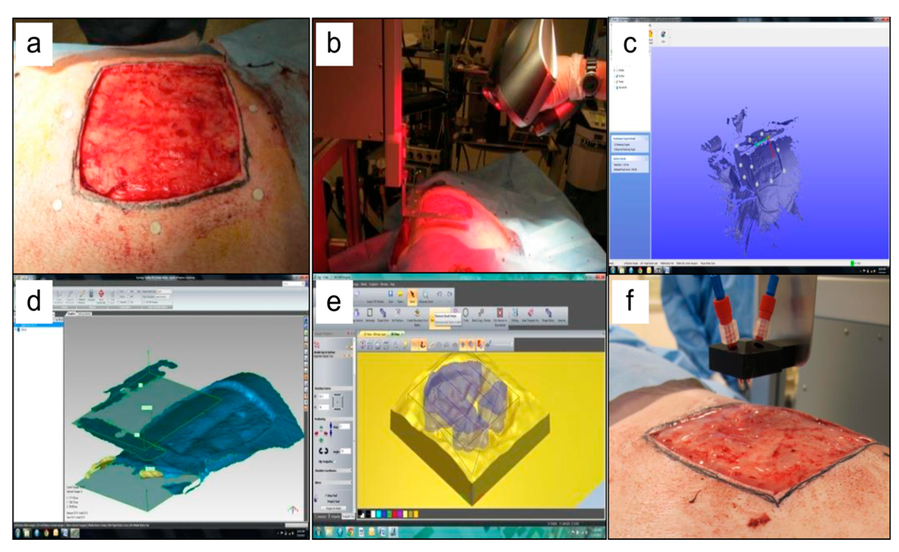
| Fibrinogen and thrombin/Collagen I | One-week post-surgery, the wound area was 66% of the original wound area in contrast to the control group wound area, which remained at 95% (n=12). Two weeks post-surgery, the wound area was 15% of the original wound area, and the control group wound area was 40% (n=8). | 10–14 days | In situ 3D bioprinting of autologous cells accelerated the process of wound healing in approximately three weeks in comparison to other treatments. | [31] |
| S-dECM | Three-weeks post-surgery, S-dECM bioink accelerated wound closure as it consists of different growth factors and cytokines capable of accelerating wound healing. Besides, cells encapsulated dECM accelerated wound re-epithelialization two weeks post-surgery. | 3 weeks | Post-implantation, the 3D bioprinted S-dECM bioink enhanced wound closure, neovascularization, and robust blood flow. | [34] |
| SS/GelMA | The immuno-histochemical observation of IL-6 and TNF-α cytokines indicated acute inflammatory on the 7th day and decreased on the 14th day and hardly found on the 28th day. | 2 weeks | Although further in vivo investigations are needed to validate the material, SS/GelMA hydrogel scaffolds represent possible candidates for the application of wound healing and tissue engineering. | [35] |
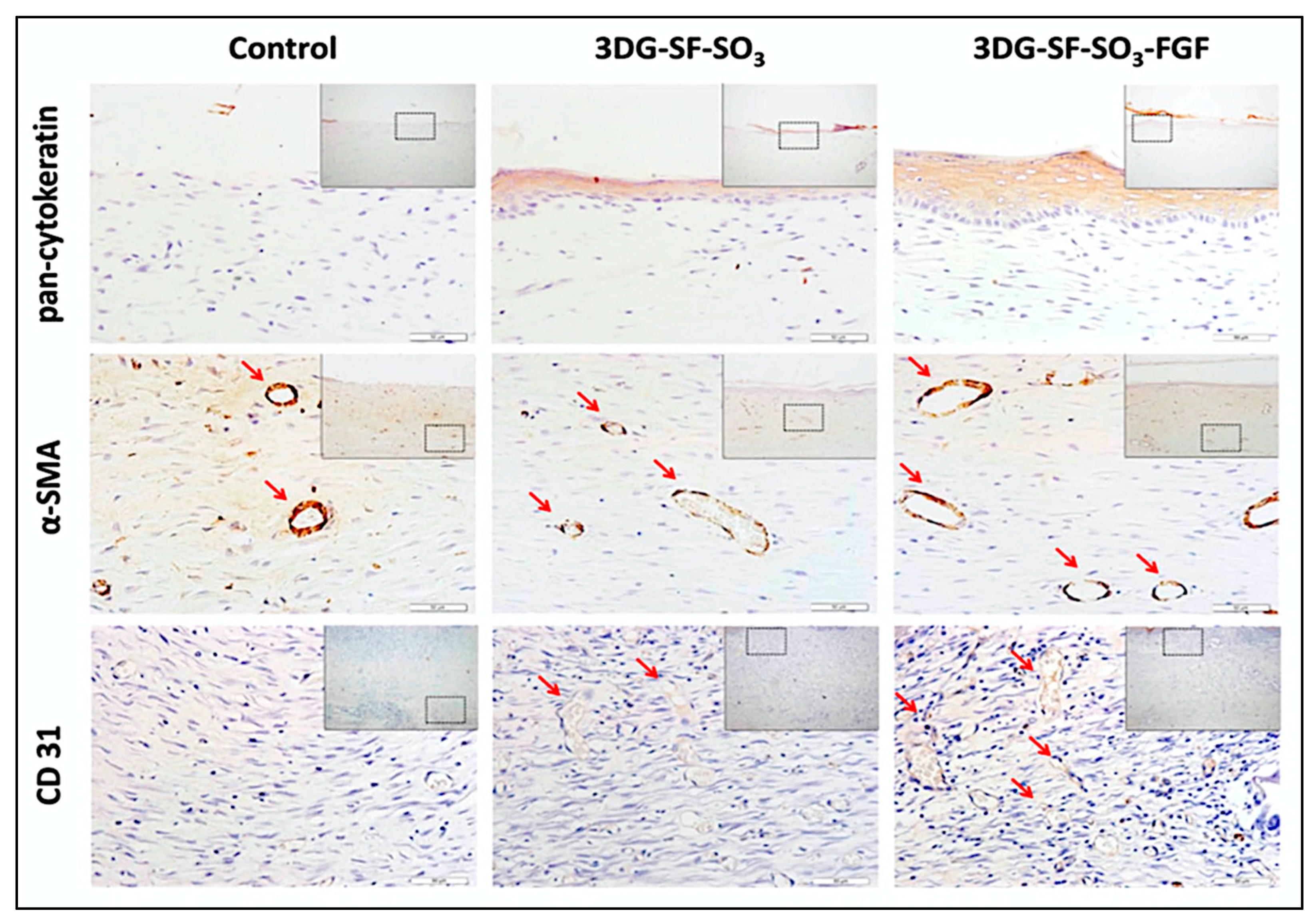
| Gel-SF-SO3-FGF2 | Two-weeks post-surgery, the epithelial cells tended to migrate from the skin edges towards the wound center in the G-SF-SO3 group. Meanwhile, the dermis and epidermis layers were almost wholly repaired in the 3D G-SF-SO3-FGF group. On the 28th day post-surgery, the wound defect was completely closed in both G-SF-SO3 and G-SF-SO3-FGF2. | 2–4 weeks | FGF2 growth factor enhanced the wound healing, re-epithelization as well as promoting blood vessel formation, and expression of various corresponding markers. | [36] |
| Gelatin-alginate | Post-surgery, the scaffold treatment group showed a significant decline in the wound area. The wound diameter decreased from 0.8 cm on the 1st day to 0.2 cm on the 14th day. The whole wound was nearly healed with almost no crust. On the 14th day, the control group seemed to be covered with hard black crusts, and the mean wound diameter was 0.7 cm. In comparison to the control group, the treatment group formed granulation tissue with uniform and layered wound thickness, which indicates that the scaffold support cell migration and proliferation. | 14 ± 1 day | The use of gelatin-alginate was found to decrease wound bleeding and perfusion post-implantation. The scaffold also found to facilitate wound maturation and healing. | [32] |
| References | ||||||||||||||||||
|---|---|---|---|---|---|---|---|---|---|---|---|---|---|---|---|---|---|---|
| A: Low Risk of Bias B: High Risk of Bias C: Not Clear D: Not Applicable | Heidenreich et al. [19] | Xu et al. [23] | Chen et al. [20] | Shi et al. [24] | Osidak et al. [25] | Liu et al. [26] | Huang et al. [33] | Albanna et al. [31] | Xu et al. [27] | Shi et al. [28] | Nocera et al. [29] | Kim et al. [34] | Datta et al. [30] | Choi et al. [21] | Chen et al. [35] | Xiong et al. [36] | Liu et al. [32] | Lee et al. [22] |
| Checklist | ||||||||||||||||||
| Clear hypothesis/objectives | A | A | A | A | A | A | A | A | A | A | A | A | A | A | A | A | A | A |
| Clear measures of outcome | A | A | A | A | A | A | A | A | A | A | A | A | A | A | A | A | A | A |
| Patient characteristics described | B | A | A | A | A | A | A | A | A | A | A | A | A | A | A | A | A | A |
| Interventions clearly described | A | A | A | B | A | A | A | A | A | A | A | A | A | A | A | A | A | A |
| Findings were clearly described | A | A | A | A | A | A | A | A | A | A | A | A | A | A | A | A | A | B |
| Adverse events were reported | B | A | A | A | B | B | B | A | A | B | B | A | A | B | A | A | B | B |
| Probability values were reported | A | B | B | A | A | A | A | A | A | A | A | A | A | A | B | B | B | B |
| Exposed and unexposed numbers were matched | C | A | C | B | A | A | B | A | A | C | A | C | A | C | A | A | A | C |
| Recruitment represents population | A | A | A | A | A | B | A | A | A | A | A | A | A | A | A | A | A | A |
| Pre-specified and reported outcomes | A | A | A | A | A | A | A | A | A | A | A | A | A | A | A | A | A | A |
| Participants represent population | B | B | B | B | B | C | A | A | B | B | B | A | B | B | A | A | A | A |
| Measuring outcomes were blinded | D | D | D | D | D | D | A | A | D | D | D | B | D | D | A | A | A | D |
| Suitable follow-up period | B | B | B | B | B | B | A | A | B | B | A | B | B | B | A | A | A | B |
| Appropriate statistical tests | C | A | A | A | A | A | A | A | A | A | A | A | A | A | A | A | A | B |
| Reliable outcome measures | B | A | A | A | A | A | A | A | A | A | A | A | B | A | A | A | A | B |
| Groups recruited from the same population | A | A | A | B | A | B | A | A | B | B | A | A | C | C | A | A | A | A |
| Subjects randomized into intervention | D | D | D | D | D | D | A | A | D | D | D | B | D | D | A | A | A | D |
| Randomized intervention concealed | D | D | D | D | D | D | A | A | D | D | D | A | D | D | A | A | A | D |
| Adjustment for confounding | D | D | D | D | D | D | A | B | D | D | D | B | D | D | A | A | A | D |
© 2020 by the authors. Licensee MDPI, Basel, Switzerland. This article is an open access article distributed under the terms and conditions of the Creative Commons Attribution (CC BY) license (http://creativecommons.org/licenses/by/4.0/).
Share and Cite
Smandri, A.; Nordin, A.; Hwei, N.M.; Chin, K.-Y.; Abd Aziz, I.; Fauzi, M.B. Natural 3D-Printed Bioinks for Skin Regeneration and Wound Healing: A Systematic Review. Polymers 2020, 12, 1782. https://doi.org/10.3390/polym12081782
Smandri A, Nordin A, Hwei NM, Chin K-Y, Abd Aziz I, Fauzi MB. Natural 3D-Printed Bioinks for Skin Regeneration and Wound Healing: A Systematic Review. Polymers. 2020; 12(8):1782. https://doi.org/10.3390/polym12081782
Chicago/Turabian StyleSmandri, Ali, Abid Nordin, Ng Min Hwei, Kok-Yong Chin, Izhar Abd Aziz, and Mh Busra Fauzi. 2020. "Natural 3D-Printed Bioinks for Skin Regeneration and Wound Healing: A Systematic Review" Polymers 12, no. 8: 1782. https://doi.org/10.3390/polym12081782
APA StyleSmandri, A., Nordin, A., Hwei, N. M., Chin, K.-Y., Abd Aziz, I., & Fauzi, M. B. (2020). Natural 3D-Printed Bioinks for Skin Regeneration and Wound Healing: A Systematic Review. Polymers, 12(8), 1782. https://doi.org/10.3390/polym12081782