Article

Bacterial Effect on the Crystallization of Mineral Phases in a Solution Simulating Human Urine

1
Crystallography Department, Institute of Earth Sciences, St. Petersburg State University, St. Petersburg 199034, Russia
2
Grebenshchikov Institute of Silicate Chemistry of the Russian Academy of Sciences, St. Petersburg 199034, Russia
3
Microbiology Department, S.M. Kirov Military Medical Academy, St. Petersburg 194044, Russia
*
Author to whom correspondence should be addressed.
Crystals 2019, 9(5), 259; https://doi.org/10.3390/cryst9050259
Submission received: 24 April 2019 / Revised: 11 May 2019 / Accepted: 16 May 2019 / Published: 18 May 2019
(This article belongs to the Special Issue Mineralogical Crystallography)

Abstract

:
The effect of bacteria that present in the human urine (Escherichia coli, Pseudomonas aeruginosa, Klebsiella pneumoniae, and Staphylococcus aureus) was studied under the conditions of biomimetic synthesis. It was shown that the addition of bacteria significantly affects both the phase composition of the synthesized material and the position of crystallization boundaries of the resulting phosphate phases, which can shift toward more acidic (struvite, apatite) or toward more alkaline (brushite) conditions. Under conditions of oxalate mineralization, bacteria accelerate the nucleation of calcium oxalates by almost two times and also increase the amount of oxalate precipitates along with phosphates and stabilize the calcium oxalate dihydrate (weddellite). The multidirectional changes in the pH values of the solutions, which are the result of the interaction of all system components and the crystallization process, were analyzed. The obtained results are the scientific basis for understanding the mechanisms of bacterial involvement in stone formation within the human body and the creation of biotechnological methods that inhibit this process.

1. Introduction

Urolithiasis is an example of pathogenic mineral formation in the human body. Various exogenous and endogenous factors are considered among the reasons for the development of urolithiasis [1,2]. The more factors act simultaneously, the more difficult the pathogenesis of urolithiasis and the worse its prognosis, which is due to frequent recurrence of the disease and the rapid growth of stones.
Currently, there are many theories explaining the causes and mechanisms of pathogenic stone formation in the human urinary system [3,4,5,6,7,8,9,10]. All theories are based on the complex interaction of biogenic and abiogenic substances, but none of them are exhaustive. The least studied is the bacterial theory [4].
It is well known that the presence of a variety of bacteria in the urine is very likely and bacterial inflammation often accompanies stone formation [11]. Assumptions about the significant effect of microorganisms on the processes of lithiasis in the human urinary system have been made in a number of works [2,3,4,5,6,7,8,9,10,11,12,13]. The crystallization system (urine) contains about a dozen bacteria species. Microbiological examination of removed urinary stones’ microflora shows that more than half of urinary stones are infected, in most cases by several types of bacteria [2,14]. Infectious diseases of the urinary tract are direct or indirect provocateurs of stone formation in the human urinary system. According to the observations of practicing urologists, infectious sequelae after lithotripsy are rather frequent, even against the background of sanitized urine, which indicates that the stones are infected by bacteria during the formation [2]. The results of urine stone sowing showed that Enterococcus faecalis, Enterococcus faecium, Staphylococcus epidermidis, Staphylococcus haemolyticus, Pseudomonas aeruginosa, Klebsiella pneumoniae, Proteus mirabilis, and Escherichia coli, as well as Streptococcus spp, Staphylococcus aureus, Acinetobacter baumanii, Candida albicans, and Morganella morganii were among the most frequently excreted microorganisms [14]. The presence of pathogens in the urine affects the parameters and composition of urine, which in turn should affect the crystallization of urinary stones’ mineral phases. A number of studies have shown that bacteria can form biofilms on the surface of a stone, which leads to the formation of chronic infection during diseases of the urinary system [2,15,16].
A substantial portion of papers on the effect of bacteria on the stone formation in the human urinary system is devoted to the so-called infectious renal stones, consisting mainly of struvite ((NH4)MgPO4·6H2O), and sometimes containing hydroxylapatite (Ca5(PO4)3(OH)) and brushite (Ca(HРО4)·2Н2О) [12]. The bacteria that cause the secondary phosphate stone formation belong to the urease-forming microflora [17]. Infectious stones are formed as a result of urea hydrolysis to ammonium ions and bicarbonate, increasing the urine pH to normal or alkaline values and binding to available cations to produce magnesium ammonium phosphate (struvite) and carbonate apatite [12]. Struvite stones are found only in a small number of patients susceptible to urinary tract infections. Thus, in our collection of renal stones of St. Petersburg and the Leningrad region residents, which consists of more than 2000 samples, only 27 belong to this "infectious" type (Figure 1). It is assumed that oxalate stones may also have an infectious origin [2,3,17]. The data on the initiation of the crystallization and aggregation of calcium oxalates in the presence of E. coli [18], as well as the work on the crystallization of weddellite (CaC2O4·2H2O) in the presence of E. coli [19], favor of this assumption. In addition, a number of papers suggest that bacteria can serve as centers of crystallization and the subsequent growth of renal stones, forming a phosphate shell around itself [20].
The results of model experiments on the crystallization of pathogenic phase analogs in the presence of bacteria have shown that bacteria change the pH of solutions and can increase the amount and alter the morphology of the resulting oxalate and phosphate crystals [3,6,7,13,21]. Unfortunately, the currently available data are insufficient to characterize the effect of the bacterial presence in the urine on the phase composition of the resulting renal stones.
In order to advance in this direction, we conducted a synthesis experiment using solutions that simulate the composition of human urine, including containing bacteria common for human urine, and revealed their role in the crystallization of urinary phosphate and oxalate stones.

2. Materials and Methods

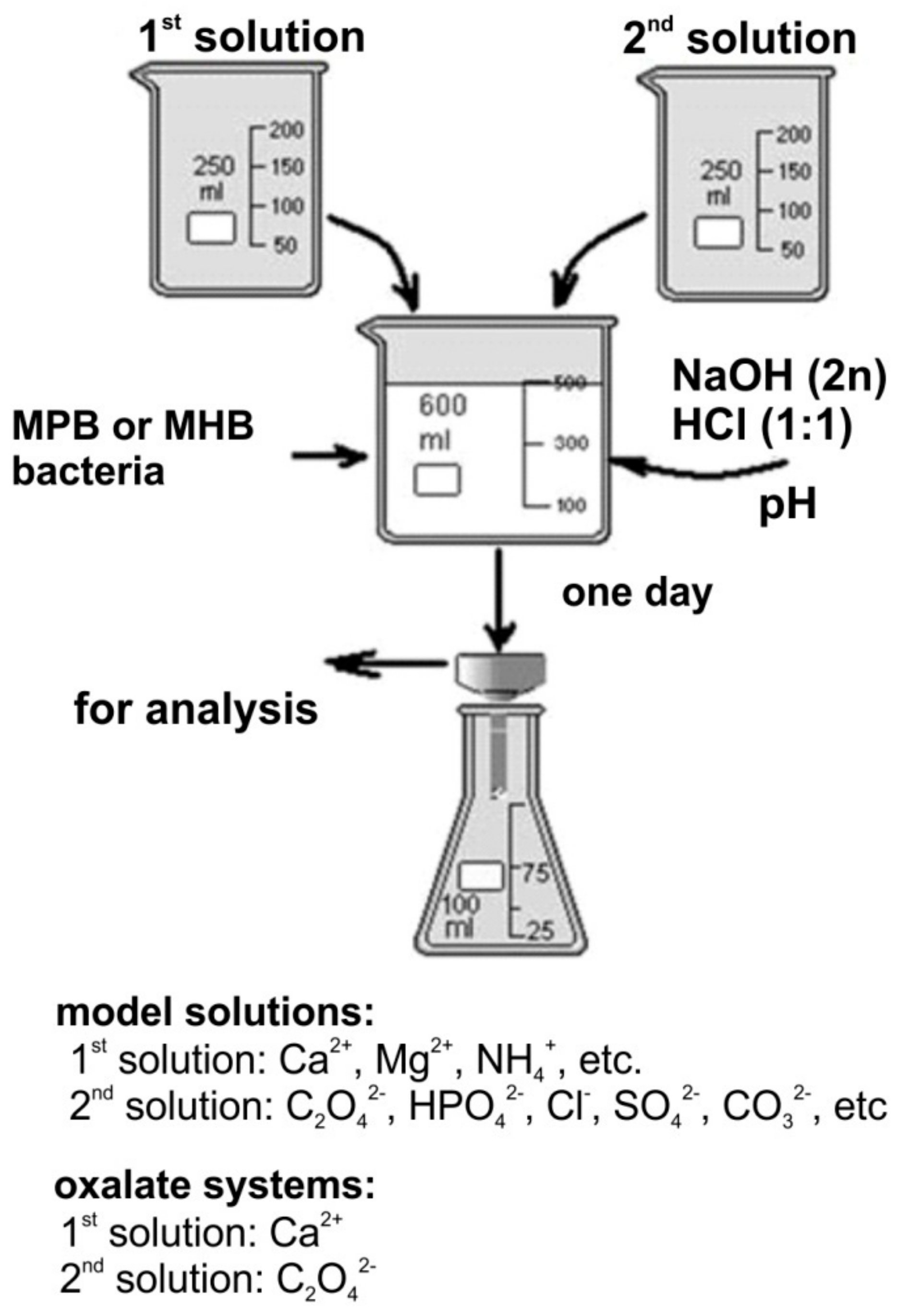
Biomimetic syntheses in the presence of bacteria were carried out by precipitation at 37 °C from solutions that simulated the composition of human urine and its inorganic components, where the content corresponded to their minimum or maximum values (Table 1). The volume of the solution after mixing of initial components in accordance with Figure 2 was 500 mL. The content of calcium cations in solutions ranged from 5 to 7.7 mmol/L, which is due to the fact that in small volumes of solution (0.2 L) and with a limited time to carry out the synthesis (1–2 days) the formation of a crystalline precipitate at lower calcium concentrations does not occur. To accelerate the crystallization of calcium and magnesium phosphates, oxalate ions (in the form of ammonium oxalate) were also added to the initial solution in a low concentration (0.1 mmol/L). Also, experiments in the so-called “oxalate system” containing only calcium ions and oxalate ions were conducted (calcium oxalate supersaturation is equal to 7, which corresponds to the physiological values of urine), since calcium oxalate does not crystallize in the system simulating the composition of urine. Ovalbumin was added to the experiments at a concentration of 10 mmol/L [22]. Syntheses were carried out by precipitation in an aqueous solution or in solutions of protein-containing nutrient media, the Müller Hinton Broth (MHB) nutrient medium or the Meat-Peptone Broth (MPB), which were prepared according to standard techniques [23,24]. In addition, bacteria associated with inflammatory processes and present in significant quantities both in the environment and in the human body were added to each of the protein media and to the model media in an amount of 106 particles per liter: Escherichia coli («e»), Klebsiella pneumoniae («kl»), Pseudomonas aeruginosa («ps»), and Staphylococcus aureus («s»). The following bacterial American Type Culture Collection (ATCC) strains were used in the experiments: 25922 («e»), 70060325922 («kl»), 27853 («ps»), and 29213 («s»). The pH of the solutions varied between 5.77–7.26 (minimum concentrations of inorganic components) and 6.10–8.07 (maximum concentrations of inorganic components). The acidity of the initial solutions was adjusted using aqueous solutions of HCl and NaOH. The crystallization start time (clouding of the solution) and phase composition of the obtained precipitates were recorded during experiments. Clouding of the solution was recorded visually. The precipitate obtained a day later was filtered, washed with distilled water, and dried at room temperature; at least three iterations were performed for each experiment.
The phase composition of precipitate products was determined by means of powder X-ray diffraction method (PXRD). The measurements were performed using a Rigaku «MiniFlex II» powder diffractometer (CuKα radiation, λ = 1.54178 Å; 30 kV/15 mA; Bragg–Brentano geometry; PSD D-Tex Ultra detector). X-ray diffraction patterns were collected at room temperature in the range of 3–60 °2θ with a step of 0.02 °2θ. Phase identification was carried out using the ICDD PDF-2 Database (release 2016). The unit cell parameters were refined by the Pawley method using TOPAS 4.2 software [26]. The background was modeled using a Chebychev polynomial of 12th order. The peak profile was described using the fundamental parameters approach.

3. Results

3.1. pH Changes of the Medium

The pH of the solutions in the crystallization process of the phosphate phases always decreased in experiments without organic additives and with the addition of nutrient media and bacteria, it either increased or decreased (Table 2). As can be seen from Table 2, the nutrient media and bacteria affected the pH values, which can be explained both by the influence of crystallization processes and bacterial activity. For instance, interaction of the solution with MHB media slightly reduced the pH value of the solution in the case of the minimum concentrations of inorganic components and in the case of maximum concentrations the pH of the solution increased. Addition of Pseudomonas aeruginosa bacteria to the MHB medium slightly increased the pH value of the solution (by 0.4), while addition of the same bacteria to the MPB medium increased the pH value of the solution by much more (by 0.6).

3.2. Model Solutions with Minimum Concentrations of Additional Ions Characteristic of a Healthy Person’s Urine Composition

In syntheses of phosphates with minimum concentrations of inorganic impurities without additives, formation of the following crystalline phases was observed: Brushite (Ca(HРО4)·2Н2О), octacalcium phosphate (Ca8(HPO4)2(PO4)4·5H2O), and whitlockite (Ca9Mg(HPO4)(PO4)6) [27,28]. Brushite formed in synthetic experiments when the initial pH of the solution ranged from 6.46 to 6.86. Octacalcium phosphate was usually observed together with brushite (less often with whitlockite) in the pH range of 6.46 to 6.95. Whitlockite was obtained in the pH range of 6.95 to 7.54.
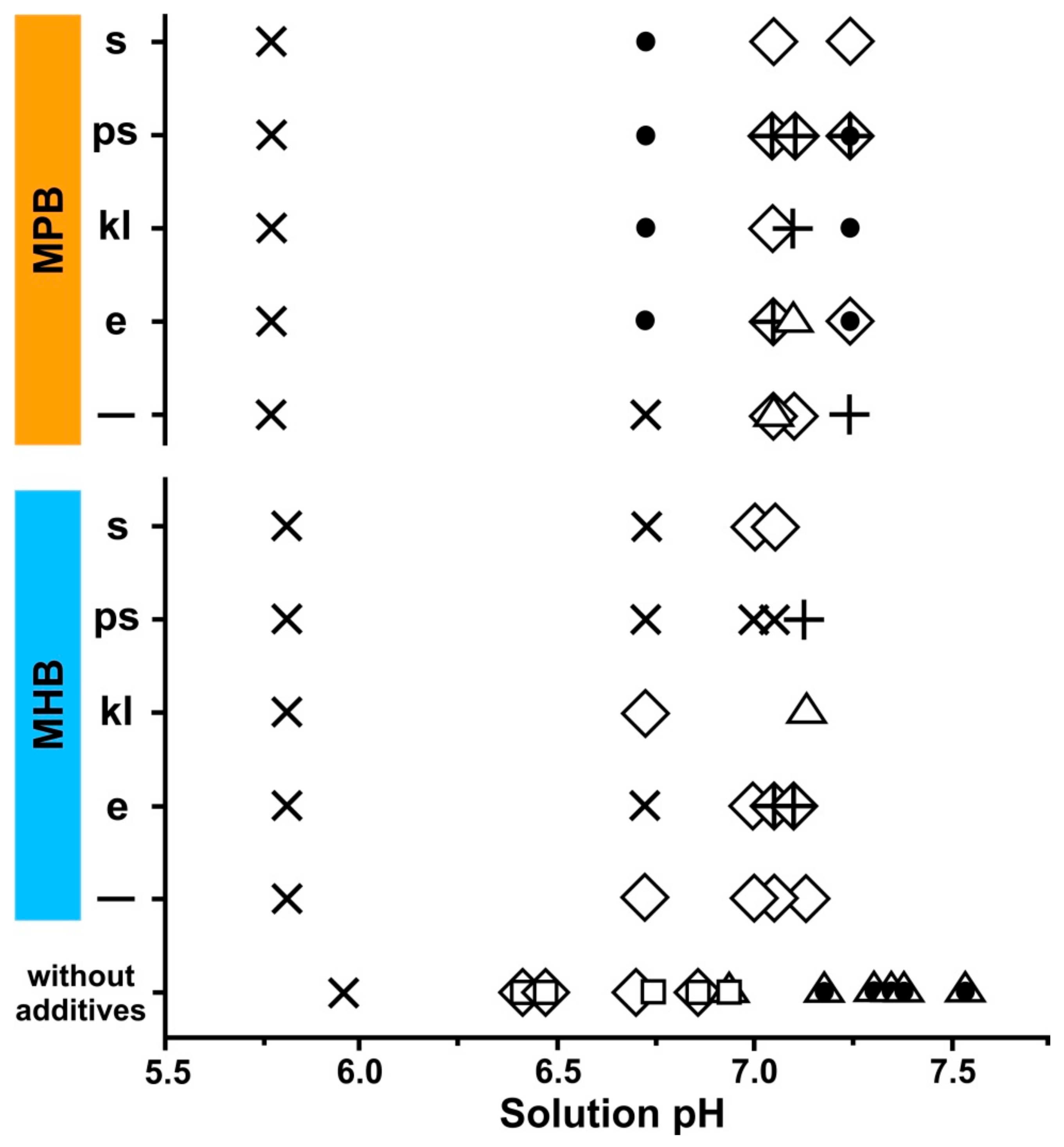
Addition of MHB medium to the model solution changed the phase composition of the sediment (Figure 3). In the pH range 6.75–7.3, the brushite phase was detected. Brushite also formed after addition of various bacteria to the solution. Moreover, the whitlockite phase was detected in the syntheses that were carried out in the presence of “kl” at a pH of 7.15. In addition, in the experiments with “e” and “ps” bacteria at pH 7.05–7.15, formation of struvite was identified (together with brushite).
Addition of the MPB medium to the model solution also led to changes in the phase composition of the sediment (Figure 3). In this case, brushite was detected at pH ~7.06. Brushite did not crystallize at such a high pH in the experiments without additives. Another difference in the phase composition of the precipitate was the formation of struvite at a pH of 7.26, which is absent in the products of syntheses without additives. The brushite phase was detected in the sediments of all syntheses, which were carried out in the presence of bacteria. Whitlockite was formed only in the synthesis in which the E. coli bacteria were present at a pH of 7.07. Struvite was formed in the syntheses with bacteria, except those experiments with the addition of Staphylococcus aureus, at a pH of 7.0 or higher. In all the syntheses with bacteria, the formation of apatite was observed at a pH of 6.72 or higher.

3.3. Model Solutions with Maximum Concentrations of Additional Ions Characteristic of a Healthy Person’s Urine Composition

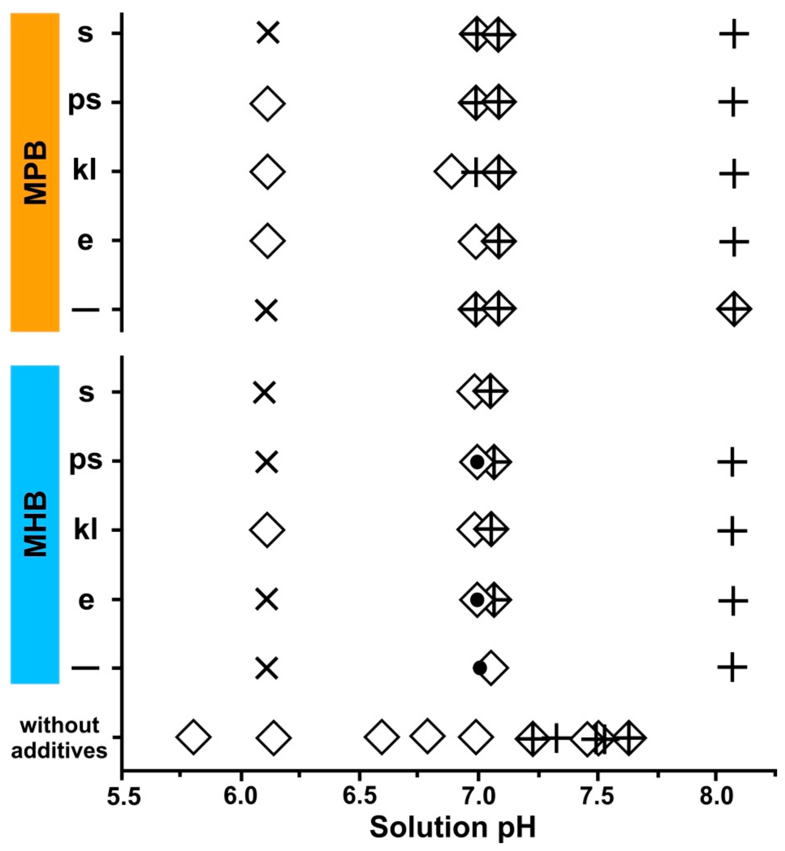
In the phosphate syntheses with maximum concentrations of inorganic impurities without additives, the formation of brushite and struvite was observed (Figure 4). Brushite was formed in a wide range of pH values of the initial solution from 5.81 to 7.63. Struvite growth occurred at higher pH values (from 7.23 to 7.73) and usually along with brushite.
When MHB medium was added to the model solution, hydroxylapatite was clearly observed in the precipitate composition, in addition to common brushite and struvite. Brushite and struvite phases also formed when various bacteria were added to the solution, while apatite was detected only in syntheses with E. coli and Pseudomonas aeruginosa. In all the systems, except for the synthesis in the presence of Klebsiella pneumoniae, there was a significant shift in the beginning of the precipitate formation toward higher pH values. Thus, brushite was obtained in syntheses within the pH range 7.0–7.03 (in the system with “kl” at a pH of 6.10), apatite was observed only at a pH of 7.0, and struvite at a pH of 7.0 or higher.
Addition of MPB medium to the model solution did not lead to changes in the phase composition of the sediment. Brushite and struvite were formed when various bacteria were added to the solution (Figure 4). The brushite phase was found in all systems, but at different pH values: Between 6.10 and 7.06 (syntheses with “st”, “kl”, and “ps”) or 6.96–7.06 (syntheses with “e”). Struvite was formed in all the systems at a pH of about 7 and higher.

3.4. Crystallization in the Oxalate System

As the result of the biomimetic syntheses, it was found that the presence of bacteria accelerates nucleation within the oxalate system (Table 3). Perhaps, bacteria can act as nucleation centers. The greatest effect (more than twice) in accelerating the crystallization rates of the calcium oxalates was observed in the presence of Pseudomonas aeruginosa.
PXRD analyses of the precipitates obtained in the presence of bacteria within the oxalate system showed formation of calcium oxalate mono and dihydrate (whewellite and weddellite, respectively), while in the syntheses without bacteria only whewellite was formed. According to the PXRD data, the whewellite/weddellite ratio in precipitants was determined along with unit cell parameters, from which the content of “zeolite” water, x, in the structure of weddellite (CaC2O4·(2 + x)H2O) was calculated (Table 4) [29]. The presence of bacteria did not practically affect the whewellite/weddellite ratio, as well as the content of “zeolite” water (x). Moreover, the results obtained for the syntheses with bacteria were close to the effect of proteins that stabilize calcium oxalate dihydrate crystallization [1,30].

4. Discussion

Perhaps, the most important results of the study are that in systems with minimum and maximum concentrations of inorganic ions, only analogs of the phosphate renal stone mineral phases were observed, while calcium oxalates were obtained under given conditions only with an increase in the concentration of oxalate ions up to the oxalatouria values, both in experiments with bacteria and without them. This result is in general agreement with the literature data on model crystallization experiments in the human urinary system [21,27,31]. Thus, according to the thermodynamic calculations and experiments in systems that simulate composition of the physiological liquid, calcium oxalates are formed in much smaller quantities than what is actually observed during pathogenic processes in the human body. Moreover, the weddellite phase (calcium oxalate dihydrate) does not form at all [31]. Introduction of bacteria and protein (ovalbumin) to the system leads to a similar result in all the experiments, increasing the portion of weddellite and increasing the amount of calcium oxalates in general. It should be also noted that, according to the unit cell parameters of weddellite crystals which are formed in the presence of bacteria, the amount of “zeolite” H2O molecules (x) falls into a rather narrow range of values, whereas those in the structures of weddellite crystals from human renal stones vary much more (from 0.13 to 0.37 p.f.u.).
According to our data, all bacteria initiate the nucleation of calcium oxalates and promote the crystallization of metastable calcium oxalate dihydrate (weddellite) in the oxalate system (containing only Ca2+ and [C2O4]2– ions). The initiation of calcium oxalate nucleation in the presence of bacteria is in agreement with the results of some recent studies, which describe an increase in the number of calcium oxalate crystals and their size in the presence of bacteria [32].
As it was shown by the results of phosphate crystallization experiments, the addition of bacteria and nutrient media leads to a change in the phase composition of the precipitate and to the shift of the phosphate phase’s formation boundaries (Figure 3 and Figure 4). The addition of the MHB medium to the model solution with the minimum concentration of inorganic impurities led to the disappearance of octacalcium phosphate and whitlockite, followed by the formation of brushite and rare struvite occurrences. The same addition to the model solution with the maximum concentration of inorganic impurities led to the crystallization of apatite, along with brushite and struvite, and to the significant shift of brushite and apatite formation areas toward higher pH values of the solution (~7.0).
The addition of the MPB medium to the model solution with a minimum concentration of inorganic impurities led to the formation of brushite and whitlockite and, in addition, crystallization of struvite was detected at a pH of 7.26, so the shift in the struvite phase formation boundary in this system moved toward being significantly more acidic. The brushite phase was observed in this system in a narrower pH range around 7.06. Since brushite did not form in experiments without organic additives at such a high pH, this suggests that the boundary of its formation expanded to the more alkaline side. The same addition to the model solution with the maximum concentration of inorganic impurities did not lead to any change in the phase composition of the synthesized products. At the same time, it can be stated that the boundary of the brushite formation area has shifted to the more alkaline region of solutions and the boundary of the struvite formation area shifted to the more acidic region (pH of 6.96).
The addition of bacteria to the appropriate media led to additional changes in the composition of the precipitates (Figure 3 and Figure 4). Thus, in the syntheses with minimal concentrations of inorganic impurities, the appearance of Escherichia coli and Pseudomonas aeruginosa in an MHB medium led to the formation of struvite and shifted its starting crystallization boundary to the more acidic region (pH of 7.05). Although struvite was initially present in the synthetic products, the appearance of bacteria in the MHB medium contributed to the displacement of its crystallization area to the more acidic region. The effect of Escherichia coli and Staphylococcus aureus bacteria on the crystallization of brushite was also well demonstrated in systems containing MHB; the shift of the brushite initial crystallization area occurred toward the more alkaline region. The effect of the bacteria addition on the crystallization of apatite was clearly visible in the MPB medium; the appearance of bacteria promoted crystallization of apatite and shifted its formation boundary to the more acidic region (pH of 6.72).
The change in the pH values of the solution during the biomimetic syntheses process occurred in different directions, due to both the crystallization process of various phases and the effect of a certain protein medium type and all types of bacteria addition. The decrease in pH in systems that modeled urine using inorganic components can be explained by the result of phosphate phase crystallization, while an increase in systems with bacteria can be explained by the influence of metabolic products. The presence of urease-producing bacteria such as Pseudomonas aeruginosa, Klebsiella pneumoniae and Staphylococcus aureus in urea led to an increase in pH [11,12].
The displacement of struvite crystallization boundaries obtained in the experiments, which led to its intensive formation, once again underlines the involvement of bacteria in the formation of "infectious" renal stones, described in a number of works [3,12]. At the same time, the expansion of the brushite crystallization area boundaries and the crystallization of apatite, as well as the formation of weddellite in the oxalate system, shows that the influence of the presence and function of bacteria in the crystallization medium was not only limited to the alkalization of the urine and the formation of ammonium ions, but significantly affected the types of growing mineral phases and the size of their crystallization areas with natural variations in urine pH.

5. Conclusions

Under the conditions of model experiments, the effect of bacteria that are present in human urine (Escherichia coli, Pseudomonas aeruginosa, Klebsiella pneumoniae and Staphylococcus aureus) on the formation of the renal stone mineral phases, such as brushite, struvite, vitlocite, octacalcium phosphate, apatite, whewellite, and weddellite, was studied in systems simulating the composition of human urine and using two types of nutrient media (Muller–Hinton Broth and Meat–Peptone Broth). Multidirectional changes in the pH values of the solutions were analyzed, which are the result of all system components’ interactions with the crystallization process.
It was shown that the presence of bacteria has a different effect on the phosphate and oxalate phases’ formation. The presence of pathogens and nutrient media significantly affect the precipitant phase composition and the position of the resulting phosphate phase’s crystallization boundaries, which can shift both to more acidic (struvite, apatite) and more alkaline (brushite) areas. Under conditions of oxalate mineralization, bacteria accelerate the nucleation of calcium oxalates by almost two times and also increase the amount of oxalate precipitates along with phosphates and stabilize the calcium oxalate dihydrate to weddellite.
As it can be seen from the reported results and the available literature data, the bacterial effect on oxalate and phosphate phase formation is different. Thus, in the case of oxalate mineralization, primarily (most likely), the inflammatory process will contribute to the decrease of oxalate supersaturation in urine due to calcium oxalate crystallization. In the case of phosphate mineralization, the change in urine pH and the products of bacterial metabolism will be of major importance. Studies aimed at identifying the specific action of certain microorganisms on the crystallization of certain mineral phases should serve to develop individual methods of treatment and prevention of urolithiasis.
The obtained results could be regarded as the scientific basis for understanding the mechanisms of bacterial participation stone formation in the human urinary system and the creation of biotechnological methods for the prevention of this disease.

Author Contributions

Conceptualization, A.R.I., O.V.F.-K., and V.V.M.; methodology, A.R.I., M.A.K., and O.V.F.-K.; investigation, A.R.I., A.M.N., M.A.K., and V.V.M.; writing—original draft preparation, A.R.I., M.A.K., and A.M.N.; writing—review and editing, A.R.I., O.V.F.-K., and V.V.M.; visualization, A.R.I. and A.M.N.

Funding

This research was funded by the Russian Science Foundation (grant 18-77-00026 to A.R.I.).

Acknowledgments

The XRD have been performed at the X-ray Diffraction Centre of the St. Petersburg State University. We are grateful to reviewers for useful comments.

Conflicts of Interest

The authors declare no conflict of interest.

References

  1. Khan, S.R.; Kok, D.J. Modulators of urinary stone formation. Frontiers Biosci. 2004, 9, 1482. [Google Scholar] [CrossRef]
  2. Seregin, A.V.; Mulabaev, N.S.; Tolordava, E.R. Urolithiasis: Current aspects of etiology and pathogenesis. Med. Buisiness 2012, 4, 4–10. [Google Scholar]
  3. Balaji, K.C.; Menon, M. Mechanism of stone formation. Urol. Clin. N. Am. 1997, 24, 1–11. [Google Scholar] [CrossRef]
  4. Bauza, J.L.; Pieras, E.C.; Grases, F.; Tubau, V.; Guimerà, J.; Sabaté, X.A.; Pizà, P. Urinary tract infection’s etiopathogenic role in nephrolithiasis formation. Med. Hypotheses 2018, 118, 34–35. [Google Scholar] [CrossRef] [PubMed]
  5. Evan, A.P. Physiopathology and etiology of stone formation in the kidney and the urinary tract. Pediatr Nephrol 2010, 25, 831–841. [Google Scholar] [CrossRef] [PubMed]
  6. Fleming, D.E.; Bronswijk, W.V.; Ryall, R.L. A comparative study of the adsorption of amino acids on to calcium minerals found in renal calculi. Clin Sci 2001, 101, 159–168. [Google Scholar] [CrossRef] [PubMed]
  7. Grases, U.F.; Costa-Bauzá, A.; García-Ferragut, L. Biopathological crystallization: A general view about the mechanisms of renal stone formation. Adv. Colloid. Interface. Sci. 1998, 74, 169–194. [Google Scholar] [CrossRef]
  8. Korago, A.A. Introduction to Biomineralogy; Nedra: St. Petersburg, Russia, 1992; p. 280. [Google Scholar]
  9. Tiktinskiy, O.L.; Aleksandrov, V.P. Urolithiasis; Liter: St. Peterburg, Russia, 2000; p. 384. [Google Scholar]
  10. Waltona, R.C.; Kavanagha, J.P.; Heywoodb, B.R.; Rao, P.N. Calcium oxalates grown in human urine under different batch conditions. J. Cryst. Growth 2005, 284, 517–529. [Google Scholar] [CrossRef]
  11. Jan, H.; Akbar, I.; Kamran, H.; Khan, J. Frequency of renal stone disease in patients with urinary tract infection. J. Ayub Med. Coll. 2008, 20, 60–62. [Google Scholar]
  12. Bichler, K.-H.; Eipper, E.; Naber, K.; Braun, V.; Zimmermann, R.; Lahme, S. Urinary infection stones. Int. J. Antimicrob. Ag. 2002, 19, 488–498. [Google Scholar] [CrossRef]
  13. Borghi, L.; Nouvenne, A.; Meschi, T. Nephrolithiasis and urinary tract infections: ‘The chicken or the egg’ dilemma? Nephrol. Dial. Transplant. 2012, 27, 3982–3985. [Google Scholar] [CrossRef]
  14. Romanova, Y.M.; Mulabaev, N.S.; Tolordava, E.R.; Seregin, A.V.; Seregin, I.V.; Alexeeva, N.V.; Stepanova, T.V.; Levina, G.A.; Barhatova, O.I.; Gamova, N.A.; et al. Microbial communities on kidney stones. Mol. Genet. Microbiol. Virol. 2015, 2, 20–25. [Google Scholar] [CrossRef]
  15. Costerton, J.W.; Stewart, P.S.; Greenberg, E.P. Bacterial biofilm: A common cause of persistent infections. Science 1999, 284, 1318–1322. [Google Scholar] [CrossRef]
  16. Musk, D.J.; Hergenrother, P.J. Chemical countermeasures for the control of bacterial biofilms: Effective compounds and promising targets. Cur. Med. Chem. 2006, 18, 2163–2177. [Google Scholar] [CrossRef]
  17. Cógain, M.R.; Lieske, J.C.; Vrtiska, T.J.; Tosh, P.K.; Krambeck, A.E. Secondarily infected non-struvite urolithiasis: A prospective evaluation. Urology 2014, 84, 1295–1300. [Google Scholar] [CrossRef]
  18. Chen, L.; Shen, Y.; Jia, R.; Xie, A.; Huang, B.; Cheng, X.; Zhang, Q.; Guo, R. The role of escherichia coliform in the biomineralization of calcium oxalate crystals. Eur. J. Inorg. Chem. 2007, 20, 3201–3207. [Google Scholar] [CrossRef]
  19. Zhao, Z.; Xia, Y.; Xue, J.; Wu, Q. Role of E. coli-secretion and melamine in selective formation of CaC2O4·H2O and CaC2O4·2H2O. Cryst. Growth Des. 2014, 14, 450–458. [Google Scholar] [CrossRef]
  20. Jamshaid, A.; Ather, M.N.; Hussain, G.; Khawaja, K.B. Single center, single operator comparative study of the effectiveness of electrohydraulic and electromagnetic lithotripters in the management of 10- to 20-mm single upper urinary tract calculi. Urol. 2008, 72, 991–995. [Google Scholar] [CrossRef]
  21. Izatulina, A.R.; Yelnikov, V. Structure, chemistry and crystallization conditions of calcium oxalates—The main components of kidney stones. In Minerals as Advanced Materials I; Krivovichev, S., Ed.; Springer: Berlin, Germany, 2008; pp. 231–241. [Google Scholar]
  22. Borodin, E.A. Biochemical Diagnosis; V. 1,2; Amuruprpoligraphizdat: Blagoveschensk, Russia, 1989; p. 77. [Google Scholar]
  23. GOST 20730-75. Nutrient Media. Meat-Peptone Broth (for Veterinary Purposes); № 899; Standards publishing house: Moscow, Russia, 1975. (in Russian) [Google Scholar]
  24. Mueller, J.H.; Hinton, J. A protein free medium for primary isolation of gonococcus and meningococcus. Proc. Soc. Exp. Biol. Med. 1941, 48, 330–333. [Google Scholar] [CrossRef]
  25. Moskalev, Y.I. Mineral Exchange; Medicine: Moscow, USSR, 1985; p. 288. [Google Scholar]
  26. Topas V4.2: General Profile and Structure Analysis Software for Powder Diffraction Data; Bruker AXS: Karlsruhe, Germany, 2009.
  27. Kuz’mina, M.A.; Nikolaev, A.M.; Frank-Kamenetskaya, O.V. The formation of calcium and magnesium phosphates of the renal stones depending on the composition of the crystallization medium. In Processes and Phenomena on the Boundary between Biogenic and Abiogenic Nature; Springer: Basel, Switzerland, 2019. (In press) [Google Scholar]
  28. Nikolaev, A.M.; Kuz’mina, M.A.; Izatulina, A.R.; Frank-Kamenetskaya, O.V.; Malyshev, V.V. Influence of the albumin substance and bacteria on formation of urinal phosphate stones (According to results of the modelling experiment). Zapiski RMO (Proceedings of the Russian Mineralogical Society, in Russian) 2014, 143, 120–133. [Google Scholar]
  29. Izatulina, A.R.; Gurzhiy, V.V.; Frank-Kamenetskaya, O.V. Weddellite from renal stones: Structure refinement and dependence of crystal chemical features on H2O content. Amer. Min. 2014, 99, 2–7. [Google Scholar] [CrossRef]
  30. Wong, T.Y.; Wu, C.Y.; Martel, J.; Lin, C.W.; Hsu, F.Y.; Ojcius, D.M.; Lin, P.Y.; Young, J.D. Detection and characterization of mineralo-organic nanoparticles in human kidneys. Sci. Rep. 2015, 5, 15272. [Google Scholar] [CrossRef] [PubMed]
  31. El’nikov, V.Yu.; Rosseeva, E.V.; Golovanova, O.A.; Frank-Kamenetskaya, O.V. Thermodynamic and experimental modeling of the formation of major mineral phases of uroliths. Russ. J. Inorg. Chem. 2007, 52, 50–157. [Google Scholar] [CrossRef]
  32. Chutipongtanate, S.; Sutthimethakorn, S.; Chiangjong, W.; Thongboonkerd, V. Bacteria can promote calcium oxalate crystal growthand aggregation. J. Biol. Inorg. Chem. 2013, 18, 299–308. [Google Scholar] [CrossRef] [PubMed]
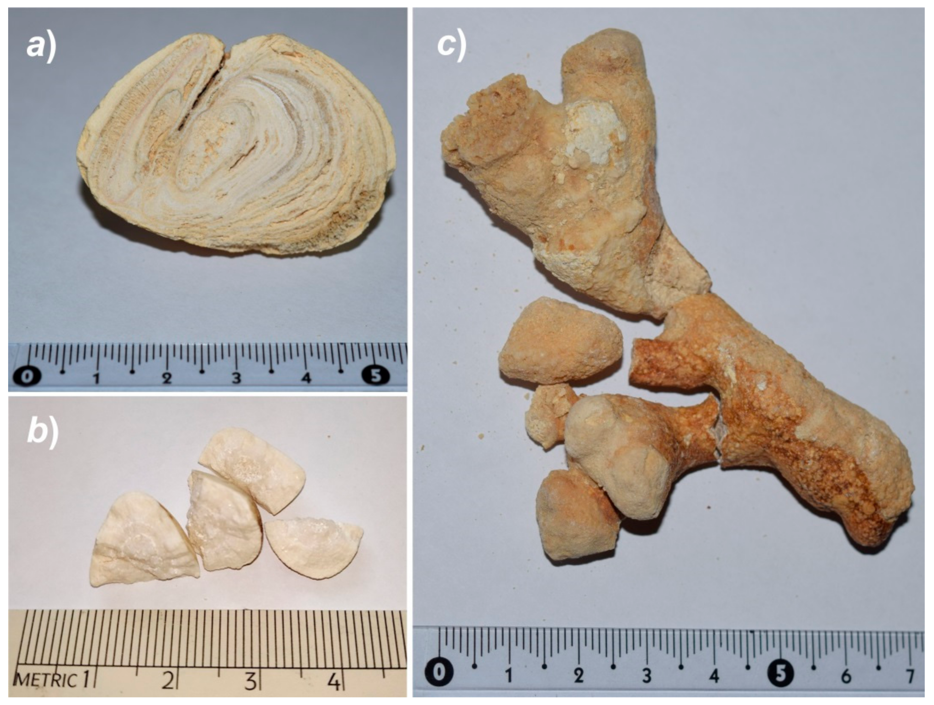
Figure 1. Infectious renal stones: (a) Apatite–struvite–brushite, (b) struvite, and (c) struvite–brushite.
Crystals 09 00259 g001
Figure 2. The scheme of the synthesis experiment in system which simulated the composition of urine by inorganic components.
Crystals 09 00259 g002
Figure 3. Phase composition of synthesized products from model solutions with minimum concentrations of additional ions characteristic of a healthy person’s urine composition. Legend: ◊—brushite, +—struvite, ∆—whitlockite, •—apatite, □—octacalcium phosphate, ×—no precipitation; Escherichia coli —«e», Klebsiella pneumoniae—«kl», Pseudomonas aeruginosa—«ps», and Staphylococcus aureus—«s».
Crystals 09 00259 g003
Figure 4. Phase composition of synthesized products from model solutions with maximum concentrations of additional ions characteristic of a healthy person’s urine composition. Legend as in Figure 3.
Crystals 09 00259 g004
Table 1. Elemental composition (mmol/L) of model solutions and urine.
ComponentModel SolutionHuman Urine [22,25]
Min ConcentrationMax Concentration
Na+607367–133
K+21.710233–47
Ca2+5–7.75–7.71.7–5
Mg2+5.3115.3–11
NH4+20.849.420–50
Cl-678067–167
CO32-0330–33
PO43-133313–33
SO42-21.76927–80
Table 2. The change in pH of the solutions during the experiment in nutrient media with the addition of bacteria.
AdditivesMinimum ConcentrationMaximum Concentration
Nutrient MediumBacteriaInitial pHFinal pHInitial pHFinal pH
nonenone5.95–7.545.94–6.715.81–7.735.75–7.50
Müller–Hinton Brothnone5.81–7.155.84–6.256.10–8.076.27–7.39
Escherichia coli (“e”)5.81–7.155.27–6.396.10–8.075.85–7.35
Klebsiella pneumoniae (“kl”)5.81–7.155.51–6.406.10–8.075.84–7.50
Pseudomonas aeruginosa (“ps”)5.81–7.156.21–6.516.10–8.076.16–7.90
Staphylococcus aureus (“s”)5.81–7.155.06–6.456.10–8.075.66–7.54
Meat–Peptone Brothnone5.77–7.265.78–7.086.10–8.036.18–7.90
Escherichia coli (“e”)5. 77–7.265.94–7.026.10–8.036.07–7.67
Klebsiella pneumoniae (“kl”)5.77–7.265.90–7.006.10–8.036.03–7.80
Pseudomonas aeruginosa (“ps”)5.77–7.266.39–7.406.10–8.036.18–7.90
Staphylococcus aureus (“s”)5.77–7.266.25–7.236.10–8.036.14–8.05
Table 3. Nucleation of calcium oxalates in the presence of bacteria at various supersaturations (γ).
BacteriaNucleation Time, s
γ = 3γ = 7γ = 10
NoneMore than 2400 (>40 min)840140
Staphylococcus aureus150051030–50
Klebsiella pneumoniae129047030–50
Escherichia coli120042030–50
Pseudomonas aeruginosa114037030–50
Table 4. Characteristics of phases synthesized within the oxalate system in the presence of bacteria and protein additives.
Additives Whewellite/Weddellite Ratio Selected Crystallographic Data for the Weddellite Phase
Unit Cell Parameter, ÅAmount of “Zeolite” Water (x), p.f.u. *
None whewellite
Ovalbumin5:212.349(1)0.26
Escherichia coli5:212.344(1)0.23
Pseudomonas aeruginosa5:212.341(2)0.21
Staphylococcus aureus5:2 12.346(2)0.24
* Per formula unit; calculations were made with regard to the a unit cell parameter, using the regression equation reported in [29].