Micro-Raman Imaging of Ferroelectric Domain Structures in the Bulk of PMN-PT Single Crystals
Abstract
1. Introduction
2. Materials and Methods
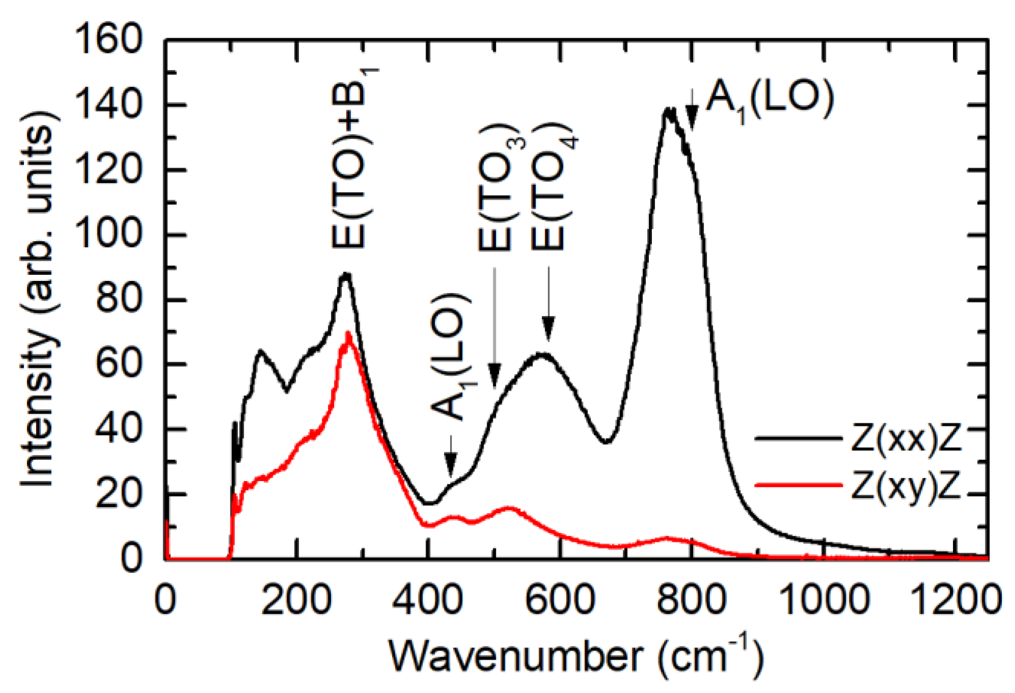
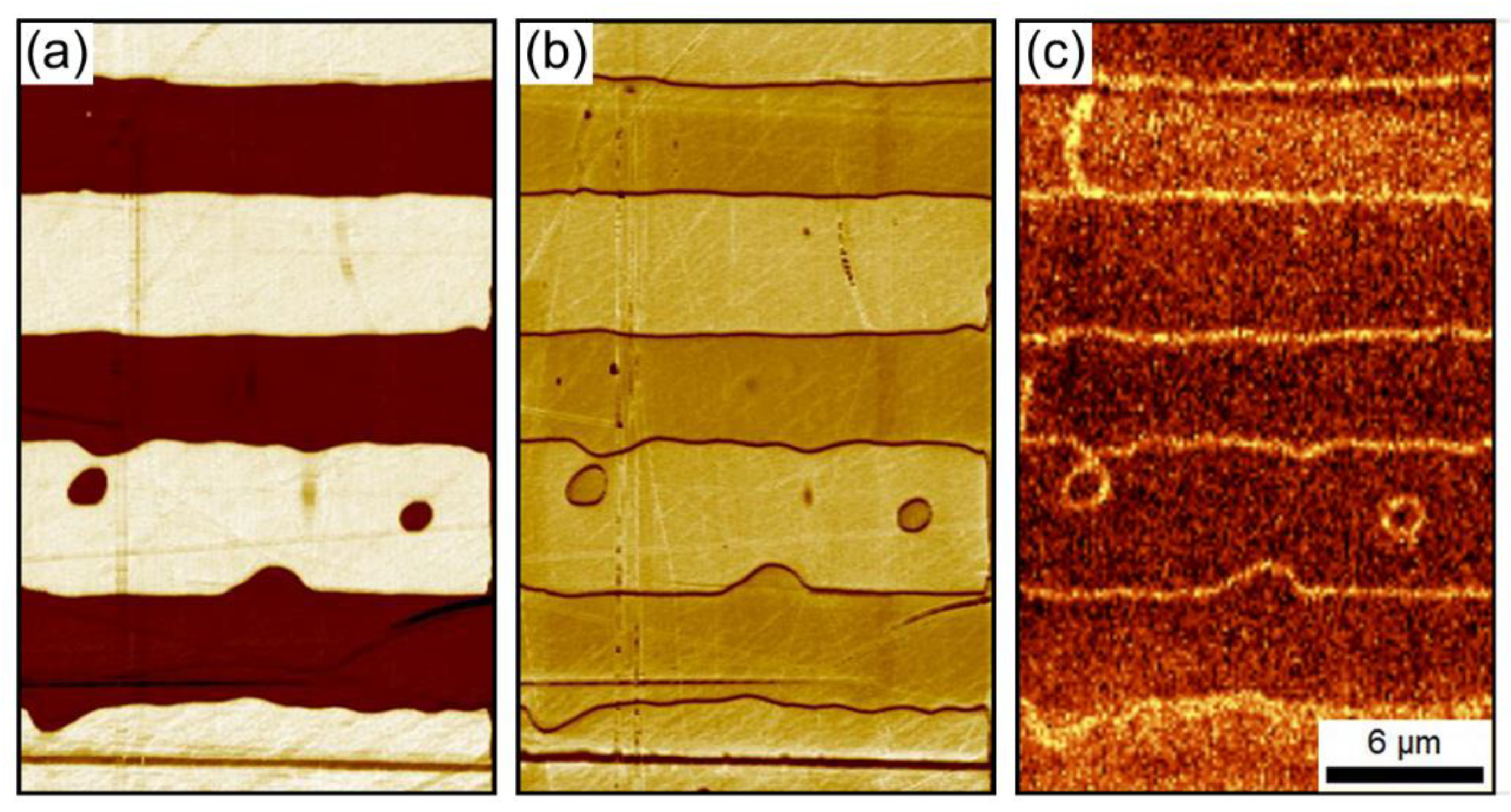
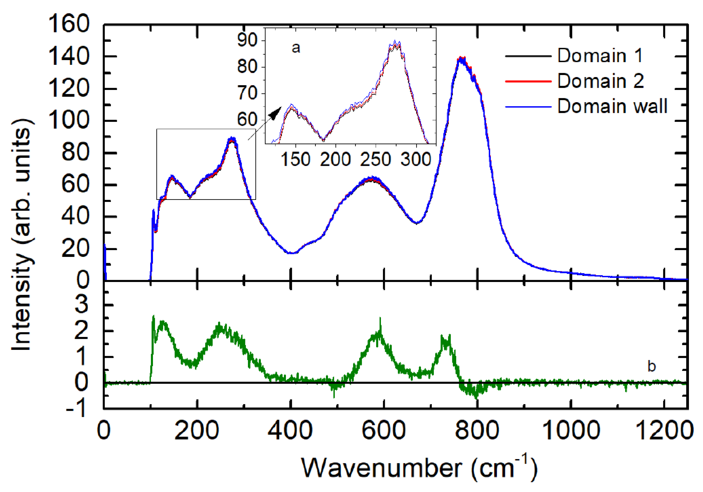
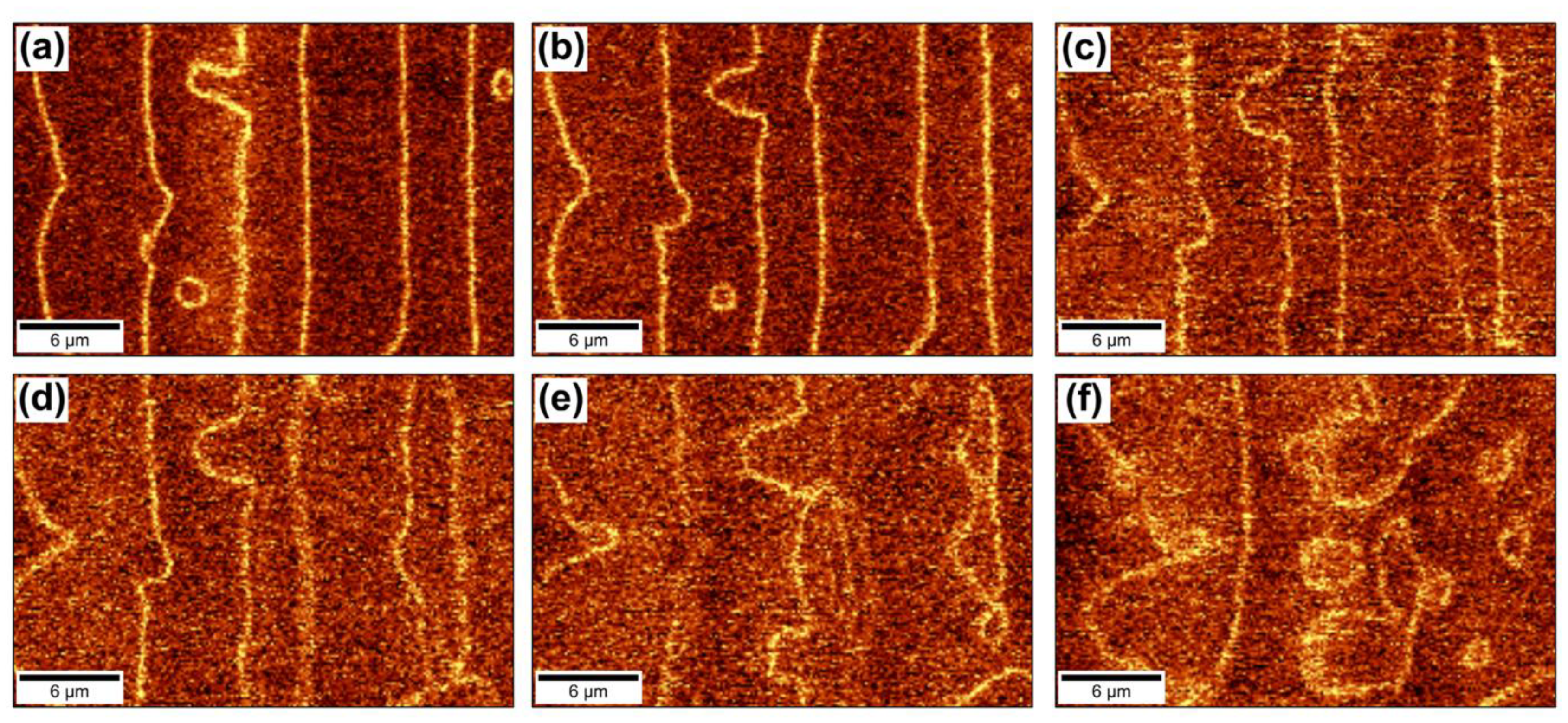
3. Results and Discussion
4. Conclusions
Author Contributions
Funding
Acknowledgments
Conflicts of Interest
References
- Zhang, S.; Li, F.; Jiang, X.; Kim, J.; Luo, J.; Geng, X. Advantages and challenges of relaxor-PbTiO3 ferroelectric crystals for electroacoustic transducers—A review. Prog. Mater. Sci. 2015, 68, 1–66. [Google Scholar] [CrossRef] [PubMed]
- Kutnjak, Z.; Petzelt, J.; Blinc, R. The giant electromechanical response in ferroelectric relaxors as a critical phenomenon. Nature 2006, 441, 956–959. [Google Scholar] [CrossRef] [PubMed]
- Li, F.; Zhang, S.; Yang, T.; Xu, Z.; Zhang, N.; Liu, G.; Wang, J.; Wang, J.; Cheng, Z.; Ye, Z.-G.; et al. The origin of ultrahigh piezoelectricity in relaxor-ferroelectric solid solution crystals. Nat. Commun. 2016, 7, 13807. [Google Scholar] [CrossRef] [PubMed]
- Wu, F.; Yang, B.; Sun, E.; Liu, G.; Tian, H.; Cao, W. Linear electro-optic properties of relaxor-based ferroelectric 0.24Pb(In1/2Nb1/2)O3-(0.76 − x)Pb(Mg1/3Nb2/3)O3-xPbTiO3 single crystals. J. Appl. Phys. 2013, 114, 027021. [Google Scholar] [CrossRef] [PubMed]
- Aillerie, M.; Fontana, M.D.; Abdi, F.; Carabatos-Nedelec, C.; Theofanous, N.; Alexakis, G. Influence of the temperature-dependent spontaneous birefringence in the electro-optic measurements of LiNbO3. J. Appl. Phys. 1989, 65, 2406. [Google Scholar] [CrossRef]
- Zhao, Y.; Wang, S.; Fu, X.; Zhuang, Y.; Yang, R.; Yang, Z.; Li, Z.; Xu, Z.; Wei, X. Thermal annealing and single–domain preparation in tetragonal Pb(In1/2Nb1/2)O3–Pb(Mg1/3Nb2/3)O3–PbTiO3 crystal for electro–optic and non-linear optical applications. J. Appl. Phys. 2018, 123, 084104. [Google Scholar] [CrossRef]
- Shur, V.Ya.; Akhmatkhanov, A.R.; Baturin, I.S. Micro- and nano-domain engineering in lithium niobate. Appl. Phys. Rev. 2015, 2, 040604. [Google Scholar] [CrossRef]
- Shur, V.Ya.; Zelenovskiy, P.S. Micro- and nanodomain imaging in uniaxial ferroelectrics: Joint application of optical, confocal Raman, and piezoelectric force microscopy. J. Appl. Phys. 2014, 116, 066802. [Google Scholar] [CrossRef]
- Soergel, E. Visualization of ferroelectric domains in bulk single crystals. Appl. Phys. B 2005, 81, 729–751. [Google Scholar] [CrossRef]
- Uesu, Y.; Kurimura, S.; Yamamoto, Y. New nonlinear optical microscope and its application to the observation of ferroelectric domain structure. Ferroelectrics 1995, 169, 249–257. [Google Scholar] [CrossRef]
- Yokota, H.; Oyama, T.; Uesu, Y. Second-harmonic-generation microscopic observations of polar state in Li-doped KTaO3 under an electric field. Phys. Rev. B 2005, 72, 144103. [Google Scholar] [CrossRef]
- Uesu, Y.; Yokota, H.; Kawado, S.; Kaneshiro, J.; Kurimura, S.; Kato, N. Three-dimensional observations of periodically poled domains in a LiTaO3 quasiphase matching crystal by second harmonic generation tomography. Appl. Phys. Lett. 2007, 91, 182904. [Google Scholar] [CrossRef]
- Sheng, Y.; Best, A.; Butt, H.-J.; Krolikowski, W.; Arie, A.; Koynov, K. Three-dimensional ferroelectric domain visualization by Čerenkov-type second harmonic generation. Opt. Express 2010, 18, 16539–16545. [Google Scholar] [CrossRef] [PubMed]
- Denev, S.A.; Lummen, T.T.A.; Barnes, E.; Kumar, A.; Gopalan, V. Probing ferroelectrics using optical second harmonic generation. J. Am. Ceram. Soc. 2011, 94, 2699–2727. [Google Scholar] [CrossRef]
- Ayoub, M.; Roedig, P.; Koynov, K.; Imbrock, J.; Denz, C. Cerenkov-type second-harmonic spectroscopy in random nonlinear photonic structures. Opt. Express 2013, 21, 8220–8230. [Google Scholar] [CrossRef]
- Kämpfe, T.; Reichenbach, P.; Schröder, M.; Haußmann, A.; Eng, L.M.; Woike, T.; Soergel, E. Optical three-dimensional profiling of charged domain walls in ferroelectrics by Cherenkov second-harmonic generation. Phys. Rev. B 2014, 89, 035314. [Google Scholar] [CrossRef]
- Ayoub, M.; Futterlieb, H.; Imbrock, J.; Denz, C. 3D imaging of ferroelectric kinetics during electrically driven switching. Adv. Mater. 2017, 29, 1603325. [Google Scholar] [CrossRef]
- Berth, G.; Hahn, W.; Wiedemeier, V.; Zrenner, A.; Sanna, S.; Schmidt, W.G. Imaging of the ferroelectric domain structures by confocal Raman spectroscopy. Ferroelectrics 2011, 420, 44–48. [Google Scholar] [CrossRef]
- Shur, V.Ya.; Zelenovskiy, P.S.; Nebogatikov, M.S.; Alikin, D.O.; Sarmanova, M.F.; Ievlev, A.V.; Mingaliev, E.A.; Kuznetsov, D.K. Investigation of the nanodomain structure formation by piezoelectric force microscopy and Raman Confocal Microscopy in LiNbO3 and LiTaO3 crystals. J. Appl. Phys. 2011, 110, 052013. [Google Scholar] [CrossRef]
- Zelenovskiy, P.S.; Fontana, M.D.; Shur, V.Ya.; Bourson, P.; Kuznetsov, D.K. Raman visualization of micro- and nanoscale domain structures in lithium niobate. Appl. Phys. A 2010, 99, 741–744. [Google Scholar] [CrossRef]
- Zelenovskiy, P.S.; Shur, V.Ya.; Bourson, P.; Fontana, M.D.; Kuznetsov, D.K.; Mingaliev, E.A. Raman study of neutral and charged domain walls in lithium niobate. Ferroelectrics 2010, 398, 34–41. [Google Scholar] [CrossRef]
- Zelenovskiy, P.S.; Shur, V.Ya.; Kuznetsov, D.K.; Mingaliev, E.A.; Fontana, M.; Bourson, P. Visualization of nanodomains in lithium niobate single crystals by scanning laser Confocal Raman Microscopy. Phys. Solid State 2011, 53, 109–113. [Google Scholar] [CrossRef]
- Rubio-Marcos, F.; Del Campo, A.; Lopez-Juarez, R.; Romeroa, J.J.; Fernandez, J.F. High spatial resolution structure of (K,Na)NbO3 lead-free ferroelectric domains. J. Mater. Chem. 2012, 22, 9714–9720. [Google Scholar] [CrossRef]
- Guennou, M.; Savinov, M.; Drahokoupil, J.; Luo, H.; Hlinka, J. Piezoelectric properties of tetragonal single-domain Mn-doped NBT-6%BT single crystals. Appl. Phys. A 2014, 116, 225–228. [Google Scholar] [CrossRef]
- Borodavka, F.; Pokorny, J.; Hlinka, J. Combined piezoresponse force microscopy and Raman scattering investigation of domain boundaries in BiFeO3 ceramics. Phase Trans. 2016, 89, 746–751. [Google Scholar] [CrossRef]
- Hammoum, R.; Fontana, M.D.; Bourson, P.; Shur, V.Ya. Characterization of PPLN-microstructures by means of Raman spectroscopy. Appl. Phys. A 2008, 91, 65–67. [Google Scholar] [CrossRef]
- Zelenovskiy, P.S.; Shikhova, V.A.; Ievlev, A.V.; Neradovskiy, M.M.; Shur, V.Ya. Micro-Raman visualization of domain structure in Strontium Barium Niobate single crystals. Ferroelectrics 2012, 439, 33–39. [Google Scholar] [CrossRef]
- Pezzotti, G.; Matsutani, A.; Zhu, W. Spectroscopic assessments of domain texture in barium titanate: I. confocal Raman polarization analysis. J. Am. Ceram. Soc. 2010, 93, 256–264. [Google Scholar] [CrossRef]
- Deluca, M.; Higashino, M.; Pezzotti, G. Raman tensor elements for tetragonal BaTiO3 and their use for in-plane domain texture assessments. Appl. Phys. Lett. 2007, 91, 091906. [Google Scholar] [CrossRef]
- Zhang, S.; Luo, J.; Hackenberger, W.; Shrout, T.R. Characterization of Pb(In1/2Nb1/2)O3–Pb(Mg1/3Nb2/3)O3–PbTiO3 ferroelectric crystal with enhanced phase transition temperatures. J. Appl. Phys. 2008, 104, 064106. [Google Scholar] [CrossRef]
- Chezganov, D.S.; Smirnov, M.M.; Kuznetsov, D.K.; Shur, V.Ya. Electron beam domain patterning of MgO-doped Lithium Niobate crystals covered by resist layer. Ferroelectrics 2015, 476, 117–126. [Google Scholar] [CrossRef]
- Shur, V.Ya.; Chezganov, D.S.; Akhmatkhanov, A.R.; Kuznetsov, D.K. Domain patterning by electron beam of MgO doped lithium niobate covered by resist. Appl. Phys. Lett. 2015, 106, 232902. [Google Scholar] [CrossRef]
- Vlasov, E.; Chezganov, D.; Chuvakova, M.; Shur, V.Ya. The ferroelectric domain structures induced by electron beam scanning in Lithium Niobate. Scanning 2018, 2018, 7809826. [Google Scholar] [CrossRef] [PubMed]
- Soergel, E. Piezoresponse force microscopy (PFM). J. Phys. Appl. Phys. 2011, 44, 464003. [Google Scholar] [CrossRef]
- Ge, W.; Zhu, W.; Pezzotti, G. Raman selection rules and tensor elements for PMN–0.3PT single crystal. Phys. Status Solidi B 2009, 246, 1340–1344. [Google Scholar] [CrossRef]
- Chan, K.Y.; Tsang, W.S.; Mak, C.L.; Wong, K.H.; Hui, P.M. Effects of composition of PbTiO3 on optical properties of (1−x)PbMg1/3Nb2/3O3−xPbTiO3 thin films. Phys. Rev. B 2004, 69, 144111. [Google Scholar] [CrossRef]
- Everall, N.J. Modeling and measuring the effect of refraction on the depth resolution of Confocal Raman Microscopy. Appl. Spectrosc. 2000, 54, 773–782. [Google Scholar] [CrossRef]


© 2019 by the authors. Licensee MDPI, Basel, Switzerland. This article is an open access article distributed under the terms and conditions of the Creative Commons Attribution (CC BY) license (http://creativecommons.org/licenses/by/4.0/).
Share and Cite
Zelenovskiy, P.; Greshnyakov, E.; Chezganov, D.; Gimadeeva, L.; Vlasov, E.; Hu, Q.; Wei, X.; Shur, V. Micro-Raman Imaging of Ferroelectric Domain Structures in the Bulk of PMN-PT Single Crystals. Crystals 2019, 9, 65. https://doi.org/10.3390/cryst9020065
Zelenovskiy P, Greshnyakov E, Chezganov D, Gimadeeva L, Vlasov E, Hu Q, Wei X, Shur V. Micro-Raman Imaging of Ferroelectric Domain Structures in the Bulk of PMN-PT Single Crystals. Crystals. 2019; 9(2):65. https://doi.org/10.3390/cryst9020065
Chicago/Turabian StyleZelenovskiy, Pavel, Evgeny Greshnyakov, Dmitry Chezganov, Lyubov Gimadeeva, Evgeny Vlasov, Qingyuan Hu, Xiaoyong Wei, and Vladimir Shur. 2019. "Micro-Raman Imaging of Ferroelectric Domain Structures in the Bulk of PMN-PT Single Crystals" Crystals 9, no. 2: 65. https://doi.org/10.3390/cryst9020065
APA StyleZelenovskiy, P., Greshnyakov, E., Chezganov, D., Gimadeeva, L., Vlasov, E., Hu, Q., Wei, X., & Shur, V. (2019). Micro-Raman Imaging of Ferroelectric Domain Structures in the Bulk of PMN-PT Single Crystals. Crystals, 9(2), 65. https://doi.org/10.3390/cryst9020065


