Article

Microgravity-Grown Crystals as Seeds for Pharmaceutical Compounds

1
Clowes Department of Chemistry and Biochemistry, Butler University, 4600 Sunset Avenue, Indianapolis, IN 46208, USA
2
Redwire, 7200 US-150, Greenville, IN 47124, USA
*
Author to whom correspondence should be addressed.
Crystals 2025, 15(9), 825; https://doi.org/10.3390/cryst15090825
Submission received: 26 August 2025 / Revised: 17 September 2025 / Accepted: 18 September 2025 / Published: 20 September 2025
(This article belongs to the Section Biomolecular Crystals)

Abstract

Polymorph formation of pharmaceutical agents continues to be a challenge for the industry. Using seeds to provide the desired polymorphic form is a practice that circumvents this obstacle. Crystals grown in a microgravity environment provide an optimal template for seeding additional crystallization. In this study, single crystals were utilized as seeds for multiple generations of the same polymorph formation for carbamazepine and atorvastatin calcium. This study shows that microgravity can provide different polymorphs than ground studies under the same conditions and that these crystals are excellent seeds for up to 10 generations of crystal growth.

1. Introduction

Polymorph formation, interconversion, and stability continue to impact pharmaceuticals, cosmetics, food, and other industries [1,2]. Given the direct impact on solubility, stability, and bioavailability, much of the research into polymorphism has been conducted in pharmaceutical science and there are several reviews [3,4,5]. Crystallization strategies for producing one polymorph vs. another have been devised [6,7,8], but a theoretical model to predict the laboratory conditions required to obtain a desired polymorph for individual crystals does not yet exist [9,10], but strides are being made toward this goal [11].
In the recent literature, specific examples of navigating polymorph formation for individual pharmaceutical agents have been reported. A recent example of a new polymorph of ritonavir, a notorious example of polymorph challenges, was formed using hot stage crystallization [12]. Cocrystal polymorphs can improve solubility, as in the example of carbamazepine + methylparaben [13]. A strategy to quantify the amount of each polymorphic form of triclabendazole present in different formulations using near FTIR spectroscopy has been developed [14]. Solubility polymorphism has been explored in sorafenib tosylate [15] and rifaximin [16]. The time–temperature polymorphism behavior of nifedipine was evaluated utilizing differential scanning calorimetry [17]. An excellent review of solid state nuclear magnetic resonance (ssNMR) spectroscopy and its application to polymorph identification [18] describes the quantitative application of this method to polymorph identification or characterization of carbamazepine, neotame, pioglitazone HCl, bambuterol, irbesartan, nifedipine, olanzapine, posaconazole, atorvastatin calcium, cimetidine, ritonavir, gabapentin, and paclitaxel among others. Another recent review evaluated time domain NMR (TD-NMR) polymorphism studies on theophylline + caffeine co-crystals, carbamazepine + indomethacin co-crystals, indomethacin + polyvinylpyrrolidone, acetaminophen + excipients, ibuprofen, diltiazem + hydrophilic matrices, and mebendazole [19]. Lastly, periodic density functional theory (DFT) calculations are being utilized to develop new polymorph synthesis strategies, and these have been recently reviewed [20].
There are also strategies to change one polymorph to another polymorph without having to resort to different crystallization conditions. High pressure has been utilized to transform one polymorph of cinchomeronic acid to another [21]. A review of compounds where mechanical milling was employed to convert one polymorph to another appeared recently [22]. Varying the drying techniques of the active ingredient can produce one polymorph preferentially [23]. Even tablet formation can induce polymorphic transformations [24].
Polymorph characterization can be performed by optical spectroscopy (in some cases), thermogravimetric analysis, differential scanning calorimetry, X-ray diffractometry (XRD), Fourier transform infrared spectroscopy (FTIR), Raman spectroscopy, nuclear magnetic resonance (NMR) spectroscopy, or a combination of these techniques. Optical spectroscopy was utilized to observe the transformation from one polymorph to another with acetaminophen (paracetamol) and aspirin [25]. Raman spectroscopy was utilized to discriminate between 10 pharmaceutical compounds [26,27]. A combination of techniques was utilized to characterize segesterone acetate [28]. A review of time domain NMR techniques used to distinguish between polymorphs [19] and other advanced NMR techniques were utilized to differentiate the polymorphs of posaconazole [29].
Microgravity is an excellent tool for making bigger, more uniform, structurally superior crystals [30,31]. On the International Space Station (ISS), the near weightlessness experienced aboard is approximately 1 × 10⁻⁶ g [32]. Crystals of low molecular weight organic molecules have been formed in microgravity [33,34,35,36,37,38,39], but the number is still significantly smaller than other materials [31] such as proteins [40] and semiconductors [41]. Microgravity is used as a tool for crystal formation because there is reduced convection near growing crystal surfaces [42,43] and diffusion-limiting kinetics slowing crystal growth [44]. Metastable polymorphs have formed in microgravity [38,39], which may have differing solubility, thus improving drug delivery [1,2,45,46]. Herein, we report on microgravity crystal studies of DL-methionine, glutamic acid, atorvastatin calcium, and hydrocortisone 21-acetate (Figure 1) and terrestrial crystallization studies of carbamazepine, atorvastatin calcium, DL-methionine, and hydrocortisone 21-acetate using space-grown crystals as seeds.
DL-methionine is an amino acid that has three polymorphic forms, α-, β-, and γ- (which is believed to be metastable) [47]. Glutamic acid, another amino acid, has two polymorphs, α- and β- [48]. Atorvastatin calcium, a cholesterol-reducing medication, is known to have at least 15 polymorphs [49]. Hydrocortisone 21-acetate, a corticosteroid for oral or topical use, has five reported polymorphs that all convert to Form I under aqueous conditions [50]. The aim of this study is to observe and characterize the small molecules crystallized in a microgravity environment and to use specific examples of microgravity-grown crystals as seeds for further terrestrial crystallization studies.

2. Materials and Methods

Compounds were purchased from commercial sources (DL-methionine, glutamic acid, atorvastatin calcium from Combi-Blocks, Inc., San Diego, CA, USA; hydrocortisone 21-acetate from Sigma-Aldrich, Inc., St. Louis, MO, USAdo) and used without further purification. Chromatography grade solvents (ethanol, dimethylformamide, dichloromethane, and ethyl acetate) were purchased from Sigma-Aldrich. Ultrapure water (resistivity: 18.2 MΩ/cm at 25 °C) prepared from a Milli-Q® Integral 5 water purification system (Millipore Corporation, Billerica, MA, USA) was used. Experiments were performed in fluid loops as described previously [39]. The chambers were pre-primed with the substrate/solvent mixture, and the antisolvent was added via syringes A & B. The conditions utilized for the official ground controls (4× each) and flight (4× each) are shown in Table 1.

2.1. Microgravity Studies

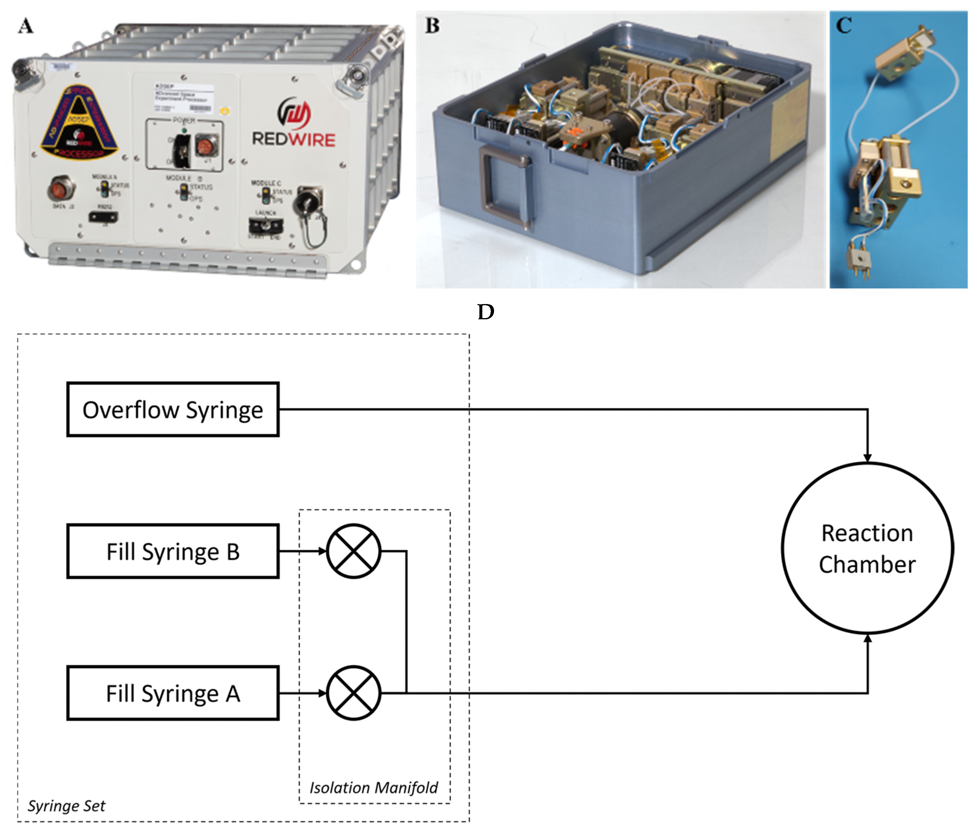
As previously described [39], the Pharmaceutical In-space Laboratory (PIL)–Biocrystal Optimization eXperiment (BOX) hardware was used to perform this experiment both on the ground and aboard the International Space Station (ISS) (Figure 2). The ADvanced Space Experiment Processor (ADSEP) (Figure 2A) is a facility on the ISS that operates cassettes (Figure 2B), which are routinely flown to and from the ISS via the National Aeronautics and Space Administration (NASA)’s Commercial Resupply (CRS) missions currently flown by SpaceX and Northop Grumman. The PIL-BOX Small Molecule Accelerated Laboratory for Structure (SMALS) (Figure 2B) contains four fluid loops (Figure 2C), which each contain two syringes (syringe A and syringe B) that are loaded with fluid and, upon experiment initiation, these syringes inject the fluids into the experiment chamber. The injection process and the crystallization process are observed via a microscope (Olympus objective, PLN 10X) and recorded via video and images at regular intervals. This ensures that the crystals that are initially formed at the onset of the experiment are the ones that are present when the experiment is complete. In addition, the video allows the scientists to watch the process of crystallization in microgravity and during ground control experiments.
Solutions were prepared for each of the target compounds as listed in Table 1. These solutions were loaded into the reaction chamber of each of the four fluid loops for each of the four experiments (Figure 2C). The antisolvent (0.4 mL; Table 1) for each experiment was loaded into syringe B (Figure 2C) and syringe A was left empty. Ground controls were placed in the ADSEP facility (Figure 2A) at ambient temperature (between 20 and 22 °C). Operations for injection of the antisolvent were initiated remotely, and syringe B was injected into the crystallization/observation chamber and observed by video (Figure 2B). In all cases, crystallization began immediately upon injection. The total volume of the crystallization/observation chamber was 0.3 mL, and excess volumes of liquid and gas (bubbles) were allowed to run into an overflow syringe (see schematic, Figure 2D). Any crystals present in the overflow syringe were also collected and evaluated.
The fluid loops for flight were loaded into the PIL-BOX SMALS cassettes and prepared for launch per NASA protocols at ambient temperatures (between 18 and 26 °C). Once on the ISS, the PIL-BOXes (Figure 2B) were removed from the packaging and inserted into the ADSEP facility (Figure 2A) by an astronaut. The antisolvent injections took place over approximately 3 min at a temperature setpoint of 22 °C and remained at that temperature for the duration of the crystallization process. In most cases, crystallization was observed upon mixing in the crystallization chamber and began immediately upon contact between the two solutions. The PIL-BOXes were processed (allowed to stand) for 2 days, observed, and images were recorded every 15 min up to the point where the PIL-BOXes were removed from the ADSEP facility. The PIL-BOXes were packaged, held on station for approximately 2 additional weeks, deorbited, and brought to a laboratory for initial return observations. During that time, the crystals remained suspended in the crystallization solvent mixture. The microgravity crystals were transported to the Butler University laboratories and compared with those grown under the same conditions terrestrially. Visualization was performed using a Leica S9D stereo microscope (Liece Mikrosyteme Vertrieb GmbH, Wetzlar, Germany) equipped with Enersight software (Flexcam_C3, v. 3.01a) at 0–5× magnification. Upon removal from the crystallization chamber and/or the overflow syringe, the crystals were placed on a glass microscope slide and evaluated visually (20× and 50× objectives of the BWTek BAC151C Raman microscope [BWTek a division of Metrohm, Plainsboro, NJ, USA]). A reference slide with a 1 mm ruler (World Precision Instruments, Sarasota, FL, USA, 500828, stage micrometer, 1 mm/200 DIV) was also photographed and used for measurements. Raman spectroscopy was obtained via an iRamanPlus fiber optic Raman system (BWTek, a division of Metrohm, Plainsboro, NJ, USA). A reference slide with a 1 mm ruler (World Precision Instruments, 500828, stage micrometer, 1 mm/200 DIV) was also photographed and used for measurements. Spectra are uncorrected and reported in Raman shift.

2.2. Seed Studies

Seed studies were performed by making up saturated solutions analyte (carbamazepine, atorvastatin calcium, methionine, and hydrocortisone 21-acetate) in the desired solvent (acetonitrile, dimethylformamide, ethanol, and dichloromethane) in a 10 mL flask. A single crystal recovered from the flight fluid loop is added to the saturated solution. The antisolvent (water, water, water, and ethyl acetate) is added dropwise in close vicinity to the seed crystal until no more crystallization takes place, approximately 1 h. These crystals are harvested and characterized by microscopy and Raman spectroscopy as above.
For the next generation, a single crystal was harvested from the previous generation and used as the seed. This crystal was added to a new saturated solution and antisolvent was added as above (see Figure 3). This sequence was repeated for up to 10 generations.

3. Results

Every effort was made to ensure that the crystallization conditions were uniform for both ground and flight; there was no way to measure the final solution concentrations post-experiment. In some cases, the overflow syringes (see Figure 2D) contained the majority of the crystals. In comparison to those crystals in the chambers, it was found that the crystals in the overflow syringes were spectroscopically identical to the crystals that formed in the crystallization chambers.

3.1. DL-Methionine

Crystallization of DL-methionine using ground conditions was complete within 5 min. The crystals that were formed were small needles, all under 0.2 mm in length. The crystals grown in microgravity were markedly larger, and of a different shape, clusters of rods (Figure 4) and grew within 10 min of injection. Evaluation by Raman spectroscopy indicated that the ground samples were β-DL-methionine. The flight samples were γ-DL-methionine as compared to the literature [51].
In three out of the four sample chambers, the DL-methionine crystals were very consistent in size, 0.1 × 0.2 to 0.1 × 0.3 mm with approximately 10–20 crystals in each chamber. In the fourth chamber, there were four very large crystals (one that was 0.7 × 1.4 mm, two that were 1.1 × 2.1 mm, and one that 1.6 × 3.0 mm), along with about 20 smaller crystals ranging in size from 0.1 × 0.2 mm to 0.2 × 0.9 mm. It is not clear why one of the chambers created such diversity in crystal sizes while the other three crystallization chambers were very uniform in size.

3.2. Glutamic Acid

Glutamic acid grown on Earth was also a small needle, all less than 0.2 mm, and all formed within 2 min. The flight samples grew in a hexagonal prism crystal habit (Figure 5), and nucleated within 2 min, and growth to their final length took up to 5 h. The hexagonal prisms were larger and more uniform, some as large as 1 mm in length. The Raman spectra of both the ground and flight samples were the same, indicating that both the ground and flight samples were both β-glutamic acid.

3.3. Atorvastatin Calcium

The atorvastatin ground and flight samples both appeared as spherulitic clusters, with the flight clusters appearing slightly larger (Figure 6), and both appeared immediately. However, Raman spectra of the ground-grown atorvastatin calcium best matches the Phase VIII polymorph [52]. A similar investigation that also used a DMF/water system for crystallization made Phase VIIIa and MD-1. The spectra of MD-1 is an excellent Raman match for our microgravity-grown crystal form [52].

3.4. Hydrocortisone 21-Acetate

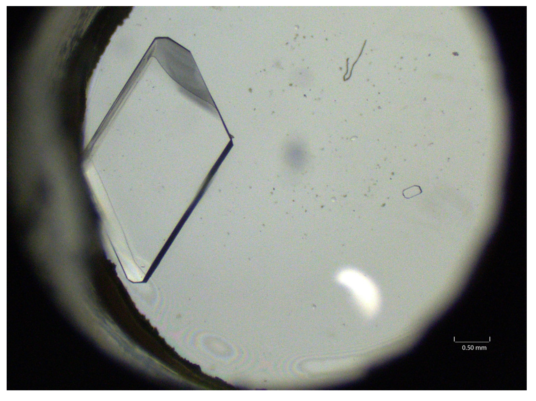
Hydrocortisone 21-acetate grew small, but nicely formed, crystals in ground studies (0.1 mm) within 5 min. The microgravity-grown crystals were orders of magnitude larger (Figure 7) and grew more slowly over the course of approximately 1 day. At the same magnification, the entire flight crystal does not fit in the frame. Zooming out (Figure 8), the size of the flight crystal can be seen. The largest crystal that was isolated was 8.0 mm in length and 5 mm wide. The ground control crystals were most consistent with Form II [53]. The flight crystals have a Raman spectrum that shares some characteristics with Form I and Form II, but it is clearly a distinct form which we are calling Form III.

3.5. Carbamazepine Seeds

The carbamazepine Form III seeds were harvested from a previous experiment (SpaceX 31) [41]. Ten generations of crystals were grown as described in the methods section with no degradation in the crystal form (Figure 9). Typical crystallization under these solvent/antisolvent conditions provides Form I (characteristic signal at 620 cm−1), which was not seen even in Generation 10.

3.6. Atorvastatin Calcium Seeds

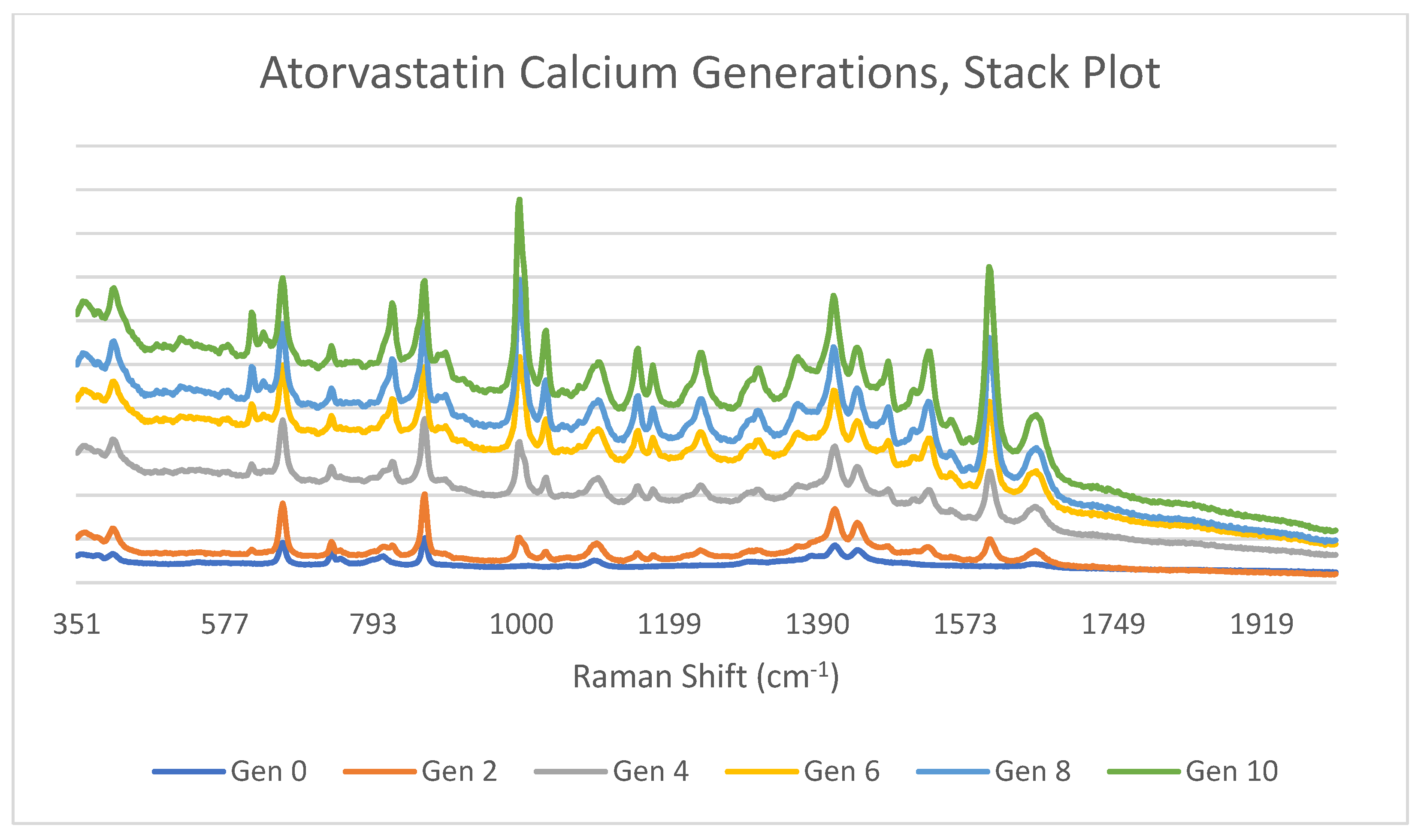
The MD-1 atorvastatin calcium crystals from this experiment were harvested as described in the methods section. Ten generations of crystals were grown with no degradation in the crystal form (Figure 10). The atorvastatin calcium ground controls created a polymorph with an unreported Raman spectrum with a key signal at 485 cm−1. There is no peak in this area even in Generation 10.

3.7. DL-Methionine Seeds

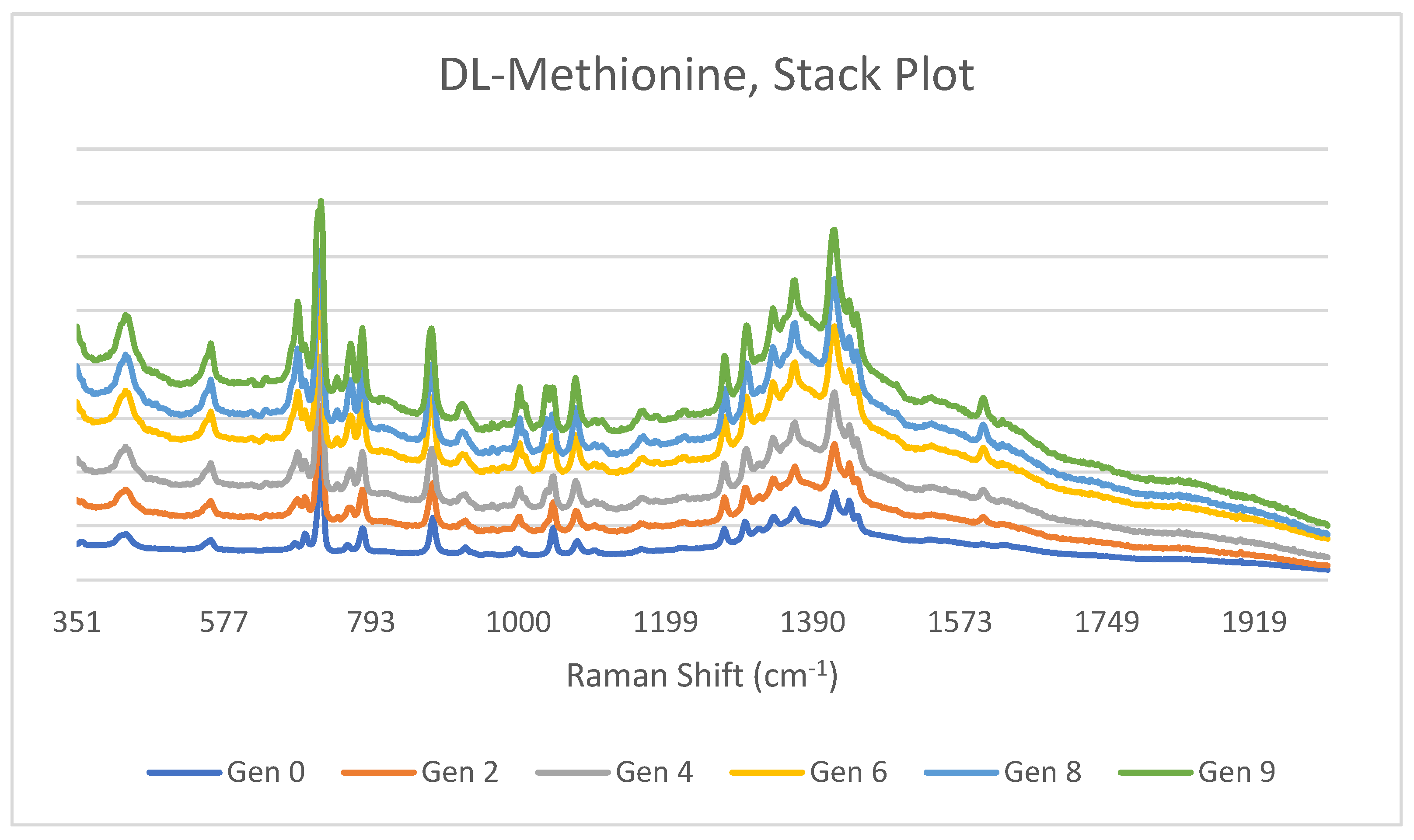
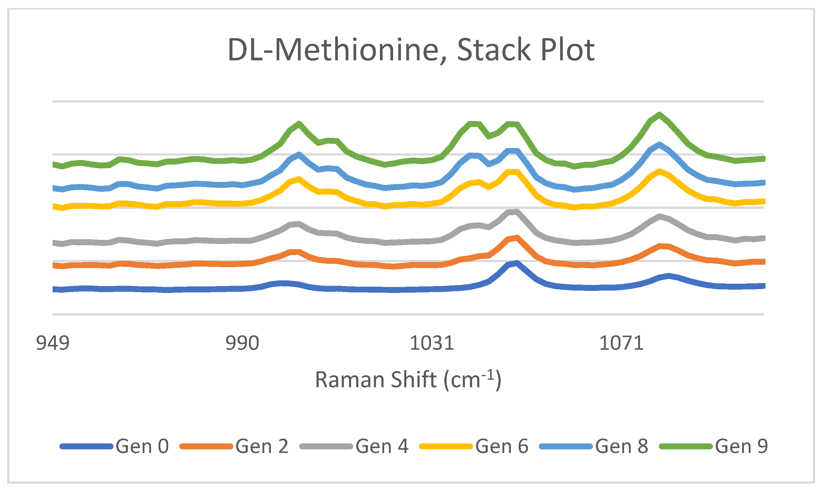
The γ-DL-methionine crystals from this experiment were harvested. Nine generations of crystals were grown (Figure 11). In Generation 4, a very small amount of the β-DL-methionine can be seen (see also Figure 12). By the ninth generation, the crystals are approximately 50% of each polymorph by Raman spectroscopy.

3.8. Hydrocortisone 21-Acetate Seeds

The hydrocortisone 21-acetate Form III crystals from this experiment were harvested. Using our crude method for seeding crystals, we found that we were able to obtain the desired (microgravity) polymorph in the first generation about half the time, with the Form II being formed the other half of the time. In the second generation, the Form II polymorph was exclusively formed.

4. Discussion

The comparisons between ground-grown and ISS-grown crystals demonstrate that the space-grown crystals were larger, had cleaner edges, and more perfect faces than their terrestrial counterparts. This was especially true for DL-methionine, glutamic acid, and hydrocortisone 21-acetate. In addition, different polymorphs were formed in space vs. ground for methionine, atorvastatin calcium, and hydrocortisone 21-acetate. The polymorph formed for hydrocortisone 21-acetate may be a known polymorph; however, this is the first Raman spectrum of this polymorph to be reported to our knowledge.

4.1. DL-Methionine

The microgravity-grown crystals of DL-methionine were up to four times larger than their ground-grown counterparts. In addition, the ground-grown crystals were β-DL-methionine, with a metastable γ-polymorph of DL-methionine formed in microgravity. Crystallization of the ground sample was immediately initiated on the edges of the chamber, particularly on the window, while crystals in the center of the chamber (free floating in solution) grew more slowly. In microgravity, crystals grew more slowly, and in the solution, and not on the sides of the chamber.
Visually, the crystals that were grown of both ground and flight DL-methionine are different from crystals reported in the literature [54]. The predicted morphology of ground samples is plate structures, while ours are more needle-like. While the microgravity-grown crystals can be described as having the form of hexagonal plates, the appearance of our hexagonal plates is elongated compared to those found in the literature [54].

4.2. Glutamic Acid

Glutamic acid also grew significantly larger (especially wider) crystals in microgravity compared to the ground-grown crystals. The ground crystals were needles and the microgravity crystals were transparent hexagonal prisms. Even though the crystals had different crystal habits, they were both β-glutamic acid. On the ground, the crystals grew quickly after nucleation, grew many small crystals, and did not change over time. On the ISS, the glutamic acid crystals nucleated quickly (as evidenced by the central point on many of the crystals) and then grew more slowly (20–40 h) over the time-period of the experiment. The slow growth could explain the very different appearance of the crystals, the fact that there were fewer (in number) crystals, as well as the length of the crystal growth.
Microscopic evaluation of the glutamic acid crystals indicates that they are consistent with crystal images in the literature [55]. The literature designation is that the β-form of glutamic acid is “needle-like” [55], which is likely appropriate for the ground-grown crystals. However, the flight crystals are the same shape, longer and wider in appearance, which we would describe as elongated prisms (Figure 4b).

4.3. Atorvastatin Calcium

The atorvastatin calcium crystals grew as spherulitic clusters in both ground and microgravity environments. They had slightly different crystal forms. The crystals nucleated and grew quickly in both ground and flight. Of the four compounds in this study, the atorvastatin calcium crystallized most quickly, with crystallization complete in 8 h. In all cases, the crystals grew on the sides of the chamber, especially the windows.
Ideally, these crystals would be candidates for PXRD analysis [56]. Unfortunately, this analysis requires 10s of milligrams of material, which are not available at this time. The samples will be archived should an instrument capable of analyzing a very small amount of material be found.
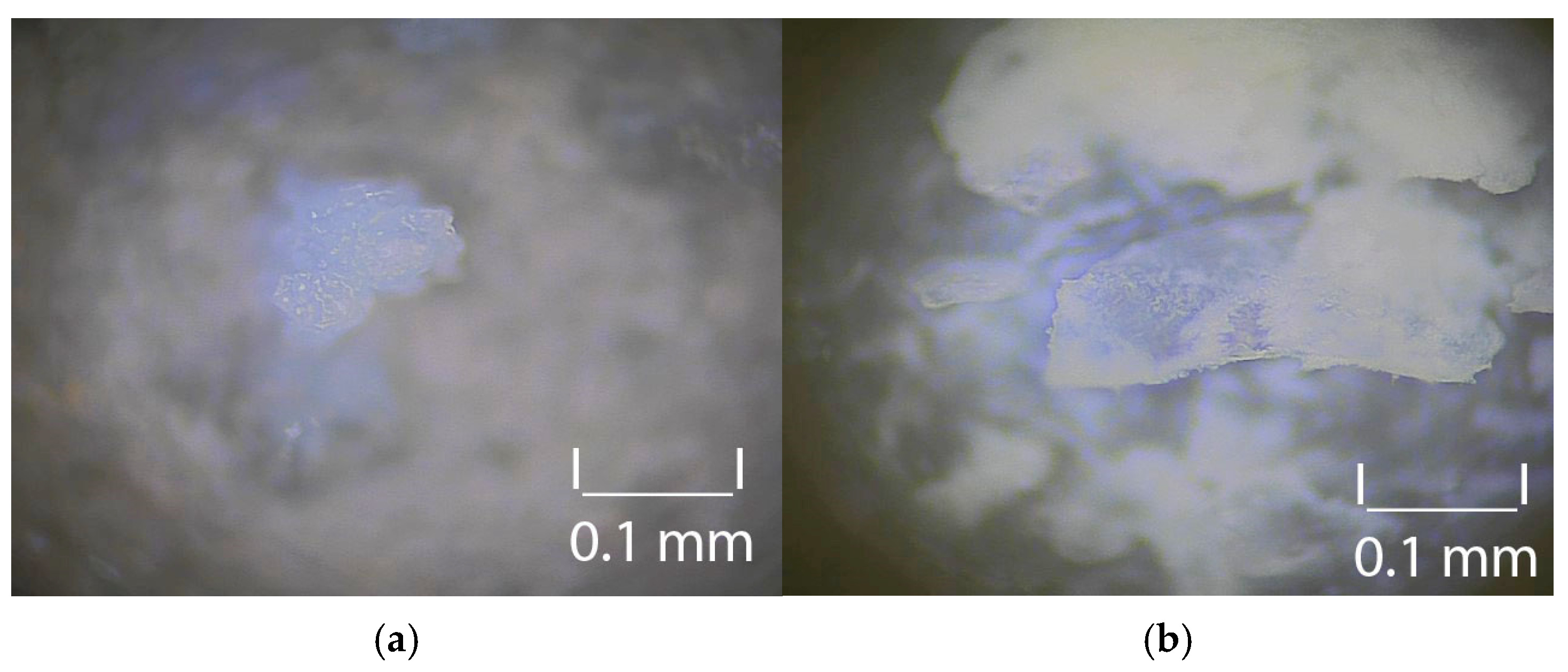
Visually, the crystals for both ground and flight are similar to the literature [56,57]. Microscopy also does not indicate that the ground or flight samples are crystalline, as they appear to both be amorphous solids (Figure 13). The Raman spectroscopy clearly indicated that these were, indeed, crystalline and gave spectra consistent with crystalline atorvastatin calcium.

4.4. Hydrocortisone 21-Acetate

Hydrocortisone 21-acetate grew significantly larger crystals, as much as 80 times larger. The microgravity-grown crystals were striking in their clarity. The ground-grown crystals were all Form II, and the microgravity grown crystals were all previously not characterized Form III hydrocortisone 21-acetate. In microgravity, the hydrocortisone 21-acetate experiments were plagued by large bubbles in the reaction chamber, making the visualization of crystal formation challenging. In two out of the four cases, the majority of the crystal growth was in the overflow syringe. Crystals were observed after 16 h in two of the four chambers.
Again, this would be an ideal candidate for PXRD to confirm this new polymorph of hydrocortisone 21-acetate. Despite being a very mature product [58], solvent-free hydrocortisone reference spectra for all five polymorphs are challenging to find [59]. The hydrocortisone 21-acetate samples will also be archived should an instrument capable of analyzing a very small amount of material be found.
Solvents are known to impact the hydrocortisone 21-acetate crystal habit [60]. The small parallelepiped crystal that was observed in the ground control study was consistent with the literature [60]. However, the extraordinary increase in size and transition to a prismatic habit of the microgravity-grown crystals was unexpected.

4.5. Seeds Studies

Our seed studies were unoptimized. There are a series of evaluations that could be undertaken to improve the process of using microgravity-grown crystals as seeds on a larger scale [61,62]. However, our limited initial studies clearly demonstrate that these crystals can be used as seeds.
In the cases of carbamazepine and atorvastatin calcium, a single crystal from each batch was used as a seed, and the same polymorph was formed in each generation, for 10 generations. While 10 generations was an arbitrary marker, the concept of using single crystals as seeds was supported. For the cases of carbamazepine and atorvastatin calcium, the polymorph formed was not significantly different in energy from other polymorphs, 1–2 kJ/mol for carbamazepine [63] and less than 2.5 kcal/mol for atorvastatin calcium [64].
However, DL-methionine gave the metastable γ-polymorph for three generations before the more stable β-polymorph began to be formed. This is consistent with the literature, which reports that γ-DL-methionine has a faster nucleation time [51], and polymorphic transformation of DL-methionine can occur through solution-mediated or solid-state mediated transformations. While the γ-form does engage in solid-state transformations even at higher temperatures [64], solution phase γ-DL-methionine can convert to β-DL methionine over time. Remarkably, half of the mixture was still the metastable γ-DL-methionine in the ninth generation. An experiment to compare the kinetics of formation of γ-DL-methionine and β-DL methionine from the solution would be helpful. Alternate forms of batch crystallization, evaporation or temperature control, could be employed by a specialist in batch seeding to provide superior results [65].
Hydrocortisone 21-acetate provided Form III upon use as a seed in a single generation for two out of five trials, with the other half providing Form II. All forms of hydrocortisone 21-acetate convert to Form I, indicating that they are metastable [50]. The Form III that was grown in microgravity may require more advanced methods [66] by a specialist using temperature control, different solvents, evaporative crystallization, and/or faster isolation in order to dependably obtain this particular polymorph of hydrocortisone 21-acetate. This may be an ideal case to design an apparatus where a Raman spectrometer can observe the formation of crystals in situ to determine the identity of polymorphs as they are formed and fall out of the solution.

5. Conclusions

Four small organic molecules (DL-methionine, glutamic acid, atorvastatin calcium, and hydrocortisone 21-acetate) have been crystallized in space, returned to Earth, harvested and characterized. In addition, a new form of hydrocortisone 21-acetate has been characterized by Raman spectroscopy. All of the crystals grew larger in microgravity than on the ground. Several of these compounds behaved differently when crystallized in space, displaying a different crystal habit from ground grown crystals, or a different polymorph despite the same concentration and solvents. While this increases the number of small molecules grown under microgravity conditions reported in the literature from 8 to 12, we are working to develop a clear picture of the impact of microgravity on the behaviors of small molecule crystal growth.
We have also demonstrated that the microgravity-grown crystals can be successfully utilized as seeds for additional crystallization studies, for up to 10 generations. This, despite the crystallizing solvent/antisolvent mixtures favoring a different form without the seed crystals. In some cases, metastable polymorphs were used as seed crystals and provided additional generations of metastable forms. Superior results in utilizing space-grown crystals as seeds would likely be obtained by a specialist in batch seeding of pharmaceutical compounds.

Supplementary Materials

The following supporting information can be downloaded at: https://www.mdpi.com/article/10.3390/cryst15090825/s1, Raman spectra of methionine, glutamic acid, atorvastatin calcium, hydrocortisone 21-acetate, carbamazepine.

Author Contributions

Conceptualization: K.A.S., S.T. and A.M.W.; methodology: S.T. and A.M.W.; validation: J.P., S.T., L.M. and A.M.W.; formal analysis: J.P., L.M.; investigation: A.M.W. and K.A.S.; resources: K.A.S.; data curation: J.P., L.M. and A.M.W.; writing—original draft preparation: A.M.W. and S.T.; writing—review and editing: M.K.M. and K.A.S.; supervision: A.M.W. All authors have read and agreed to the published version of the manuscript.

Funding

This research was funded by NASA Task Orders 80JSC022F0133 and 80JSC025FA075; CASIS provided launch and astronaut support through the CASIS User Agreement for flight sponsorship, UA-2022-8785 and UA-2025-9722; Butler University for their support of undergraduate research; and Redwire for their support of the project.

Data Availability Statement

The original contributions presented in this study are included in the article/Supplementary Material. Further inquiries can be directed to the corresponding author.

Acknowledgments

We gratefully acknowledge the work of NASA astronaut Butch Wilmore for his assistance in performing the microgravity studies.

Conflicts of Interest

Authors Molly K. Mulligan, Kenneth A. Savin, and Stephen Tuma were employed by the company Redwire. The remaining authors declare that the research was conducted in the absence of any commercial or financial relationships that could be construed as a potential conflict of interest.

Abbreviations

The following abbreviations are used in this manuscript:
XRDX-ray diffractometry
ISSInternational Space Station
PIL-BOXPharmaceutical In-space Laboratory (PIL)–Biocrystal Optimization eXperiment
ADSEPADvanced Space Experiment Processor
SMALSSmall Molecule Accelerated Laboratory for Structure

References

  1. Singh, P.; Sharma, S.; Kumar Sharma, P.; Alam, A. Drug Polymorphism: An Important Pre-formulation Tool in the Formulation Development of a Dosage Form. Curr. Phys. Chem. 2024, 14, 2–19. [Google Scholar] [CrossRef]
  2. Brittain, H.G. Polymorphism and Solvatomorphism 2008. J. Pharm. Sci. 2010, 99, 3648–3664. [Google Scholar] [CrossRef] [PubMed]
  3. Saifee, M.; Inamdar, N.; Dhamecha, D.L.; Rathi, A.A. Drug Polymorphism: A Review. Int. J. Health Res. 2009, 2, 291–306. [Google Scholar] [CrossRef]
  4. Shi, Q.; Chen, H.; Wang, Y.; Xu, J.; Liu, Z.; Zhang, C. Recent advances in drug polymorphs: Aspects of pharmaceutical properties and selective polymorphs. Int. J. Pharm. 2022, 611, 121320. [Google Scholar] [CrossRef]
  5. Variankaval, N.; Cote, A.S.; Doherty, M.F. From Form to Function: Crystallization of Active Pharmaceutical Ingredients. AIChE J. 2008, 54, 1682–1688. [Google Scholar] [CrossRef]
  6. Sachin Kumar Sing, R.; Kumar Yadav, A.; Gulati, M.; Mittal, A.; Narang, R.; Garg, V. Polymorph control: Success so far and future expectations. Int. J. PharmTech Res. 2016, 9, 144–165. [Google Scholar]
  7. Corvis, Y. Solid State Development and Processing of Pharmaceutical Molecules: Salts, Cocrystals, and Polymorphism; Gruss, M., Ed.; Wiley-VCH: Weinheim, Germany, 2022. [Google Scholar]
  8. Zhou, Y.; Lv, C.; Liu, X.; Gao, J.; Gao, Y.; Wang, T.; Huang, X. An Overview on Polymorph Preparation Methods of Active Pharmaceutical Ingredients. Cryst. Growth Des. 2024, 24, 584–600. [Google Scholar] [CrossRef]
  9. Lowe, D. Stalking Polymorphs. Science, 14 March 2023. Available online: https://www.science.org/content/blog-post/stalking-polymorphs (accessed on 25 July 2025).
  10. Heng, T.; Yang, D.; Wang, R.; Zhang, L.; Lu, Y.; Du, G. Progress in Research on Artificial Intelligence Applied to Polymorphism and Cocrystal Prediction. ACS Omega 2021, 6, 15543–15550. [Google Scholar] [CrossRef]
  11. Burcham, C.L.; Doherty, M.F.; Peters, B.G.; Price, S.L.; Salvalagilio, M.; Reutzel-Edens, S.M.; Price, L.S.; Reddy Addula, R.K.; Francia, N.; Khanna, V.; et al. Pharmaceutical Digital Design: From Chemical Structure through Crystal Polymorph to Conceptual Crystallization Process. Cryst. Growth Des. 2024, 24, 5417–5438. [Google Scholar] [CrossRef]
  12. Parent, S.D.; Smith, P.A.; Purcell, D.K.; Smith, D.T.; Bogdanowich-Knipp, S.J.; Bhavsar, A.S.; Chan, L.R.; Croom, J.M.; Bauser, H.C.; McCalip, A.; et al. Ritanovir Form III: A Coincidental Concurrent Discovery. Cryst. Growth Des. 2023, 23, 320–325. [Google Scholar] [CrossRef]
  13. Surov, A.O.; Drozd, K.V.; Ramazanova, A.G.; Churakov, A.V.; Vologzhania, A.V.; Kulikova, E.S.; Perlovich, G.L. Polymorphism of Carbamazepine Pharmaceutical Cocrystal: Structural Analysis and Solubility Performance. Pharmaceutics 2023, 15, 1747. [Google Scholar] [CrossRef]
  14. Salazar-Rojas, D.; Kaufman, T.S.; Maggio, R.M. A comprehensive approach toward concomitant triclabendazole polymorphism in pharmaceutical products. J. Drug Deliv. Sci. Tech. 2021, 62, 102386. [Google Scholar] [CrossRef]
  15. Wirgowska, G.; Stasilowicz, A.; Miklaszewski, A.; Lewandowska, K.; Cielecka-Piontek, J. Structural Polymorphism of Sorafenib Tosylate as a Key Factor in Its Solubility Differentiation. Pharmaceutics 2021, 13, 384. [Google Scholar] [CrossRef] [PubMed]
  16. Toukabri, I.; Bahri, S.; Sfar, S.; Ali Lassoued, M. Impact of crystal polymorphism of rifaximin on dissolution behavior. Heliyon 2024, 10, e27131. [Google Scholar] [CrossRef] [PubMed]
  17. Cheng, S.; McKenna, G.B. Isothermal Crystallization and Time-Temperature Transformation of Amorphous Nifedipine: A Case of Polymorphism Formation and Conversion. Molec. Pharm. 2021, 18, 2786–2802. [Google Scholar] [CrossRef]
  18. Li, M.; Xu, W.; Su, Y. Solid-state NMR spectroscopy in pharmaceutical sciences. Trends Anal. Chem. 2021, 135, 116152. [Google Scholar] [CrossRef]
  19. Souza Almeida, L.; Carneiro, J.; Alberto Colnago, L. Time domain NMR for polymorphism characterization: Current status and future prospects. Int. J. Pharm. 2025, 669, 125027. [Google Scholar] [CrossRef]
  20. Helena Mazurek, A.; Szeleszczuk, Ł.; Maciej Pisklak, D. Periodic DFT Calculations—Review of Applications in the Pharmaceutical Sciences. Pharmaceutics 2020, 12, 415. [Google Scholar] [CrossRef]
  21. Yan, T.; Deng, Y.; Yu, Z.; John, E.; Han, R.; Yao, Y.; Liu, Y. Exploring the Polymorphism of Cinchomeronic Acid at High Pressure. J. Phys. Chem. C 2021, 125, 8582–8588. [Google Scholar] [CrossRef]
  22. Guerain, M.; Willart, J.-F. Polymorphic Transformations of Pharmaceutical Materials Induced by Mechanical Milling: A Review. Pharmaceutics 2025, 17, 946. [Google Scholar] [CrossRef]
  23. Chakraborty, J.; Subash, M.; Thorat, B.N. Drying induced polymorphic transformation of pharmaceutical ingredients: A critical review of recent progresses and challenges. Dry. Technol. 2022, 40, 2817–2835. [Google Scholar] [CrossRef]
  24. Park, H.; Kim, J.-S.; Hong, S.; Ha, E.-S.; Nie, H.; Zhou, Q.T.; Kim, M.-S. Tableting process-induced solid-state polymorphic transition. J. Pharm. Investig. 2022, 52, 175–194. [Google Scholar] [CrossRef]
  25. Maruyama, M.; Yoshikawa, H.Y.; Takano, K.; Yoshimura, M.; Mori, Y. Solution-mediated phase transition of pharmaceutical compounds: Case studies of acetaminophen and aspirin. J. Cryst. Growth 2023, 602, 126990. [Google Scholar] [CrossRef]
  26. Roy, S.; Chamberlin, B.; Matzger, A.J. Polymorph Discrimination Using Low Wavenumber Raman Spectroscopy. Org. Proc. Res. Dev. 2013, 17, 976–980. [Google Scholar] [CrossRef] [PubMed]
  27. Fateixa, S.; Nogueira, H.I.S.; Trindade, T. Carbamazepine polymorphism: A re-visitation using Raman imaging. Int. J. Pharm. 2022, 617, 121632. [Google Scholar] [CrossRef]
  28. Aragon, F.F.H.; Haeck, C.M.; Morals, P.C.; Variano, B. Polymorphism characterization of segesterone acetate: A comprehensive study using CRPD, FT-IR and Raman spectroscopy. Int. J. Pharm. 2021, 596, 120234. [Google Scholar] [CrossRef]
  29. Du, Y.; Frank, D.; Chen, Z.; Struppe, J.; Su, Y. Ultrafast magic angle spinning NMR characterization of pharmaceutical solid polymorphism: A posaconazole example. J. Magn. Reson. 2023, 346, 107352. [Google Scholar] [CrossRef]
  30. Wright, H.; Williams, A.; Wilkinson, A.; Harper, L.; Savin, K.; Wilson, A.M. An Analysis of Publicly Available Microgravity Crystallization Data; Emergent Themes Across Crystal Types. Cryst. Growth Des. 2022, 22, 6849–6851. [Google Scholar] [CrossRef]
  31. Jackson, K.; Brewer, F.; Wilkinson, A.; Williams, A.; Whiteside, B.; Wright, H.; Harper, L.; Wilson, A.M. Microgravity Crystal Formation. Crystals 2024, 14, 12. [Google Scholar] [CrossRef]
  32. NASA; Sands, K.; Bowman, A. “What Is Microgravity?”. 2023. Available online: https://www.nasa.gov/centers-and-facilities/glenn/what-is-microgravity/#:~:text=But%20they’re%20not%20falling,%C3%9710%2D6%20g (accessed on 4 September 2025).
  33. Yoo, H.-D.; Wilcox, W.R.; Lal, R.; Trolinger, J.D. Modeling the growth of triglycine sulphate crystals in spacelab 3. J. Cryst. Growth 1988, 92, 101–117. [Google Scholar] [CrossRef]
  34. Lal, R.B.; Batra, A.K.; Trolinger, J.D.; Wilcox, W.R.; Steiner, B. Growth and characteristics of tgs crystals grown aboard first international microgravity laboratory (IML-1). Ferroelectrics 1994, 158, 81–85. [Google Scholar] [CrossRef]
  35. Aggarwal, M.D.; Batra, A.K.; Lal, R.B.; Penn, B.G.; Frazier, D.O. Growth and Characteristics of Bulk Single Crystals Grown from Solution on Earth and in Microgravity. NASA Technical Reports Server. Available online: https://ntrs.nasa.gov/citations/20110006347 (accessed on 10 September 2024).
  36. Kroes, R.L.; Reiss, D.A.; Lehoczky, S.L. Nucleation of Crystals from Solution in Microgravity-USML-1 Glovebox (GBX) Investigation. In Proceedings of the Joint Launch + One Year Science Review of USML-1 and USMP-1 with the Microgravity Measurement Group, Huntsville, AL, USA, 22–24 September 1993; Volume 2. [Google Scholar]
  37. Nielsen, K.F.; Lind, M.D. Results of the TTF-TCNQ- and the Calcium Carbonate-Crystallization on the Long Duration Exposure Facility. In Proceedings of the First LDEF Post-Retrieval Symposium Abstracts, Hyatt Orlando, Kissimmee, FL, USA, 2–8 June 1991; NASA Langley Research Center: Hampton, VA, USA, 1991; pp. 725–731. [Google Scholar]
  38. Bauser, H.C.; Smith, P.A.; Parent, S.D.; Chan, L.R.; Bhavsar, A.S.; Condon, K.H.; McCalip, A.; Croom, J.M.; Purcell, D.K.; Bogdanowich-Knipp, S.J.; et al. Return of Ritanovir: A Study on the Stability of Pharmaceuticals Processed in Orbit and Returned to Earth. ChemRxiv 2024, 3. Available online: https://chemrxiv.org/engage/chemrxiv/article-details/65faecbee9ebbb4db91b4ac1 (accessed on 10 September 2024).
  39. Miller, L.; Mulligan, M.K.; Savin, K.A.; Tuma, S.; Wilson, A.M. Crystallization of Small Molecules in Microgravity Using Pharmaceutical In-Space Laboratory-Biocrystal Optimization eXperiment (PIL-BOX). Crystals 2025, 15, 527. [Google Scholar] [CrossRef]
  40. Jackson, K.; Hoff, R.; Wright, H.; Wilkinson, A.; Brewer, F.; Williams, A.; Whiteside, B.; Macbeth, M.R.; Wilson, A.M. An Analysis of Protein Crystals Grown under Microgravity Conditions. Crystals 2024, 14, 652. [Google Scholar] [CrossRef]
  41. Wilkinson, A.; Brewer, F.; Wright, H.; Whiteside, B.; Williams, A.; Harper, L.; Wilson, A.M. A meta-analysis of semiconductor materials fabricated in microgravity. npj Microgravity 2024, 10, 73. [Google Scholar] [CrossRef]
  42. DeLucas, L.J.; Smith, C.D.; Smith, H.W.; Vijay-Kumar, S.; Senadhi, S.E.; Ealick, S.E.; Carter, D.C.; Salemme, F.R.; Ohlendorf, D.H.; Einspahr, H.M.; et al. Protein Crystal Growth in Microgravity. Science 1989, 246, 651–654. [Google Scholar] [CrossRef]
  43. McPherson, A.; DeLucas, L.J. Microgravity protein crystallization. npj Microgravity 2015, 1, 15010. [Google Scholar] [CrossRef]
  44. Maes, D.; Decanniere, K.; Zegers, I.; Vanhee, C.; Sleutel, M.; Willaert, R.; Weerdt, C.; Martial, J.; Declercq, J.; Evrard, C.; et al. Protein crystallization under microgravity conditions: What did we learn on TIM crystallization from the Soyuz missions? Microgravity Sci. Technol. 2007, 19, 90–94. [Google Scholar]
  45. Nicoud, N.; Licordari, F.; Myerson, A.S. Estimation of the Solubility of Metastable Polymorphs: A Critical Review. Cryst. Growth Des. 2018, 18, 7228–7237. [Google Scholar] [CrossRef]
  46. Singhal, D.; Curatolo, W. Drug polymorphism and dosage form design: A practical perspective. Adv. Drug Deliv. Rev. 2004, 56, 335–347. [Google Scholar] [CrossRef]
  47. Li, Z.; Ma, Y.; Lin, J.; Gao, Z.; Wu, S.; Li, W.; Han, D.; Gong, J.; Wang, J. The polymorph and crystal habit control of dl-methionine assisted by ultrasound. J. Cryst. Growth 2022, 596, 126818. [Google Scholar] [CrossRef]
  48. Srinivasan, K.; Dhanasekaran, P. Nucleation control and crystallization of l-glutamic acid polymorphs by swift cooling process and their characterization. J. Cryst. Growth 2011, 318, 1080–1084. [Google Scholar] [CrossRef]
  49. Skorda, D.; Kontoyannis, C.G. Identification and quantitative determination of atorvastatin calcium polymorph in tablets using FT-Raman spectroscopy. Talanta 2008, 74, 1066–1070. [Google Scholar] [CrossRef] [PubMed]
  50. Callow, R.K.; Kennard, O. Polymorphism of Cortisone Acetate. J. Pharm. Pharmacol. 1961, 13, 723–733. [Google Scholar] [CrossRef]
  51. Wantha, L.; Flood, A.E. Crystal growth rates and secondary nucleation threshold for γ-DL-methionine in aqueous solution. J. Cryst. Growth 2011, 318, 117–121. [Google Scholar] [CrossRef]
  52. Jin, Y.S.; Ulrich, J. New Crystalline Solvates of Atorvastatin Calcium. Chem. Eng. Technol. 2010, 33, 839–844. [Google Scholar] [CrossRef]
  53. Deeley, C.M.; Spragg, R.A.; Threlfall, T.L. A comparison of Fourier transform infrared and near-infrared Fourier transform Raman spectroscopy for quantitative measurements: An application in polymorphism. Spectrochim. Acta 1991, 47A, 1217–1223. [Google Scholar] [CrossRef]
  54. Matsuoka, M.; Yamanobe, M.; Tezuka, N.; Takiyama, H.; Ishii, H. Polymorphism, morphologies and bulk densities of DL-methionine agglomerate crystals. J. Cryst. Growth 1999, 198/199, 1299–1306. [Google Scholar] [CrossRef]
  55. Hou, Y.; Liu, T.; Yang, Y.; Ma, C.Y.; Wang, X.E.; Ni, X. In Situ Measurement of 3D Crystal Size Distribution by Double-View Image Analysis with Case Study on L-Glutamic Acid Crystallization. Ind. Eng. Chem. Res. 2020, 59, 4646–4658. [Google Scholar]
  56. Shete, G.; Puri, V.; Kumar, L.; Bansal, A.K. Solid State Characterization of Commercial Crystalline and Amorphous Atorvastatin Calcium Samples. AAPS PharmSciTech 2010, 11, 598–609. [Google Scholar] [CrossRef]
  57. Kurakula, M.; El-Helw, A.M.; Sobahi, T.R.; Abdelaal, M.Y. Chitosan based atorvastatin nanocrystals: Effect of cationic charge on particle size, formulation stability, and in-vivo efficacy. Int. J. Nanomed. 2015, 15, 321–334. [Google Scholar] [CrossRef]
  58. Hench, P.S.; Kendall, E.C.; Slocumb, C.H.; Polley, H.F. The effect of a hormone of the adrenal cortex (17-hydroxy-11-dehydrocorticosterone; compound E and of pituitary adrenocorticotropic hormone on rheumatoid arthritis. Proc. Staff Meet. Mayo Clin 1949, 24, 181–197. [Google Scholar] [CrossRef]
  59. Suitchmezian, V.; Jeß, I.; Näther, C. Structural, Thermodynamic, and Kinetic Aspects of the Trimorphism of Hydrocortisone. J. Pharm. Sci. 2008, 97, 4516–4527. [Google Scholar] [CrossRef] [PubMed]
  60. Chen, J.; Wang, J.; Ulrich, J.; Yin, Q.; Xue, L. Effect of Solvent on the Crystal Structure and Habit of Hydrocortisone. Cryst. Growth Des. 2008, 8, 1490–1494. [Google Scholar] [CrossRef]
  61. He, Y.; Gao, Z.; Zhang, T.; Sun, J.; Ma, Y.; Tian, N.; Gong, J. Seeding Techniques and Optimization of Solution Crystallization Processes. Org. Proc. Res. Dev. 2020, 24, 1839–1849. [Google Scholar] [CrossRef]
  62. Long, Y.; Ment, A.; Xu, Q.; Shan, B.; Wang, Y.; Zhang, F.; Yu, Z.-Q. Uncertainty analysis of seed recipe for optimal control of crystal size distribution in batch cooling crystallization. Chem. Eng. Res. Des. 2024, 204, 601–611. [Google Scholar] [CrossRef]
  63. Czernicki, W.; Baranska, M. Carbamazepine polymorphs: Theoretical and experimental vibrational spectroscopy studies. Vib. Spect. 2013, 65, 12–23. [Google Scholar] [CrossRef]
  64. Van de Streek, J.; Firaha, D.; Kaduk, J.A.; Blanton, T.N. From ‘crystallographic accuracy’ to ‘thermodynamic accuracy’: A redetermination of the crystal structure of calcium atorvastatin trihydrate (Lipitor®). Acta Cryst. 2024, B80, 682–687. [Google Scholar] [CrossRef]
  65. Corvis, Y. Impact of Solid Forms on API Scale-Up. In Solid State Development and Processing of Pharmaceutical Molecules: Salts, Cocrystals, and Polymorphism; Gruss, M., Ed.; Wiley-VCH: Weinheim, Germany, 2022; p. 307. [Google Scholar]
  66. Yamanobe, M.; Takiyama, H.; Matsuoka, M. Polymorphic transformation of DL-methionine crystals in aqueous solutions. J. Cryst. Growth 2002, 237–239 Pt 3, 2221–2226. [Google Scholar] [CrossRef]
Figure 1. Target molecules for crystallization in microgravity at the International Space Station (ISS).
Crystals 15 00825 g001
Figure 2. Images of the ADSEP facility (A), a PIL-BOX SMALS (B), a fluid loop (C), and the basic schematic of the fluid loop (D).
Crystals 15 00825 g002
Figure 3. Schematic of how seed studies were performed.
Crystals 15 00825 g003
Figure 4. (a) Methionine grown on the ground. (b) Methionine grown at the ISS. Images taken by a Leica S9D stereo microscope (Leica Mikrosyteme Vertrieb GmbH, Wetzlar, Germany) equipped with Enersight software (Flexcam_C3, v. 3.01a) at 5.5×.
Crystals 15 00825 g004
Figure 5. (a) Glutamic acid grown on the ground. (b) Glutamic acid grown at the ISS. Images taken by a Leica S9D stereo microscope (Leica Mikrosyteme Vertrieb GmbH, Wetzlar, Germany) equipped with Enersight software (Flexcam_C3, v. 3.01a) at 5.5×.
Crystals 15 00825 g005
Figure 6. (a) Atorvastatin calcium grown on the ground. (b) Atorvastatin calcium grown at the ISS. Images taken by a Leica S9D stereo microscope (Leica Mikrosyteme Vertrieb GmbH, Wetzlar, Germany) equipped with Enersight software (Flexcam_C3, v. 3.01a) at 5.5×.
Crystals 15 00825 g006
Figure 7. (a) Hydrocortisone 21-acetate grown on the ground. (b) Hydrocortisone 21-acetate grown at the ISS. Images taken by a Leica S9D stereo microscope (Leica Mikrosyteme Vertrieb GmbH, Wetzlar, Germany) equipped with Enersight software (Flexcam_C3, v. 3.01a) at 5.5×.
Crystals 15 00825 g007
Figure 8. Image of the whole crystal of hydrocortisone 21-acetate. Images taken by a Leica S9D stereo microscope (Leica Mikrosyteme Vertrieb GmbH, Wetzlar, Germany) equipped with Enersight software (Flexcam_C3, v. 3.01a) at 1×.
Crystals 15 00825 g008
Figure 9. Carbamazepine Raman spectra. Generation 0—seed crystal from microgravity experiment through Generation 10.
Crystals 15 00825 g009
Figure 10. Atorvastatin Calcium Raman spectra. Generation 0—seed crystal from microgravity experiment through Generation 10.
Crystals 15 00825 g010
Figure 11. DL-Methionine Raman spectra. Generation 0—seed crystal from microgravity experiment through Generation 9.
Crystals 15 00825 g011
Figure 12. DL-Methionine Raman expansion. β-DL-methionine growing over generations.
Crystals 15 00825 g012
Figure 13. Atorvastatin calcium ground (a) and flight (b) crystals. Images taken on BWTek microscope at 50×.
Crystals 15 00825 g013
Table 1. Solution conditions for microgravity experiments.
Substrate (Amount)Solvent (Amount)Antisolvent
DL-Methionine (24.6 mg)Water (1 mL)Ethanol
Glutamic Acid (7.9 mg)Water (1 mL)Ethanol
Atorvastatin Calcium (28.9 mg)Dimethylformamide (1 mL)Water
Hydrocortisone 21-Acetate (58.8 mg)Dichloromethane (1 mL)Ethyl Acetate
Disclaimer/Publisher’s Note: The statements, opinions and data contained in all publications are solely those of the individual author(s) and contributor(s) and not of MDPI and/or the editor(s). MDPI and/or the editor(s) disclaim responsibility for any injury to people or property resulting from any ideas, methods, instructions or products referred to in the content.