Article

Supramolecular Stabilisation Leads to Challenging Coordination in Fe(III) Hydrazinylpyrazine Schiff Base Complexes

by
Omar Coughlin
,
Sophie L. Benjamin
and
Anthony J. Fitzpatrick
*
Department of Chemistry and Forensic Science, School of Science and Technology, Nottingham Trent University, Clifton Ln, Nottingham, NG1 8NS, UK
*
Author to whom correspondence should be addressed.
Crystals 2025, 15(9), 805; https://doi.org/10.3390/cryst15090805
Submission received: 21 July 2025 / Revised: 8 September 2025 / Accepted: 10 September 2025 / Published: 12 September 2025
(This article belongs to the Section Crystal Engineering)

Abstract

The coordination chemistry of a hydrazinylpyrazine-derived Schiff base ligand (L1), formed in situ from salicylaldehyde and 2-hydrazinopyrazine, with Fe(III) salts has been systematically investigated under varied synthetic conditions. Six discrete Fe(III) complexes (1a1e and 2) were isolated and structurally characterised via single-crystal X-ray diffraction, revealing diverse coordination geometries ranging from five-coordinate pseudo-trigonal bipyramidal to six-coordinate pseudo-octahedral environments. The supramolecular architectures are governed by a rich interplay of non-covalent interactions, including hydrogen bonding, halogen bonding, and π–π stacking, which significantly influence the crystallisation pathways and final solid-state structures. Continuous shape measure (CShM) analysis highlights substantial geometric distortion in the bis-tridentate complexes, attributed to the steric and electronic constraints imposed by the ligand. Powder X-ray diffraction and infrared spectroscopy confirm the presence of multiple phases in bulk samples, underscoring the kinetic competition between crystallisation and coordination. The results demonstrate that supramolecular stabilisation of monoligated species can kinetically inhibit bis-ligation, with ligand excess and solvent polarity serving as key parameters to direct complex speciation. These findings provide insight into the delicate balance between coordination geometry, ligand strain, and supramolecular assembly in Fe(III) Schiff base complexes.

1. Introduction

Coordination chemistry is a foundational pillar of inorganic chemistry, underpinning the design of functional materials, catalysts, and molecular devices [1,2,3,4]. Central to this field is the ability to control and understand the coordination environment of metal complexes. Factors such as ligand denticity, bite angle, electronic effects, and solvent interactions govern the geometry, stability, and reactivity of metal centres. Even subtle changes in these parameters can lead to profound differences in the physicochemical properties of the resulting complexes, including their redox behaviour [5], magnetic susceptibility [6], and optical characteristics [7,8,9].
Pyrazine-based ligands are widely used in coordination chemistry due to their rigid, planar structure and ability to act as π-acceptors and bridging units [10]. These features make them ideal for constructing coordination complexes, from simple mononuclear complexes [11] to Hofmann-type clathrates [12,13] and discrete electron transfer systems [14]. Schiff base ligands, particularly those derived from salicylaldehyde, are similarly versatile and have been extensively used in the synthesis of coordination compounds with applications in catalysis, magnetism, and photochemistry [15,16,17,18,19]. Their modularity allows for precise tuning of steric and electronic properties, making them indispensable tools in the rational design of metal complexes.
Single-crystal X-ray diffraction (SCXRD) provides precise information on coordination geometry, bond lengths, angles, and intermolecular interactions, which are critical for interpreting spectroscopic data and predicting reactivity. Jahn–Teller distortions in Cu(II) and Mn(III) can be directly observed and quantified through crystallographic analysis [20,21,22,23]. However, caution should be used when looking at a single crystal versus possible bulk properties.
In this study, the coordination chemistry of the Schiff base ligand derived from salicylaldehyde and 2-hydrazinopyridine (Sal-hypy) with Fe(III) salts is investigated, Figure 1. The ligand, L1, has been previously synthesised [24], with only the Zn(II) complexes being published to date [25]. These exhibit unusual ligation with a mixed phenol/phenolate coordination motif, leading to an extended H-bonded supramolecular network. This provides a crystallisation-induced emission enhancement where the crystalline materials are more strongly luminescent than the solution of the same material. By synthesising and structurally characterising Sal-hypy iron complexes, we aim to elucidate their coordination modes and assess how ligand design influences the resulting molecular architecture, coordination motifs, spin state and homogeneity of the final structural arrangement of the complexes.
Here we present six iron(III) complexes that produce varying coordination motifs. The same ligand, HL1, based on the Schiff base condensation of salicylaldehyde and 2-hydrazinopyrazine, is used in all cases.
All six complexes resulted from similar synthetic methodologies. The in situ reaction forming L1 was followed by the addition of the metal source. Crystals of both 1a and 1b were isolated from the same batch, showing that there is competitive formation of both a five-coordinate and a six-coordinate system. Under different conditions, single crystals of 1c1e were isolated; however, multiple components are evident in the powder X-ray analyses of the bulk products. Changes in stoichiometry, reaction solvent, base addition, and iron source all affect the final product isolated and have been fully investigated to elucidate the effect on coordination in the reaction mixture. The complexes may also yield interesting magnetic properties, as similar systems have promoted spin switching phenomena [26]; however, we have not undertaken these studies due to the difficulty in obtaining pure samples.

2. Materials and Methods

2.1. Synthesis

All chemicals were purchased from either Sigma-Alrich/Merck (UK) or Fluorochem (UK) and used as received.
  • Complex 1a and 1b
Salicylaldehyde (0.732 g, 5.99 mmol) was added neat to a stirring suspension of 2-hydrazinopyrazine (0.6638 g, 6.03 mmol) in EtOH/MeCN (1:1, 120 mL). The reaction mixture was stirred for 30 min, during which a cloudy yellow precipitate formed. FeCl3·6H2O (0.8114 g, 3.00 mmol) was added as a solid, causing an instant colour change to deep brown/black. The reaction solution was stirred for 10 min and then filtered under gravity. Then, 1 mL was removed and placed in a slow diffusion chamber with diethyl ether anti-solvent; the remaining solution was slowly evaporated until the formation of black crystalline precipitate, which was removed by filtration and washed with diethyl ether. Crystals of FeLCl2·0.229MeCN (1a) and FeLCl2(EtOH) (1b) were obtained from the slow diffusion crystallisation of the same solution.
  • Complex 1c
A suspension of ligand (L1) was prepared as above from salicylaldehyde (0.1278 g, 1.05 mmol) and 2-hydrazinopyrazine (0.1092 g, 0.99 mmol) in MeOH (20 mL). FeCl3·6H2O (0.0873 g, 0.32 mmol) was added as a solid, causing an instant colour change to deep brown/black. The solution was stirred for 10 min and then filtered under gravity. Then, 1 mL was removed from the solution and placed in a slow diffusion chamber with diethyl ether anti-solvent; the remaining solution was slowly evaporated until the formation of a precipitate, which was removed by filtration and washed with diethyl ether. Crystals of FeL1Cl2(MeOH)·MeOH (1c) were obtained from the slow diffusion crystallisation.
  • Complex 1d
A suspension of ligand (L1) was prepared as above from salicylaldehyde (0.4240 g, 3.47 mmol) and 2-hydrazinopyrazine (0.3829 g, 3.48 mmol) in EtOH/MeCN (1:1, 70 mL). Et3N (~0.5 mL) was added, and then 20 mL of the solution was removed and added to solid FeCl3·6H2O (0.1310 g, 0.48 mmol). The solution was stirred for 10 min and then filtered under gravity. Then, 1 mL was removed from the solution and placed in a slow diffusion chamber with diethyl ether anti-solvent; the remaining solution was slowly evaporated until the formation of a precipitate, which was removed by filtration and washed with diethyl ether. Crystals of [Et3NH][FeL1Cl3] (1d) were obtained from the slow diffusion crystallisation.
  • Complex 1e
A suspension of ligand (L1) was prepared from salicylaldehyde (0.1220 g, 1.00 mmol) and 2-hydrazinopyrazine (0.1100 g, 1.00 mmol) in EtOH/MeCN (1:1, 20 mL). FeCl3·6H2O (0.0801 g, 0.30 mmol) was added as a solid. The solution was stirred for 10 min and then filtered under gravity. Then, 1 mL was removed from the solution and placed in a slow diffusion chamber with diethyl ether anti-solvent; the remaining solution was slowly evaporated until the formation of a precipitate, which was removed by filtration and washed with diethyl ether. Crystals of [Fe(L1)2]Cl (1e) were obtained from the slow evaporation crystallisation.
  • Complex 2
A suspension of ligand (L1) was prepared as above from salicylaldehyde (0.2502 g, 2.05 mmol) and 2-hydrazinopyrazine (0.2214 g, 2.01 mmol) in EtOH/MeCN (1:1, 40 mL). Fe(NO3)3·9H2O (0.2697 g, 0.67 mmol) was added as a solid, causing an instant colour change to deep brown/black. Then, 20 mL of this solution was removed and added to solid NaB(3,5-(CF3)2C6H3)4 (0.8886 g, 1.00 mmol). The solution was stirred for 10 min and then filtered under gravity. Then, 1 mL was removed from the solution and placed in a slow diffusion chamber with diethyl ether anti-solvent; the remaining solution was slowly evaporated until the formation of a precipitate, which was removed by filtration and washed with diethyl ether. Crystals of [FeL2]NO3·H2O (2) were obtained from the slow diffusion crystallisation.

2.2. Crystallography

Suitable single crystals were mounted using a MiTeGen sample holder on an Xcalibur, Sapphire3, Gemini diffractometer (Oxford Diffraction, Oxford, UK). The crystal was kept at 150 K during data collection. Using Olex2 [27], the structure was solved with the SHELXT [28] structure solution program using Intrinsic Phasing and refined with the SHELXL-2018/3 [29] refinement package using Least Squares minimisation.

3. Results and Discussion

3.1. Crystallographic Analysis

All complexes crystallise with similar habits, forming dark purple, almost black crystals with block-like morphologies. It is not possible to discern each crystal’s identity visually through the mixtures isolated from complexes 1a2.
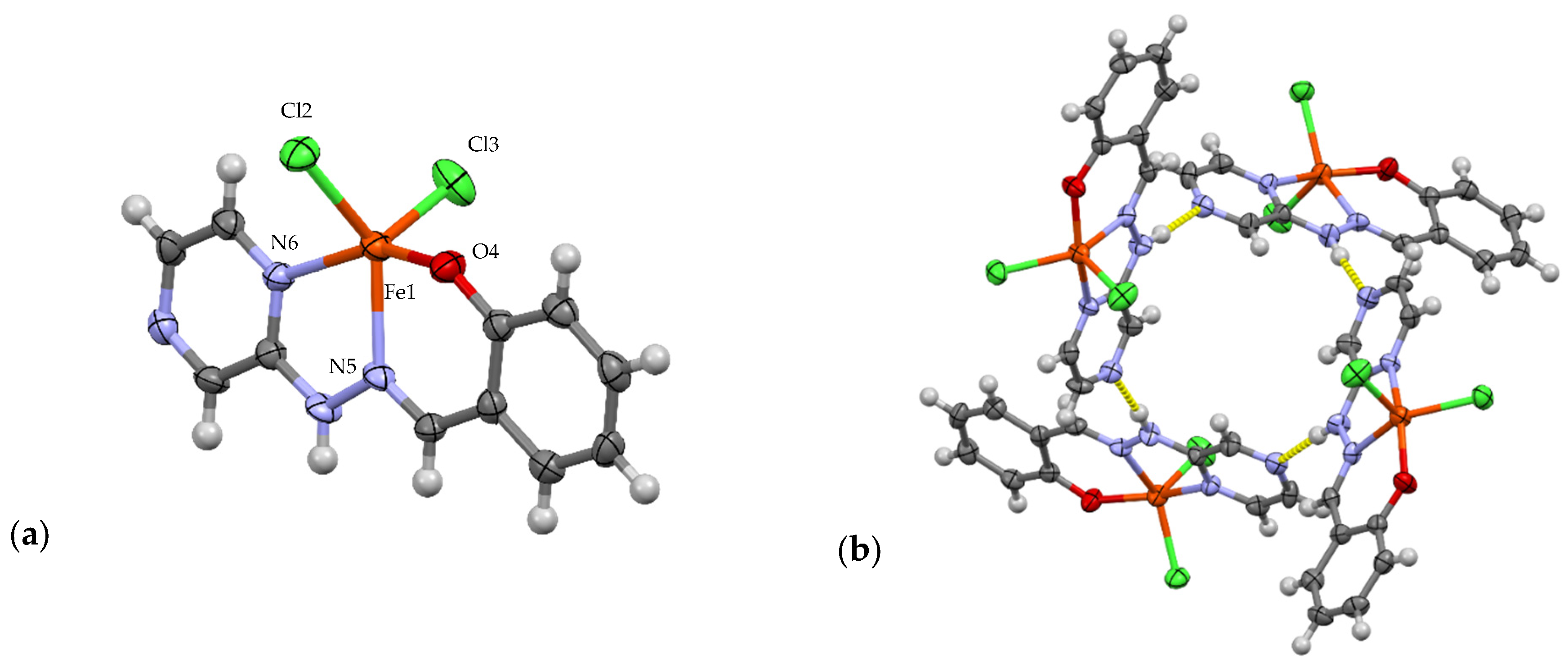
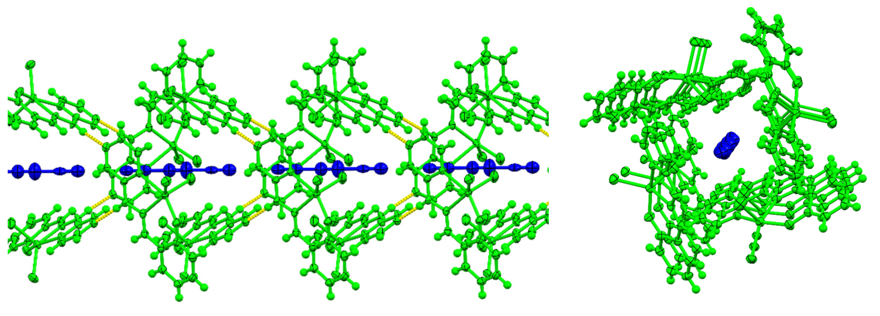
Complex 1a, [Fe(L1)Cl2]·CH3CN, crystallises in the tetragonal space group P42/n. The asymmetric unit consists of a neutral iron(III) complex with a fully coordinated NNO ligand (L1) and two chloride ligands in a strained pseudo-trigonal bipyramidal coordination (Figure 2a). The complex co-crystallises with one partially occupied molecule of acetonitrile, disordered over an inversion centre. The bond lengths of the phenol, imine, and pyrazine metal bond are 1.886(4) Å, 2.139(5) Å, and 2.149(5), respectively, indicating a high-spin Fe(III) ion [30]. The bond lengths of Fe(III) NNO complexes have been extensively studied, especially with respect to spin-switchable systems. The longer bond lengths indicate the population of the higher energy antibonding d-orbitals. This may influence the coordination possibilities due to the repulsion between the ligand and the metal centre. There is hydrogen bonding present, shown in yellow, between the hydrazinyl proton and the non-coordinated pyrazine nitrogen, 1.968 Å, and due to the tetragonal arrangement of the complex, this forms a tetrameric structure (Figure 2b). The hydrogen-bonded tetramer has a 1,3-alternate structure and stacks with its neighbouring tetramers, forming a channel in which the disordered acetonitrile sits (Figure 3).
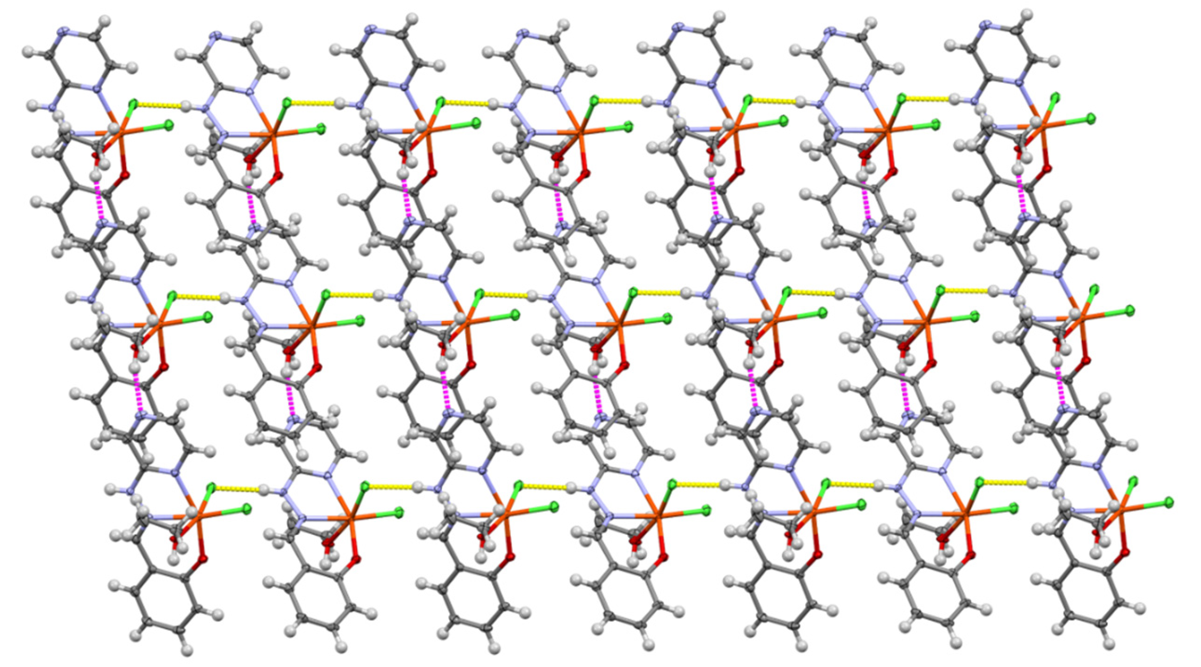
Complex 1b, [Fe(L1)Cl2EtOH], crystallises in the monoclinic space group P21/c. Complex 1b is also neutral; however, the vacant coordination site is now taken up by an exogenous ethanol molecule completing the hexacoordinate system, leading to a pseudo-octahedral Fe(III) ion (Figure 4a). The metal–ligand bond lengths again indicate a high-spin Fe(III) species. This complex crystallises without any solvent molecules in the lattice. The bond length for the ethanol O-Fe bond is 2.136(2) Å, verifying the protonated state rather than an ethoxide coordination. The hydrogen bonding present in complex 1b is of interest as hydrogen–halogen interactions are now observed, as well as traditional hydrogen bonding motifs. The chloride ligand trans to the ethanol oxygen interacts with the hydrazinyl proton of the neighbouring complex, 2.298 Å, and this repeats to form a one-dimensional non-covalent chain along the b-axis of the lattice (Figure 4b). There is a linking of these 1-D chains to form a 2-D sheet (Figure 5). The non-coordinated pyrazine nitrogen hydrogen bonds to the proton on the coordinated ethanol oxygen linking the halogen–hydrogen-bonded chains, 1.943 Å. Due to the position of the ethanol tails in the chains, a 3-D non-covalent architecture is not possible, as they act as a capping agent for the 2-D sheets formed by the hydrogen/halogen bonding.
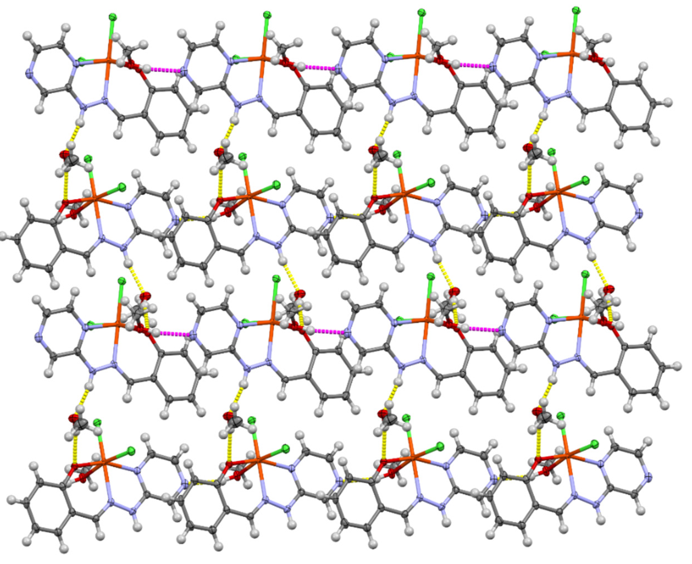
Complex 1c, [Fe(L1)Cl2MeOH]·MeOH, also crystallises in the monoclinic space group P21/c. Complex 1c is also neutral, with an exogenous methanol molecule completing the hexacoordinate system, rather than an ethanol molecule as with complex 1b, leading to a slightly distorted octahedral Fe(III) ion (Figure 6a). The metal–ligand bond length indicates a high-spin Fe(III) species. The methanol oxygen–metal bond length of 2.1539(17) Å agrees with a protonated donor. Complex 1c displays a more complex hydrogen bonding system than complex 1b. The hydrazinyl proton interacts with the methanol solvent molecule, 1.942 Å, which then forms a hydrogen bond with the phenolate oxygen of the neighbouring molecule, 2.167 Å, forming a one-dimensional chain (Figure 6b). This 1-D chain is then linked to the neighbouring chains through the interactions of the non-coordinated pyrazine of one complex bonding to the proton of the coordinated methanol oxygen, 1.965 Å, on the neighbouring species chain, forming a two-dimensional sheet (Figure 7). The coordinated methanol and the solvent methanol act as a barrier to forming a 3-D hydrogen-bonded architecture throughout the solid.
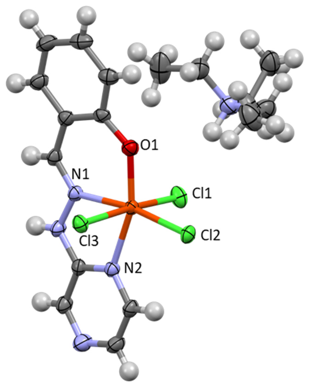
Complex 1d, HNEt3[Fe(L1)Cl3], also crystallises in the monoclinic space group P21/c. With the addition of triethylamine in the synthesis, complex 1d is an anionic complex with three coordinated chloride ligands and a deprotonated L1. The iron centre is therefore formally tetra-anionic with a triethylammonium cation balancing the overall Fe(III) charge, Figure 8. The asymmetric unit consists of two anions and two cations. The bond lengths around the metal centres once again indicate a high-spin Fe(III) configuration. Complex 1d is the first complex that exhibits no hydrogen- or halogen-based interactions from the non-ligating nitrogen in the pyrazine ring to a neighbouring molecule. A one-dimensional chain, along the a-axis, is present through halogen–hydrogen interactions (Figure 9). There are two motifs present in the 1-D chain. A single hydrogen bond between the chloride ligand and hydrazinyl proton, 2.372 Å, makes one bridge (blue bonds), while there is a bifurcated hydrogen bond between the hydrazinyl proton and the chloride of the next molecule, 2.746 Å, along with an interaction between the same proton and the coordinated phenolate oxygen atom, 2.310 Å (yellow bonds). There is no further linking into a sheet or 3-D architecture.
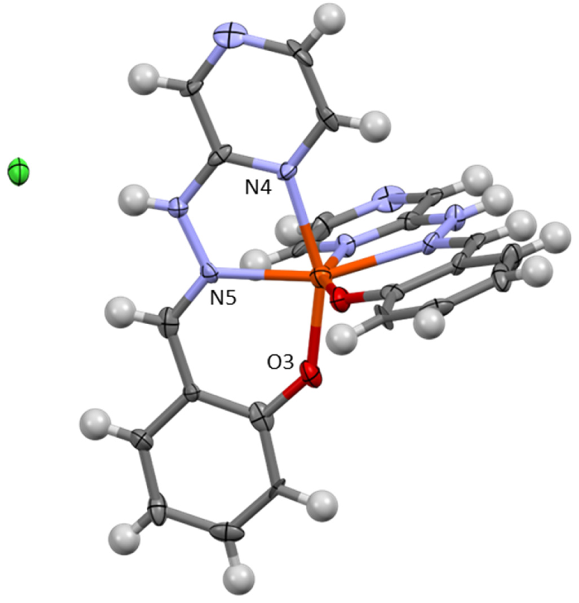
Complex 1e, [Fe(L1)2]Cl, crystallises in the monoclinic space group P2/c. Here, both deprotonated L1 ligands coordinate in a tridentate meridional fashion, completing the coordination sphere of the metal ion. The pseudo-octahedral cationic complex is charge-balanced by a chloride ion. A centre of symmetry exists at the Fe(III) centre; therefore, there are only three distinct Fe–ligand bond lengths, N4Pyrazine-Fe, N5Imine-Fe, and O3Phen-Fe, with distances of 2.158(3) Å, 2.147(3) Å, and 1.913(3) Å, respectively. These bond lengths are indicative of a high-spin Fe(III) ion (Figure 10). Halogen–hydrogen interactions are dominant in the lattice. The chloride spectator ion acts as a bridge between two complex cations through interactions with the hydrazinyl proton on the ligand backbone, 2.247 Å, creating a one-dimensional non-covalent chain along the c-axis (yellow). A double-layered chain structure occurs due to offset π–π stacking between the pyrazine ring of one complex and the phenyl ring of the neighbouring complex (blue). While a direct overlap of the rings does not occur, the distances between phenyl and pyrazine rings are 3.42–3.47 Å, and the angle between the planes of the rings is 2.43° (Figure 11).
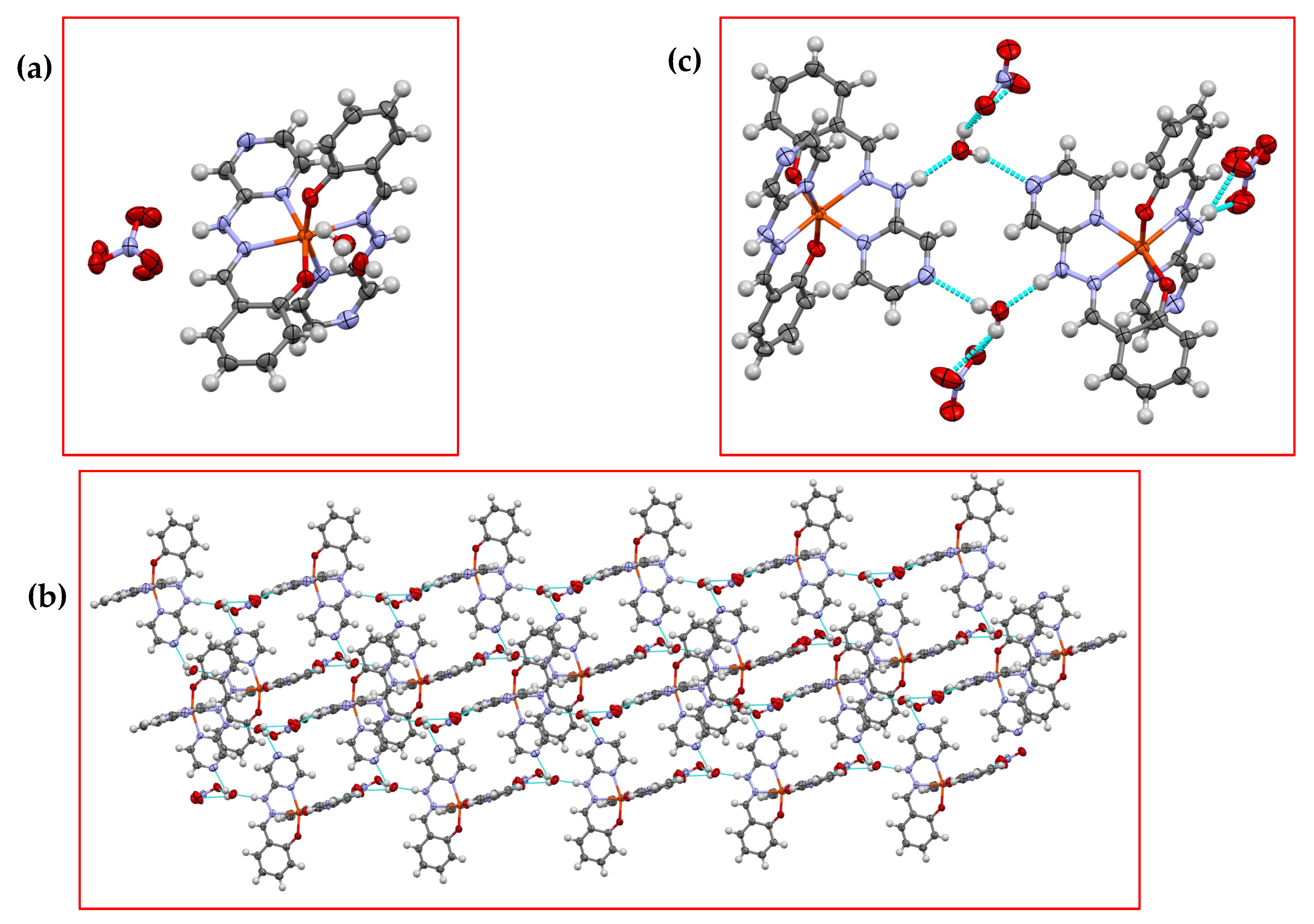
Complex 2 crystallises in a triclinic P-1 space group. As in complex 1d, both sets of L1 are fully coordinated in a meridional coordination. The asymmetric unit consists of one cationic Fe(III) complex, one nitrate anion, disordered over two positions, and one water molecule, again disordered over two positions (Figure 12a). The bond lengths are indicative again of a distorted octahedral high-spin Fe(III) system. The solvent molecule is intrinsically important to the hydrogen bonding network present in complex 2. A layered hydrogen-bonded one-dimensional chain, along the c-axis, is formed by interactions between the hydrazinyl proton and the water molecule, 1.817 Å, which in turn interacts with a neighbouring molecule’s non-ligating pyrazine nitrogen, 2.188 Å (Figure 12b). There is a complex arrangement of hydrogen bonding forming the layered chain. The nitrate has a bifurcated hydrogen bond with the hydrazinyl proton on one of the coordinated ligands, 2.002 Å and 2.634 Å, and with the proton of the water molecule, 2.483 Å and 2.210 Å. The hydrazinyl proton on the other coordinated L1 ligand with a hydrogen bond with the water molecule. This nitrate-based hydrogen bonding links the macrocyclic hydrogen bonding motif found between the hydrazinyl proton and the free pyrazine nitrogen and two water molecules (Figure 12c).

3.2. Continuous Shape Measure Analysis

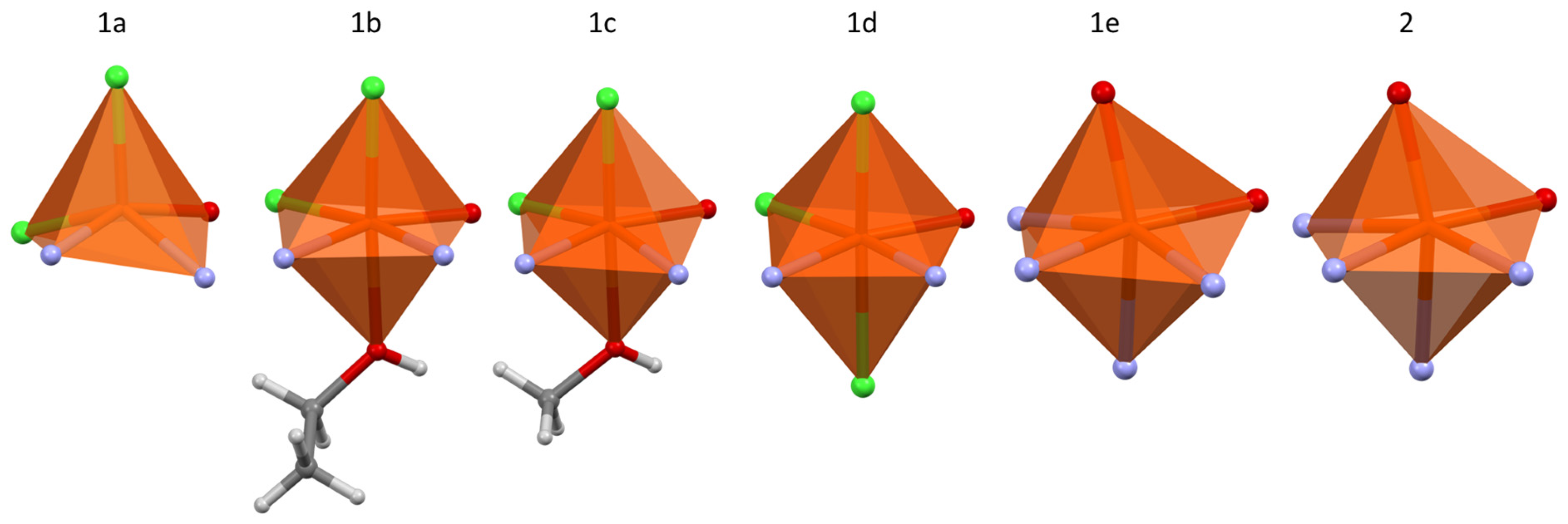
The various motifs observed in the six complexes, described above, all take distorted octahedral geometries, bar complex 1a, which has a five-coordinate geometry. The octahedral nature of these complexes allows for investigation of ligand strain induced by L1 on the coordination geometry in complexes 1b, 1c, 1d, 1e, and 2. Continuous shape measures (CShMs) were calculated using Shape 2.0 to investigate the degree of deviation from an ideal octahedron, Table 1 [31]. The ideal value for a perfect octahedron is 0, with that for a perfect trigonal prism being 16 [32,33]. From these values, we can see, even visually, that there is a significant strain in taking the bis-tricoordinate coordination motif (Figure 13). The N,N,O coordination from L1 in all cases exhibits a very tight angle and distorts the meridional coordination far from the ideal angles of 90° and 180° for the cis and trans coordination sites, respectively.

3.3. Powder X-Ray Analysis

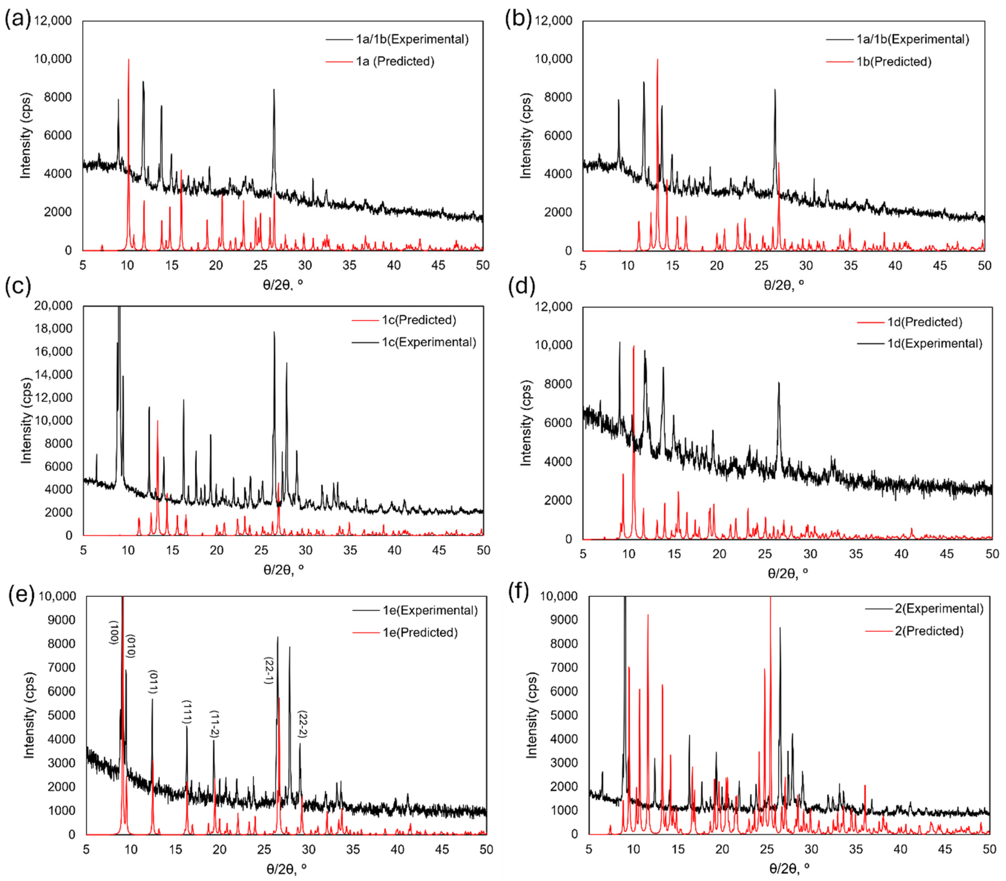
To further probe the heterogeneity of the complexes, powder X-ray diffraction experiments were performed. The experimental powder X-ray diffractograms for the bulk material isolated from each synthetic pathway are compared to the predicted powder pattern generated from the single-crystal structure obtained by picking crystals from each batch (Figure 14). The diffractogram of the bulk material from which crystals of 1a and 1b were picked contains only very small contributions from these phases, with the major peaks likely corresponding to a third, unknown phase (Figure 14a,b). This unknown phase also seems to dominate the bulk from which 1d was isolated, once again in contrast to the single-crystal structure obtained. For 1e, the experimental and predicted powder patterns match well, and the pattern can be indexed to the structural data, with only one significant unindexed peak, demonstrating that an excess of ligand can be used to control crystallisation and cleanly isolate the 2:1 complex. A significant quantity of 1e also appears to be present in the bulk that yielded single crystals of 1c, 1d, and 2, though these also contain minor peaks corresponding to other phases.

3.4. Infrared Analysis

Due to the mixture of complexes present in each reaction mixture, the IR spectra of 1a2 are complicated (Figure S1). This reflects the difficulty in isolating one coordination motif from these reaction mixtures on a bulk scale.
For all complexes, there are similarities in the modes associated with the pyrazine ring, C=C and C=N, and the imine, C=N, in the regions of 1350–1600 cm−1 and 1550–1650 cm−1, respectively. There is splitting of the peaks, representative of the slightly differing coordination environments and, therefore, the back bonding to the coordinated systems and downshifts in wavenumber. A N-H stretch can be observed in all cases due to the hydrazinyl proton of L1, 3300–3450 cm−1. In the mixed complex 1a/b, the large broad peak centred at 3300 cm−1 is the O-H moiety from the protonated and coordinated alcohol solvent found in the six-membered, L1Cl2ROH, coordination motif. A strong peak around 1450 cm−1 is also present in compound mixture 1a/b, which represents the non-coordinating acetonitrile molecule found in 1a. Complex 1e appears to exhibit the least amount of peak splitting with well-defined single peaks and no evidence of obvious solvent coordination in the bulk material.

3.5. Synthetic Insights

The speciation of the FeL1 complex system would seem to depend on the ability to coordinate a second ligand in preference to the crystallisation of the chloride-coordinated species isolated in the examples above. One way of affecting this was to change the reaction stoichiometry (1e). Solvent choice will also affect the hydrogen bonding motifs possible and proton sources such as ammonium counterions (1a/b/c/d). Addition of other possible counterions such as (3,5-bis(trifluoromethyl)phenyl)borate and tetrakis(pentafluorophenyl)borate to abstract or replace the chloride yielded no difference to the resulting mixture of isolated structures. The solvent and stoichiometry remained the deciding factors for which structures were observed.

4. Conclusions

We have probed the effect of varying the synthetic conditions on complex formation and speciation, using infrared spectroscopy as well as single-crystal and powder X-ray diffraction techniques. L1 can and does form several different high-spin Fe(III) complexes in situ. We postulate that this is due, in part, to the presence of strong intramolecular non-covalent interactions. We hypothesise that non-covalent interactions stabilise the 1:1 coordination complex, allowing the crystallisation process to occur before the second ligand coordination takes place. This competitive crystallisation versus coordination can be directed to some extent through the synthetic conditions employed. Solvent choice plays some role, but still allows for several phases to appear as observed in complexes 1a, 1b, and 1c. The addition of a base, as in 1d, to try to facilitate the coordination through deprotonation of the phenols to phenolates, yielded an anionic complex charge-balanced by a triethylammonium cation with no evidence of the bis-tridentate motif. Only when the excess addition of ligand to metal is performed does the [Fe(L1)2]Cl complex preferentially form, 1e. The difficulty in formation of this complex can be ascribed to several factors: the large networks of non-covalent interactions that stabilise the 1:1 complexes; the strain the ligand itself puts on the octahedral geometry, as evidenced by the continuous shape measures; and the need for excess ligand to overcome the obviously slow coordination of the second ligand in light of the previous competitive processes.

Supplementary Materials

The following supporting information can be downloaded at https://www.mdpi.com/article/10.3390/cryst15090805/s1. Table S1: Crystallographic Parameters; Figure S1: ATR-IR spectra for complexes 1a2.

Author Contributions

Conceptualization, A.J.F.; methodology, O.C. and S.L.B.; formal analysis, O.C., S.L.B., and A.J.F.; investigation, O.C.; data curation, O.C. and S.L.B.; writing—original draft preparation, O.C. and A.J.F.; writing—review and editing, S.L.B. and A.J.F.; supervision, S.L.B. and A.J.F. All authors have read and agreed to the published version of the manuscript.

Funding

This research received no external funding.

Data Availability Statement

Crystallographic data can be accessed at www.ccdc.cam.ac.uk (CCDC 2473920-2473925).

Acknowledgments

The authors acknowledge Nottingham Trent University for support and funding for X-ray facilities (NTU/20/1076/MAL and NTU/21/2126/ML) and for the NTU VC PhD fellowship granted to O.C.

Conflicts of Interest

The authors declare no conflicts of interest.

References

  1. Martinho, P.N.; Martins, F.F.; Bandeira, N.A.G.; Calhorda, M.J. Spin Crossover in 3D Metal Centers Binding Halide-Containing Ligands: Magnetism, Structure and Computational Studies. Sustainability 2020, 12, 2512. [Google Scholar] [CrossRef]
  2. Boda, S.K.; Pandit, S.; Garai, A.; Pal, D.; Basu, B. Bacterial Siderophore Mimicking Iron Complexes as DNA Targeting Antimicrobials. RSC Adv. 2016, 6, 39245–39260. [Google Scholar] [CrossRef]
  3. Atanasov, M.; Aravena, D.; Suturina, E.; Bill, E.; Maganas, D.; Neese, F. First Principles Approach to the Electronic Structure, Magnetic Anisotropy and Spin Relaxation in Mononuclear 3d-Transition Metal Single Molecule Magnets. Coord. Chem. Rev. 2015, 289–290, 177–214. [Google Scholar] [CrossRef]
  4. Batten, S.R.; Champness, N.R.; Chen, X.-M.; Garcia-Martinez, J.; Kitagawa, S.; Öhrström, L.; O’Keeffe, M.; Suh, M.P.; Reedijk, J. Coordination Polymers, Metal–Organic Frameworks and the Need for Terminology Guidelines. CrystEngComm 2012, 14, 3001. [Google Scholar] [CrossRef]
  5. Esra, D.; Pilia, L.; Marchiò, L.; Mercuri, M.L.; Serpe, A.; Barsella, A.; Fort, A.; Dalgleish, S.J.; Robertson, N.; Deplano, P. Redox-Switchable Chromophores Based on Metal (Ni, Pd, Pt) Mixed-Ligand Dithiolene Complexes Showing Molecular Second-Order Nonlinear-Optical Activity. Inorg. Chem. 2011, 50, 2058–2060. [Google Scholar] [CrossRef]
  6. Brooker, S. Spin Crossover with Thermal Hysteresis: Practicalities and Lessons Learnt. Chem. Soc. Rev. 2015, 44, 2880–2892. [Google Scholar] [CrossRef] [PubMed]
  7. Büldt, L.A.; Wenger, O.S. Chromium Complexes for Luminescence, Solar Cells, Photoredox Catalysis, Upconversion, and Phototriggered NO Release. Chem. Sci. 2017, 8, 7359–7367. [Google Scholar] [CrossRef] [PubMed]
  8. Belair, S.D.; Maupin, C.L.; Logue, M.W.; Riehl, J.P. Analysis of the Temperature Dependence of the Racemization of Eu(III) Complexes through Measurement of Steady-State Circularly Polarized Luminescence. J. Lumin. 2000, 86, 61–66. [Google Scholar] [CrossRef]
  9. Bünzli, J.C.G. On the Design of Highly Luminescent Lanthanide Complexes. Coord. Chem. Rev. 2015, 293–294, 19–47. [Google Scholar] [CrossRef]
  10. Rajput, A.; Mukherjee, R. Coordination Chemistry with Pyridine/Pyrazine Amide Ligands. Some Noteworthy Results. Coord. Chem. Rev. 2013, 257, 350–368. [Google Scholar] [CrossRef]
  11. Abedin, N.; Alshehri, A.H.A.; Almughrbi, A.M.A.; Moore, O.; Alyza, S.; Rusbridge, E.K.; Masood, N.; Egbowon, B.F.; Hargreaves, A.J.; Dafhnis-Calas, F.; et al. Expanding the Family of Tetrahalide Iron Complexes: Synthesis, Structure and Biological Applications. Polyhedron 2020, 190, 114755. [Google Scholar] [CrossRef]
  12. Li, J.-Y.; He, C.-T.; Chen, Y.-C.; Zhang, Z.-M.; Liu, W.; Ni, Z.-P.; Tong, M.-L. Tunable Cooperativity in a Spin-Crossover Hoffman-like Metal–Organic Framework Material by Aromatic Guests. J. Mater. Chem. C 2015, 3, 7830–7835. [Google Scholar] [CrossRef]
  13. Shepherd, H.J.; Bartual-Murgui, C.; Molnar, G.; Real, J.A.; Munoz, M.C.; Salmon, L.; Bousseksou, A. Thermal and Pressure-Induced Spin Crossover in a Novel Three-Dimensional Hoffman-like Clathrate Complex. New J. Chem. 2011, 35, 1205–1210. [Google Scholar] [CrossRef]
  14. Goze, C.; Dupont, N.; Beitler, E.; Leiggener, C.; Jia, H.; Monbaron, P.; Liu, S.; Neels, A.; Hauser, A.; Decurtins, S. Ruthenium (II) Coordination Chemistry of a Fused Donor—Acceptor Ligand: Synthesis, Characterization, and Photoinduced Electron-Transfer Reactions of [{Ru(bpy)2}n(TTF-ppb)](PF6)2n (n = 1, 2). Inorg. Chem. 2008, 47, 11010–11017. [Google Scholar] [CrossRef]
  15. Beddoe, S.V.F.; Fitzpatrick, A.J.; Price, J.R.; Mallo, N.; Beves, J.E.; Morgan, G.G.; Kitchen, J.A.; Keene, T.D. A Bridge Too Far: Testing the Limits of Polypyridyl Ligands in Bridging Soluble Subunits of a Coordination Polymer. Cryst. Growth Des. 2017, 17, 6603–6612. [Google Scholar] [CrossRef]
  16. Fitzpatrick, A.J.; Trzop, E.; Müller-Bunz, H.; Dîrtu, M.M.; Garcia, Y.; Collet, E.; Morgan, G.G.; Muller-Bunz, H.; Dirtu, M.M.; Garcia, Y.; et al. Electronic vs. Structural Ordering in a Manganese(III) Spin Crossover Complex. Chem. Commun. 2015, 51, 17540–17543. [Google Scholar] [CrossRef]
  17. Kelly, C.T.; Dunne, S.; Kühne, I.A.; Barker, A.; Esien, K.; Felton, S.; Müller-Bunz, H.; Ortin, Y.; Morgan, G.G. Proton-Induced Spin State Switching in an FeIII Complex. Angew. Chem. 2023, 135, e202217388. [Google Scholar] [CrossRef]
  18. Gualandi, A.; Calogero, F.; Potenti, S.; Cozzi, P.G. Al(Salen) Metal Complexes in Stereoselective Catalysis. Molecules 2019, 24, 1716. [Google Scholar] [CrossRef] [PubMed]
  19. Shyu, H.-L.; Wei, H.-H.; Wang, Y. Structure and Magnetic Properties of Dinuclear [Mn(III)(salen)(H2O)]2(ClO4)2 and polynuclear [Mn(III)(salen)(NO3)]n. Inorganica Chim. Acta 1999, 290, 8–13. [Google Scholar] [CrossRef]
  20. Fitzpatrick, A.J.; Stepanovic, S.; Müller-Bunz, H.; Gruden-Pavlović, M.A.; García-Fernández, P.; Morgan, G.G.; Falvello, L.R.; Kliment, I.K.; Khomskiĭ, D.I.; Rao, C.N.R.; et al. Challenges in Assignment of Orbital Populations in a High Spin Manganese(III) Complex. Dalton Trans. 2016, 45, 6702–6708. [Google Scholar] [CrossRef]
  21. Ammeter, J.; Buergi, H.B.; Gamp, E.; Meyer-Sandrin, V.; Jensen, W.P. Static and Dynamic Jahn-Teller Distortions in CuN6 Complexes. Crystal Structures and EPR Spectra of Complexes between Copper(II) and Rigid, Tridentate Cis,Cis-1,3,5-Triaminocyclohexane (Tach: Cu(Tach)2(ClO4)2, Cu(Tach)2(NO3)2. Crystal Structure of Ni(Tach)2(NO3)2. Inorg. Chem. 1979, 18, 733–750. [Google Scholar] [CrossRef]
  22. Persson, I.; Persson, P.; Sandstroem, M.; Ullstroem, A.-S. Structure of Jahn-Teller Distorted Solvated Copper(II) Ions in Solution, and in Solids with Apparently Regular Octahedral Coordination Geometry. J. Chem. Soc. Dalton Trans. 2002, 1256–1265. [Google Scholar] [CrossRef]
  23. Realista, S.; Fitzpatrick, A.J.; Santos, G.; Ferreira, L.P.; Barroso, S.; Pereira, L.C.J.; Bandeira, N.A.G.; Neugebauer, P.; Hrubý, J.; Morgan, G.G.; et al. A Mn(III) Single Ion Magnet with Tridentate Schiff-Base Ligands. Dalton Trans. 2016, 45, 12301–12307. [Google Scholar] [CrossRef] [PubMed]
  24. Coimbra, E.S.; Antinarelli, L.M.R.; de A. Crispi, M.; Nogueira, T.C.M.; Pinheiro, A.C.; de Souza, M.V.N. Synthesis, Biological Activity, and Mechanism of Action of 2-Pyrazyl and Pyridylhydrazone Derivatives, New Classes of Antileishmanial Agents. ChemMedChem 2018, 13, 1387–1394. [Google Scholar] [CrossRef] [PubMed]
  25. Golding, D.J.L.; Carter, N.; Robinson, D.; Fitzpatrick, A.J. Crystallisation-Induced Emission Enhancement in Zn(II) Schiff Base Complexes with a Tuneable Emission Colour. Sustainability 2020, 12, 9599. [Google Scholar] [CrossRef]
  26. Tang, J.; Sánchez Costa, J.; Smulders, S.; Molnár, G.; Bousseksou, A.; Teat, S.J.; Li, Y.; van Albada, G.A.; Gamez, P.; Reedijk, J. Two-Step Spin-Transition Iron(III) Compound with a Wide [High Spin-Low Spin] Plateau. Inorg. Chem. 2009, 48, 2128–2135. [Google Scholar] [CrossRef]
  27. Dolomanov, O.V.; Bourhis, L.J.; Gildea, R.J.; Howard, J.A.K.; Puschmann, H. OLEX2: A Complete Structure Solution, Refinement and Analysis Program. J. Appl. Crystallogr. 2009, 42, 339–341. [Google Scholar] [CrossRef]
  28. Sheldrick, G.M. IUCr SHELXT—Integrated Space-Group and Crystal-Structure Determination. Acta Crystallogr. Sect. Found. Adv. 2015, 71, 3–8. [Google Scholar] [CrossRef]
  29. Sheldrick, G.M. Crystal Structure Refinement with SHELXL. Acta Crystallogr. Sect. C Struct. Chem. 2015, 71, 3–8. [Google Scholar] [CrossRef]
  30. Kelly, C.T.; Griffin, M.; Esien, K.; Felton, S.; Müller-Bunz, H.; Morgan, G.G. Crystallographic Detection of the Spin State in FeIII Complexes. Cryst. Growth Des. 2022, 22, 6429–6439. [Google Scholar] [CrossRef]
  31. Llunell, M.; Casanova, D.; Cirera, J.; Bofill, J.M.; Alvarez, S.; Pinsky, M.; AvnirJerusalem, D.U.d.B. The University of Barcelona. 2003. SHAPE, Version 2.0 2003. Available online: https://www.ee.ub.edu/ (accessed on 1 August 2025).
  32. Alvarez, S.; Avnir, D.; Llunell, M.; Pinsky, M. Continuous Symmetry Maps and Shape Classification. The Case of Six-Coordinated Metal Compounds. New J. Chem. 2002, 26, 996–1009. [Google Scholar] [CrossRef]
  33. Alvarez, S.; Alemany, P.; Casanova, D.; Cirera, J.; Llunell, M.; Avnir, D. Shape Maps and Polyhedral Interconversion Paths in Transition Metal Chemistry. Coord. Chem. Rev. 2005, 249, 1693–1708. [Google Scholar] [CrossRef]
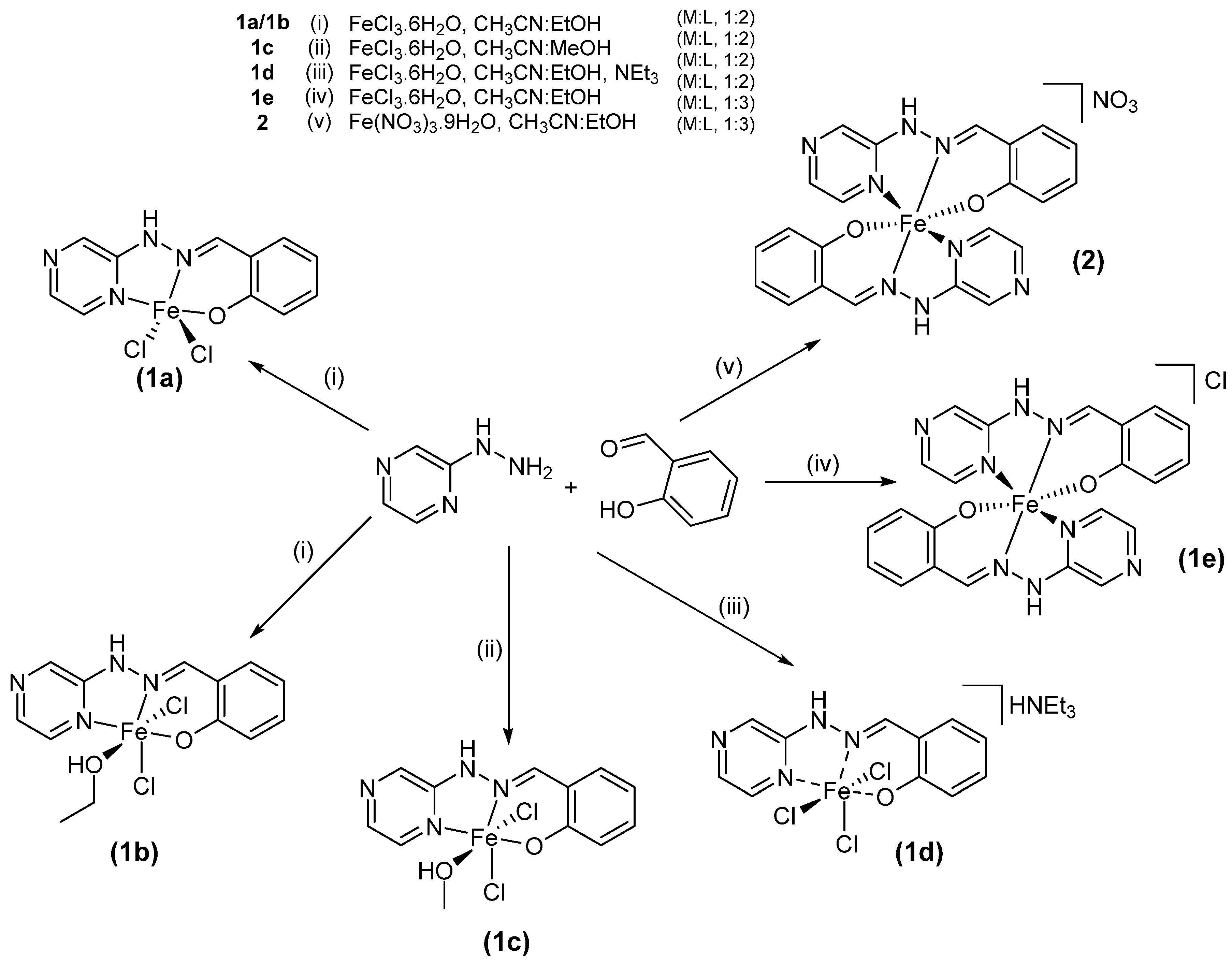
Figure 1. Synthesis methods and conditions used for complexes 1ae and 2.
Crystals 15 00805 g001
Figure 2. (a) Crystal structure of complex (1a) at 150 K. Thermal ellipsoids at 50% probability. Acetonitrile solvent molecule omitted for clarity. (b) Hydrogen-bonded tetramer of complex (1a); hydrogen bonding shown in yellow.
Crystals 15 00805 g002
Figure 3. View down the b-axis (left) and c-axis (right); green displays stacked tetramers of complex (1a) that form a continuous cavity, and blue displays disordered acetonitrile that sits within this cavity.
Crystals 15 00805 g003
Figure 4. (a) Crystal structure of complex (1b) at 150 K. Thermal ellipsoids at 50% probability. (b) One-dimensional hydrogen–halogen bonded chain, shown in yellow.
Crystals 15 00805 g004
Figure 5. Two-dimensional hydrogen-bonded sheet, viewed down the b-axis. One-dimensional chain halogen–hydrogen bonding, shown in yellow; pyrazine-ethanol interaction links the chains to form a 2-D sheet, with hydrogen bonding shown in pink.
Crystals 15 00805 g005
Figure 6. (a) Crystal structure of complex 1c at 150 K. Thermal ellipsoids at 50% probability. Methanol solvent molecule omitted for clarity. (b) One-dimensional hydrogen-bonded chain including the methanol solvent molecule, shown in yellow.
Crystals 15 00805 g006
Figure 7. Two-dimensional hydrogen-bonded sheet in 1c. One-dimensional chain as described above, with hydrogen bonding shown in yellow; pyrazine–methanol interaction links the chains to form a 2-D sheet, along the b and c axes, with hydrogen bonding shown in pink.
Crystals 15 00805 g007
Figure 8. Molecular structure of complex 1d at 150 K. Thermal ellipsoids at 50% probability.
Crystals 15 00805 g008
Figure 9. One-dimensional chain formed through hydrogen–halogen interactions in 1d. Single interactions between chloride and hydrazinyl proton shown in blue. Bifurcated interactions between hydrogen, chloride, and phenolate oxygen shown in yellow.
Crystals 15 00805 g009
Figure 10. Molecular structure of complex 1e at 150 K. Thermal ellipsoids at 50% probability.
Crystals 15 00805 g010
Figure 11. Double-layered one-dimensional chain supported by hydrogen halogen interactions (yellow) and π-π stacking (blue).
Crystals 15 00805 g011
Figure 12. (a) Molecular structure of complex 2 at 150 K. Thermal ellipsoids at 50% probability. (b) Hydrogen-bonded (in blue) layered one-dimensional chain. (c) Expansion of the macrocyclic hydrogen-bonded ring formed between Fe(III) complex units.
Crystals 15 00805 g012
Figure 13. Coordination polyhedra of complexes 1a2, left to right.
Crystals 15 00805 g013
Figure 14. Diffractograms of the experimental powder patterns (black) and the predicted patterns from the single-crystal structures (red) for complexes 1a (a), 1b (b), 1c (c), 1d (d), 1e (e), and 2 (f).
Crystals 15 00805 g014
Table 1. Continuous shape measures for hexa-coordinate complexes 1be, 2.
ComplexCShM (OCT)% Deviation from Ideal
1b1.3127.8
1c1.3458.0
1dFe1 1.362
Fe 2 1.281
8.0
8.1
1e3.52221.0
22.85117.0
Disclaimer/Publisher’s Note: The statements, opinions and data contained in all publications are solely those of the individual author(s) and contributor(s) and not of MDPI and/or the editor(s). MDPI and/or the editor(s) disclaim responsibility for any injury to people or property resulting from any ideas, methods, instructions or products referred to in the content.