Article

Preferential Adsorption of Single-Stranded DNA on Graphene Oxide with Hydroxyl and Epoxy Groups

1
School of Electrical Engineering, Changzhou Vocational Institute of Mechatronic Technology, Changzhou 213164, China
2
School of Intelligent Manufacturing, Changzhou Vocational Institute of Textile and Garment, Changzhou 213164, China
3
School of Physical Science and Technology, Yangzhou University, Yangzhou 225009, China
4
School of Physics, East China University of Science and Technology, Shanghai 200237, China
*
Authors to whom correspondence should be addressed.
Crystals 2025, 15(9), 800; https://doi.org/10.3390/cryst15090800
Submission received: 17 August 2025 / Revised: 2 September 2025 / Accepted: 6 September 2025 / Published: 10 September 2025
(This article belongs to the Section Biomolecular Crystals)

Abstract

The interaction between DNA and two-dimensional materials, such as graphene oxide (GO), has aroused significant research interest due to its potential applications, including biosensors, drug delivery, and gene therapy. However, the difference in interaction between DNA and oxygen functional groups on GO remains unclear, and direct observation at the experimental level is still challenging. In this work, we investigated the adsorption process of a single-stranded DNA (ssDNA) onto GO exhibiting a series of oxidation degrees by molecular dynamics simulations. We found that the ssDNA preferentially binds to hydroxyl groups (-OH) over epoxy groups (-O-) on the GO surface. This preferential adsorption feature may be attributed to the stronger tendency of ssDNA to form hydrogen bonds (HBs) with hydroxyl groups compared to epoxy groups in aqueous solutions. Further analysis indicates that the affinity interaction between ssDNA and hydroxyl groups presumably increases the oxidation degree of GO, thus suggesting a better binding between ssDNA and GO. This work is not only expected to provide the underlying mechanism of ssDNA onto graphene-based interfaces but also offers a deeper understanding of the structures of DNA-two-dimensional complexes, which may potentially contribute to designing new molecular structures for bio-sensing-related nano-devices and nanostructures.

1. Introduction

DNA, as the fundamental carrier of genetic information in living organisms, serves as the cornerstone of the entire biological world. Its unique double-helix structure encodes the blueprints for life’s diversity and complexity, governing processes from organismal morphology to cellular function. Research on DNA has driven remarkable breakthroughs in fields such as genetics, medicine, biotechnology, and data storage [1,2,3,4,5,6,7,8], and its aqueous structural behavior has also been carefully characterized [9]. In particular, the integration of DNA with two-dimensional (2D) materials has unlocked novel avenues for both fundamental science and practical applications [10]. Understanding how DNA molecules interact with these two-dimensional materials provides crucial insights into nanoscale interfacial phenomena, including molecular adsorption, charge transfer, and conformational changes of DNA. Such investigations reveal fundamental physicochemical processes at bio-nanointerfaces, bridging biological and inorganic nanomaterial systems.
GO, as one of the prominent two-dimensional materials, exhibits extraordinary properties, such as large surface area, excellent mechanical strength, unique electrical, and thermal conductivity, and supports its potential applications in various fields, including materials science, carbocatalysts, and nanotechnology [11,12,13,14,15,16,17,18,19,20,21,22]. Critically, DNA-GO interaction provides new opportunities in bio-sensing, drug delivery, and gene therapy [23,24,25,26,27,28,29]. For instance, GO can be used as a carrier for DNA delivery to enhance the specificity of genetic material transport [30]. In bio-sensing, DNA-GO composites enable highly sensitive and selective sensors for detecting biomolecules, including DNA, proteins, and small molecules [31], where analyte binding modulates electrical conductivity to generate measurable signals [32]. These systems leverage DNA’s programmable recognition of cells/tissues while utilizing GO to improve payload stability and solubility. Furthermore, DNA-GO composites facilitate the fabrication of tailored nano-devices and nanostructures for applications spanning electronics, photonics, and catalysis [24,33].
The functionality of DNA-GO materials is governed by their specific adsorption structures, which arise from multiple interactions, such as electrostatic interactions, π-π stacking, and hydrogen bonding [34,35]. Extensive experimental work has characterized DNA-GO interactions using techniques, including X-ray photoelectron spectroscopy (XPS), Fourier transform infrared spectroscopy (FTIR), and fluorescence spectroscopy [36,37,38,39]. However, these characterization techniques cannot directly probe molecular-level interaction mechanisms. Electrostatic forces play a significant role, as the negatively charged DNA can interact with the charged surface of GO under appropriate conditions, like suitable pH and ionic strength. Meanwhile, π-π interaction between DNA nucleobases and GO’s delocalized π-electron system significantly stabilizes adsorption [40]. While our prior work examined these interactions broadly, the distinct binding mechanisms of DNA with hydroxyl groups over epoxy groups on GO remain unresolved.
In this paper, we perform molecular dynamics (MD) simulations to investigate ssDNA adsorption onto GO in aqueous solutions. Our results show that ssDNA preferentially forms HBs with hydroxyl groups rather than epoxy groups on the GO surface, across a range of oxidation degrees of GO from 10% to 25%. This preferential adsorption feature is expected to arise from stronger attraction energies due to more numerous and longer-lived HBs between ssDNA and hydroxyl groups. Furthermore, analysis indicates that ssDNA-hydroxyl groups’ affinity presumably increases with higher GO oxidation degrees. Despite the absence of experimental data, this work provides the atomistic-level insight into the differential binding affinities of ssDNA to various oxidation sites on GO and may potentially contribute to the rational design of DNA-GO composite materials.

2. Method and Model

2.1. Simulation Workflow

Based on the systematic workflow outlined in Figure 1, the molecular simulation procedure was conducted as follows.
(a)
All essential input files and parameters were prepared, including the configuration file, simulation parameter file, and force field file.
(b)
Energy minimization was carried out to eliminate unfavorable atomic contacts, followed by a 2 ns isothermal-isobaric (NPT) equilibration at 300 K and 1 atm to stabilize the system density.
(c)
The production phase consisted of a 400 ns canonical ensemble (NVT) simulation at 300 K to investigate the adsorption mechanism of ssDNA onto GO. The carbon atoms of GO were frozen at their initial positions while the other atoms were free to move. Five independent simulation replicates with identical initial configurations were performed to obtain statistically averaged results.
(d)
The simulation outcomes were then validated to ensure physical plausibility and consistency with the initial research objectives.
(e)
Finally, statistical analyses were performed using multiple independent replicas to extract reliable metrics, including HBs, HB lifetimes, and interaction energies, and so on.

2.2. System Settings

The 12-mer ssDNA molecule with the base sequence 5′-ATGCATGCATGC-3′, denoted by (ATGC)3, was used in this study. The initial (ATGC)3 structure was built using the nucleic acid builder (NAB) in the AMBER 14 package. The model of GO was constructed based on the high correlation between oxidation loci, as reported by Yang et al. [41]. The oxidation degree of GO, dO, was defined by dO = nO/nC, where nO and nC were the number of oxygen atoms and carbon atoms of GO. We built the GO models with oxidation degrees of 10% (GO-10%), 15% (GO-15%), 20% (GO-20%), and 25% (GO-25%), covering the typical experimental value of 15% for various applications [42,43,44]. Given that over-oxidation disrupts GO’s sp2 structure, e.g., compromising mechanical properties and electronic conductivity, thereby limiting practical utility, we selected the GO-15% system as the representative case, extending our investigation to systems with oxidation degrees up to 25% as GO-25%. The number of hydroxyl/epoxy groups for these GO models was 197/197, 311/290, 395/393, and 493/487 for GO-10%, GO-15%, GO-20%, and GO-25%, respectively. Initially, the ssDNA molecule was positioned 3 nm above the GO basal plane, measured from its center of mass (COM).

2.3. Simulation Parameters

All MD simulations were performed using GROMACS 5.1.2 at a temperature of 300 K. Configurations from the simulation system were visualized using the Visual Molecular Dynamics (VMD) software, version 1.9.1. The box for the NVT simulation was measured 10.084 nm × 7.000 nm × 10.224 nm. Periodic boundary conditions were applied in all three Cartesian directions. The Amber03 force field was employed to describe the parameters of the ssDNA molecule. For GO, all parameters followed the settings in the force field. The van der Waals (vdW) parameters for the carbon atom were set to σCC = 3.580 Å and εCC = 0.066 kcal/mol. The C-C bond was described by the harmonic potentials with the bond length of 0.142 nm and spring constant of 322.550 kcal/(mol∙Å2). The C-C-C angle was simulated by the harmonic potentials with a bond angle of 120° and a spring constant of 53.350 kcal/(mol∙rad2). The planar dihedral angles between C-C-C-C were maintained by a spring constant of 3.150 kcal/mol. The vdW parameters of the oxygen atom in -OH were σOO = 2.950 Å, εOO = 0.203 kcal/mol, and σHH = 2.370 Å, εHH = 0.028 kcal/mol, and those in -O- were σOO = 2.850 Å, εOO = 0.253 kcal/mol. The charges (q) for -OH were qO = −0.674 e and qH = +0.408 e, and that for -O- was qO = −0.400 e. Water molecules were represented using the TIP3P model. Long-range electrostatic interactions were treated by the particle-mesh Ewald (PME) method using a real-space cutoff of 1.200 nm. The van der Waals cutoff distance was also set to 1.200 nm.

3. Result and Discussion

Building upon our previous findings that oxidation degree critically governs ssDNA assembly on GO [45,46], this study aims to investigate the roles of distinct oxygen-containing functional groups in adsorption. Although both π-π stacking and electrostatic forces contribute significantly to DNA-GO interactions, we focus here on elucidating the differential contributions of hydrophilic moieties, particularly hydroxyl groups over epoxy groups, during ssDNA adsorption.
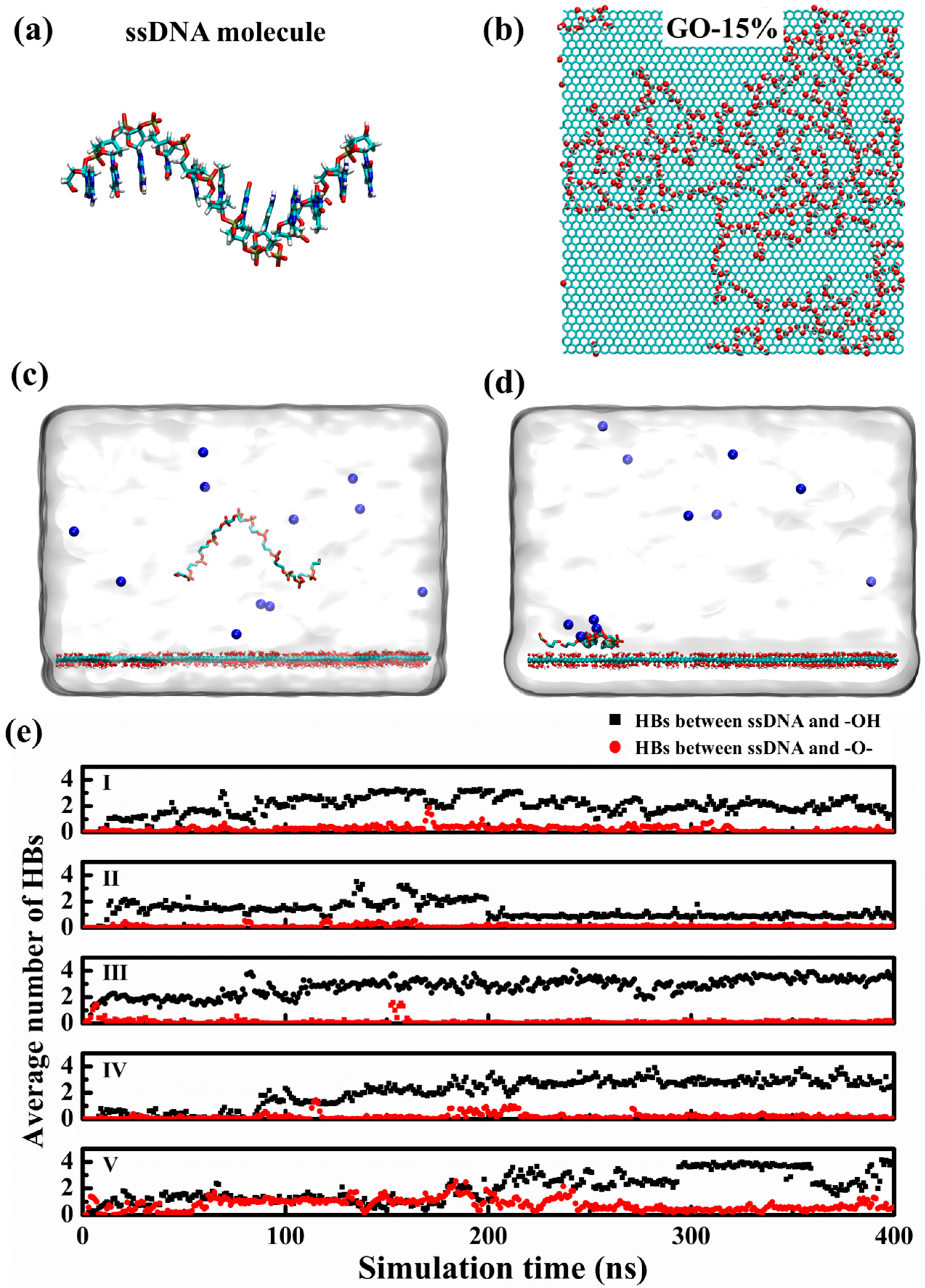
Figure 2 illustrates ssDNA adsorption dynamics on GO-15% and the time evolution of the average number of HBs between ssDNA and two types of hydrophilic groups on GO. HBs were identified using geometric criteria, with oxygen atoms serving as both donor (D) and acceptor (A). An HB was considered formed if the distance between the D and A oxygen atoms was less than 3.5 Å and the D–H–A angle was less than 30°. Initially, the ssDNA was extended in aqueous solutions, with the centroid distance of 3 nm above the GO basal plane (Figure 2a,c). Two types of hydrophilic groups, hydroxyl and epoxy groups, dominated the basal plane of GO and coexisted in a highly correlated manner (Figure 2b) [41]. After 400 ns of simulation, the system reached the equilibrium state with ssDNA adsorbed on the GO. As shown in Figure 2d, ssDNA showed a curved configuration and was localized at highly oxidized domains. These regions were enriched in clustered hydroxyl and epoxy groups, which drove the bending or folding of ssDNA through hydrogen bonding. Five independent simulations at the GO-15% had been performed. It was shown in Figure 2e that the average number of HBs between ssDNA and hydroxyl groups was larger than that between ssDNA and epoxy groups, despite the fact that the values differed from each other as 1.216/0.038, 0.680/0.276, 3.268/0.148, 2.756/0.134, and 3.774/0.592 in the equilibrium state. For systems I, III, IV, and V, the average numbers of HBs between ssDNA and hydroxyl groups were above 1.2, while system II showed a smaller number of 0.680. This variability was expected to correlate with the adsorption configurations of ssDNA on GO [45], which was dictated by the spatial distribution of oxygen-containing functional groups at adsorption sites.
Next, we analyzed the time evolution of Coulomb interactions between ssDNA and hydroxyl/epoxy groups of GO-15%. The values were averaged per nanosecond. Figure 3a revealed that the Coulomb interactions between ssDNA and hydroxyl groups remained negative for all five systems from 0 ns to 400 ns, suggesting an attractive interaction between them. In contrast, Coulomb interactions between ssDNA and epoxy groups fluctuated near zero over the same period across all systems, indicating configuration-dependent alternation between attraction and repulsion. Thermodynamic analysis showed ssDNA adsorption was less favorable on epoxy groups than hydroxyl groups across all systems. For example, for system Ⅰ, the Coulomb interaction between ssDNA and hydroxyl groups increased attraction from 0 kJ/mol at 0 ns to −42.99 kJ/mol at 400 ns. In comparison, the Coulomb interaction between ssDNA and epoxy groups increased the repulsion interaction from 0 kJ/mol at 0 ns to 10.34 kJ/mol at 400 ns despite fluctuations.
Figure 3a further revealed trajectory-dependent variability in Coulomb interactions across GO-15% replicas despite identical initial conditions. For example, as for system II, the steady Coulomb interaction between ssDNA and hydroxyl/epoxy groups was measured −37.29 kJ/mol/5.84 kJ/mol, which was different from that of −42.99 kJ/mol/10.34 kJ/mol for system I. This divergence stemmed from stochastic adsorption configurations emerging during system evolution, consistent with our prior findings on oxygen-group spatial heterogeneity.
Given that both Coulomb and π-π stacking interactions played critical roles in the adsorption of ssDNA onto the GO surface, we further examined the π-π stacking behavior across five independent simulation systems. Figure 3b illustrates a representative configuration of π-π stacking between ssDNA and GO. The time-dependent change in the number of π-π stacking events is shown in Figure 3c. As evident from the data, all systems exhibited an increasing number of π-π stacking over the simulation time. At 400 ns, the average number of π-π stacking reached 11.0, 7.0, 5.9, 5.6, and 10.0, respectively. Although variations were observed among individual replicas, the collective results clearly demonstrate that π-π stacking interactions consistently exceeded the number of HBs formed between ssDNA and GO.
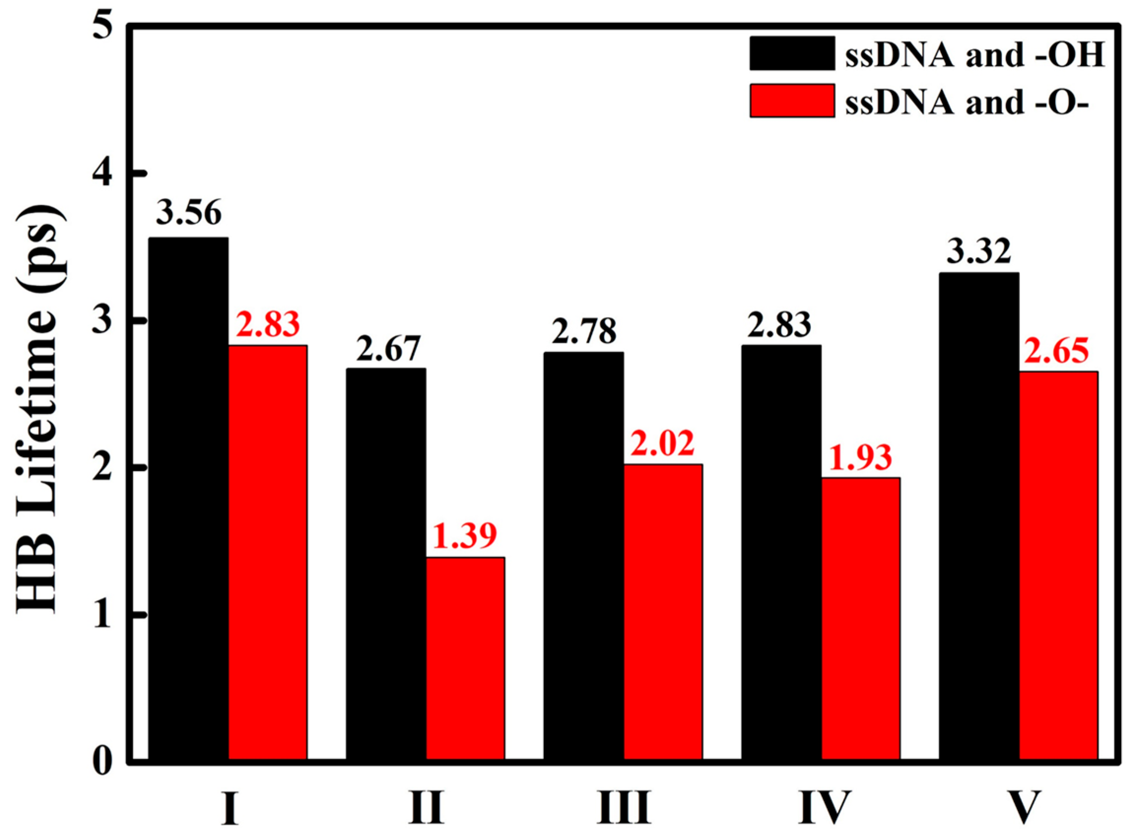
To further differentiate interactions of ssDNA with hydrophilic groups, we computed the average HB lifetimes formed between ssDNA and hydroxyl groups over epoxy groups on GO-15% after systems reached thermodynamic equilibrium. The results indicated that the HB lifetimes between ssDNA and hydroxyl groups were longer than those between ssDNA and epoxy groups. As shown in Figure 4, the HB lifetimes between ssDNA and hydroxyl groups were 3.56 ps, 2.67 ps, 2.78 ps, 2.83 ps, and 3.32 ps, which were significantly longer than those between ssDNA and epoxy groups of 2.83 ps, 1.39 ps, 2.02 ps, 1.93 ps, and 2.65 ps, respectively. Analysis consistently revealed longer HB lifetimes for hydroxyl groups over epoxy groups across all GO-15% replicas. These extended lifetimes (Δ = 0.73~1.28 ps) confirmed enhanced stability of ssDNA-hydroxyl interactions, reflecting stronger binding of ssDNA at graphene oxide interfaces.
Furthermore, consistent with the trends observed in Coulomb interactions, the HB lifetimes exhibited considerable variation across replicas, despite identical simulation setups. This divergence was expected to stem from the stochastic nature of the adsorption configurations emerging during the energy-minimization process. Although the initial DNA-GO configurations and energy minimization principles were consistent across all systems, kinetic randomness during adsorption led to distinct trajectories and interfacial arrangements.
To validate the universality of preferential hydroxyl binding, we extended our analysis to systems with varying oxidation degrees: GO-10%, GO-20%, and GO-25% systems. For each oxidation degree, five independent replicas underwent 400 ns of simulation. Upon reaching thermodynamic equilibrium, quantification of HBs revealed consistent preferential binding to hydroxyl groups over epoxy groups across all oxidation degrees, in agreement with the trends observed for GO-15%.
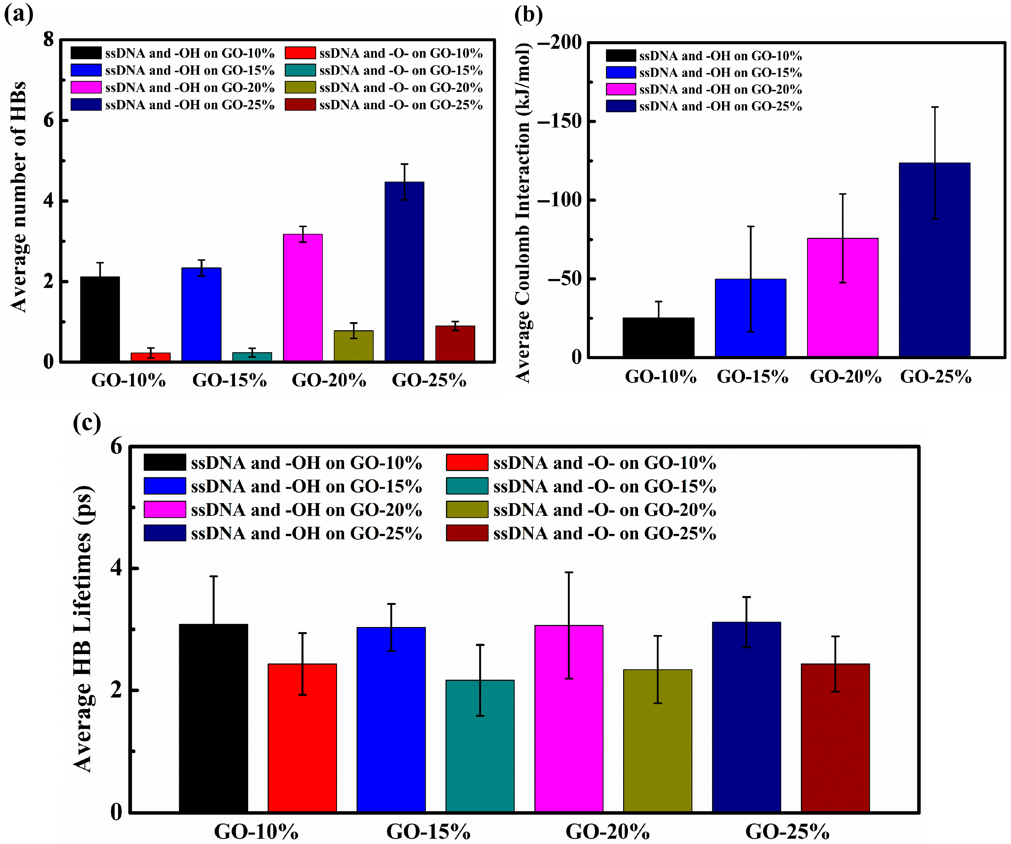
Comparative analysis of HBs characteristics across different oxidation degrees consistently revealed preferential binding of ssDNA to hydroxyl groups over epoxy groups in all systems. As shown in Figure 5a, the average numbers of HBs with hydroxyl/epoxy groups were 2.121 ± 0.348/0.229 ± 0.124, 2.339 ± 0.197/0.238 ± 0.108, 3.175 ± 0.194/0.781 ± 0.192, and 4.473 ± 0.443/0.898 ± 0.110 at GO-10%, GO-15%, GO-20%, and GO-25%. Consistent with the trend observed in GO-15%, ssDNA exhibited a higher propensity to form HBs with hydroxyl groups compared to epoxy groups across GO-10%, GO-20%, and GO-25%. Importantly, as the oxidation degrees increased from GO-10% to GO-25%, the number of HBs formed between ssDNA and GO also increased for both hydroxyl and epoxy groups. As shown in Figure 5b, the Coulomb interactions were −25.25 ± 10.37 kJ/mol, −49.90 ± 33.43 kJ/mol, −75.80 ± 28.18 kJ/mol, and −123.69 ± 35.46 kJ/mol between ssDNA and hydroxyl groups on GO-10%, GO-15%, GO-20%, and GO-25%, respectively. This oxidation-dependent reinforcement indicated that higher oxidation degrees simultaneously strengthened HB networks and electrostatic forces between ssDNA and hydroxyl groups, thereby thermodynamically favoring ssDNA adsorption.
Figure 5c presents the HB lifetimes between ssDNA and hydroxyl/epoxy groups across GO systems with oxidation degrees ranging from 10% to 25%. It was observed that the HB lifetimes between ssDNA and hydroxyl groups were approximately 3.0 ps, independent of the oxidation degree of GO. In contrast, HBs involving epoxy groups exhibited shorter lifetimes, averaging around 2.2 ps, which were significantly shorter than those with hydroxyl groups. These results confirmed thermodynamically preferential binding to hydroxyl groups, presumably attributed to superior hydrogen-bonding geometry and charge complementarity at DNA-GO interfaces.
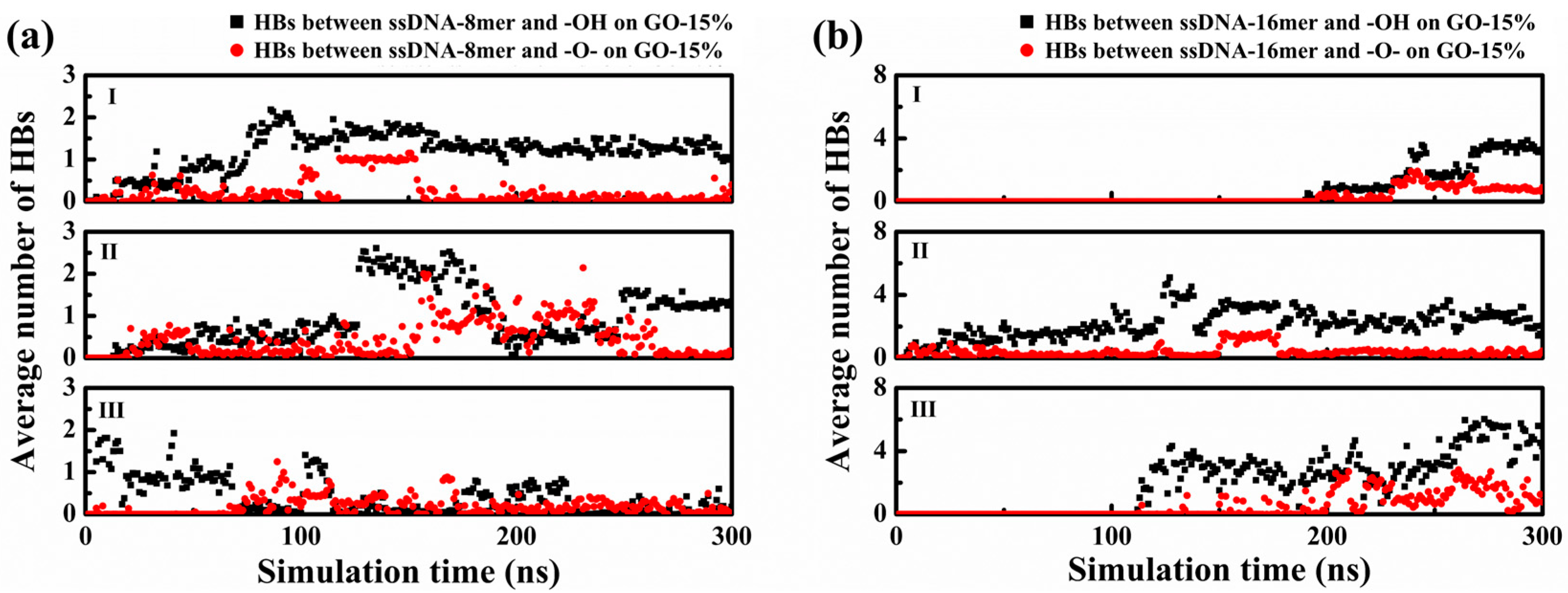
To further validate the generality of the conclusion mentioned above, we investigated the adsorption behavior on GO-15% with varied chain lengths of ssDNA from the original 12-mer to 8-mer and 16-mer, donated as ssDNA-8mer and ssDNA-16mer. For each system, three independent replicas (I~III) were simulated for 300 ns under identical parameters. As shown in Figure 6a,b, a consistent preference for binding to hydroxyl groups over epoxy groups was observed across all chain lengths. In the ssDNA-8mer systems, a higher number of HBs with hydroxyl groups compared to epoxy groups was maintained in all replicas, despite some conformational variations. Similarly, the ssDNA-16mer systems also exhibited the same preferential binding behavior. These results demonstrated that the preferential adsorption toward hydroxyl groups was expected to be independent of the chain length of ssDNA.

4. Conclusions

In summary, ssDNA adsorption onto GO is driven by electrostatic interactions, π-π stacking, and hydrogen bonding. Although π-π stacking between ssDNA bases and aromatic domains of GO plays a key role in the adsorption process, our simulations demonstrate that hydrogen bonding with hydroxyl groups dominates the interfacial recognition process. Specifically, hydroxyl groups facilitate preferential adsorption via stable hydrogen bonds and strong electrostatic attraction between ssDNA and GO, whereas epoxy groups contribute less effectively due to their transient hydrogen bonding capability and electrostatically neutral nature. This mechanistic understanding of group-specific interactions provides critical design principles for GO-based bio-nanotechnologies and may potentially contribute to the rational optimization of biosensors, drug delivery, and so on.
It should be noted that the current findings are primarily based on MD simulations and have not yet been validated experimentally. Although the computational approach provides valuable atomistic insights into the DNA-GO interaction mechanisms, further experimental studies, such as spectroscopic characterization or surface analysis, are necessary to confirm these predictions. Nonetheless, our simulation results establish a theoretical foundation and offer practical guidance for designing future experiments, including the selection of functional groups, environmental conditions, and measurement techniques. This will enable more efficient and targeted experimental exploration of DNA–GO interactions.

Author Contributions

Conceptualization, H.W. and G.S.; methodology, H.M., M.W., L.Z., and X.L.; software, H.M., X.H., L.Z., and X.L.; validation, S.W.; data curation, H.W. and G.S.; writing—original draft preparation, H.M., L.Z., and X.L.; writing—review and editing, H.M., L.Z., and X.L.; visualization, H.M. and L.Z.; supervision, S.W.; project administration, H.M., M.W., L.Z., and X.L.; funding acquisition, X.H. All authors have read and agreed to the published version of the manuscript.

Funding

This work is funded by the Jiangsu Province High-level Innovative and Entrepreneurial Talent Introduction Program (Double Innovation Program, No. JSSCBS20221163), Changzhou Basic Research Program (Applied Basic Research)—Young Doctor Project (No. CJ20235006), Changzhou Basic Research Program (Applied Basic Research)—Young Doctor Project (No. CJ20235003), and the Major scientific research project of Changzhou Vocational Institute of Mechatronic Technology (No. 2023-ZDKYXM-04).

Data Availability Statement

The original contributions presented in the study are included in the article; further inquiries can be directed to the corresponding authors.

Acknowledgments

This work is supported by the Supercomputer Center of the Chinese Academy of Sciences and the Shanghai Supercomputer Center of China.

Conflicts of Interest

The authors declare no conflicts of interest.

References

  1. An, H.; Jin, B. Prospects of nanoparticle–DNA binding and its implications in medical biotechnology. Biotechnol. Adv. 2012, 30, 1721–1732. [Google Scholar] [CrossRef] [PubMed]
  2. Glick, B.R.; Patten, C.L. Molecular Biotechnology: Principles and Applications of Recombinant DNA; John Wiley & Sons: Washington DC, USA, 2022. [Google Scholar]
  3. Hirao, I. Unnatural base pair systems for DNA/RNA-based biotechnology. Curr. Opin. Chem. Biol. 2006, 10, 622–627. [Google Scholar] [CrossRef] [PubMed]
  4. Yang, D.; Hartman, M.R.; Derrien, T.L.; Hamada, S.; An, D.; Yancey, K.G.; Cheng, R.; Ma, M.; Luo, D. DNA materials: Bridging nanotechnology and biotechnology. Acc. Chem. Res. 2014, 47, 1902–1911. [Google Scholar] [CrossRef] [PubMed]
  5. Wang, G.; Vasquez, K.M. Dynamic alternative DNA structures in biology and disease. Nat. Rev. Genet. 2023, 24, 211–234. [Google Scholar] [CrossRef]
  6. Wang, S.; Mao, X.; Wang, F.; Zuo, X.; Fan, C. Data storage using DNA. Adv. Mater. 2024, 36, 2307499. [Google Scholar] [CrossRef]
  7. Ladabaum, M.; Tikhomirov, G. Growing moiré with DNA. Nat. Nanotechnol. 2025. [Google Scholar] [CrossRef]
  8. Pan, C.; Tabatabaei, S.K.; Tabatabaei Yazdi, S.H.; Hernandez, A.G.; Schroeder, C.M.; Milenkovic, O. Rewritable two-dimensional DNA-based data storage with machine learning reconstruction. Nat. Commun. 2022, 13, 2984. [Google Scholar] [CrossRef]
  9. Crane-Robinson, C. Role of water in defining the structure and properties of B-Form DNA. Crystals 2022, 12, 818. [Google Scholar] [CrossRef]
  10. Cao, Z.; Yadav, P.; Barati Farimani, A. Which 2D material is better for DNA detection: Graphene, MoS2, or MXene? Nano Lett. 2022, 22, 7874–7881. [Google Scholar] [CrossRef]
  11. Situ, B.; Zhang, Z.; Zhao, L.; Tu, Y. Graphene oxide-based large-area dynamic covalent interfaces. Nanoscale 2023, 15, 17739–17750. [Google Scholar] [CrossRef]
  12. Hu, Y.; Feng, Z.; Xie, Y.; Wang, H.; Ji, Q.; Wang, J.; Xu, C. Surface modification of graphene oxide and its strengthening and toughening mechanism for alumina-based ceramic materials. Crystals 2024, 14, 949. [Google Scholar] [CrossRef]
  13. Chen, L.; Shi, G.; Shen, J.; Peng, B.; Zhang, B.; Wang, Y.; Bian, F.; Wang, J.; Li, D.; Qian, Z. Ion sieving in graphene oxide membranes via cationic control of interlayer spacing. Nature 2017, 550, 380–383. [Google Scholar] [CrossRef] [PubMed]
  14. Chung, C.; Kim, Y.K.; Shin, D.; Ryoo, S.R.; Hong, B.H.; Min, D.H. Biomedical applications of graphene and graphene oxide. Acc. Chem. Res. 2013, 46, 2211–2224. [Google Scholar] [CrossRef] [PubMed]
  15. Dimiev, A.M.; Tour, J.M. Mechanism of graphene oxide formation. ACS Nano 2014, 8, 3060–3068. [Google Scholar] [CrossRef]
  16. Dreyer, D.R.; Park, S.; Bielawski, C.W.; Ruoff, R.S. The chemistry of graphene oxide. Chem. Soc. Rev. 2010, 39, 228–240. [Google Scholar] [CrossRef] [PubMed]
  17. Eigler, S.; Hirsch, A. Chemistry with graphene and graphene oxide—Challenges for synthetic chemists. Angew. Chem. Int. Ed. 2014, 53, 7720–7738. [Google Scholar] [CrossRef]
  18. Su, C.; Loh, K.P. Carbocatalysts: Graphene oxide and its derivatives. Acc. Chem. Res. 2013, 46, 2275–2285. [Google Scholar] [CrossRef]
  19. Zhu, Y.; James, D.K.; Tour, J.M. New routes to graphene, graphene oxide and their related applications. Adv. Mater. 2012, 24, 4924–4955. [Google Scholar] [CrossRef]
  20. Hutapea, J.A.A.; Manik, Y.G.O.; Ndruru, S.T.C.L.; Huang, J.; Goei, R.; Tok, A.I.Y.; Siburian, R. Comprehensive review of graphene synthesis techniques: Advancements, challenges, and future directions. Micro 2025, 5, 40. [Google Scholar] [CrossRef]
  21. Wakchaure, M.B.; Menezes, P.L. Advances in the tribological performance of graphene oxide and its composites. Materials 2025, 18, 3587. [Google Scholar] [CrossRef]
  22. Chen, S.; Lee, C.J.M.; Tan, G.S.X.; Ng, P.R.; Zhang, P.; Zhao, J.; Novoselov, K.S.; Andreeva, D.V. Ultra-tough graphene oxide/DNA 2D hydrogel with intrinsic sensing and actuation functions. Macromol. Rapid. Commun. 2025, 46, 2400518. [Google Scholar] [CrossRef]
  23. Zhang, H.; Jia, S.; Lv, M.; Shi, J.; Zuo, X.; Su, S.; Wang, L.; Huang, W.; Fan, C.; Huang, Q. Size-dependent programming of the dynamic range of graphene oxide–DNA interaction-based ion sensors. Anal. Chem. 2014, 86, 4047–4051. [Google Scholar] [CrossRef]
  24. Gao, L.; Lian, C.; Zhou, Y.; Yan, L.; Li, Q.; Zhang, C.; Chen, L.; Chen, K. Graphene oxide–DNA based sensors. Biosens. Bioelectron. 2014, 60, 22–29. [Google Scholar] [CrossRef] [PubMed]
  25. Liu, B.; Salgado, S.; Maheshwari, V.; Liu, J. DNA adsorbed on graphene and graphene oxide: Fundamental interactions, desorption and applications. Curr. Opin. Colloid Interface Sci. 2016, 26, 41–49. [Google Scholar] [CrossRef]
  26. Mo, R.; Jiang, T.; Sun, W.; Gu, Z. ATP-responsive DNA-graphene hybrid nanoaggregates for anticancer drug delivery. Biomaterials 2015, 50, 67–74. [Google Scholar] [CrossRef] [PubMed]
  27. Tang, L.; Wang, Y.; Liu, Y.; Li, J. DNA-directed self-assembly of graphene oxide with applications to ultrasensitive oligonucleotide assay. ACS Nano 2011, 5, 3817–3822. [Google Scholar] [CrossRef] [PubMed]
  28. Xue, T.; Cui, X.; Guan, W.; Wang, Q.; Liu, C.; Wang, H.; Qi, K.; Singh, D.J.; Zheng, W. Surface plasmon resonance technique for directly probing the interaction of DNA and graphene oxide and ultra-sensitive biosensing. Biosens. Bioelectron. 2014, 58, 374–379. [Google Scholar] [CrossRef]
  29. Chiticaru, E.A.; Damian, C.M.; Pilan, L.; Ioniță, M. Label-free DNA biosensor based on reduced graphene oxide and gold nanoparticles. Biosensors 2023, 13, 797. [Google Scholar] [CrossRef]
  30. Mukherjee, S.; Bytesnikova, Z.; Ashrafi, A.M.; Adam, V.; Richtera, L. Graphene oxide as a nanocarrier for biochemical molecules: Current understanding and trends. Processes 2020, 8, 1636. [Google Scholar] [CrossRef]
  31. Li, Q.; Wang, Q.; Yang, X.; Wang, K.; Zhang, H.; Nie, W. High sensitivity surface plasmon resonance biosensor for detection of microRNA and small molecule based on graphene oxide-gold nanoparticles composites. Talanta 2017, 174, 521–526. [Google Scholar] [CrossRef]
  32. Jin, J.; Lin, Y.; Song, M.; Gui, C.; Leesirisan, S. Enhancing the electrical conductivity of polymer composites. Eur. Polym. J. 2013, 49, 1066–1072. [Google Scholar] [CrossRef]
  33. Liu, B.; Sun, Z.; Zhang, X.; Liu, J. Mechanisms of DNA sensing on graphene oxide. Anal. Chem. 2013, 85, 7987–7993. [Google Scholar] [CrossRef] [PubMed]
  34. Lee, J.; Yim, Y.; Kim, S.; Choi, M.H.; Choi, B.S.; Lee, Y.; Min, D.H. In-depth investigation of the interaction between DNA and nano-sized graphene oxide. Carbon 2016, 97, 92–98. [Google Scholar] [CrossRef]
  35. Zeng, S.; Chen, L.; Wang, Y.; Chen, J. Exploration on the mechanism of DNA adsorption on graphene and graphene oxide via molecular simulations. J. Phys. D Appl. Phys. 2015, 48, 275402. [Google Scholar] [CrossRef]
  36. Balapanuru, J.; Yang, J.X.; Xiao, S.; Bao, Q.; Jahan, M.; Polavarapu, L.; Wei, J.; Xu, Q.H.; Loh, K.P. A graphene oxide–organic dye ionic complex with DNA-sensing and optical-limiting properties. Angew. Chem. Int. Ed. 2010, 49, 6549–6553. [Google Scholar] [CrossRef]
  37. Park, J.S.; Goo, N.I.; Kim, D.E. Mechanism of DNA adsorption and desorption on graphene oxide. Langmuir 2014, 30, 12587–12595. [Google Scholar] [CrossRef]
  38. Tang, L.; Chang, H.; Liu, Y.; Li, J. Duplex DNA/graphene oxide biointerface: From fundamental understanding to specific enzymatic effects. Adv. Funct. Mater. 2012, 22, 3083–3088. [Google Scholar] [CrossRef]
  39. Guo, J.; Zhang, H.; Yang, J.; Zhang, Y.; Wang, J.; Yan, G. ssDNA-QDs/GO multicolor fluorescence system for synchronous screening of hepatitis virus DNA. Arabian J. Chem. 2023, 16, 104582. [Google Scholar] [CrossRef]
  40. Xu, Z.; Lei, X.; Tu, Y.; Tan, Z.J.; Song, B.; Fang, H. Dynamic cooperation of hydrogen binding and π stacking in ssDNA adsorption on graphene oxide. Chem. Eur. J. 2017, 23, 13100–13104. [Google Scholar] [CrossRef]
  41. Yang, J.; Shi, G.; Tu, Y.; Fang, H. High correlation between oxidation loci on graphene oxide. Angew. Chem. Int. Ed. 2014, 126, 10354–10358. [Google Scholar] [CrossRef]
  42. Ye, C.; Wang, G.; Yuan, H.; Li, J.; Ni, K.; Pan, F.; Guo, M.; Wu, Y.; Ji, H.; Zhang, F.; et al. Microfluidic oxidation of graphite in two minutes with capability of real-time monitoring. Adv. Mater. 2022, 34, 2107083. [Google Scholar] [CrossRef]
  43. Chu, S.; Huang, W.; Shen, F.; Li, T.; Li, S.; Xu, W.; Lv, C.; Luo, Q.; Liu, J. Graphene oxide-based colorimetric detection of organophosphorus pesticides via a multi-enzyme cascade reaction. Nanoscale 2020, 12, 5829–5833. [Google Scholar] [CrossRef]
  44. Liou, T.H.; Tseng, Y.K.; Zhang, T.Y.; Liu, Z.S.; Chen, J.Y. Rice husk char as a sustainable material for the preparation of graphene oxide-supported biocarbons with mesoporous structure: A characterization and adsorption study. Fuel 2023, 344, 128042. [Google Scholar] [CrossRef]
  45. Ma, H.; Chen, J.; Fang, H.; Lei, X. Oxidation degree dependent adsorption of ssDNA onto graphene-based surface. Chin. Phys. B 2021, 30, 106806. [Google Scholar] [CrossRef]
  46. Ma, H.; Xu, Z.; Fang, H.; Lei, X. Unexpected sequence adsorption features of polynucleotide ssDNA on graphene oxide. Phys. Chem. Chem. Phys. 2020, 22, 11740–11746. [Google Scholar] [CrossRef]
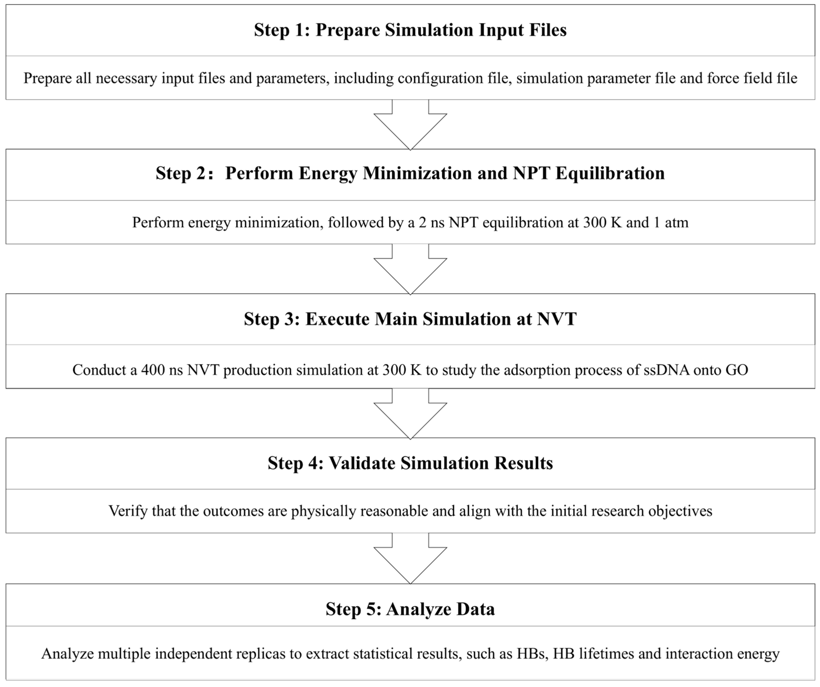
Figure 1. Workflow of simulation methodology.
Crystals 15 00800 g001
Figure 2. Adsorption dynamics of ssDNA onto GO-15%. (a) Initial configuration of ssDNA visualized by the VMD software. The green, blue, red, and white parts represent the carbon, nitrogen, oxygen, and hydrogen atoms, respectively. (b) Initial configuration of GO visualized by the VMD software. The green hexagonal structure is the carbon plane of GO. The red and white spheres are the oxygen and hydrogen atoms on GO. Configurations of the system at (c) 0 ns and (d) 400 ns. The blue spheres represent the sodium atoms. (e) Time evolution of the average number of HBs formed between ssDNA and hydroxyl groups (-OH, black squares) over epoxy groups (-O-, red dots) on GO across five systems (I~V) from 0 ns to 400 ns.
Crystals 15 00800 g002
Figure 3. (a) Time evolution of Coulomb interactions between ssDNA and hydroxyl (-OH, black squares)/epoxy (-O-, red dots) groups for five GO-15% systems (I~V) from 0 ns to 400 ns. (b) Top view of the π-π stacking between benzene ring structure on ssDNA and GO-15% visualized by the VMD software. The green, blue, red, and white parts represent the carbon, nitrogen, oxygen, and hydrogen atoms on ssDNA. The red and white spheres represent the oxygen and hydrogen atoms on the GO, respectively. (c) Average numbers of π-π stacking for systems I~V between ssDNA and GO-15%.
Crystals 15 00800 g003
Figure 4. Average HB lifetimes of ssDNA and -OH (black bar), -O- (red bar) in five systems (I~V) of GO-15% after simulation systems reached the thermodynamic equilibrium.
Crystals 15 00800 g004
Figure 5. (a) Average number of HBs between ssDNA and hydroxyl/epoxy groups. (b) Average Coulomb interaction between ssDNA and hydroxyl groups. (c) Average HB lifetimes between ssDNA and hydroxyl/epoxy groups.
Crystals 15 00800 g005
Figure 6. Average number of HBs between (a) ssDNA-8mer and (b) ssDNA-16mer and GO-15% in three systems (I~III). The black squares and red dots represent the HBs between ssDNA and hydroxyl groups (-OH, black squares) and epoxy (-O-, red dots) groups, respectively.
Crystals 15 00800 g006
Disclaimer/Publisher’s Note: The statements, opinions and data contained in all publications are solely those of the individual author(s) and contributor(s) and not of MDPI and/or the editor(s). MDPI and/or the editor(s) disclaim responsibility for any injury to people or property resulting from any ideas, methods, instructions or products referred to in the content.