Article

The Influence of Precursor pH on the Synthesis and Morphology of AuNPs Synthesized Using Green Tea Leaf Extract

by
Oksana Velgosova
1,*,
Zuzana Mikulková
1 and
Maksym Lisnichuk
2,3
1
Institute of Materials, Faculty of Materials Metallurgy and Recycling, Technical University of Košice, Letná 9/A, 042 00 Košice, Slovakia
2
Faculty of Science, Institute of Physics, Pavol Jozef Šafárik University in Košice, Park Angelinum 9, 040 01 Košice, Slovakia
3
Institute of Materials Research of the Slovak Academy of Sciences, Watsonova 47, 04 001 Košice, Slovakia
*
Author to whom correspondence should be addressed.
Crystals 2025, 15(8), 682; https://doi.org/10.3390/cryst15080682
Submission received: 20 June 2025 / Revised: 8 July 2025 / Accepted: 24 July 2025 / Published: 26 July 2025

Abstract

This study investigates the effect of precursor pH (1.3, 2, 4, 6, 8, and 10) on the synthesis of gold nanoparticles (AuNPs) via a green synthesis approach using an aqueous extract of green tea (Camellia sinensis) leaves. The formation of AuNPs was monitored using UV-vis spectrophotometry and confirmed using transmission electron microscopy (TEM). The results confirmed that the morphology and size of the AuNPs are strongly dependent on the pH of the reaction medium. Based on spectral features, the color of the colloids, and TEM analysis, the synthesized samples were classified into three groups. The first (pH 8 and 10) contained predominantly spherical nanoparticles with an average diameter of ~18 nm, the second (pH 1.3 and 2) contained different shaped nanoparticles (20–250 nm in diameter), and the third (pH 4 and 6) contained flower-like nanostructures with a mean diameter of ~60 nm. UV-vis analysis revealed good stability of all AuNP colloids, except at pH 1.3, where a significant decrease in absorbance intensity over time was observed. These findings confirm that tuning the precursor pH allows for controlled manipulation of nanoparticle morphology and stability in green synthesis systems.

1. Introduction

Noble metal nanoparticles possess unique properties, including a high surface-to-volume ratio, tunable optical features, and bioactivity, making them suitable for diverse applications such as diagnostics, targeted drug delivery, therapies, and antibacterial or antiviral coatings [1]. Gold and silver nanoparticles serve as sensors for disease biomarkers and as drug carriers, notably in cancer treatment [2,3]. They are also applied in regenerative medicine, water purification, and catalysis [4,5].
Metal nanoparticles are commonly synthesized through physical or chemical methods [6,7,8]. However, these methods pose limitations: chemical synthesis involves toxic substances, while physical methods require extreme conditions and expensive equipment. Biological synthesis offers a greener and cost-effective alternative. This approach uses natural materials (especially plant extracts) as reducing and stabilizing agents, producing biocompatible nanoparticles with minimal waste and energy input.
Numerous studies have demonstrated the successful green synthesis of AuNPs using various plant extracts such as Syzygium aromaticum [9], Mangifera indica [10], Martynia annua [11], or lavender [12]. However, in many cases, the resulting nanoparticles are predominantly spherical, and precise control over shape and size remains limited. Achieving non-spherical morphologies (e.g., rods, triangles, flowers, or decahedra) is essential, as shape greatly influences optical [13], catalytic, and biological properties [14,15].
Among the factors influencing nanoparticle morphology in green synthesis, such as temperature [16], light exposure, concentration [17], and extract composition, the pH of the reaction medium plays a particularly critical role, as it affects both the redox potential of the metal precursor and the ionization state of active phytochemicals involved in reduction and capping [18]. Despite its significance, systematic investigations of pH-driven shape control during green synthesis remain scarce. Most existing studies focus on general synthesis optimization rather than targeted morphological engineering. For instance, Raghunandan et al. observed changes in size with pH during neem-mediated synthesis but without detailed morphological control [19]. Similarly, Song and Kim reported size variation in Magnolia kobus-mediated AuNPs with changing pH, but the study lacked insight into anisotropic shape formation [20].
In this context, our study provides a novel contribution by systematically investigating the influence of precursor pH on the morphology, size distribution, and stability of AuNPs synthesized using an aqueous green tea (Camellia sinensis) extract. Green tea was selected due to its well-known richness in polyphenols, particularly catechins, which serve as strong reducing and stabilizing agents [21]. By modulating the pH of the precursor solution, we demonstrate the reproducible formation of various nanoparticle shapes, including spheres, rods, triangles, and flower-like structures, without the use of toxic chemicals or external shape-directing agents.
The synthesized AuNPs were characterized using UV-vis spectrophotometry and transmission electron microscopy (TEM), providing insight into their optical behavior and morphological diversity under different pH conditions. This study offers a reproducible, environmentally sustainable approach for tailoring nanoparticle morphology via pH control alone, which has strong implications for the scalable production of AuNPs with application-specific features.

2. Materials and Methods

2.1. Materials

A solution of HAuCl4 (1000 mg/L, Merck, Darmstadt, Germany) was used as the gold precursor. A commercially available green tea (Zen Chai, Teekanne brand, Düsseldorfe, Germany), purchased from a local store, was used to prepare the extract, which served as both the reducing and stabilizing agent. All solutions were prepared and diluted with deionized water.

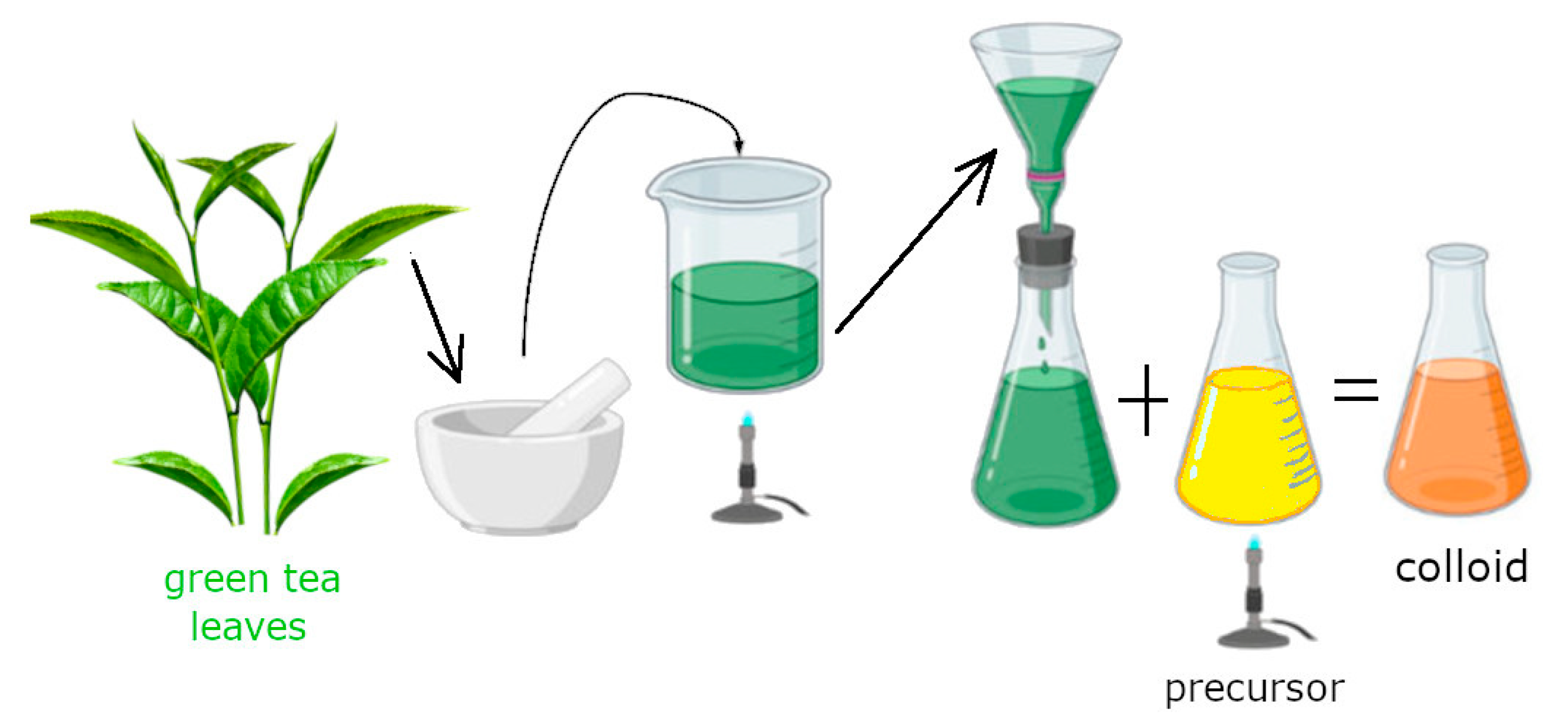
2.2. Solution Preparation and Synthesis of AuNPs

The extract was prepared by adding 2 g of dry green tea leaves to 100 mL of distilled water. The mixture was heated to 70–80 °C and maintained at this temperature for 10 min. After heating, it was allowed to cool and filtered through Whatman filter paper to remove solid components. Finally, the filtrate was centrifuged at 9000 rpm for 15 min to remove all solid components. The prepared extract was used to synthesize AuNPs.
The gold precursor was prepared by diluting the HAuCl4 solution with distilled water to a concentration of 50 mg/L (0.25 mM). The pH of the solution and extract was measured.
The AuNP colloidal solution (50 mL) was prepared by heating the Au precursor to 70 °C with constant stirring. Subsequently, the extract (5 mL) was added to the heated precursor, and the mixture was stirred at 70 °C for 5 min. The ratio of precursor to extract was 100:10. After synthesis, the pH was measured, and UV-vis analysis was performed.
To observe the effect of pH on the synthesis, the pH of the gold precursor solution was adjusted using sodium hydroxide (NaOH). To ensure that only a few drops were sufficient for pH adjustment, thus avoiding any significant dilution or change in the concentration of the precursor, NaOH solutions of varying concentrations (5%, 10%, 20%, and 30%) were prepared. The pH of the precursor was adjusted to values of 2, 4, 6, 8, and 10. Subsequently, gold nanoparticles were synthesized using these pH-adjusted precursor solutions, following the procedure described above.

2.3. Methods

AuNPs were characterized using UV-vis spectrophotometry (UNICAM UV4), and the size and morphology of nanoparticles were analyzed using TEM (JEOL JEM-2000FX, an accelerating voltage of 200 kV, Tokyo, Japan). 1.54g ImageJ software was used for size distribution analysis. pH was measured with an Orion Star A214 (Thermo Scientific, Ayer Rajah Crescent, Singapore).
The combination of UV-vis spectroscopy and transmission electron microscopy (TEM) is essential for the comprehensive characterization of metallic nanoparticles. UV-vis spectroscopy provides rapid, non-destructive insight into the surface plasmon resonance (SPR), which is sensitive to particle size, shape, and aggregation state. This allows for real-time monitoring of nanoparticle formation and stability. TEM, on the other hand, enables direct visualization of individual particles, offering detailed information about size distribution, morphology, and structural uniformity at the nanoscale. Together, these methods offer complementary perspectives, ensuring a reliable interpretation of nanoparticle properties and synthesis conditions.

3. Results and Discussion
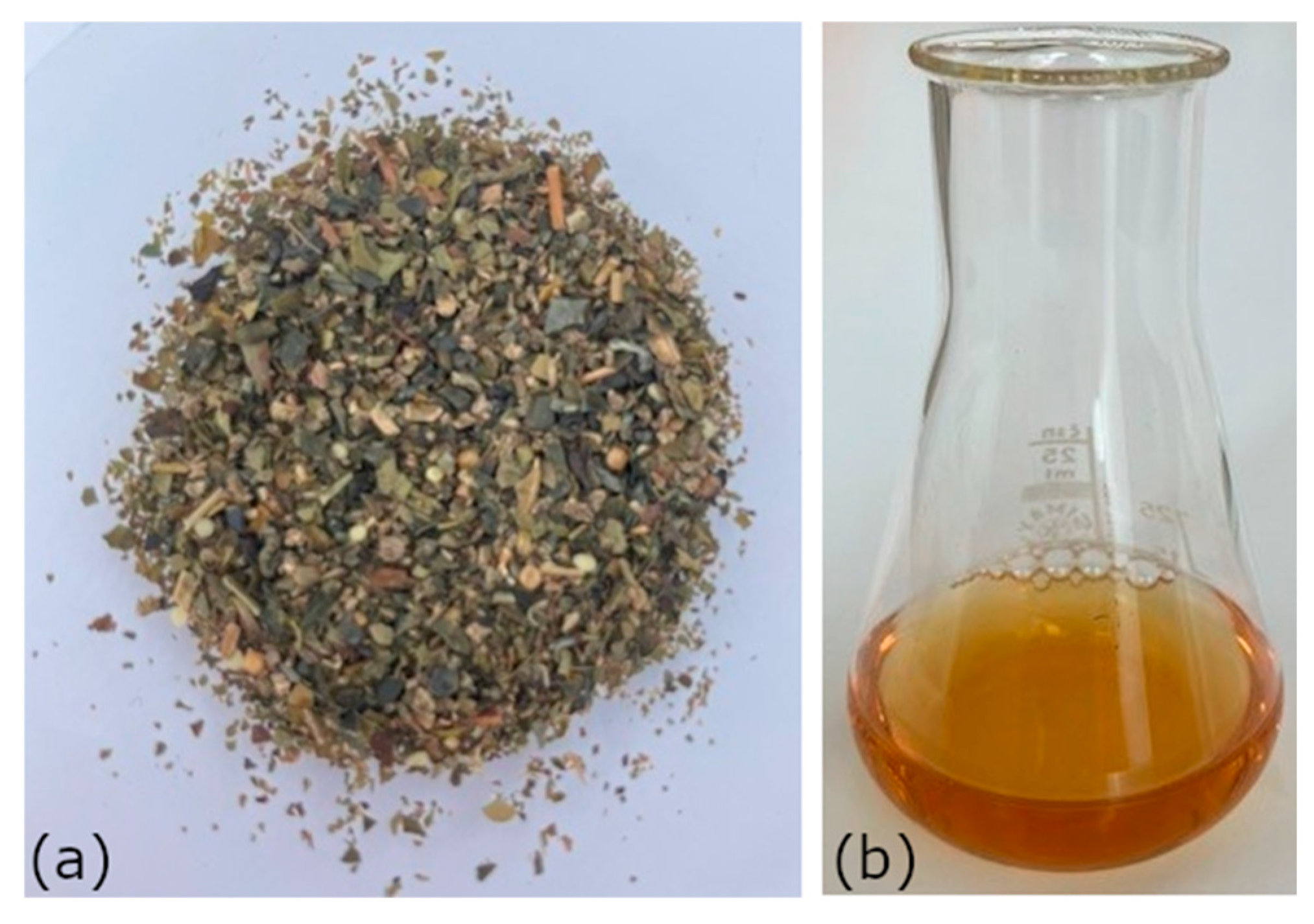
3.1. Green Tea Leaf

Green tea leaves, Figure 1a, are rich in biologically active compounds, primarily polyphenols (such as epigallocatechin gallate, epicatechin, and epicatechin gallate), alkaloids (e.g., caffeine), proteins, polysaccharides, flavonoids, theobromine, and theophylline [22]. They also contain amino acids (e.g., L-theanine), essential vitamins (C, B2, E, and K), and minerals (potassium, magnesium, calcium, zinc, and fluoride). Saponins and tannins contribute to the characteristic bitterness and exhibit antibacterial properties.
There are various methods for preparing plant extracts. In this study, we employed a simple, rapid, and cost-effective approach, as outlined in the scheme shown in Figure 2. The composition of the extract depends on the method used, which includes factors such as extraction time, temperature, stirring, and the solvent. For example, as demonstrated by [23], ethanol enables the extraction of certain compounds that are not water-soluble.
The aqueous extract of green tea leaves, Figure 1b, contains a high concentration of bioactive compounds, especially when prepared using hot-water infusion [24,25]. These compounds are believed to play a key role in nanoparticle synthesis. Many of them possess hemiacetal reducing ends and hydroxyl groups, which contribute to both the reduction and stabilization of metallic nanoparticles [26]. Polyphenols, in particular, act as reducing agents by donating electrons or hydrogen atoms to convert Au3+ to elemental gold (Au0). The resulting atoms nucleate and grow into nanoparticles.
Stabilization and capping are primarily attributed to functional groups, such as free amine groups in proteins and carbonyl groups in flavonoids, which form protective layers around the nanoparticles and help maintain their dispersion in solution. Moreover, some biomolecules may influence the shape of the nanoparticles during synthesis [22,27].

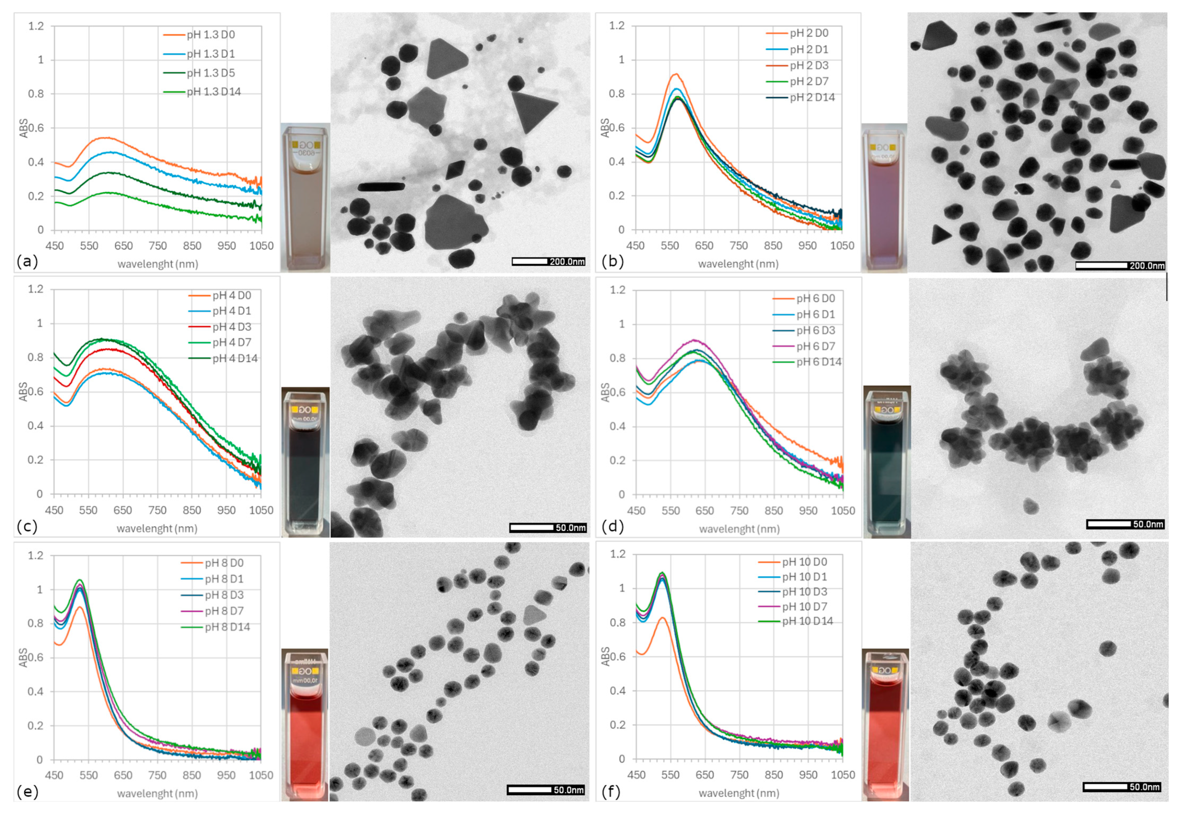
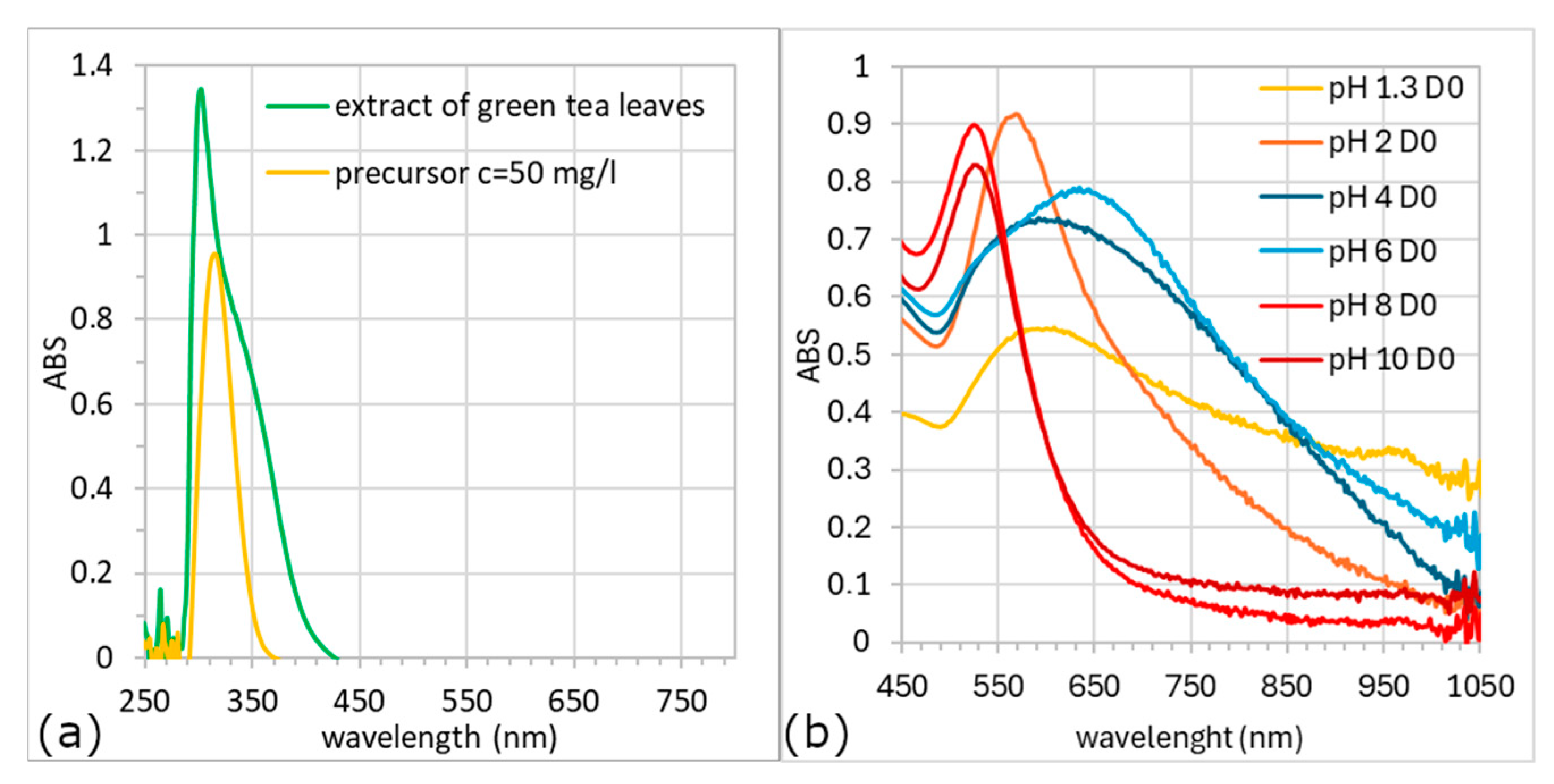
3.2. UV-Vis Analysis

UV-vis spectrophotometry is a key technique for characterizing colloidal nanoparticle solutions. A primary feature is the Surface Plasmon Resonance (SPR) band [28], caused by the collective oscillation of conduction electrons upon light excitation. The SPR band’s position, shape, and intensity are sensitive to the dielectric environment, particle size and morphology, and aggregation [28,29,30]. Thus, UV-vis analysis not only provides optical data but also provides information on nanoparticle size and shape. Moreover, based on the SPR band information, it is possible by applying the Lambert–Beer law (Equation (1)) to approximate the concentration of nanoparticles in the colloidal solution:
A = log 10 I 0 I = ε λ · c · l ,
  • I I 0 —intensity of the transmitted and incident light (W)
  • ε λ —molar absorption coefficient (L/mol·cm)
  • c—concentration in (mol/L)
  • l—the length of the light path in (cm)
  • A—absorbance
Shifts in the SPR band, as well as peak symmetry and intensity, serve as indicators of size and morphological changes [31]. It is well established that colloidal solutions of spherical gold nanoparticles (AuNPs) typically display a red-to-reddish-violet coloration, with an absorption maximum (ABSmax) near 530 nm. Such values of ABSmax of AuNPs were observed by Ha Pham et al. and Anik et al. [32,33]. A blue shift in the ABSmax is generally associated with a reduction in particle size, whereas a red shift indicates an increase in nanoparticle dimensions.
Figure 3a shows the UV-vis spectra of the extract and precursor; no spectra in the interval 400–800 nm, where AuNPs typically show the absorption peaks, can be observed. After mixing the extract (pH 5.41) with the precursors (original pH 1.3, and adjusted to pH 2, 4, 6, 8, and 10), the solution’s color changed within 5 min. Color change is the first sign of successful nanoparticle synthesis, and differences in the color of individual colloids indicate differences in the shape/size of the resulting nanoparticles.
The UV-vis spectra of colloids prepared with the original (pH 1.3) and adjusted pH (2, 4, 6, 8, and 10) on the day of synthesis (D0) are shown in Figure 3b. The UV-vis spectra confirm the formation of gold nanoparticles. Differences between the samples are evident not only in the position of the surface plasmon resonance (SPR) bands (i.e., shifts in the absorption maxima, λmax) but also in the intensity (height) of the absorption peaks. The intensity of the ABSmax can serve as an indicator of synthesis efficiency, reflecting the relative concentration of nanoparticles formed in each reaction. Among the tested conditions, the lowest nanoparticle yield was observed at pH 1.3, while the differences among the other pH values were less pronounced.
A study by A. K. Chaurasia et al. investigated the influence of precursor concentration on AuNPs synthesis and found no direct correlation between precursor concentration and nanoparticle yield. Their findings suggest that the yield is more dependent on the reducing capacity of the plant extract, assuming that sufficient Au3+ ions are present in the reaction medium. Consistent with this, our results demonstrate that pH plays a significant role in modulating the extract’s reducing ability and thus strongly affects nanoparticle formation efficiency [28].
The UV-vis spectra of colloids recorded from the day of synthesis (D0) to the 14th day (D14) are shown in Figure 4. Based on the colloid’s color and spectra profiles, the samples can be categorized into three types:
  • Type I—tall and narrow spectra; ABSmax ~530 nm; colloids in the shades of violet–red (solutions with pH 8 and 10),
  • Type II—ABSmax ~580 nm; beige and violet–beige solutions (pH 1.3 and 2),
  • Type III—broad spectrum; ABSmax of more than 600 nm; colloids in the shades of blue (pH 4 and 6).
Both UV-vis spectroscopy and TEM analysis demonstrated a significant effect of pH on the morphology, size distribution, and optical properties of the synthesized nanoparticles.
Type I colloids (pH 8 and 10) exhibited narrow, symmetrical SPR bands, indicating monodisperse, spherical AuNPs in a narrow size distribution. TEM confirmed uniform spherical shapes with average diameters of 17 nm (pH 8) and 19 nm (pH 10), Figure 4e,f. Size distribution analysis, Figure 5b, showed that 90% of particles were ≤20 nm in size, with no particles exceeding 30 nm. The spectra remained unchanged over time, indicating high colloidal stability. A.K. Chaurasia et al. achieved similar results, a narrow SPR bend with ABSmax around 540 nm, and the TEM analysis confirmed uniform and spherical nanoparticles [28]. M. Shirzadi-Ahodashti et al. synthesized a purple–red colloid using the extract of Pistacia vera. The UV-vis spectrum showed an ABSmax wavelength near 570 nm. Through TEM analysis, mostly spherical-like, homogeneous, and regular AuNPs with a size ranging from 20 to 35 nm were confirmed [34]. This supports the claim that the red shift of ABSmax signifies an increase in nanoparticle size.
Figure 4. UV-vis spectra, colloid color, and TEM microphotographs: pH 1.3 (a), pH 2 (b), pH 4 (c), pH 6 (d), pH 8 (e), and pH 10 (f).
Crystals 15 00682 g004
Type II colloids (pH 1.3 and 2) display broad and shallow SPR bands with ABSmax wavelengths around 580 nm, suggesting a heterogeneous mix of shapes and sizes of nanoparticles, Figure 4a. At pH 1.3, TEM revealed spherical (average diameter of ~80 nm, 80%), triangular (~180 nm, 4.4%), decahedral (~125 nm, 4.7%), rod-like (~200 nm, 1%), trapezohedral (~104 nm, 1.7%), and irregular prism-shaped particles (~230 nm, 8.4%). The size distribution histogram, Figure 5a, showed three populations of nanoparticles: small spherical (~45 nm), large spherical (~115 nm), and large non-spherical nanoparticles (>200 nm). This morphological and size diversity likely contributes to the observed dichroic effect (the solution appears beige in transmission and brick-red in reflection light). The low value of ABSmax and its decline indicated low yield and partial degradation over time. The reason for the synthesis of different morphologies may be the formation of large nanoparticles [35]. The system tries to minimize the excess Gibbs free energy associated with the formation of nanoparticles [17,36]. Therefore, these large nanoparticles change to form more energetically favorable shapes, such as hexagonal and truncated triangular shapes.
At pH 2, nanoparticles were mostly quasi-spherical (~60 nm, 87%), with fewer non-spherical shapes, Figure 4b. In addition, the presence of trapezohedral particles (~55 nm, 1%), decahedral particles (~67 nm, 3.1%), various irregularly shaped nanoparticles (~114 nm, 5.5%), and triangular or truncated prisms (~84 nm, 3.1%) was confirmed, as shown in Figure 4b and Figure 5a. The presence of non-quasi-spherical nanoparticles is also indicated by a less steep slope of the SPR band at wavelengths above 600 nm, with the spectrum being less symmetrical than, for example, the colloid in Figure 4f. This colloid showed a similar dichroic effect as the sample at pH 1.3 but greater spectral and morphological stability.
Figure 5. The size distribution histograms. Histograms of AgNPs size distribution of samples with pH 1.3 and 2 (a) and samples with pH 4, pH 6, pH 8, and pH 10 (b).
Crystals 15 00682 g005
Type III colloids (pH 4 and 6) exhibited a significant red shift of the ABSmaxmax > 600 nm), Figure 4c,d. Based on the shape of the SPR band, the nonuniformity in the size and shape of AuNPs was expected. TEM revealed predominantly “flower-like” morphologies. At pH 4, both quasi-spherical (~15 nm) and flower-like (~34 nm) nanoparticles were present; at pH 6, the average particle size was ~46 nm with increased structural symmetry compared to pH 4. These colloids were dark gray to anthracite in color, corresponding to larger, complexly shaped nanoparticles and a broader size distribution, as also confirmed by other authors [13,14,16].
High-resolution transmission electron microscopy (HRTEM) analysis was performed to confirm the internal structure and crystallinity of the flower-like AuNPs, Figure 6. The HRTEM image revealed that the particle is a single crystalline nanoparticle with an overall size of approximately 70 nm and not an aggregate of smaller nanostructures. Upon further magnification, distinct lattice fringes were observed, with an interplanar spacing of 0.235 nm, which corresponds to the (111) lattice planes of face-centered cubic (fcc) gold. The measured distance of 4.91 nm across 21 atoms supports this, as it yields an average atomic spacing of ~0.234 nm. These results unambiguously confirm the crystalline nature of the nanoparticle and provide structural evidence that the material is indeed elemental gold.
From the experimental results, under these experimental conditions, the pH is the main factor influencing the shape and size of AuNPs. The increase/decrease in OH ions in the solutions affected the reaction properties of the extract. The extract probably contains substances that are sensitive to pH changes, which indicates that phytochemicals can control the shape of AuNPs. Various authors have confirmed that polyphenols are mainly responsible for the isotropic stabilization of AuNPs, while flavonoids are involved in the anisotropic growth of AuNPs [23].
Based on experimental results and comparisons with findings from other studies, it can be concluded that pH is a critical parameter influencing the morphology, size, and stability of gold nanoparticles synthesized using plant extracts. Changes in hydroxide ion (OH) concentration substantially affect the chemical environment of the reaction, modulating the reduction kinetics of Au3+ ions and thereby influencing both nucleation and growth mechanisms [37].
Phytochemicals present in the green tea extract, particularly polyphenols and flavonoids, play a critical role in these processes. Their activity is governed by the pH-dependent ionization of functional groups (-OH, -COOH, and -NH2), which determines both their metal-binding affinity and redox potential. While polyphenols tend to promote isotropic stabilization, flavonoids are more likely to support anisotropic crystal growth, contributing to the formation of non-spherical morphologies such as rods and triangular prisms [38].
In addition, pH directly affects the structural integrity and antioxidant activity of key bioactive compounds such as rosmarinic acid, carnosic acid, and caffeic acid. Under highly acidic or strongly alkaline conditions, these molecules may undergo hydrolytic degradation or chemical transformation into less active forms, thereby reducing their capacity to stabilize and reduce gold ions. This phenomenon may account for observed trends in particle size distribution: acidic environments (e.g., pH 2) typically produce larger and more polydisperse nanoparticles, whereas alkaline conditions (e.g., pH 9) favor the formation of smaller, more uniform particles [39], which aligns with our observations.
Interestingly, at near-neutral pH (approximately pH 4–6), a distinctive shift in nanoparticle morphology was detected, characterized by the emergence of complex, flower-like structures. We assume that this unusual formation likely results from a delicate balance between reduction and stabilization mechanisms. At this pH range, the slower reduction kinetics of Au3+ prolong the growth phase, while the partially ionized functional groups of phytochemicals exhibit increased binding specificity to certain crystallographic planes of gold. This favors anisotropic and branched growth rather than the thermodynamically stable spherical forms. Additionally, the competition between nucleation and surface passivation may induce transient instabilities, further promoting the formation of asymmetric and dendritic nanostructures.
A review of recent studies, Table 1, also confirms that the morphology and size of gold nanoparticles (AuNPs) synthesized via green methods strongly depend on synthesis parameters such as pH, temperature, precursor concentration, and the ratio of plant extract to metal salt. Reported shapes vary widely, from spherical and rod-like to star-shaped and irregular structures, with particle sizes ranging from a few nanometers up to over 100 nm. These differences are primarily attributed to the biochemical composition of the plant extract and reaction conditions. Our results align with this trend, demonstrating that even commonly available plant materials, such as commercial green tea, can effectively act as reducing and stabilizing agents when the synthesis conditions are properly optimized.
Overall, these findings underscore the role of pH in shaping the physicochemical properties of green-synthesized AuNPs. The system’s complexity highlights the need for case-specific optimization, as the interplay between pH, extract composition, and nanoparticle growth dynamics is highly dependent on the type of plant material and target application.

4. Conclusions

This study demonstrates the successful green synthesis of gold nanoparticles using an aqueous extract of green tea leaves, with a focus on the influence of precursor pH on nanoparticle morphology and stability. The results reveal that simple modulation of pH enables the controlled formation of distinct nanoparticle shapes, ranging from spherical to anisotropic forms such as triangular, rod-like, decahedral, and even flower-like morphologies, without the need for additional chemical agents or complex procedures.
The novelty of this work lies in the demonstration that a single, eco-friendly biological extract can be used to reproducibly generate a wide variety of AuNP shapes solely through pH adjustment. This approach offers a sustainable and easily tunable method for tailoring the physicochemical properties of nanoparticles, which is particularly advantageous for applications requiring specific morphologies, such as in plasmonic sensing, catalysis, or antimicrobial coatings.

Author Contributions

Validation, writing—original draft, writing—review and editing, supervision, O.V.; investigation, formal analysis, and data curation, Z.M.; data curation, methodology, and investigation, M.L. All authors have read and agreed to the published version of the manuscript.

Funding

This research received no external funding.

Data Availability Statement

All data are available from the corresponding author upon reasonable request.

Acknowledgments

KEGA 011TUKE-4/2025 Modern Materials for Energy: Innovative Education for Enhancing the Quality of Teaching Materials Engineering Subjects.

Conflicts of Interest

The authors declare no conflicts of interest.

References

  1. Dreaden, E.C.; Alkilany, A.M.; Huang, X.; Murphy, C.J.; El-Sayed, M.A. The golden age: Gold nanoparticles for biomedicine. Chem. Soc. Rev. 2012, 41, 2740–2779. [Google Scholar] [CrossRef]
  2. Aziz, F.; Ihsan, A.; Nazir, A.; Ahmad, I.; Bajwa, S.Z.; Rehman, A.; Diallo, A.; Khan, W.S. Novel route synthesis of porous and solid gold nanoparticles for investigating their comparative performance as contrast agent in computed tomography scan and effect on liver and kidney function. Int. J. Nanomed. 2017, 12, 1555–1563. [Google Scholar] [CrossRef]
  3. Rai, M.; Ingle, A.P.; Birla, S.; Yadav, A.; Santos, C.A.D. Strategic role of selected noble metal nanoparticles in medicine. Crit. Rev. Microbiol. 2016, 42, 696–719. [Google Scholar] [CrossRef]
  4. Kumar, B.; Smita, K.; Angulo, Y.; Debut, A.; Cumbal, L. Honeybee pollen assisted biosynthesis of nanogold and its application as catalyst in reduction of 4-nitrophenol. Heliyon 2022, 8, e10191. [Google Scholar] [CrossRef]
  5. Daniel, M.C.; Astruc, D. Gold nanoparticles: Assembly, supramolecular chemistry, quantum-size-related properties, and applications toward biology, catalysis, and nanotechnology. Chem. Rev. 2004, 104, 293–346. [Google Scholar] [CrossRef]
  6. Al-Harbi, N.; Abd-Elrahman, N.K. Physical methods for preparation of nanomaterials, their characterization and applications: A review. J. Umm Al-Qura Univ. Appl. Sci. 2025, 11, 356–377. [Google Scholar] [CrossRef]
  7. Hussain, M.H.; Abu Bakar, N.F.; Mustapa, A.N.; Low, K.-F.; Othman, N.H.; Adam, F. Synthesis of Various Size Gold Nanoparticles by Chemical Reduction Method with Different Solvent Polarity. Nanoscale Res. Lett. 2020, 15, 140. [Google Scholar] [CrossRef]
  8. Fuentes-García, J.A.; Santoyo-Salzar, J.; Rangel-Cortes, E.; Goya, G.F.; Cardozo-Mata, V.; Pescador-Rojas, J.A. Effect of ultrasonic irradiation power on sonochemical synthesis of gold nanoparticles. Ultrason. Sonochem. 2021, 70, 105274. [Google Scholar] [CrossRef]
  9. Raghunandan, D.; Bedre, M.D.; Basavaraja, S.; Sawle, B.; Manjunath, S.Y.; Venkataraman, A. Rapid biosynthesis of irregular shaped gold nanoparticles from macerated aqueous extracellular dried clove buds (Syzygium aromaticum) solution. Colloids Surf. B Biointerfaces 2010, 79, 235–240. [Google Scholar] [CrossRef]
  10. Philip, D. Rapid green synthesis of spherical gold nanoparticles using Mangifera indica leaf. Spectrochim. Acta Part A Mol. Biomol. Spectrosc. 2010, 77, 807–810. [Google Scholar] [CrossRef]
  11. Palaniswamy, S.; Bepari, A.; Rajan, S.; Assiri, R.A.; Shibu, A.; Narayasamy, A.; Krishnamoorthy, K. Biosynthesis of gold nanoparticles from Martynia annua aqueous leaves extract, its antioxidant potential and antimicrobial ability against selected wound pathogens. Inorg. Chem. Comm. 2024, 70, 113241. [Google Scholar] [CrossRef]
  12. Bedlovičová, Z.; Siksa, P.; Kováčová, M.; Bureš, R.; Tkáčiková, Ľ.; Džunda, R.; Tampubolon, I.O.; Balážová, Ľ.; Baláž, M. Lavender-mediated solvent-free biomechanochemical synthesis of antibacterially active Ag/AgCl nanoparticles using a Taguchi design. Adv. Nat. Sci. Nanosci. Nanotechnol. 2025, 16, 015018. [Google Scholar] [CrossRef]
  13. Nehl, C.L.; Liao, H.; Hafner, J.H. Optical Properties of Star-Shaped Gold Nanoparticles. Nano Lett. 2006, 6, 683–688. [Google Scholar] [CrossRef]
  14. Silvestri, A.; Lay, L.; Psaro, R.; Polito, L.; Evangelisti, C. Fluidic Manufacture of Star-Shaped Gold Nanoparticles. Chemistry 2017, 23, 9732–9735. [Google Scholar] [CrossRef]
  15. Sau, T.K.; Murphy, C.J. Seeded High Yield Synthesis of Short Au Nanorods in Aqueous Solution. Langmuir 2004, 20, 6414–6420. [Google Scholar] [CrossRef]
  16. Barbosa, S.; Agrawal, A.; Rodríguez-Lorenzo, L.; Pastoriza-Santos, I.; Alvarez-Puebla, R.A.; Kornowski, A.; Weller, H.; Liz-Marzán, L.M. Tuning Size and Sensing Properties in Colloidal Gold Nanostars. Langmuir 2010, 26, 14943–14950. [Google Scholar] [CrossRef]
  17. Huo, C.; Khoshnamvand, M.; Liu, P.; Liu, C.; Yuan, C.-G. Rapid mediated biosynthesis and quantification of AuNPs using persimmon (Diospyros Kaki, L.f) fruit extract. J. Mol. Struc. 2018, 1178, 366–374. [Google Scholar] [CrossRef]
  18. Ahmad, N.A.; Adaila, K.; Omar, A.A. Effect of pH in the Biosynthesis of Gold Nanoparticles: A Review. Afr. J. Adv. Pure Appl. Sci. 2023, 2, 360–368. Available online: https://aaasjournals.com/index.php/ajapas/index (accessed on 1 May 2025).
  19. Hadi, H.T.; Ibrahim, O.M.S. Green Synthesis and Characterization of Gold Nanoparticles Using Crushed Clove Buds (Syzygium aromaticum) Oil Extracted by Hydrodistillation. J. Res. Pharm. 2024, 28, 1883–1891. [Google Scholar] [CrossRef]
  20. Song, J.Y.; Jang, H.-K.; Kim, B.S. Biological synthesis of gold nanoparticles using Magnolia kobus and Diopyros kaki leaf extracts. Process Biochem. 2009, 44, 1133–1138. [Google Scholar] [CrossRef]
  21. Nadagouda, M.N.; Varma, R.S. Green synthesis of silver and palladium nanoparticles at room temperature using coffee and tea extract. Green Chem. 2008, 10, 859. [Google Scholar] [CrossRef]
  22. Ariski, R.T.; Lee, K.K.; Kim, Y.; Lee, C.-S. The impact of pH and temperature on the green gold nanoparticles preparation using Jeju Hallabong peel extract for biomedical applications. RSC Adv. 2024, 14, 14582–14592. [Google Scholar] [CrossRef]
  23. Baesso, A.S.; Silva, D.J.; Soares, A.K.; Paula, M.M.S.; Gonzalez de Cademartori, P.H. Biosynthesis of gold nanoparticles using papaya seed extract for the functionalization of nanocellulose membranes. Ind. Crops Prod. 2023, 197, 116601. [Google Scholar] [CrossRef]
  24. Patra, S.; Golder, A.K.; Uppaluri, R.V. Green synthesis of carbon dots from mature green tea leaves for label-free fluorescence sensing of chromium(VI). Opt. Mater. 2024, 154, 115767. [Google Scholar] [CrossRef]
  25. Sahu, S.K.; Mansoori, A.; Jana, S.K.; Kumar, A.; Ghorai, T.K. Biosynthesis of silver nanoparticles using green tea aqueous leaf extract and their biological and chemotherapeutic activity. J. Molec. Struct. 2025, 1320, 139690. [Google Scholar] [CrossRef]
  26. Datta, D.; Deepak, K.S.; Das, B. Progress in the synthesis, characterisation, property enhancement techniques and application of gold nanoparticles: A review. MRS Commun. 2022, 12, 700–715. [Google Scholar] [CrossRef]
  27. Zhao, T.; Li, C.; Wang, S.; Song, X. Green Tea (Camellia sinensis): A Review of its Phytochemistry, Pharmacology, and Toxicology. Molecules 2022, 27, 3909. [Google Scholar] [CrossRef]
  28. Chaurasia, A.K.; Singh, R.K.; Kumar, A. Synthesis, optimization, characterization, antioxidant and anti-cancerous activity analysis of nanoparticles IA-AuNPs synthesized using leaf extract of soil grown Ipomoea aquatica. J. Mol. Struct. 2025, 1327, 141212. [Google Scholar] [CrossRef]
  29. El-Borady, O.M.; Fawzy, M.; Hosny, M. Antioxidant, anticancer and enhanced photocatalytic potentials of gold nanoparticles biosynthesized by common reed leaf extract. Appl. Nanosci. 2023, 13, 3149–3160. [Google Scholar] [CrossRef]
  30. Rahman, A.U.; Khan, A.U.; Yuan, Q.; Wei, Y.; Ahmad, A.; Ullah, S.; Khan, Z.U.H.; Shams, S.; Tariq, M.; Ahmad, W. Tuber extract of Arisaema flavum eco-benignly and effectively synthesize silver nanoparticles: Photocatalytic and antibacterial response against multidrug resistant engineered E. coli QH4. J. Photochem. Photobiol. B Biol. 2019, 193, 31–38. [Google Scholar] [CrossRef]
  31. Meng, X.-H.; Li, N.; Zhu, H.-T.; Wang, D.; Yang, C.-R.; Zhang, Y.-J. Plant Resources, Chemical Constituents and Bioactivities of Tea Plants from the Genus Camellia Section Thea. J. Agric. Food Chem. 2018, 67, 5318–5349. [Google Scholar] [CrossRef]
  32. Ha Pham, T.T.; Dien, N.D.; Vu, X.H. Facile synthesis of silver/gold alloy nanoparticles for ultra-sensitive rhodamine B detection. RSC Adv. 2021, 11, 21475–21488. [Google Scholar] [CrossRef]
  33. Anik, M.I.; Mahmud, N.; Al Masud, A.; Hasan, M. Gold nanoparticles (GNPs) in biomedical and clinical applications: A review. Nano Sel. 2022, 3, 792. [Google Scholar] [CrossRef]
  34. Shirzadi-Ahodashti, M.; Mizwari, Z.M.; Mohammadi-Aghdam, S.; Ahmadi, S.; Ebrahimzadeh, M.A.; Mortazavi-Derazkola, S. Optimization and evaluation of anticancer, antifungal, catalytic, and antibacterial activities: Biosynthesis of spherical-shaped gold nanoparticles using Pistacia vera hull extract (AuNPs@PV). Arab. J. Chem. 2023, 16, 104423. [Google Scholar] [CrossRef]
  35. Lukman, A.I.; Gong, B.; Marjo, C.E.; Roessner, U.; Harris, A.T. Facile synthesis, stabilization, and anti-bacterial performance of discrete Ag nanoparticles using Medicago sativa seed exudates. J. Colloid Interface Sci. 2011, 353, 433–444. [Google Scholar] [CrossRef]
  36. Narayanan, K.B.; Sakthivel, N. Biological synthesis of metal nanoparticles by microbes. Adv. Colloid Interface Sci. 2010, 156, 1–13. [Google Scholar] [CrossRef]
  37. Ahmed, S.; Ahmad, M.; Swami, B.L.; Ikram, S. A review on plants extract mediated synthesis of silver nanoparticles for antimicrobial applications: A green expertise. J. Adv. Res. 2016, 7, 17–28. [Google Scholar] [CrossRef]
  38. Miličevič, A. Flavonoid Oxidation Potentials and Antioxidant Activities-Theoretical Models Based on Oxidation Mechanisms and Related Changes in Electronic Structure. Int. J. Mol. Sci. 2024, 25, 5011. [Google Scholar] [CrossRef]
  39. Timoszyk, A.; Grochowalska, R. Mechanism and Antibacterial Activity of Gold Nanoparticles (AuNPs) Functionalized with Natural Compounds from Plants. Pharmaceutics 2022, 14, 2599. [Google Scholar] [CrossRef]
Figure 1. Chopped green tea leaves (a) and extract prepared using green tea leaves (b).
Crystals 15 00682 g001
Figure 2. Scheme of the extraction process and nanoparticle colloid preparation.
Crystals 15 00682 g002
Figure 3. UV-vis spectra of the extract and precursor (a) and prepared colloids (b).
Crystals 15 00682 g003
Figure 6. TEM and HRTEM microphotographs of flower-like Au nanoparticles.
Crystals 15 00682 g006
Table 1. Summary of some reported AuNP syntheses using various methods and conditions.
ReferenceReducing Agent/Plant SourceSynthesis Method/ConditionsShape of AuNPsSize Range
[4]Honeybee pollenGreen synthesisMostly spherical7–42 nm
[5]Various (overview)Multiple methods (review)Various (spheres, rods, and cages)1–100+ nm
[9]Clove bud extractMacerated extract, room tempIrregular5–100 nm
[10]Mangifera indica leaf extractRapid green synthesisSpherical~18 nm
[11]Martynia annua leaf extractRoom temp biosynthesisSpherical21 nm
[15]Seed-mediated growthHigh-yield synthesisShort rods20–100 nm length
[16]Seed-mediated growthSize tuning with silver ionsNanostars40–150 nm
[17]Diospyros kaki fruit extractBiosynthesisVarious shapes20–100+ nm
[18]Review (multiple plant types)Focus on the pH effectVarious shapes15–90 nm
[20]Magnolia kobus,
Diospyros kaki
Biological synthesisVarious shapes5–300 nm
Disclaimer/Publisher’s Note: The statements, opinions and data contained in all publications are solely those of the individual author(s) and contributor(s) and not of MDPI and/or the editor(s). MDPI and/or the editor(s) disclaim responsibility for any injury to people or property resulting from any ideas, methods, instructions or products referred to in the content.

