Article

Magnesia Partially Stabilized Zirconia/Hydroxyapatite Biocomposites: Structural, Morphological and Microhardness Properties

by
Liliana Bizo
1,*,
Adriana-Liana Bot
1,
Marieta Mureșan-Pop
2,3,*,
Lucian Barbu-Tudoran
4,5,
Claudia Andreea Cojan
1 and
Réka Barabás
6
1
Department of Chemical Engineering, Faculty of Chemistry and Chemical Engineering, Babeş-Bolyai University, 11 Arany János Str., 400028 Cluj-Napoca, Romania
2
Nanostructured Materials and Bio-Nano-Interfaces Center, Interdisciplinary Research Institute in Bio-Nano-Sciences, Babeş-Bolyai University, 42 Treboniu Laurian Str., 400271 Cluj-Napoca, Romania
3
Contrast Agents and Specific Therapeutics Center-INSPIRE Platform, Interdisciplinary Research Institute in Bio-Nano-Sciences, Babeş-Bolyai University, 42 Treboniu Laurian Str., 400271 Cluj-Napoca, Romania
4
Electron Microscopy Center, Faculty of Biology and Geology, Babeș-Bolyai University, 5-7 Clinicilor Street, 400006 Cluj-Napoca, Romania
5
Electron Microscopy Integrated Laboratory, National Institute for Research and Development of Isotopic and Molecular Technologies, 67-103 Donath Street, 400293 Cluj-Napoca, Romania
6
Department of Chemistry and Chemical Engineering of Hungarian Line of Study, Faculty of Chemistry and Chemical Engineering, Babeş-Bolyai University, 11 Arany János Str., 400028 Cluj-Napoca, Romania
*
Authors to whom correspondence should be addressed.
Crystals 2025, 15(7), 608; https://doi.org/10.3390/cryst15070608
Submission received: 31 May 2025 / Revised: 27 June 2025 / Accepted: 29 June 2025 / Published: 30 June 2025

Abstract

Hydroxyapatite (HAP) is the most widely accepted biomaterial for repairing bone tissue defects, demonstrating excellent biocompatibility and bioactivity that promote new bone formation. Zirconia (ZrO2), known for its strength and fracture toughness, is commonly used to reinforce ceramics. In this study, magnesium oxide (MgO) served as a stabilizer for zirconia, resulting in magnesia partially stabilized zirconia (Mg-PSZ). Both Mg-PSZ and HAP were synthesized via coprecipitation and mixed in specific ratios to create composites through a ceramic method involving mixing, compaction, and sintering at 1100 °C. The samples were characterized using techniques such as X-ray powder diffraction (XRPD), Fourier-transform infrared spectroscopy (FTIR), and scanning electron microscopy/energy-dispersive X-ray spectroscopy (SEM/EDS). Structural analyses confirmed the presence of both monoclinic and tetragonal zirconia phases. Besides, the increased wt.% HAP in the composites produced distinct peaks for hexagonal HAP. Crystallite sizes ranged from 27.45 nm to 31.5 nm, and surface morphology was homogeneous with small pores. Elements such as calcium, phosphorus, magnesium, zirconium, and oxygen were detected in all samples. This research also examined microhardness changes in the materials. The findings revealed enhancement in microhardness for the biocomposite with higher zirconia content, 90Mg-PSZ/10HAP sample, with the smallest average pore size, highlighting its potential for biomedical applications.

1. Introduction

Zirconia-based ceramics have been extensively studied over the years and continue to be of significant interest in the development of advanced materials. This interest stems from their excellent properties, including good fracture toughness, mechanical strength, high ionic conductivity, and resistance to corrosion [1,2,3]. Zirconium dioxide (ZrO2) exhibits a complex polymorphism, consisting of three phases: the monoclinic phase (m-ZrO2, stable at room temperature up to 1170 °C), which is the most stable but has poor mechanical properties; the tetragonal phase (t-ZrO2, between 1170 and 2370 °C), which is considered the most promising due to its favorable overall properties; and the cubic phase (c-ZrO2, above 2370 °C), which possesses moderate characteristics. Phase transformations can occur with significant volume changes, potentially leading to microcracks and affecting the quality of the final product. For instance, the transformation from the t-ZrO2 to m-ZrO2 results in a volume change of approximately 3 to 5% [4,5]. Partially stabilized zirconia (PSZ) has gained increased attention in the last decade, owing to its excellent biological, chemical, physical, mechanical, thermal, and electrical properties. These attributes make it suitable for various applications, including dental and bone implants, thermal barrier coatings, refractory materials, and membranes. PSZ is characterized by the presence of a mixture of two or more ZrO2 phases: t-ZrO2, c-ZrO2, and m-ZrO2. Zirconia can be stabilized by adding metal oxide dopants such as MgO. Systems based on zirconia with MgO and CaO are of interest due to their effectiveness in stabilizing ZrO2 and due to similar properties at much lower production costs compared to yttrium-stabilized zirconia (YSZ) ceramics [6,7]. In addition to its low cost and high availability, MgO is recognized as an effective antibacterial agent, which makes it an excellent candidate for biological applications [8,9]. About 10 mol% magnesium is the percentage most used in industry to obtain partially magnesium-stabilized zirconia (Mg-PSZ) [10]. Mg-PSZ exhibits satisfactory thermal shock resistance, high mechanical properties, and a small thermal expansion coefficient.
Hydroxyapatite (HAP) is the main inorganic component of human bones and teeth. It possesses excellent properties like biocompatibility and bioactivity, which improve its use in various biomedical applications. These include bone tissue engineering, dental implants, orthopedic implants, and drug delivery systems (DDSs) [11,12,13,14,15,16]. Due to its biocompatibility and similarity to the mineral component of natural bone, HAP is a promising material for bone regeneration. It can be used as a scaffold material to support bone growth, promote osteogenesis (bone formation), and enhance the overall regeneration process [15]. HAP can be fabricated into porous scaffolds that mimic the structure of natural bone, providing a template for new bone tissue to grow into [17]. HAP coatings on implants can improve bone cell growth on the implant surface, enhancing osseointegration (the process of bone bonding to the implant) [18]. HAP can be combined with other materials, such as polymers or other bioactive ceramics, to create composites with enhanced mechanical properties and biological activity for bone regeneration.
HAP exhibits poor mechanical properties, so the challenge is to bring the mechanical properties as close as possible to those of the human bone/teeth. This can be achieved through advanced manufacturing techniques and the development of novel composite materials. Consequently, one approach to combining the advantageous properties of HAP and ZrO2 biomaterials could be the preparation of composites [19,20]. Additionally, ZrO2-HAP composites are considered promising materials due to the combination of the inert ZrO2 and the active HAP, which could enhance the bonding ability with natural bone in various medical fields. Consequently, many ZrO2-HAP composites have been developed as coatings or substrates to achieve both bone reconstruction and regeneration needed to treat bone defects [21].
In recent research by Fan et al., ZrO2-doped HAP ceramics were prepared using VAT photopolymerization and microwave sintering [22]. They explored how different zirconia levels and sintering temperatures affected the ceramics’ properties. Results showed that zirconia increased porosity and reduced shrinkage, leading to more anisotropic behavior, especially with higher porosity. The best doping level for mitigating anisotropy was 4 wt%, and the optimal sintering temperature was 1200 °C. At 6 wt% doping, the ceramics showed impressive strength with a flexural strength of 51.01 ± 5.61 MPa and a compressive strength of 116.15 ± 7.73 MPa, along with 31.40% porosity. Thus, a 6 wt% zirconia doping effectively achieves high porosity while maintaining excellent mechanical properties. Zhang et al. [23] employed DLP 3D printing to create zirconia-toughened hydroxyapatite bioceramic composites. Their study achieved a composite with 6 wt% zirconia (ZrO2) doping and a relative density of 90.7% after sintering at 1200 °C. They demonstrated that the incorporation of zirconia significantly enhanced the toughness and mechanical properties of HAP ceramics. Xing et al. [24] studied a series of zirconia/hydroxyapatite composites with varied compositions, prepared under different conditions in order to find the optimal composites for the target application. The results indicate that both the component ratio and sintering temperature significantly influence the properties of the composites. Increasing HAP content tends to enhance bioactivity, but it can also reduce mechanical strength. Composites with 10 wt% HAP maintain adequate mechanical strength under optimal sintering conditions while exhibiting excellent bioactivity. This demonstrates that hydroxyapatite-modified zirconia has potential as a dental implant material. Sintering tests show that the desired mechanical strength is achieved at 1400 °C for 2 h in the composite containing 10 wt% HAP. In the investigation of Sivaperumal et al. [25], the reinforcement of zirconia in biomimetic HAP using a specially designed stir-type hydrothermal reactor to improve the biocompatibility and mechanical stability of bare hydroxyapatite was performed. The formation of a nanocrystalline HAP-Zirconia composite without any intermediate phases was confirmed with the size of the synthesized nanocomposite of 30 nm. The study demonstrated excellent biocompatibility with MG-63 human osteoblasts, indicating the materials’ potential in various hard tissue engineering applications. HAP/ZrO2 porous composites were successfully fabricated by Mohammad et al. [26] mixing TCP (tri calcium phosphate) and Ca(OH)2 via polymeric sponge methods. The porosity of the ceramic can be controlled by varying the concentration of hydroxyapatite (HAP) and zirconia (ZrO2), with porosity levels ranging from 65.7% to 84.4%. The compressive strength of 10.1 MPa achieved by porous scaffolds with a composition of 25 wt% zirconia and 75 wt% HAP is optimal, as it closely resembles that of cancellous bone. Additionally, these composites exhibit not only the best physical and mechanical properties but also enhanced bioactive characteristics, making them very comparable to cancellous bone. Overall, this study presents a novel method for creating HAP/ZrO2 porous ceramic composites, which hold promise as candidates for bone replacement and regeneration.
According to the literature, the amount of zirconia added has an influence on the mechanical properties of the materials, and higher amounts of ZrO2 are shown to inhibit the shrinkage of HAP [27]. For hydroxyapatite/zirconia nanocomposites obtained via the low-temperature mineralization sintering process (LMSP), Vickers hardness values between 2.5 and 3.6 GPa were identified and were attributed to an increased mechanical functionality of pure HAP [28]. HAP-ZrO2-hBN biocomposites obtained by a simple solid-state reaction technique showed better mechanical properties at a lower % of HAP [29]. Another study identified a mean value of 2336 MPa for zirconia-hydroxyapatite bioceramics and an expected range according to the literature review between 4000 and 5000 MPa [30]. It is also stated that ZrO2 at higher ratios with HAP could cause the formation of secondary phases with different thermal expansion coefficients, that specifically β-TCP could affect the mechanical properties of the materials, and that the volume expansion at the transition between m- and t-ZrO2 could lead to cracks in the samples. Another study stated Knoop hardness range values between 5 and 8 GPa for HAP/ZrO2 nanocomposites [31]. For hydroxyapatite/yttrium-stabilized zirconia bioceramics synthesized by the plasma method, microhardness values between 3.9 and 3.95 GPa were obtained [32]. Another study specified that for ZrO2/HAP biomaterials obtained by the solid-state method, the shrinkage rate increased with the percentage of HAP in the sample, a fact that can influence the mechanical properties of the materials [33].
While HAP provides excellent biocompatibility and bioactivity, and ZrO2 offers enhanced mechanical properties, further research is needed to tailor the material’s properties for optimal performance in various load-bearing and regenerative scenarios. In conclusion, while HAP/ZrO2 biocomposites hold great promise for various biomedical applications, further research is crucial to address the specific challenges related to optimizing their composition, microstructure, biocompatibility, bioactivity, and mechanical properties for specific clinical needs.
Briefly, this paper presents the synthesis of ceramic-ceramic composites comprising Mg-PSZ and HAP, which could be particularly useful in bone regeneration and reconstruction due to their ability to promote bone cell adhesion and growth. It is designed that Mg-PSZ serves as the matrix to provide mechanical integrity and physical functions, whereas HAP is the dispersed phase that imparts bioactivity. The research will focus on obtaining new composites by solid-state synthesis with enhanced structural, morphological, and microhardness performance. Moreover, the impact of the HAP addition to Mg-PSZ on the structure and the mechanical strength of the composites was investigated. The correlation between the microhardness and structural properties was established.

2. Materials and Methods

2.1. Mg-PSZ Preparation

Zirconia doped with 10 mol% MgO (Mg-PSZ) was obtained by the coprecipitation method [34,35]. The starting materials ZrOCl2·8H2O (Riedel-de Haën AG, Seelze, Germany) and 10 mol% MgCl2·6H2O (Reactivul, Bucharest, Romania) were dissolved in distilled water until homogeneous solutions were obtained. NH4OH (Poch Basic, Gliwice, Poland) was used as precipitant and added dropwise to the constantly stirred MgCl2-doped ZrOCl2 solution to produce a white and gelatinous Mg(OH)2-doped zirconium hydroxide precipitate. The precipitate containing the Zr4+ and Mg2+ cations in the form of hydroxides was separated from the solution by filtration. After filtration, washing with distilled water, drying at 105 °C for 24 h (Memmert incubator, Schwabach, Germany), and calcining at 800 °C for 2 h (Barnstead Thermolyne furnace, Thermo Scientific, Waltham, MA, USA), the Mg(OH)2-doped zirconium hydroxide gel was transformed into MgO-doped ZrO2.

2.2. HAP Preparation

Hydroxyapatite (HAP) was obtained by the coprecipitation method as previously described [36,37,38]. The precursors used were 1.5 mol/L Ca(NO3)2·4H2O (Merck, Darmstadt, Germany) and 0.9 mol/L (NH4)2HPO4 (Merck, Darmstadt, Germany). Diammonium hydrogen phosphate and ammonium solution were slowly added to the calcium nitrate solution. The pH of the reaction mixture was adjusted to 11 by adding a 25% NH4OH solution (Poch Basic, Gliwice, Poland). The solution was homogenized under continuous stirring for 22 h. The obtained precipitate was filtered and washed with ethanol. The filtered and washed precipitate was subjected to drying for 6 h at 90 °C in a Fistreem vacuum oven (Fistreem International, Cambridge, England) and further thermally treated at 1000 °C with a 1 h dwell time (Barnstead Thermolyne furnace).

2.3. Biocomposites Preparation

Mg-PSZ/HAP composites are prepared by a ceramic processing route [20,39], from the obtained Mg-PSZ and HAP powders in predetermined ratios. The powders are well mixed using an agate mortar and pestle, using 5% PVA as a binder, and then pressed into pellets using a die of 10 mm diameter. The compaction was carried out with a hydraulic press (Carver Inc., Wabash, IN, USA), and the pressure was 400 kgf. The resulting compacts were then sintered at 1100 °C to avoid HAP decomposition. The furnace (LHT 04/16 High-Temperature Furnace, Nabertherm GmbH, Lilienthal, Germany) followed a cycle from room temperature to 1100 °C with a 1 h dwell time and heating and cooling rates of 5 °C/min. Composite samples prepared in this research are listed in Table 1 below.

2.4. Characterization Methods

The obtained materials and composites were characterized by a series of techniques, including thermal analysis that allowed the evaluation of the thermal stability and the heating behavior of the compounds, X-ray powder diffraction (XRPD) and Fourier Transform Infrared Spectroscopy (FTIR) to determine the crystalline phases and analyze the absorption bands related to crystallinity, scanning electron microscopy coupled with energy dispersive X-ray spectroscopy (SEM-EDX) provided detailed information about the surface morphology and elemental composition. To evaluate the changes in the surface microhardness of the materials, Vickers microhardness tests were conducted on the sintered biocomposites.
For the thermal analysis of the previously obtained biocomposites’ precursors, simultaneous DSC/TGA SDT-Q600 equipment from TA Instruments (New Castle, DE, USA) was used. In the apparatus, approximately 10 mg from each sample was placed in a 90 μL alumina pan, and the measurements were conducted in air at a constant rate of 10 °C/min, with a temperature ranging from room temperature to 1000 °C. XRPD was performed using a Shimadzu XRD-6000 X-ray diffractometer (Shimadzu Corporation, Kyoto, Japan) to investigate the structure of the samples. The equipment operated at 40 kV, 30 mA, with a Ni-filter and graphite monochromator for CuKα (λ = 1.54060 Å), with the diffraction patterns recorded in the 2θ range of 10–80° at a scan speed of 2 °/min. For measurements and diffraction data processing, XRD-6000 V5.21 software was used. FTIR absorption spectra were recorded in KBr pellets with a Bruker Vector 22 FTIR spectrometer (Bruker, Karlsruhe, Germany), in the range of 600 to 4000 cm−1. Microscopy images were obtained using a scanning electron microscope coupled with energy dispersive X-ray spectroscopy, type SU8230, HITACHI, and an AZtecLive EDX spectrometer, from Oxford Instruments. The Vickers microhardness analysis was performed using a FALCON 600G2FAO2 (INNOVATEST, Maastricht, The Netherlands), designed to measure hardness at small test forces ranging from 0.1 to 30 kgf. The dwell time was 10 s, and the force was 3 kgf. Only well-defined indents, free from chipping or cracks, were considered for all experiments. Each sample was carefully inspected with a magnifying lens to eliminate any specimens with surface defects. The Vickers microhardness (VMH) value for each indent was calculated automatically. Each sample underwent five indentations, and the mean VMH value was determined in kgf/mm2.

3. Results and Discussion

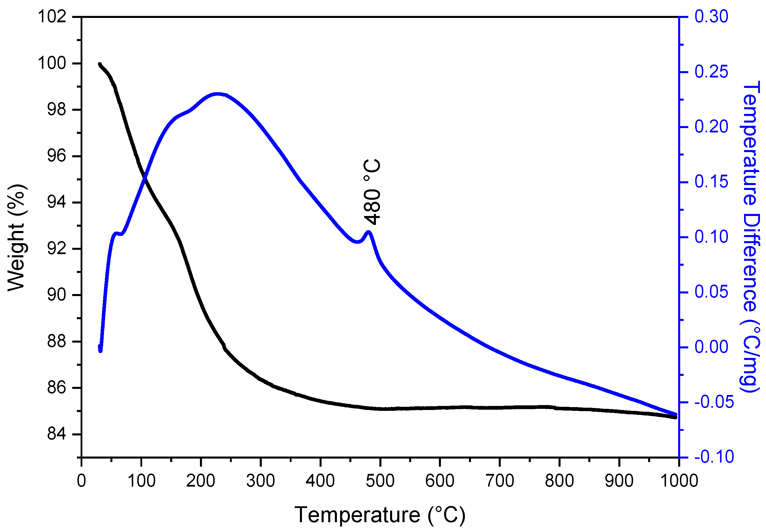
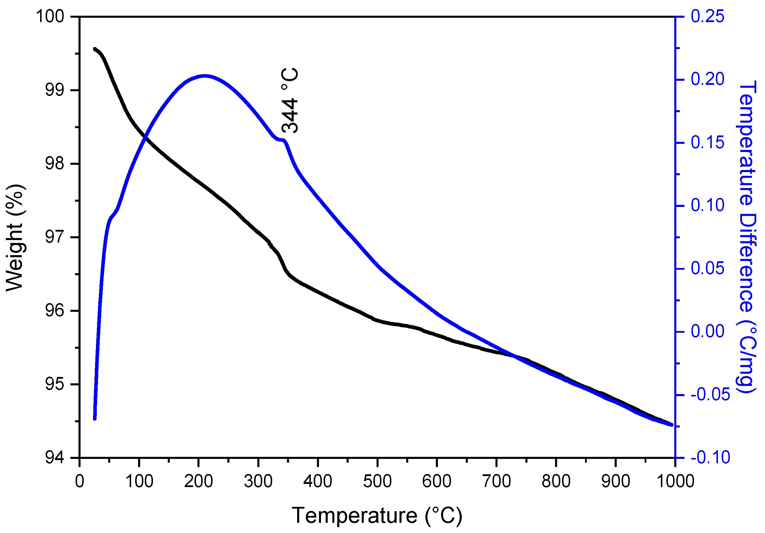
3.1. Thermal Analysis

The advantages of thermal analysis include specifying the temperature range in which bioceramics are applicable and determining the exact temperature of phase transformation.
Thermal analyses were carried out under the same conditions to determine the thermal degradation behaviors. Thermogravimetry (TG), differential thermal analysis (DTA), and DSC profiles of composite uncalcined precursor materials Mg-PSZ and HAP are displayed in Figure 1, Figure 2, and Figure S1, respectively.
Theoretical calculations suggest that the weight loss of H2O from the precursor hydroxide of Mg-PSZ, due to the condensation of bridging hydroxyl groups, should be around 13.55% [34]. The TG curves presented in Figure 1 indicate an H2O weight loss of 15.26%. This difference between the experimental and calculated mass loss values implies that the dried precursor still contains a small amount of absorbed water. Furthermore, the TG curves revealed that condensation between the bridging hydroxyl groups occurred at approximately 480 °C. After the 600 °C temperature, no significant mass loss was observed, indicating that the material has reached thermal stability and does not undergo any further significant decomposition, thereby confirming the formation of Mg-PSZ. The DTA curve shown in Figure 1 indicates that the transformation from amorphous to crystalline phase occurs at around 480 °C, which is consistent with the findings of Wang and Zhai [31]. This transformation is also supported by DSC results (Figure S1, Supplementary Materials), where the exothermic peaks observed at approximately 480 °C can be attributed to the crystallization of amorphous zirconia [28].
In the HAP sample synthesized in this study, weight loss occurring in the range of 25–110 °C, which is attributed to physically and weakly chemically bound water, is reflected in the TG curve with a weight loss of 1.62% (see Figure 2). All dissociation products are released as gas between 170 and 210 °C. Additionally, a small amount of water is released from the pores up to 500 °C, with a slight exothermic peak observed on the DTA curve at 344 °C. As previously mentioned, the thermal decomposition of calcium phosphates occurs between 550 and 800 °C. Finally, between 700 and 1000 °C, the last adsorbed water is released, which may indicate the decomposition of HAP into CaO and Ca3(PO4)2 (beta tricalcium phosphate, β-TCP) [37]. If HAP had been synthesized in its pure form, a gradual mass loss would be expected starting at around 600 °C relative to its dehydroxylation [40,41,42]. The exact decomposition temperature can vary depending on factors like the presence of impurities and the heating rate.
Generally, for the HAP prepared by the wet precipitation method, there is a consensus that the thermal behavior occurs in a 4-step process involving dehydroxylation and decomposition. Dehydroxylation incorporates the loss of water, which proceeds via the provisional formation of firstly oxyhydroxyapatite (OHAP) and then oxyapatite (OHA).
The process of HAP dehydroxylation with the release of water starts at a noticeable rate at 850–900 °C and leads to the formation of vacancies in the position of the hydroxyl groups in the HAP structure. As a result, the formation of oxyhydroxyapatite (OHA) takes place by the following reaction:
Ca10(PO4)6(OH)2 → Ca10(PO4)6(OH)2−2xOxx + xH2O
where ☐ is the OH group vacancy [43].
Decomposition of OHA then proceeds to secondary phases such as β-TCP and tetracalcium phosphate (T-TCP). The transformation of HAP has important consequences in bone engineering and plasma-coated implants, since β-TCP is a resorbable calcium phosphate, and while it will enhance the resorption of HAP implants, decomposition of HAP will also reduce the mechanical properties of the material [34].

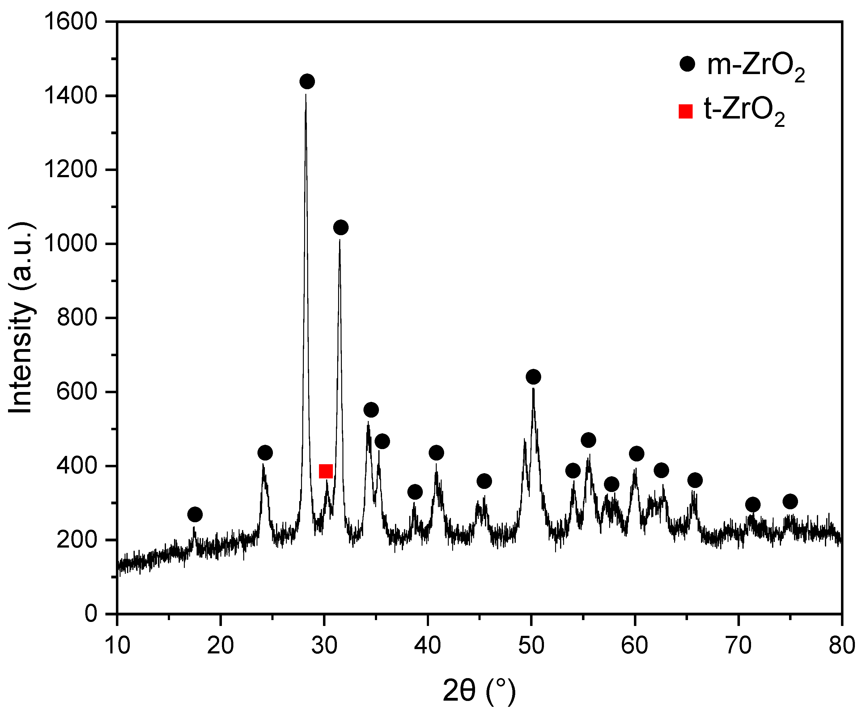
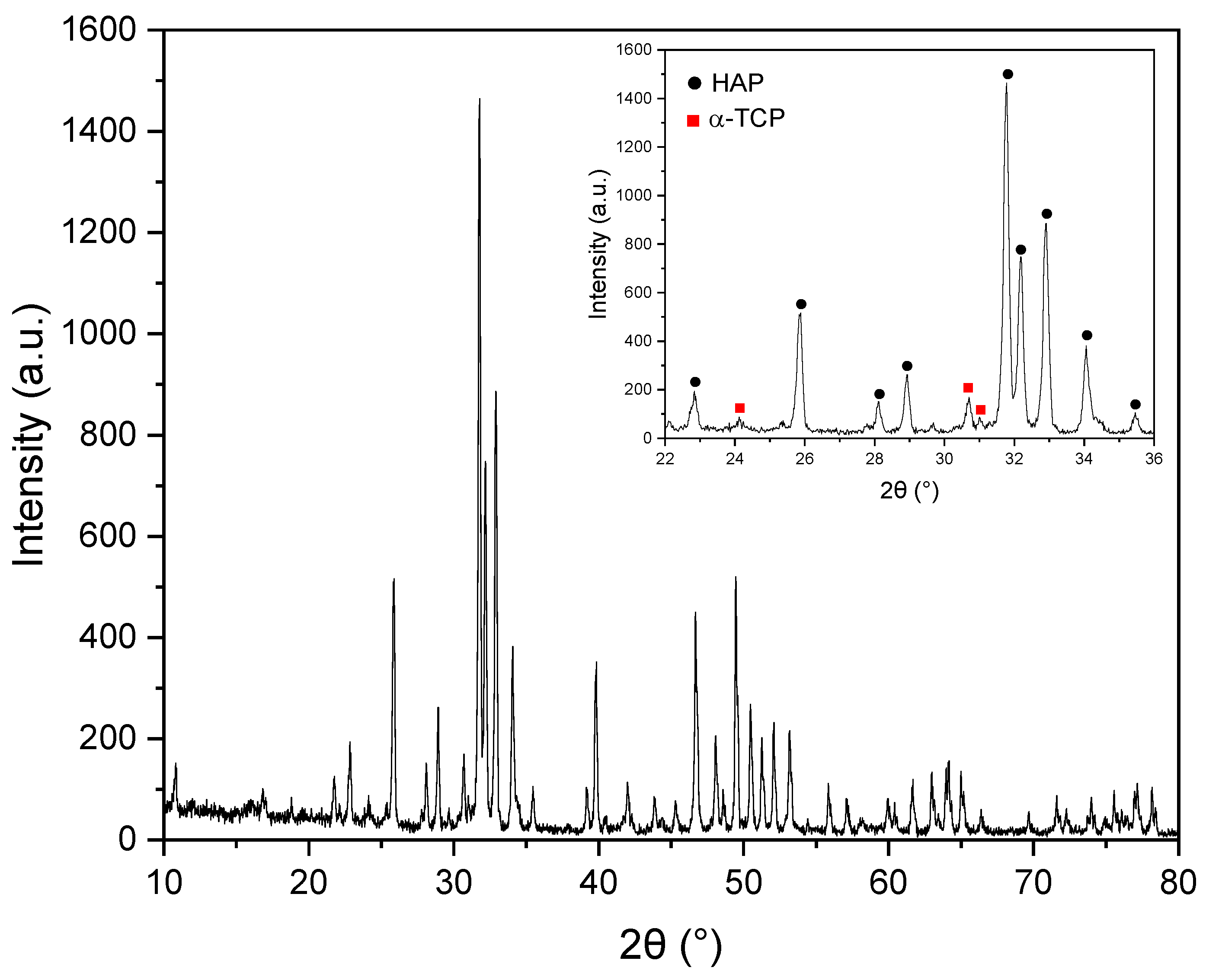
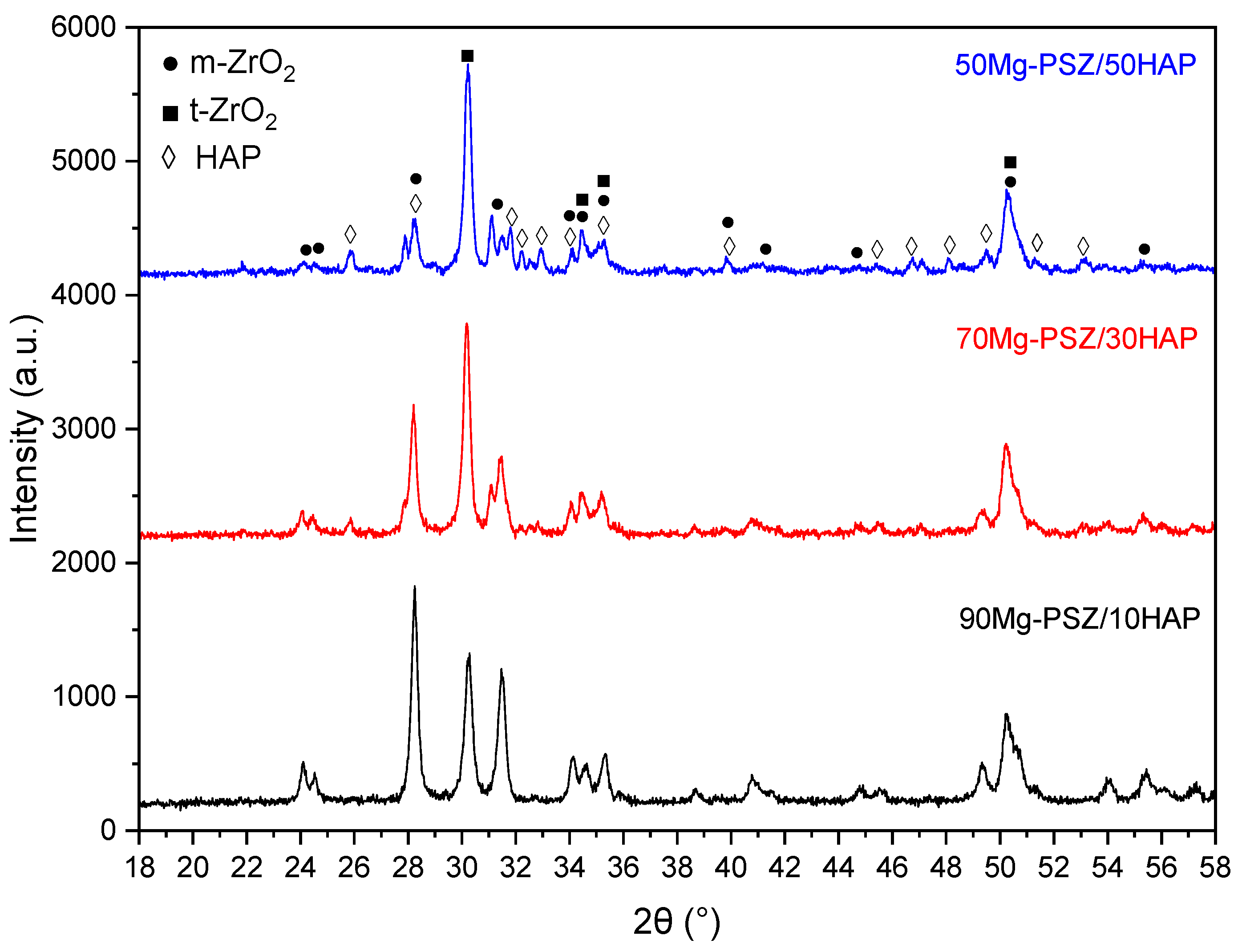
3.2. Structural Analyses

XRPD analyses were performed on calcined Mg-PSZ, HAP, and sintered biocomposites to resolve the phase composition of the prepared samples. The XRPD patterns are presented in Figure 3 for Mg-ZrO2, Figure 4 for HAP, and Figure 5 for biocomposites, respectively.
In the XRPD analysis performed on Mg-ZrO2 calcined at 800 °C (Figure 3), two distinct structures of zirconia have been identified: monoclinic (m-ZrO2) and tetragonal (t-ZrO2). The m-ZrO2 (97.6%), identified as the main phase, was accompanied by a small amount of t-ZrO2 (2.4%). Phase identification was done with Match! 3 software (version 3.13 Build 220), the crystallographic information corresponding to the monoclinic (m-ZrO2, PDF #96-230-0545, S.G. P121/c1, a= 5.14870 Å, b = 5.20230 Å, c = 5.32310 Å, β = 99.164 °) and tetragonal (t-ZrO2, PDF #96-152-6428, S.G. P42/nmc (137), a = 3.61200 Å, c = 5.21200 Å) phases was selected from the Crystallography Open Database (COD).
In the diffractogram registered on HAP calcined at 1000 °C (Figure 4), the main phase HAP in hexagonal form (according to COD data: PDF #96-900-2217) was observed. In addition, α-Ca3(PO4)2 (α-TCP, COD: PDF #96-210-6195) was identified. It is known that HAP starts to decompose at high temperatures. Above 800 °C, HAP can lose water and other volatile components, leading to the formation of different phases, like dicalcium phosphate (DCP), tetracalcium phosphate (TTCP) or β-TCP, [44]
Ca10(PO4)6(OH)2 ⇌ 3 β-Ca3(PO4)2 + CaO + H2O
Further, the thermal decomposition of beta tricalcium phosphate (β-TCP) involves a phase transition to alpha tricalcium phosphate (α-TCP) at temperatures around 1150–1200 °C. This transition is accompanied by a volume change, which can lead to cracking if sintering is done at higher temperatures. Pure β-TCP undergoes this phase transition to α-TCP, and sintering should be done below 1200 °C to avoid it [44,45,46,47]. Our results indicate that the decomposition of the HAP structure led to the formation of the α-TCP phase at a relatively low temperature.
The XRPD analysis of composite samples was performed to determine possible structural changes induced by the sintering process. The XRPD patterns of the composites after sintering at 1100 °C are displayed in Figure 5. According to the XRPD analysis, the presence of t-ZrO2 (COD: PDF #96-152-6428) and m-ZrO2 (COD: PDF #96-230-0545), with an increase in the intensity of the t-ZrO2 phase as the amount of zirconia decreases on the sintered samples, was observed. In addition, for samples 70Mg-PSZ/30HAP and 50Mg-PSZ/50HAP, peaks belonging to the HAP (COD: PDF #96-900-2217) were also identified.
One observation should be emphasized. As the amount of the HAP is increasing on the sintered samples, the splitting of the diffraction maxima at 31.40° 2θ, attributed to the (111) plane of m-ZrO2, clearly visible on the pattern of the 90Mg-PSZ/10HAP sample, was observed. As the composite samples contain multiple crystalline phases, such as m-ZrO2, t-ZrO2, and HAP, each phase will contribute to its own set of peaks, as shown in Figure 5. Some of these peaks might overlap as the HAP amount increases. The diffraction peaks analogous to the HAP structure become combined into the peaks of the ZrO2 phase. However, the most intense peaks for HAP are noticeable in the 50Mg-PSZ/50HAP composite, while no extra diffraction peaks appear, signifying the safeguarding of the apatite structure.
The Scherrer formula (Equation (3)) was adopted to calculate the crystallite size of the sintered samples
Dhkl = (K∙λ)/(β∙cosθ)
where Scherrer’s constant (k) = 0.9, wavelength (λ) = 0.154056 nm for Cu-Kα radiation, θ is the Bragg diffraction angle, and β is the broadening of the hkl diffraction peak measured at half of its maximum intensity in radians [48].
Using Scherrer’s formula, the estimated crystallite sizes in the biocomposites are in the nanometric range, 31.5 nm for sample 90Mg-PSZ/10HAP, 30.71 nm for sample 70Mg-PSZ/30HAP, and 27.45 nm for sample 50Mg-PSZ/50HAP. It is observed that the crystallite size decreases as the amount of Mg-PSZ decreases. Thus, 90Mg-PSZ/10HAP has the largest crystallites among the three composite samples, while 50Mg-PSZ/50HAP has the smallest crystallites. In HAP-zirconia nanocomposites, the crystallite size tends to decrease with increasing zirconia content, at least up to a certain point, after which it may increase. The addition of zirconia to HAP can initially refine the microstructure of the HAP, leading to smaller crystallite sizes. This can be attributed to zirconia particles acting as nucleation sites, hindering HAP grain growth. As the zirconia content increases further, the strain and dislocation density within the HAP lattice can increase, potentially leading to larger crystallites. This is because the zirconia particles may start to agglomerate or create regions of high strain, disrupting the HAP structure and promoting larger crystallite formation. This tendency of decreasing the crystallite size with increasing HAP content contributes to reduced crystallinity (Table S1, Supplementary Materials). The changing crystallite size with zirconia content can have significant implications for the overall properties of the composite, including mechanical strength, biocompatibility, and bioresorbability. For example, smaller crystallites can enhance mechanical properties by increasing the grain boundary density, while larger crystallites can influence biocompatibility and bioresorbability [25,31]. Furthermore, annealing temperature can also influence crystallite size in zirconia, with larger crystallites observed at higher annealing temperatures.

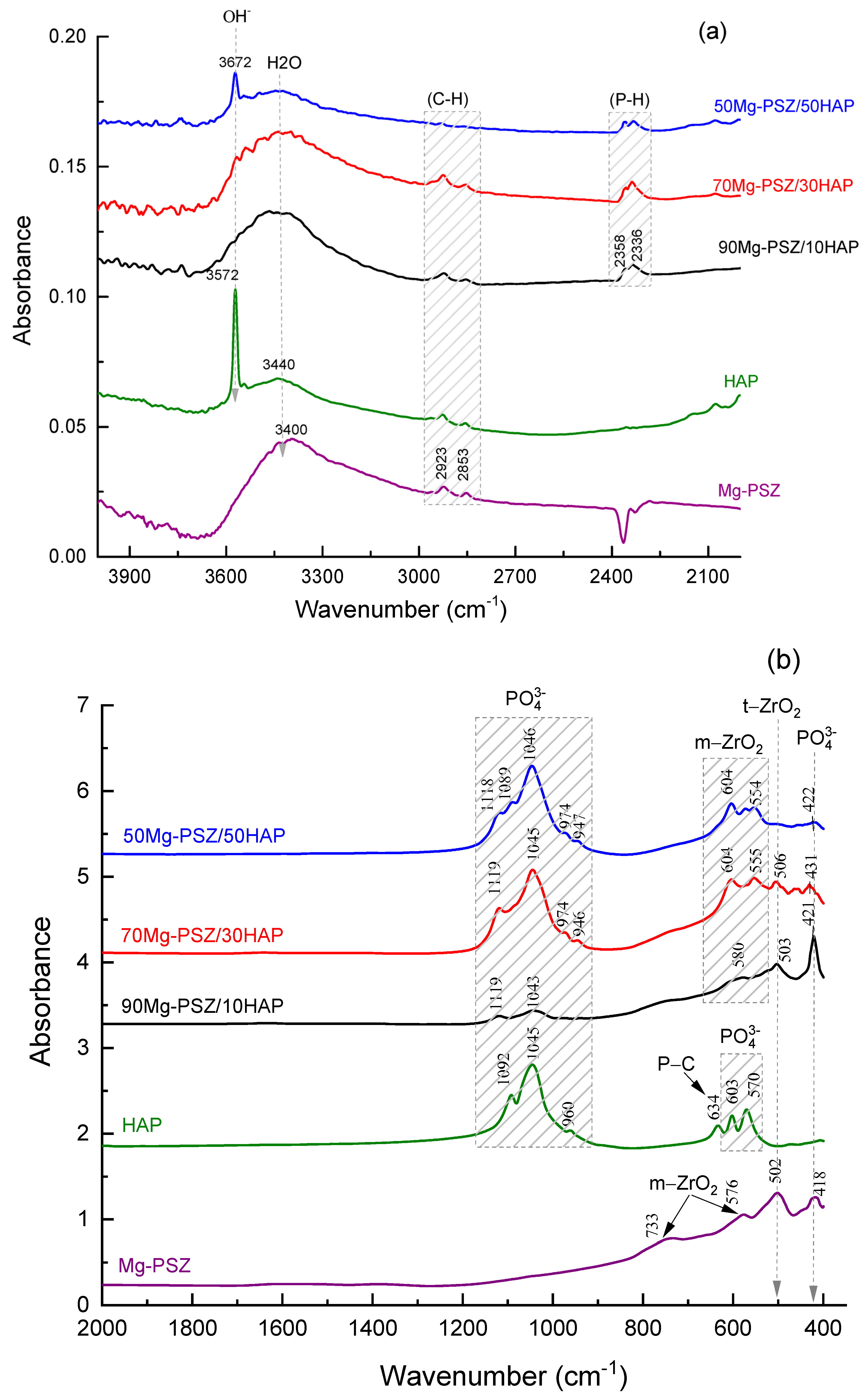
3.3. FTIR Spectroscopy Investigation

The FTIR spectra of Mg-PSZ, HAP, and Mg-PSZ/HAP composites were analyzed to identify changes in the characteristic groups during the synthesis of Mg-PSZ/HAP composite (see Figure 6).
In the spectrum of the HAP, the broadband at 3572 cm−1 is associated with the OH- vibration. This hydroxyl group of hydroxyapatites appeared in the spectrum of 50Mg-PSZ/HAP composite at 3642 cm−1, but of weaker intensity [49]. This vibration does not appear in the spectra of the composite 70Mg-PSZ/HAP and 90Mg-PSZ/HAP.
Between 3000 and 3500 cm−1 in the HAP spectrum, a broad band is observed assigned to adsorbed water, due to the high specific surface area. This peak, from HAP, is observed in all composites’ spectra, and it is attributed to the hydroxyl group vibration from the adsorbed water [49]. The peaks at ~2923 and 2853 cm−1 are present in all spectra and could be assigned to the stretching vibrations of the CH group [49]. In the spectra of the three composites, two vibrational bands are observed, identified at 2358 and 2336 cm−1, attributed to the phosphate groups vibration (P-H).
In the spectral range 2000–4000 cm−1, the FT-IR spectrum of HAP shows two characteristic absorption bands, located at 1092 and 1045 cm−1, attributed to the vibration of the phosphate group [30]. In the spectrum of the Mg-PSZ sample, two characteristic bands at 733 and 576 cm−1 are observed due to m-ZrO2 vibration. The band at 502 cm−1 is attributed to t-ZrO2 vibration [6]. In the spectra of the Mg-PSZ/HAP composite samples, zirconium phases are observed at frequencies shifted towards lower values, due to the deformation of the crystal lattice and bonds with the introduction of HAP. In the frequency range 1100–900 cm−1, the spectrum of HA and the composite samples shows vibration bands assigned to phosphate groups PO43− vibration [50]. In the composite samples, the vibration of the P-C group from HAP is missing; an explanation could be the decarbonization of HA after applying the sintering at high temperature.

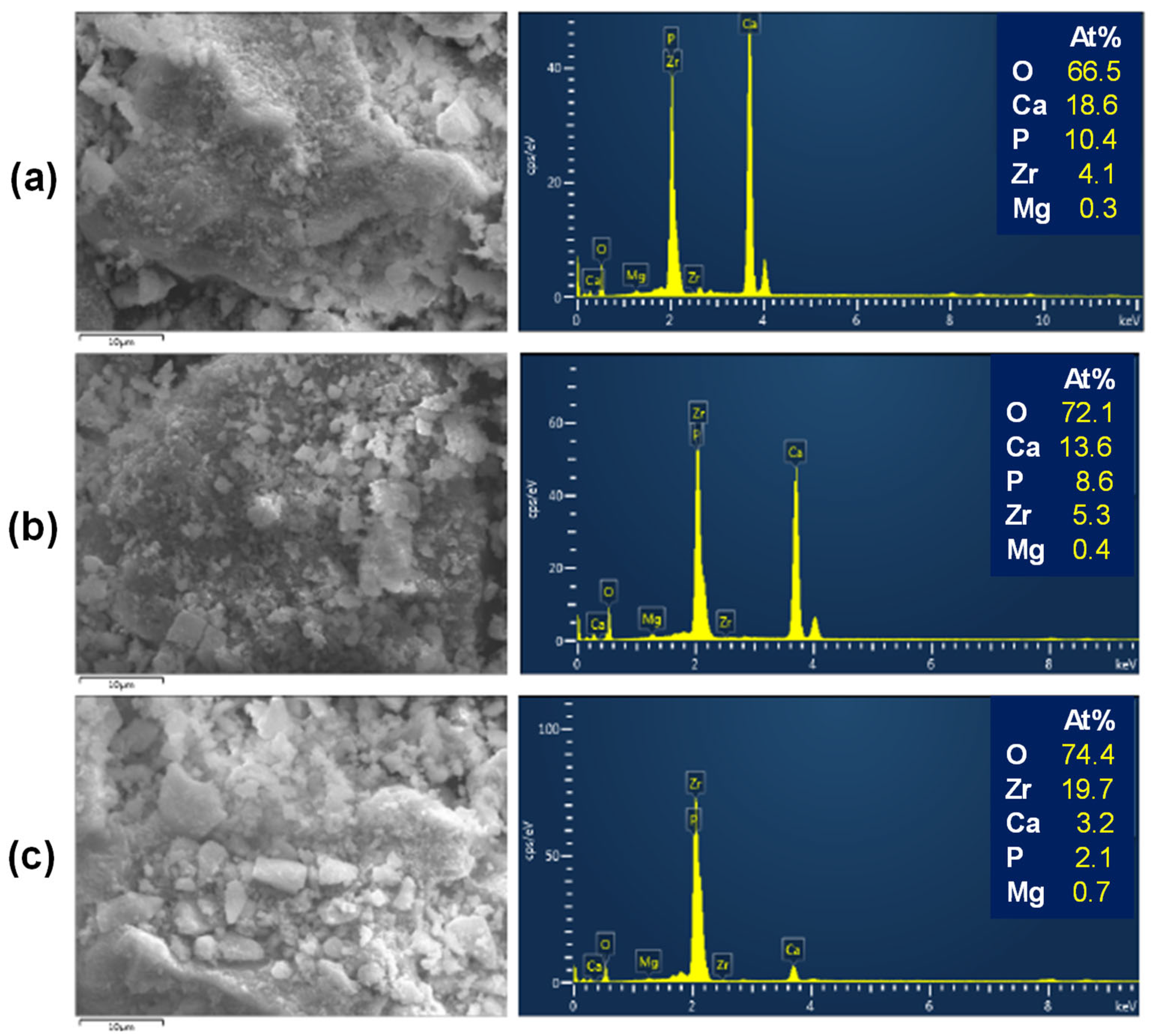
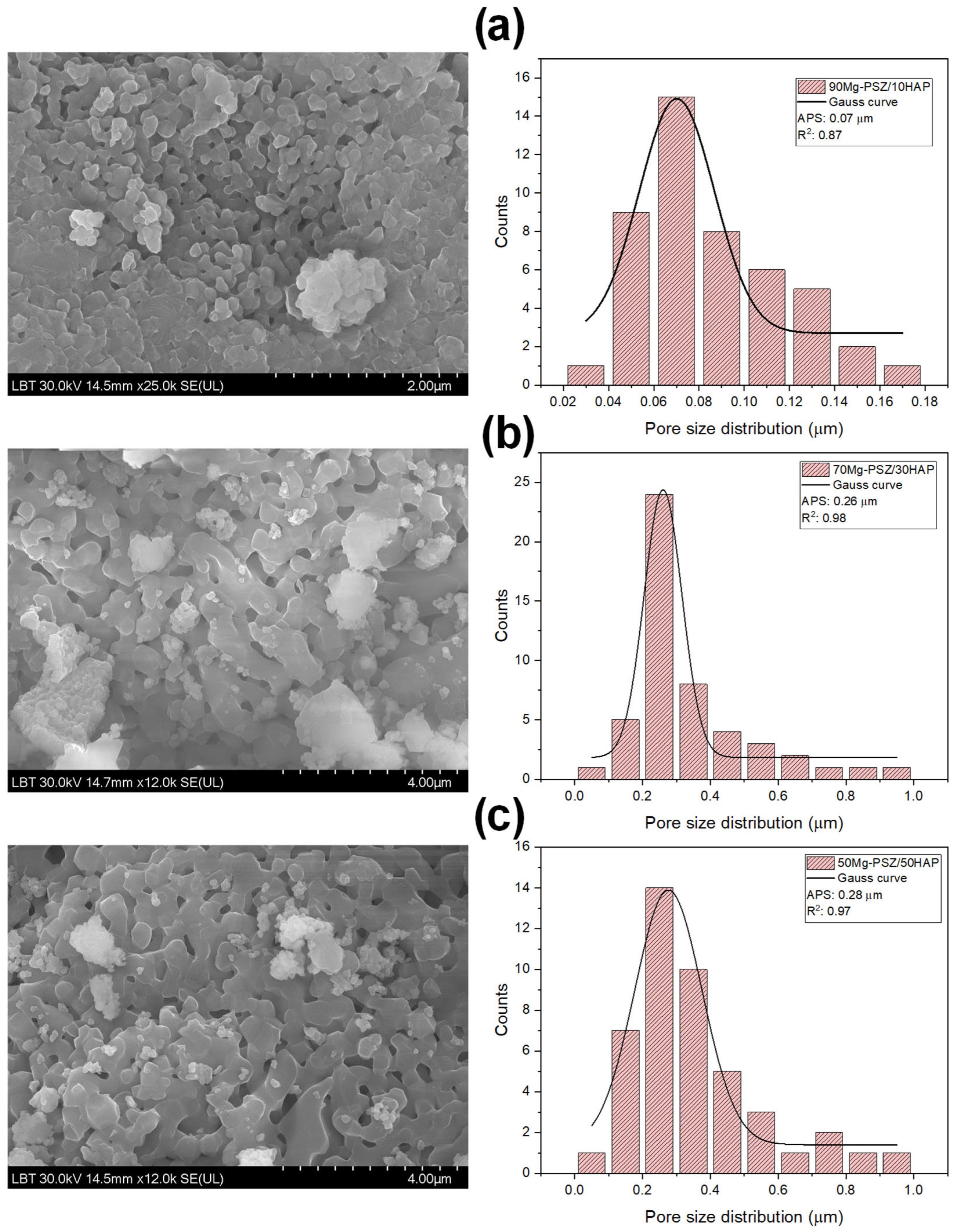
3.4. Morphological Analyses

The biocomposites were subjected to analysis using SEM-EDX to obtain critical insights into their structural characteristics and perform qualitative assessments. The resultant micrographs, together with the EDX spectra and elemental mapping, are presented in Figure 7, Figure 8, Figure S1, and Figure S2, corresponding to samples 90Mg-PSZ/10HAP, 70Mg-PSZ/30HAP, and 50Mg-PSZ/50HAP, respectively.
Figure 7 illustrates the SEM micrographs of the sintered surfaces of the biocomposites, highlighting a decrease in crystallite size with an increasing amount of ZrO2. The grain size varied from 50 nm for 90Mg-PSZ/10HAP to 100 nm for 50Mg-PSZ/50HAP, suggesting that the presence of ZrO2 inhibits crystal growth, as revealed in Figure 7 and Figure 8, respectively.
Furthermore, microstructural examinations indicate the presence of pores (Figure 9). In the context of biomedical applications, the presence of finer pores and reduced porosity fractions is advantageous for enhancing the attachment of biological cells. Smaller pores may facilitate the initial adhesion of osteoblast cells through mechanical anchoring. As a result, the attachment of cells during the early stages of implantation can promote the formation of multiple cells, thereby fostering tissue formation in vivo [32,51]. Pore size distribution in HAP/ZrO2 composites is crucial for their application in bone tissue engineering, influencing mechanical properties and biocompatibility. The distribution is affected by fabrication methods and can be tailored by controlling factors like pore-forming agents and sintering temperatures [31]. In the present work, pore size data were obtained from at least 35 pores using SEM images analyzed with the Nano Measurer 1.2.5 image analysis program. The pore size distribution histograms and average pore sizes (APS) of the composites were analyzed through a Gaussian curve fitting procedure. The SEM-derived statistical histograms of the pore size distribution are shown in Figure 3. As visible, the morphology of the samples displayed a macroporous structure with average pore sizes (APS) of 0.07, 0.26, and 0.28 μm, for 90Mg-PSZ/10HAP, 70Mg-PSZ/30HAP, and 50Mg-PSZ/50HAP composites, respectively. The increasing amount of HAP in the composites resulted in an enlarged pore size distribution.
The chemical composition of the biocomposites in the Mg-PSZ/HAP system was analyzed using EDX, as shown in Figure 8. The EDX results confirmed the presence of several elements, including calcium, phosphorus, magnesium, oxygen, and zirconium, indicated by the peaks corresponding to the elements Ca, P, Mg, Zr, and O. This confirms the purity of the product, with no signals from other elements detected. The Ca/P ratio is an important parameter for HAP, as both the mechanical properties and biodegradation rate strongly depend on it. Raynaud et al. conducted a study on the transformations of HAP based on the Ca/P ratio. They found that for HAP heat-treated at 1000 °C with a stoichiometric Ca/P ratio of 1.67, no secondary phases were detected. However, for HAP that was deficient in calcium (Ca/P < 1.67) or rich in calcium (Ca/P > 1.67), the appearance of tricalcium phosphate (TCP) and calcium oxide (CaO) phases was observed, respectively [52].
The qualitative analysis of the elements in the Mg-PSZ/HAP biocomposites was assessed using elemental mapping. The resulting micrographs, shown in Figure S2 (Supplementary Materials), confirmed a homogeneous distribution of elements throughout the sample. It was observed that HAP is evenly distributed across the area depicted in the images, suggesting that Mg-PSZ is modified at the surface by HAP. Additionally, Mg-PSZ is uniformly dispersed within HAP, indicating an effective interaction between Mg-PSZ and HAP. This interaction may enhance the mechanical and biological properties of the biocomposite.

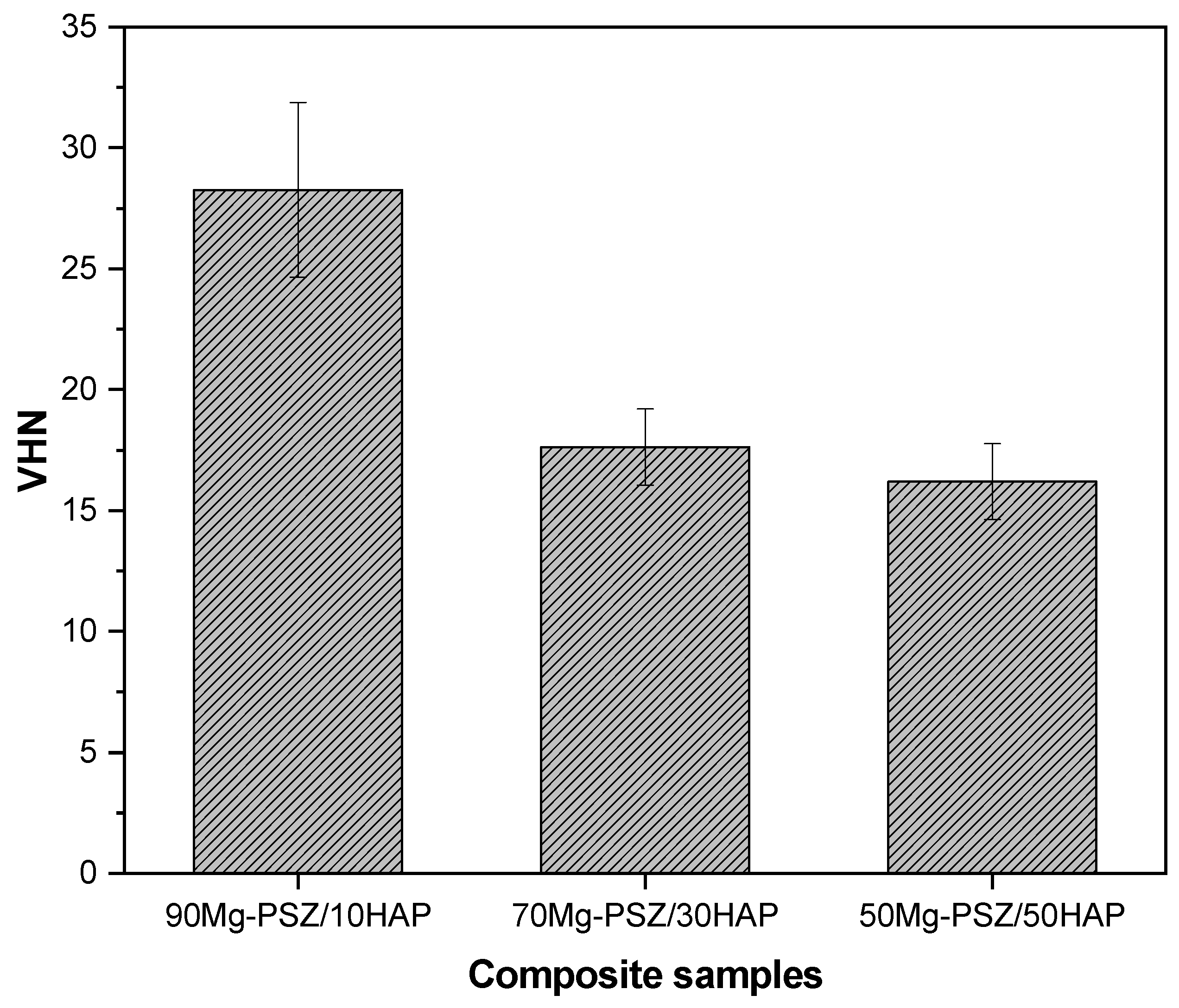
3.5. Vickers Microhardness

The obtained microhardness data are presented in Figure 9. In the samples presented in this paper, the Vickers microhardness value increased with the increasing amount of Mg-PSZ. The composite 90Mg-PSZ/10HAP presents a mean value of 28.26 HV3, while for 70Mg-PSZ/30HAP and 50Mg-PSZ/50HAP, the values measured were 17.63 HV3 and 16.19 HV3, respectively (these are values between 277 and 159 MPa). For all the samples, the main phases were t- and m-ZrO2. The presence of the tetragonal phase of zirconia is generally attributed to better mechanical properties in the materials. In addition, HAP could contribute to the biological properties of the materials, but can also alter their mechanical performance.
Compared to HAP, β-TCP shows a faster degradation and resorption rate and a different crystal structure that could present lower crystallographic density. This secondary phase also presents higher porosity, a fact that could reduce the microhardness values obtained. In the sample with 90Mg-PSZ/10HAP, the amount of HAP is too low to have a HAP degradation that could lead to secondary phases. This fact seems to have the main influence on the mechanical performance of this material. The shrinkage of HAP could be expected in all the samples and could influence the mechanical properties, but the reduced amount of HAP leads to a decreased influence in the 90Mg-PSZ/10HAP composite.
When sintered at 1100 °C, the addition of ZrO2 did not significantly enhance the microhardness of the composite ceramics. This suggests that ZrO2 did not provide a noticeable toughening effect under conditions where the sintering temperature was insufficient for the densification of HAP ceramics. This finding aligns with the research conducted by Zhang et al. [53], who observed that when the sintering temperature was increased to 1200 °C and 1250 °C, the mechanical properties of pure HAP ceramics were significantly improved, making the toughening effect of zirconia more pronounced.

4. Conclusions

Due to the fact that HAP is a biocompatible material with bioactivity and osteoconductive properties suitable for bone applications, while Mg-PSZ, despite its excellent mechanical properties, faces challenges related to biocompatibility and stability, the design of new composites with enhanced mechanical properties and biological activity for bone regeneration represents a promising approach to overcome the limitations of each material. In this work, magnesia partially stabilized zirconia/hydroxyapatite biocomposites were successfully prepared using the ceramic method from Mg-PSZ and HAP synthesized via coprecipitation, and sintered at 1100 °C. The structural analyses confirmed the presence of monoclinic and tetragonal zirconia phases, accompanied by hexagonal HAP. The crystallite sizes ranged in the nanometric domain, and surface morphology was homogeneous with small pores, with the elements such as Ca, P, Mg, Zr, and O consistently detected. The examination of microhardness analyses showed that sintering at 1100 °C does not significantly improve the microhardness of the composite ceramics, with the best value of 28.26 HV3 obtained for the composite 90Mg-PSZ/10HAP. In conclusion, the biocomposites obtained could be used as restorative materials, presenting sufficient mechanical performance for specific esthetic dental applications and biological functionality due to the HAP.

Supplementary Materials

The following supporting information can be downloaded at https://www.mdpi.com/article/10.3390/cryst15070608/s1: Figure S1: DSC curves of the uncalcined HAP and Mg-PSZ materials; Figure S2: SEM/EDS elemental mapping of the composite samples. In the elemental mapping images, the assignment of color for each element is the following: cyan for O, green for P, orange for Zr, blue for Ca, and red for Mg, respectively; Figure S3: SEM images of the composites sample (a) 90Mg-PSZ/10HAP, (b) 70Mg-PSZ/30HAP and (c) 50Mg-PSZ/50HAP at different magnifications ×25.0 k (left) and ×100 k (right); Table S1: Calculated crystallite size (Dhkl), degree of crystallinity (DC), Ca/P and Zr/Mg ratios for Mg-PSZ/HAP composites.

Author Contributions

Conceptualization, L.B.; methodology, L.B., M.M.-P., and R.B.; software, L.B., A.-L.B., M.M.-P., L.B.-T., and C.A.C.; validation, L.B., M.M.-P., and R.B.; investigation, A.-L.B., M.M.-P., C.A.C., L.B.-T., and R.B.; writing—original draft preparation, L.B., M.M.-P., and C.A.C.; writing—reviewing and editing, L.B.; visualization, L.B., A.-L.B., C.A.C., M.M.-P., L.B.-T., and R.B.; supervision, L.B. All authors have read and agreed to the published version of the manuscript.

Funding

This research received no external funding.

Data Availability Statement

The data presented in this study are available on request from the corresponding author due to privacy.

Conflicts of Interest

The authors declare no conflicts of interest.

References

  1. Zhao, Y.; Xu, L.; Guo, M.; Li, Z.; Xu, Z.; Ye, J.; Li, W.; Wei, S. Effects of calcination temperature on grain growth and phase transformation of nano-zirconia with different crystal forms prepared by hydrothermal method. J. Mater. Res. Technol. 2022, 19, 4003–4017. [Google Scholar] [CrossRef]
  2. Guo, M.; Wang, G.; Zhao, Y.; Li, H.; Tang, K.; Zhao, Y.; Burgess, K. Preparation of Nano-ZrO2 powder via a microwave-assisted hydrothermal method. Ceram. Int. 2021, 47, 12425–12432. [Google Scholar] [CrossRef]
  3. Hussain, A.; Gautam, C.; Jafri, A.; Mishra, V.K.; Madheshiya, A.; Gautam, A.; Singh, M.K.; Gautam, R.K.; Gupta, M.; Arshad, M.; et al. Formation of multifunctional ZrO2–MgO-hBN nanocomposite for enhanced bone regeneration and E coli bacteria filtration applications. Ceram. Int. 2020, 46, 23006–23020. [Google Scholar] [CrossRef]
  4. Ngashangua, S.; Vasanthavel, S.; Ponnilavan, V.; Kannan, S. Effect of MgO additions on the phase stability and degradation ability in ZrO2-Al2O3 composite systems. Ceram. Int. 2015, 41, 3814–3821. [Google Scholar] [CrossRef]
  5. Keerthana, L.; Sakthivel, C.; Prabha, I. MgO-ZrO2 mixed nanocomposites: Fabrication methods and applications. Mater. Today Sustain 2019, 3–4, 100007. [Google Scholar] [CrossRef]
  6. Bizo, L.; Mureşan-Pop, M.; Barabás, R.; Barbu-Tudoran, L.; Berar, A. In Vitro Degradation of Mg-Doped ZrO2 Bioceramics at the Interface with Xerostom® Saliva Substitute Gel. Materials 2023, 16, 2680. [Google Scholar] [CrossRef] [PubMed]
  7. Cojan, C.A.; Barabás, R.; Mureșan-Pop, M.; Bizo, L. Comparative Phase Evolution, Morphological and Optical Analysis of Partially Stabilized Zirconia Ceramics. Studia UBB Chem. 2024, LXIX, 7–17. [Google Scholar] [CrossRef]
  8. Godwin, E.R.; Kumar I, P.A.; Mathows, J.; Govindasamy, C.; Al-Numair, K.S.; Sana, S.S.; Chandrasekaran, K.; Arulselvan, P. Biomedical potential of green-engineered chitosan-magnesium oxide nanoparticles: An in vitro study on antibacterial and anticancer activities. Ceram. Int. 2024, 50, 39775–39786. [Google Scholar] [CrossRef]
  9. Gatou, M.-A.; Skylla, E.; Dourou, P.; Pippa, N.; Gazouli, M.; Lagopati, N.; Pavlatou, E.A. Magnesium Oxide (MgO) Nanoparticles: Synthetic Strategies and Biomedical Applications. Crystals 2024, 14, 215. [Google Scholar] [CrossRef]
  10. Cho, J.; Yang, B.; Shen, C.; Wang, H.; Zhang, X. Micromechanical properties and microstructure evolution of magnesia partially stabilized zirconia prepared by spark plasma sintering. J. Eur. Ceram. Soc. 2023, 43, 1098–1107. [Google Scholar] [CrossRef]
  11. Mysore, T.H.M.; Patil, A.Y.; Hegde, C.; Sudeept, M.A.; Kumar, R.; Soudagar, M.E.M.; Fattah, I.M.R. Apatite insights: From synthesis to biomedical applications. Eur. Polym. J. 2024, 209, 112842. [Google Scholar] [CrossRef]
  12. Liu, C.; Xu, M.; Wang, Y.; Yin, Q.; Hu, J.; Chen, H.; Sun, Z.; Liu, C.; Li, X.; Zhou, W.; et al. Exploring the potential of hydroxyapatite-based materials in biomedicine: A comprehensive review. Mater. Sci. Eng. R Rep. 2024, 161, 100870. [Google Scholar] [CrossRef]
  13. Mondal, S.; Park, S.; Choi, J.; Vu, T.T.H.; Doan, V.H.M.; Vo, T.T.; Lee, B.; Oh, J. Hydroxyapatite: A journey from biomaterials to advanced functional materials. Adv. Colloid Interface Sci. 2023, 321, 103013. [Google Scholar] [CrossRef]
  14. Ielo, I.; Calabrese, G.; De Luca, G.; Conoci, S. Recent Advances in Hydroxyapatite-Based Biocomposites for Bone Tissue Regeneration in Orthopedics. Int. J. Mol. Sci. 2022, 27, 9721. [Google Scholar] [CrossRef]
  15. Liu, W.; Cheong, N.; He, Z.; Zhang, T. Application of Hydroxyapatite Composites in Bone Tissue Engineering: A Review. J. Funct. Biomater. 2025, 16, 127. [Google Scholar] [CrossRef] [PubMed]
  16. Bystrov, V.S.; Paramonova, E.V.; Avakyan, L.A.; Eremina, N.V.; Makarova, S.V.; Bulina, N.V. Effect of Magnesium Substitution on Structural Features and Properties of Hydroxyapatite. Materials 2023, 16, 5945. [Google Scholar] [CrossRef] [PubMed]
  17. Rehner, A.M.G.; Moldoveanu, E.-T.; Niculescu, A.-G.; Bîclesanu, F.C.; Pangică, A.M.; Grumezescu, A.M.; Croitoru, G.-A. Advances in Dental Implants: A Review of In Vitro and In Vivo Testing with Nanoparticle Coatings. J. Compos. Sci. 2025, 9, 140. [Google Scholar] [CrossRef]
  18. Vishwakarma, V.; Kaliaraj, G.S.; Amirtharaj Mosas, K.K. Multifunctional Coatings on Implant Materials-A Systematic Review of the Current Scenario. Coatings 2023, 13, 69. [Google Scholar] [CrossRef]
  19. Rapacz-Kmita, A.; Slósarczyk, A.; Paszkiewicz, Z. Mechanical properties of HAp–ZrO2 composites. J. Eur. Ceram. Soc. 2006, 26, 1481–1488. [Google Scholar] [CrossRef]
  20. Barabás, R.; Fort, C.I.; Turdean, G.L.; Bizo, L. Influence of HAP on the Morpho-Structural Properties and Corrosion Resistance of ZrO2-Based Composites for Biomedical Applications. Crystals 2021, 11, 202. [Google Scholar] [CrossRef]
  21. Antoniac, I. Bioceramics and Biocomposites: From Research to Clinical Practice; Wiley-VCH: Weinheim, Germany, 2019. [Google Scholar]
  22. Fan, H.; Wang, L.; Dou, R. Properties of zirconia-doped hydroxyapatite ceramics prepared by vat photopolymerization additive manufacturing technology and microwave sintering. Ceram. Int. 2025. [Google Scholar] [CrossRef]
  23. Zhang, J.; Huang, D.; Liu, S.; Dong, X.; Li, Y.; Zhang, H.; Yang, Z.; Su, Q.; Huang, W.; Zheng, W.; et al. Zirconia toughened hydroxyapatite biocomposite formed by a DLP 3D printing process for potential bone tissue engineering. Mat. Sci. Eng. C-Mater. 2019, 105, 110054. [Google Scholar] [CrossRef] [PubMed]
  24. Xing, Z.; Pang, Y.; Li, E.; Zhang, J.Y.; Xu, D. Preparation and characterisation of zirconia/hydroxyapatite bioactive composites as potential dental implants. J. Mater. Sci. Mater. Eng. 2024, 19, 43. [Google Scholar] [CrossRef]
  25. Sivaperumal, V.R.; Mani, R.; Polisetti, V.; Aruchamy, K.; Oh, T. Synthesis of Hydroxyapatite (HAp)-Zirconia Nanocomposite Powder and Evaluation of Its Biocompatibility: An In Vitro Study. Appl. Sci. 2022, 12, 11056. [Google Scholar] [CrossRef]
  26. Mohammad, M.; Taha, S.; Shahatha, A. Preparation of Hydroxyapatite/Zirconia Porous Composites via Polymeric Sponge Method and Study the Physical and Bioactivity Properties. IOP Conf. Ser. Mater. Sci. Eng. 2020, 757, 012046. [Google Scholar] [CrossRef]
  27. Zaw, L.H.; Nurazreena, A.; Aye Aye, T.; Ahmad-Fauzi, M.N. Characterization of CaO-ZrO2 Reinforced Hap Biocomposite for Strength and Toughness Improvement. Procedia Chem. 2016, 19, 510–516. [Google Scholar] [CrossRef]
  28. Yeongjun, S.; Shiori, N.; Tomoyo, G.; Sunghun, C.; Tohru, S. Densification of hydroxyapatite/zirconia nanocomposites fabricated via low-temperature mineralization sintering process and their mechanical properties. Sci. Rep. 2025, 15, 2479. [Google Scholar] [CrossRef]
  29. Gautam, A.; Gautam, C.; Mishra, M.; Sahu, S.; Nanda, R.; Kisan, B.; Gautam, R.K.; Prakash, R.; Sharma, K.; Singh, D.; et al. Synthesis, structural, mechanical, and biological properties of HAp-ZrO2-hBN biocomposites for bone regeneration applications. Ceram. Int. 2021, 47, 30203–30220. [Google Scholar] [CrossRef]
  30. Es-saddik, M.; Laasri, S.; Taha, M.; Laghzizil, A.; Guidara, A.; Chaari, K.; Bouaziz, J.; Hajjaji, A.; Nunzi, J.M. Effect of the surface chemistry on the stability and mechanical properties of the Zirconia-Hydroxyapatite bioceramic. Surf. Interfaces 2021, 23, 100980. [Google Scholar] [CrossRef]
  31. Kumar, V.A.; Rama Murty Raju, P.; Ramanaiah, N.; Siriyala, R. Effect of ZrO2 content on the mechanical properties and microstructure of HAp/ZrO2 nanocomposites. Ceram. Int. 2018, 44, 10345–10351. [Google Scholar] [CrossRef]
  32. Ayoub, G.; Veljovic, D.; Lezaja Zebic, M.; Miletic, V.; Palcevskis, E.; Petrovic, R.; Janackovic, D. Composite nanostructured hydroxyapatite/yttrium stabilized zirconia dental inserts-The processing and application as dentin substitutes. Ceram. Int. 2018, 44, 18200–18208. [Google Scholar] [CrossRef]
  33. Matsumoto, T.J.; An, S.-H.; Ishimoto, T.; Nakano, T.; Matsumoto, T.; Imazato, S. Zirconia-hydroxyapatite composite material with micro porous structure. Dent. Mater. 2011, 27, e205–e212. [Google Scholar] [CrossRef] [PubMed]
  34. Wang, S.; Zhai, Y.; Li, X.; Li, Y.; Wang, K. Coprecipitation Synthesis of MgO-Doped ZrO2 Nano Powder. J. Am. Ceram. Soc. 2006, 89, 3577–3581. [Google Scholar] [CrossRef]
  35. Yuan, L.; Xiang, D.; Yu, J. Effect of solvents on the properties of co-precipitated MgO-ZrO2 nano powders. J. Ceram. Process. Res. 2013, 14, 517–520. [Google Scholar] [CrossRef]
  36. Barabás, R.; de Souza Ávila, E.; Ladeira, L.O.; Mosqueira Antônio, L.; Tötös, R.; Simedru, D.; Bizo, L.; Cadar, O. Graphene Oxides/Carbon Nanotubes–Hydroxyapatite Nanocomposites for Biomedical Applications. Arab. J. Sci. Eng. 2020, 45, 219–227. [Google Scholar] [CrossRef]
  37. Barabás, R.; Deemter, D.; Katona, G.; Batin, G.; Barabás, L.; Bizo, L.; Cadar, O. Comparative study on physicochemical and mechanical characterization of new nanocarbon-based hydroxyapatite nanocomposites. Turk. J. Chem. 2019, 43, 809–824. [Google Scholar] [CrossRef]
  38. Barabás, R.; Czikó, M.; Dékány, I.; Bizo, L.; Bogya, E.S. Comparative study of particle size analysis of hydroxyapatite-based nanomaterials. Chem. Pap. 2013, 67, 1414–1423. [Google Scholar] [CrossRef]
  39. Bizo, L.; Sabo, K.; Barábas, R.; Katona, G.; Barbu-Tudoran, L.; Berar, A. Structural, morphological and dissolution properties of ZrO2-based biocomposites for dental applications. Studia UBB Chem. 2020, 1, 137–148. [Google Scholar] [CrossRef]
  40. Murray, M.G.S.; Wang, J.; Ponton, C.B.; Marquis, P.M. An improvement in processing of hydroxyapatite ceramics. J. Mater. Sci. 1995, 30, 3061–3074. [Google Scholar] [CrossRef]
  41. Ficai, A.; Albu, M.G.; Birsan, M.; Sonmez, M.; Ficai, D.; Trandafir, V.; Andronescu, E. Collagen hydrolysate based collagen/hydroxyapatite composite materials. J. Mol. Struct. 2013, 1037, 154–159. [Google Scholar] [CrossRef]
  42. Senra, M.R.; Barbosa de Lima, R.; de Holanda Saboya Souza, D.; de Fátima Vieira Marques, M.; Neves Monteiro, S. Thermal characterization of hydroxyapatite or carbonated hydroxyapatite hybrid composites with distinguished collagens for bone graft. J. Mater. Res. Technol. 2020, 9, 7190–7200. [Google Scholar] [CrossRef]
  43. Bulina, N.V.; Makarova, S.V.; Baev, S.G.; Matvienko, A.A.; Gerasimov, K.B.; Logutenko, O.A.; Bystrov, V.S. A Study of Thermal Stability of Hydroxyapatite. Minerals 2021, 11, 1310. [Google Scholar] [CrossRef]
  44. Jamil, M.; Elouahli, A.; Abida, F.; Assaoui, J.; Gourri, E.; Hatim, Z. Apatitic calcium phosphate/montmorillonite nano-biocomposite: In-situ synthesis, characterization and dissolution properties. Heliyon 2022, 8, e10042. [Google Scholar] [CrossRef] [PubMed]
  45. Kantana, W.; Jarupoom, P.; Pengpat, K.; Eitssayeam, S.; Tunkasiri, T.; Rujijanagul, G. Properties of hydroxyapatite/zirconium oxide nanocomposites. Ceram. Int. 2013, 39, S379–S382. [Google Scholar] [CrossRef]
  46. Hashimoto, K.; Baba, M.; Shibata, H.; Yoshida, K. Enhanced sinterability and mechanical strength of beta-type tricalcium phosphates ceramics through reaction sintering with hydroxyapatite. J. Eur. Ceram. Soc. 2025, 45, 117023. [Google Scholar] [CrossRef]
  47. Ryu, H.-S.; Youn, H.-J.; Hong, K.S.; Chang, B.-S.; Lee, C.-K.; Chung, S.-S. An improvement in sintering property of β-tricalcium phosphate by addition of calcium pyrophosphate. Biomaterials 2002, 23, 909–914. [Google Scholar] [CrossRef] [PubMed]
  48. Patterson, A.L. The Scherrer Formula for X-Ray Particle Size Determination. Phys. Rev. 1939, 56, 978–982. [Google Scholar] [CrossRef]
  49. Shaltout, A.A.; Allam, M.A.; Moharram, M.A. FTIR spectroscopic, thermal and XRD characterization of hydroxyapatite from new natural sources. Spectrochim. Acta A Mol. Biomol. Spectrosc. 2011, 83, 56–60. [Google Scholar] [CrossRef]
  50. Uysal, I.; Severcan, F.; Evis, Z. Characterization by Fourier transform infrared spectroscopy of hydroxyapatite co-doped with zinc and fluoride. Ceram. Int. 2013, 39, 7727–7733. [Google Scholar] [CrossRef]
  51. Mishchenko, O.; Yanovska, A.; Kosinov, O.; Maksymov, D.; Moskalenko, R.; Ramanavicius, A.; Pogorielov, M. Synthetic Calcium–Phosphate Materials for Bone Grafting. Polymers 2023, 15, 3822. [Google Scholar] [CrossRef]
  52. Raynaud, S.; Champion, E.; Bernache-Assollant, D.; Thomas, P. Calcium phosphate apatites with variable Ca/P atomic ratio I. Synthesis, characterisation and thermal stability of powders. Biomaterials 2002, 23, 1065–1072. [Google Scholar] [CrossRef] [PubMed]
  53. Zhang, J.; Iwasa, M.; Kotobuki, N.; Tanaka, T.; Hirose, M.; Ohgushi, H.; Jiang, D. Fabrication of Hydroxyapatite-Zirconia Composites for Orthopedic Applications. J. Am. Ceram. Soc. 2006, 89, 3348–3355. [Google Scholar] [CrossRef]
Figure 1. TG−DTA curve for Mg-PSZ precipitate (exo-up).
Crystals 15 00608 g001
Figure 2. TG−DTA curve for HAP precipitate (exo-up).
Crystals 15 00608 g002
Figure 3. XRPD pattern for Mg-PSZ calcined at 800 °C for 2 h.
Crystals 15 00608 g003
Figure 4. XRPD pattern for HAP calcined at 1000 °C for 1 h. The 22–36° 2θ region is enlarged in the inset to show the presence of the α-TCP phase.
Crystals 15 00608 g004
Figure 5. XRPD patterns for the composite samples sintered at 1100 °C for 1 h.
Crystals 15 00608 g005
Figure 6. FTIR spectra of precursors and composite samples in the spectral range 4000−2000 cm−1 (a) and 2000−400 cm−1, respectively (b).
Crystals 15 00608 g006
Figure 7. SEM images (left) and corresponding EDS spectrum (right) for (a) 90Mg-PSZ/10HAP, (b) 70Mg-PSZ/30HAP, and (c) 50Mg-PSZ/50HAP, respectively.
Crystals 15 00608 g007
Figure 8. SEM images of the composite samples (a) 90Mg-PSZ/10HAP, (b) 70Mg-PSZ/30HAP, and (c) 50Mg-PSZ/50HAP (left) and corresponding statistical histograms of the pore size distribution derived from the SEM images (right). In the histograms, the Gauss curves indicate the average pore size (APS) of the data.
Crystals 15 00608 g008
Figure 9. Results of the Vickers microhardness tests for the composite samples.
Crystals 15 00608 g009
Table 1. Details of the composites’ preparation by solid-state reaction using different weight percentages of Mg-PSZ and HAP, after sintering at 1100 °C.
Sample IDMg-PSZ (wt.%)HAP (wt.%)
90Mg-PSZ/10HAP9010
70Mg-PSZ/30HAP7030
50Mg-PSZ/50HAP5050
Disclaimer/Publisher’s Note: The statements, opinions and data contained in all publications are solely those of the individual author(s) and contributor(s) and not of MDPI and/or the editor(s). MDPI and/or the editor(s) disclaim responsibility for any injury to people or property resulting from any ideas, methods, instructions or products referred to in the content.

